DOI:
10.1039/D4RA06398H
(Paper)
RSC Adv., 2024,
14, 39027-39039
Retracted Article: Molecular docking and in vivo/in vitro studies of a novel thiadiazole Schiff base as a hepatoprotective drug against angiogenesis induced by breast cancer†
Received
4th September 2024
, Accepted 17th October 2024
First published on 10th December 2024
Abstract
Two new thiadiazole imidazolium salicylidene Schiff bases (TISSBs) were successfully synthesized, and their structures were analyzed comprehensively using spectroscopic techniques. The results of the MTT assay showed that TISSB2 was the safest and most effective anti-breast cancer agent. The anti-angiogenic activity of TISSB2 was evaluated using in vivo tests in Ehrlich ascites carcinoma (EAC)-bearing Swiss albino mice. The degree of angiogenesis was assessed by measuring the levels of vascular endothelial growth factor (VEGF), tumor necrosis factor-α (TNF-α), and transforming growth factor-β1 (TGF-β1). The results of biochemical, immunohistochemical, and histopathological examinations indicated that TISSB2 could restore the normal functional indices of the injured liver, as evident from the downregulated TGF-β1, TNF-α, and VEGF levels reverting to normal values. Moreover, in the molecular docking study, TISSB2 exhibited stronger interactions with VEGFR-2 and NF-κB proteins, with binding affinity scores of −11.79 and −9.25 kcal mol−1, respectively. These stronger interactions involved H-bonding, ionic bonds, and hydrophobic π-interactions. Overall, TISSB2 can be a promising therapeutic option for the treatment of EAC-induced tumour angiogenesis.
1 Introduction
Liver is a master regulator of numerous physiological and biochemical processes, including the metabolism of macronutrients, the maintenance of lipid and glucose homeostasis, the regulation of the immune system and its function, and endocrine control of different signalling pathways.1 Many studies have established that angiogenesis is one of the basic physiological and pathological processes involved in the promotion of carcinogenesis as well as in its progression and metastasis.2 It also facilitates the migration of immune cells from one organ to another, aids in the healing of tissue damage, and leads to the restoration of damaged tissue during tissue homeostasis.3 Angiogenesis is a very intricate physiological process that is closely controlled by a variety of growth factors, inflammatory cytokines, and hypoxic conditions in the body.4 Angiogenesis is a crucial step in the processes leading to the spread of tumours in nearly all types of cancer.5 Numerous signalling pathways contribute to liver damage and fibrosis caused by inflammation. According to the majority of studies, tumour angiogenesis is one of the characteristics of cancer that contributes to fibrosis and inflammation of the liver in chronic liver disorders, and it also promotes the development of hepatocellular carcinoma (HCC).6 One of the deadliest cancers that affect women worldwide is breast cancer.7 A well-known model in cancer biology is Ehrlich ascites mammary carcinoma, which is a spontaneous murine mammary adenocarcinoma.8 Moreover, there is a wealth of research on the metastasis of breast cancer to the heart, lung, brain, and bones.9 Angiogenesis has been identified as a critical characteristic of several malignancies, including HCC, in a number of research works, including our previous study.10
Molecular hybridization (MH) of various pharmacophores is a powerful technique in the field of multitarget drug development. Due to the increasing prevalence of complex illnesses, developing medications with a single target is no longer sufficient. The effectiveness of treatment can be enhanced by drugs that target many disease pathways simultaneously. One way to improve the pharmacological properties of a drug is to combine two or more pharmacophores into one molecule.11–13 In addition to its potential in overcoming drug resistance, the ability to concurrently target several disease pathways, including cancer, Alzheimer's, and diabetes, is a key advantage of MH.
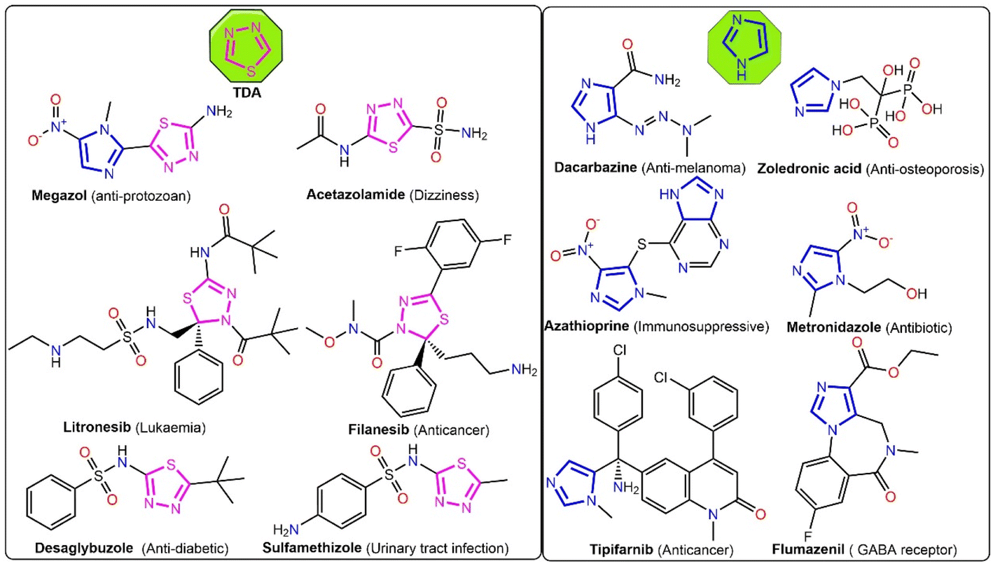
The thiadiazole (TDA) ring is a key pharmacophoric molecule used in the development of several drugs (Fig. 1). Although tiny, this ring features many bioactive groups and active binding sites (H-bonding donor/acceptor, HBD/HBA).14 Furthermore, the TDA ring has been found to exhibit a diverse range of biological activities, which make it a valuable building block in drug design. For instance, the TDA ring has been shown to possess anti-inflammatory,15,16 antiviral,17 antibacterial,18,19 antifungal,18 and anticancer16,19 properties. Its versatile nature, structural tunability, diverse biological activities, stability, and bioavailability make it an attractive scaffold for the design of novel and effective drugs. Interestingly, thiadiazole and its derivatives have shown significant potential as angiogenesis inhibitors. These compounds exhibit anti-angiogenic properties through different mechanisms, including the inhibition of vascular endothelial growth factor (VEGF) signalling and its receptor (VEGFR-2), the suppression of matrix metalloproteinases (MMPs), nuclear factor-kappa B (NF-κB) inhibition, and the disruption of microtubule function.20–22
 |
| | Fig. 1 Examples for thiadiazole- and imidazole-based clinical drugs. | |
On the other hand, imidazole derivatives and imidazolium ionic liquids (ImILs) hold great potential as promising pharmacophores in drug design (see Fig. 1).23,24 Imidazole derivatives have been found to exhibit broad-spectrum pharmacological capabilities, including antitumor,13,25–27 anti-inflammatory,28 anticonvulsant,29 anti-biofouling,12 antihypertensive,30 and antimicrobial31–34 activities. On the other hand, ImILs have received increasing attention in recent years due to their tunable physicochemical properties, which can be easily modified by changing the substituents on the imidazolium ring. This allows for the customization of these compounds for specific drug targets and enhances their potential as pharmacophores in drug design.35 Moreover, ImILs have shown promising potential as antimicrobial, anticancer, and antihypertensive agents.35
Inspired by these remarkable findings, our work focuses on creating hybrid bioactive molecules with diverse pharmacological applications. The objective of this study is to design novel multifunctional hybrid molecules (TISSBs) that combine three potent pharmacophores (thiadiazole, imidazolium ionic liquid, and a Schiff base) for the purpose of cancer therapy. An investigation was conducted to explore the impact of TISBs on Ehrlich solid tumor (EST) artificially produced in mice. This study also assessed the influence of TISBs on VEGV, TNF-α, and TGF-B1.
2 Materials and methods
The ESI† provides details about the solvents and key starting materials used in this work, as well as their suppliers. It also details the protocols used for preparing vanillyl butyl imidazolium ionic liquids (VBIILs) (2a, b) and 2,5-diaminothiadiazole (2,5-H2N-TDA), and the instrumental techniques used to fully characterize the prepared compounds.
2.1. Synthesis of TISSBs
A solution containing 7 mmol VBIILs in 35 mL of ethanol was slowly added under vigorous stirring to a solution of 0.35 g (3.5 mmol) 2,5-H2N-TDA in 15 mL of ethanol, which also contained three drops of glacial CH3COOH. Thin-layer chromatography (TLC) was used to track the progress of the reaction, and the mixture was then agitated at the reflux temperature for a further 5–7 h. After the reaction was complete, the mixture was cooled to room temperature, and the resulting precipitates were gathered by filtration. The collected solids were then rinsed twice with 3 mL of cold ethanol and subsequently thrice with 5 mL of diethyl ether. After drying, the crude products were purified by recrystallization from ethanol to obtain the desired products.
2.1.1 2,5-Bis-(5-(1-butylimidazolium chloride)-3-methoxysalicylideneimino)-1,3,4-thiadiazole (TISSB1). A canary yellow powder with a 84% yield; MP 189–191 °C. FTIR (KBr, cm−1): 3436 (s, br), 3074 (m, sh), 2933 (s, sh), 2862 (m, sh), 1634 (vs, sh), 1558 (m, sh), 1461 (s, sh), 1270 (s, sh), 1162 (s, sh), 1043 (m, sh), 752 (m, sh). 1H NMR (200 MHz, DMSO-d6) δ 10.31 (s, 2H), 9.26 (s, 2H), δ 7.79 (dd, J = 6.8, 2.0 Hz, 2H), 7.62 (dd, J = 8.5, 2.4 Hz), 7.59–7.48 (m, 2H), 7.07 (d, J = 8.5 Hz, 2H), 5.37 (s, 4H), 4.18 (t, J = 7.2 Hz, 4H), 3.39 (s, 3H), 1.79 (p, J = 7.3 Hz, 4H), 1.27 (h, J = 7.2 Hz, 4H), 0.91 (t, J = 7.2 Hz, 6H). 13C NMR (126 MHz, DMSO-d6) δ 163.76, 161.42, 151.62, 149.29, 136.28, 125.79, 123.08, 122.93, 122.81, 119.94, 117.82, 56.66, 52.03, 49.06, 31.61, 31.04, 19.37, 19.16, 13.61. ESI-MS: m/z at 694.20 and 329.40 corresponding to C34H42ClN8O4S+ [M– Cl−]+ and C34H42N8O4S2+ [M–2 Cl−]2+, respectively. Anal. Calcd for C34H42Cl2N8O4S (M = 729.72 g mol−1): C, 55.96; H, 5.80; N, 15.36; S, 4.39%. Found: C, 55.87; H, 5.83; N, 15.31; S, 4.34%.
2.1.2 2,5-Bis-(5-(1-butylimidazolium tetrafluoroborate)-3-methoxysalicylideneimino)-1,3,4-thiadiazole (TISSB2). A canary yellow powder (82%). FTIR (KBr, cm−1): 3438 (s, br), 3112 (m, sh), 2937 (s, sh), 2868 (m, sh), 1634 (vs, sh), 1563 (m, sh), 1468 (s, sh), 1276 (s, sh), 1168 (s, sh), 1056 (m, sh), 753 (m, sh). 1H NMR (500 MHz, DMSO-d6) δ 10.31 (s, 2H), 9.25 (s, 2H), 7.84 (s, 2H), 7.80 (s, 2H), 7.41 (d, J = 2.1 Hz, 2H), 7.34 (d, J = 2.1 Hz, 2H), 5.34 (s, 4H), 4.17 (t, J = 7.2 Hz, 4H), 3.36 (s, 3H), 1.78 (p, J = 7.3 Hz, 4H), 1.26 (h, J = 7.3 Hz, 4H), 0.90 (t, J = 7.3 Hz, 6H). 13C NMR (126 MHz, DMSO-d6) δ 163.68, 161.65, 151.62, 149.29, 136.28, 136.28, 125.81, 123.09, 122.92, 122.80, 119.94, 117.84, 56.66, 52.02, 49.06, 31.61, 19.16, 13.62. 19F NMR (471 MHz, DMSO-d6) δ −148.67 (s). 11B NMR (96 MHz, DMSO-d6) δ −1.30 (s). ESI-MS: m/z at 745.60 and 329.40 corresponding to C34H42BF4N8O4S+ [M– BF4−]+ and C34H42N8O4S2+ [M–2 BF4−]2+, respectively. Anal. Calcd for C34H42B2F8N8O4S (M = 832.43 g mol−1): C, 49.06; H, 5.09; N, 13.46; S, 3.85%. Found: C, 48.98; H, 5.11; N, 13.39; S, 3.80%.
2.2. In vitro MTT cytotoxicity assay in breast cancer cell lines (MCF-7)
The 3-(4,5-dimethylthiazol-2-yl)-2,5-diphenyltetrazolium bromide (MTT) assay was used to evaluate the efficacy of the new compounds (TISSBs) against breast cancer cell lines (MCF-7 and MDA-MB-231, a model of triple-negative breast cancer) and human skin fibroblasts (HSF) by comparing the cell viability of both cell types after each treatment.36 The cytotoxicity markers monitored were the alterations in cell viability and IC50 values as they can provide valuable insights into the therapeutic potential and safety profiles of these compounds.
2.3. The median lethal dosage (LD50)
The median lethal dose (LD50) of each test compound was ascertained using the procedure outlined by Reed.37 Briefly, the procedure involved intraperitoneally injecting single doses of varying dilutions of each test drug to groups of ten mice each. The injected mice were then monitored for behaviour and death for a full day. The percentage of mortality in each group was determined by counting the number of mice that survived and died. The formula for LD50 was as follows: log![[thin space (1/6-em)]](https://www.rsc.org/images/entities/char_2009.gif) LD50 = logLD next below 50% + [log increasing factor × proportional distance], where proportional distance is equal to [50 − dilution next below mortality]/[next above mortality − next below mortality]. The LD50 value of TISSB2 was found to be 450 mg kg−1 body weight.
LD50 = logLD next below 50% + [log increasing factor × proportional distance], where proportional distance is equal to [50 − dilution next below mortality]/[next above mortality − next below mortality]. The LD50 value of TISSB2 was found to be 450 mg kg−1 body weight.
2.4. Experimental animals
Swiss albino mice weighing 120–140 g were kept enclosed in cages of plastic with standard pellet food and unlimited water in standard laboratory settings (25 ± 2 °C, 70–80% humidity, and a 12 hours light/dark cycle). The animals were obtained from the animal house of the National Research Centre at Dokki, Giza, Egypt. The National Hepatology & Tropical Medicine Research Institute (NHTMRI) ethics committee approved all the animal experiments performed in this study (Serial: A10-2023). The experiments were conducted in accordance with applicable laws and regulations based on the ARRIVE guidelines 2.0: updated guidelines for reporting research using animals.38 The ethical treatment and care criteria for animals were also followed. The National Cancer Institute (Cairo, Egypt) provided the initial inoculum for Ehrlich ascites carcinoma (EAC). Through repeated intraperitoneal (IP) injection, 106 cells were transplanted using 0.2 mL of PBS to each animal. The EAC cells were multiplied in the peritoneal cavity of mice and then transferred every seven days to ordinary animals.
2.5. Ehrlich solid tumour model (EST)
Three groups were formed with male fifteen albino mice randomly selected from a total of forty five. While the solid tumour was produced in the two groups, Group I was maintained as the control. From the ascitic fluid of a female Swiss albino mouse with an 8 days-old ascetic tumour, Ehrlich cells were recovered on the induction day; the National Cancer Institute in Cairo, Egypt provided the animal. Normal saline was used to dilute the ascitic fluid (1:10 v/v). The thigh to the right back limb of each mouse was intramuscularly inoculated with the ascitic fluid (0.2 mL), which contained roughly 2.5 × 10⁶ EAC cells, to create solid tumours.39 Mice were given test material TISSB2 intraperitoneally every other day after two weeks of tumour inoculation in accordance with the following protocol: Group I: saline control group of normal mice: for a period of 13 days, fifteen normal mice received intraperitoneal injections (IP) of 0.2 mL physiological saline (0.9 g dL−1) every other day. Group II: Ehrlich group consisting of fifteen tumorized animals. Group III, TISSB2 (1/10 LD50; 450 mg kg−1) was administered to fifteen tumorized mice every other day for thirteen days.
2.5.1 Weight fluctuations. The body weights of the mice in each group were found to be different, and the formula used to compute the percentage changes in body weight was [(final weight − initial weight/initial weight)] × 100.40
2.6. Biochemical parameters
The mice were weighed the day following the final dosage,41 and the retroorbital venous plexus was utilised to draw 3 mL of blood from each mouse using capillary tubes. The serum obtained after centrifuging the blood at 4000 rpm for 10 minutes was kept at −20 °C for the subsequent analysis of liver function parameters. All mice were given normal saline after they were anaesthetized with sodium pentobarbital (80 mg kg−1, i.p.) for transcardial perfusion. Blood samples were collected using the Frankenberg method42 in tubes devoid of anticoagulant for biochemical serum examination. The sera were separated and kept in aliquots at −80 °C for the ELISA of vascular endothelial growth factor (VEGF) and the investigation of biochemical parameters, including AST, ALT, albumin, and total bilirubin. After the mice were sacrificed, the livers were promptly removed and cleaned with a cold saline solution (0.9% NaCl). Then, half of each liver was homogenised in accordance with the protocol outlined by Hussein et al.43 to measure the tumour necrosis factor α (TNF-α) level using the ELISA technique. As directed by the manufacturer, the remaining half of the liver was preserved in a 10% neutral buffered formalin solution for the immunohistochemical investigation of transforming growth factor-β1 (TGF-β1) and histopathological analysis.
2.7. Immunohistochemistry (IHC) analysis
As previously indicated,44 slices from the selected paraffin blocks were cut into 4 micrometer-thick sections for immunohistochemical (IHC) labelling. The prepared slides were incubated with the primary anti-TGF-beta antibody (1:50 μg mL; Leader in Biomolecular Solutions for Life Science, Catalogue No. A2124). The biotinylated goat anti-mouse IgG secondary antibody (1:200) (Vector Laboratories Catalogue No. BA-9200-1.5) and streptavidin peroxidase complex (1:200) (Vector Laboratories Catalogue No. SA-5004-1) were then added and incubated in the following steps. All slides were lightly counterstained for thirty seconds with hematoxylin before being dried and mounted.
2.8. Immunohistochemical scoring
When liver tissue was examined, TFG-β1-expressing cells were identified as positive. The stained tissue slices were subjected to semi-quantitative analysis in accordance with the Allred scoring system.45 The final grades were calculated by adding the individual values for the staining intensity of the reaction (0–3) and the percentage of the positive region (0–5). Three separate high-power fields HPFs (40×) were used to evaluate the proportion of positive areas. The average score was derived using the standard deviation (±SD), and each region was assigned a value of (1) less than 10%, (2) between 10% and 25%, (3) between 26% and 50%, (4) between 51% and 75%, and score (5) greater than 75%. Three scores were assigned to the staining intensity: faint, medium, and strong.
2.9. Histopathological evaluation
The liver tissue was removed as soon as possible, kept for a day in 10% neutral buffered formalin, dried in increasing amounts of alcohol, cleaned with xylene, and then immobilised in paraffin. Hematoxylin-eosin (H&E) stain was then applied to 4 micrometer-thick tissue slices cut out from the paraffin blocks by following the protocol described by Ogunsuyi et al.46 Light microscopy was used to assess and analyse the morphology of the liver tissue (OPTIKA B-150 microscope, Italy). According to International Consensus Group for Hepatocellular Neoplasia,47 the liver tissue slices were inspected for the emergence of dysplastic alterations, fibrosis, cirrhosis, or architectural abnormalities. Furthermore, liver lesions were staged and graded in accordance with Ishak et al.48
2.10. Docking studies
Molecular docking was performed to study the binding patterns of the tested compound within the target pockets of VEGFR-2 and NF-κB. The 3D crystal structures of VEGFR-2 and NF-κB were downloaded from the Protein Data Bank, http://www.rcsb.org/pdb (PDB ID: 4asd and 5t8p). At first, the water molecules bound to the protein structures were removed, hydrogen atoms were added during molecular preparation, and energy minimization was carried out using the CHARMM force field. Subsequently, the 2D structure of TISSB2 was drawn using ChemBioDraw 14.0 and saved in the MDL-SD file format. Energy minimization was performed using the MMFF94 force field. The docking process was carried out using Autodock Vina 1.5.7, and approximately twenty poses were predicted.
2.11. Detection of apoptosis
Breast cancer MCF-7 cells were incubated for 48 hours with TISSB2 at the IC50 concentration dosage (2 × 10⁵ cells per well). After treatment, the cells were collected and washed twice at 4 °C for 10 minutes using 100 μL of PBS. Subsequently, 5 μL of Annexin V-FITC and 100 μL of a binding buffer were added to every well. Following a 10 minutes incubation period at room temperature, 400 μL of PBS was added. Prior to analysis, the cells were immediately stained with PI.
2.12. Statistical analysis
To facilitate comparison between the EAC and treatment groups, all data were presented as mean ± standard error of the mean (SEM). Using SPSS 16.0 software (SPSS Corp, Chicago, IL), one-way analysis of variance was used to determine the statistical significance of the data, and Duncan's multiple range test was used for individual comparisons. Values with p < 0.05 were considered statistically significant; when p < 0.01, they were deemed highly significant; and those with p < 0.001 were deemed extremely highly significant.
3 Results and discussion
3.1. Synthesis protocol
The target hybrid molecules (TISSBs) integrating thiadiazole, imidazolium ionic liquid, and Schiff bases, were prepared through a stepwise protocol, as depicted in Scheme 1. 2,5-H2N-TDA was first prepared via heterocyclization of the as-prepared bis-thiourea in the presence of H2O2. Meanwhile, the vanillyl butyl imidazolium ionic liquids (VBIILs) (2a, b) were prepared using consecutive chemical reactions, including the chloromethylation of O-vanillin to yield 5-chloromethyl vanillin (CMVal), which was then quaternized by the reaction with 1-butylimidazole, affording vanyl butyl-imidazolium chloride (VBIIL1, 2a). It finally underwent an anion metathesis reaction to form the corresponding tetrafluoroborate ionic liquid (VBIIL2, 2b). The intended hybrid Schiff base molecules (TISSBs) were eventually produced via the condensation of the VBIILs (2a, b) with 2,5-H2N-TDA (Scheme 1). These compounds were produced in high to excellent yields. Elemental and spectral investigations (FTIR, 1H-NMR, 13C-NMR, 11B-NMR, 19F-NMR, ESI-MS) were used to determine their structures.
 |
| | Scheme 1 Step-by-step protocol used for the preparation of 2,5-H2N-TDA, VBIILs, and TISSBs (TISSB1, X = Cl; TISSB2, X = BF4). | |
3.2. Structural characterization
The microanalytical results obtained from the elemental analysis (CHNS) of the TISSB2 were completely in agreement with their suggested structural formula, as shown in the area designated for experimentation. In addition, the electrospray ionisation mass spectrometry (ESI-MS) of these hybrid molecules revealed prominent peaks at m/z 694.20 and 745.60 in their positive-mode spectra (see Fig. S1 and S2, ESI†), corresponding to the singly-charged cations [M-Cl−] and [M-BF4−] of TISSB1 and TISSB2, respectively. Additionally, a peak at m/z 329.40 was observed, indicating the presence of a doubly-charged cation [M-2X−] (X = Cl or BF4). These results provide further evidence supporting the proposed structures.
In the FTIR spectra of the new compounds (TISSBs), significant changes were observed in the characteristic functional group peaks compared with the starting materials (2,5-H2N-TDA and VBIILs). Evidently, the amine-related stretches observed in the 2,5-H2N-TDA spectrum at 3489 and 3419 cm−1 (Fig. S3, ESI†) were conspicuously absent in the TISSBs spectra (Fig. S5 and S6, ESI†). Similarly, the characteristic aldehydic carbonyl stretch of VBIILs at around 1679 ± 4 cm−1 (Fig. S4 and S5, ESI†) also vanished in the spectra of TISSBs. The disappearance of these peaks suggests that the amino groups of 2,5-H2N-TDA react with the carbonyl group of VBIILs to form imine groups in the TISSB compounds, contributing to alterations in the spectral features. Our assumptions were further validated by the emergence of new absorption bands at around 1437 ± 1, 1634, 1273 ± 3, and 847 ± 2 cm−1, which could be assigned to the vibration of different functional groups, including phenolic O–H, H–C![[double bond, length as m-dash]](https://www.rsc.org/images/entities/char_e001.gif) N azomethine, aryl–O, and the imidazolium ring, respectively.
N azomethine, aryl–O, and the imidazolium ring, respectively.
Compared with the 1H NMR spectra of VBIILs (Fig. S8 and S10, ESI†), four common distinct peak sets could be seen in the TISSBs spectra, and one of them had newly emerged (see Fig. 2A and S12 (ESI†)). The first characteristic peak consisted of two singlets in the low-field region at chemical shift values of around 10.5 and 9.5 ppm, which could be assigned to the resonances of the phenolic and azomethine protons. Another set consisted of several peaks with various splitting patterns in the chemical shift range of 7.84–7.34 ppm, which are characteristic of the imidazolium and salicylidene protons. Notably, the benzylic protons (Ar-CH2-Im) stood out as a singlet peak in the median-field region at 5.34 ppm. Lastly, the high-field region revealed a peak set ranging from 4.17 to 0.90 ppm, which is likely due to the resonances of the methoxy and butyl protons. Meanwhile, in addition to the native peaks of VBIILs (Fig. S9 and S11, ESI†), new peaks were seen in the 13C NMR spectra of TISSBs (Fig. 2B and S13, ESI†). For example, the 13C NMR spectrum of TISSB2 showed a total of 18 carbon peaks (Fig. 2B) categorized into four distinct sets. The first set was observed as two downfield peaks at 163.68 and 161.65 ppm, which could be attributed to the resonances of the thiadiazole and azomethine carbon atoms, respectively. The second set of downfield peaks were seen in the range of 151.62–117.84 ppm and could be assigned to the imidazolium and salicylidene carbon atoms. Notably, the benzylic carbon atom (Ar-CH2-Im) produced a peak in the median-field region at 56.66 ppm. Lastly, the upfield set of peaks observed in the range of 52.02–13.62 ppm was likely due to the resonances of the methoxy and butyl carbon atoms. The presence of BF4 as a counter anion in TISSB2 was confirmed by the 11B and 19F NMR spectral analysis. The 11B NMR spectrum of TISSB2 showed a sharp singlet peak at −1.30 ppm, while the 19F NMR spectrum showed another sharp singlet peak at −148.67 ppm, both pointing to the existence of BF4.
 |
| | Fig. 2 NMR spectra of TISSB2 in d6-DMSO: (A) 1H NMR (500 MHz); (B) 13C NMR (126 MHz); (C) 11B NMR (96 MHz); (D) 19F NMR (471 MHz). | |
3.3. Cytotoxicity
This in vitro study focused on three specific cell lines: MCF-7 and MDA-MB-231, which are breast cancer cells representing different hormonal profiles and aggressiveness levels, as well as HSF, a normal human fibroblast line used to assess the potential off-target effects of the drugs on healthy cells. A comparative analysis with 5-fluorouracil (5-Fu), a widely used chemotherapeutic agent, provided an important benchmark for evaluating the efficacy of TISSBs. The essential indices of cytotoxicity for these compounds, the IC50 values and cell viability, were also determined. Notably, upon exposure of MCF-7 cells to various concentrations of TISSBs, their survival ratio significantly diminished. The effectiveness of a chemotherapeutic agent is influenced by both its structural composition and concentration. In terms of effectiveness against breast cancer, TISSB2, which is backed by a tetrafluoroborate anion, outperformed all other agents (TISSB1 and 5-Fu). At a dosage of 250 μg mL−1 (see Fig. S14, ESI†), it reduced the number of surviving MCF-7 cells from 100% to approximately 3%, yielding a growth reduction of about 97%. Moreover, the in vitro experiments revealed the significant cytotoxic effects of these new hybrid molecules on MDA-MB-231 cells, as evidenced by their lower IC50 values compared to 5-Fu. Interestingly, the IC50 values of TISSB1, TISSB2, and 5-Fu were 16.45 ± 1.25, 3.68 ± 0.31, and 12.03 ± 1.97 μg mL−1 against MCF-7, and 32.75 ± 1.91, 7.23 ± 1.17, and 29.85 ± 2.25 μg mL−1 against MDA-MB-231, respectively. Our findings suggest that TISSB2 is a potent cytotoxic against the human breast cancer cell lines MCF-7 and MDA-MB-231 and may provide useful anti-breast cancer treatment options. Meanwhile, the increased susceptibility of MCF-7 to TISSB2 suggests that this molecule might interact synergistically or antagonistically with pathways more prominent in receptor-positive cell lines. In order to determine if the TISSBs specifically target tumor cells and not healthy cells, the viability of HSF cells was examined after 24 h of treatment with increasing doses of TISSBs. Unlike their effect on cancer cells, the TISSBs had a smaller dose-dependent impact on the viability of HSF cells, as shown in Fig. S15 (ESI†). Satisfactorily, compared with the existing drug (5-Fu), TISSBs are not harmful to healthy cells. According to a previous work, the selectivity index (SI) was calculated to determine the most effective chemical for killing malignant MCF-7 cells compared with healthy cells. Table S1 (ESI†) shows that, of all the anticancer candidates, the most potent drug TISSB2 had the best selectivity (SI = 32.57) for cancer cells (MCF-7) rather than healthy cells (HSF). As a result, TISSB2 is a potential candidate for non-toxic and, very effective treatment of breast cancer. Thus, this study not only underscores the promising anticancer potential of TISSB2 but also highlights its relevance in selective anti-tumor therapies that minimize harm to healthy tissues. These results prompted us to carry out the following investigations with TISSB2.
3.4. Body weight changes
The liver plays a major role in the metabolism of most xenobiotics, including drugs and foreign substances, and hence is highly susceptible to poisoning. A significant marker of toxicity is a decrease in body weight. Body weight alterations may result from hepatitis caused by breast cancer in both human and mouse models.49 The initial weights of the mice did not differ significantly across the groups under investigation (p = 0.849). Mice in the EAC-group had a significantly lower final body weight than those in the control group (p < 0.001). Although body weight loss was marginally reversed in the TISSB2-treated mice, it was still considerably lower than those in the control group (p < 0.001). Mice in the EAC group showed a substantial decrease in body weight change (%) when compared to the control group (p < 0.001), while body weight change (%) slightly increased in mice administered with TISSB2, although it was still considerably lower than that of the control group (p < 0.001) (Table 1). In our investigation, mice with EAC showed a statistically significant decrease in body weight and a significant decrease in body weight change (%) compared with the control group. Additionally, our research demonstrates that administering TISSB2 to the mice corrected their body weight loss to some extent, with a marginal increase in body weight change (%).
Table 1 Body weights of mice and body weight change (%) among the various study groupsa
| Groups |
Final weight |
Initial weight |
Weight change |
| Data are expressed as mean ± SE. *denotes significant difference between test groups and control group. *** P < 0.001. |
| Control |
152.60 ± 3.58 g |
129.90 ± 1.83 g |
17.55 ± 2.64% |
| EAC |
123.70 ± 2.71*** g |
131.00 ± 1.15 g |
−5.58 ± 1.88***% |
| EAC + TISSB2 |
135.80 ± 5.00*** g |
130.00 ± 1.43 g |
4.58 ± 4.03***% |
3.5. The impact of TISSB2 on the biochemical parameters of mice with ehrlich tumours
Table 2 demonstrates that, in comparison with the untreated Ehrlich control group, the TISSB2 treatment exhibited considerable modifications of biochemical parameters caused by Ehrlich. The present study's results showed that, in contrast to the normal control group, Ehrlich-tumorized mice had higher liver activities of AST and ALT along with a total bilirubin value. Compared to the normal control group, the Ehrlich-tumorized mice had lower albumin levels. Ehrlich has been shown to cause some liver impairment. In this work, Ehrlich mice with tumours showed enhanced activity of liver enzymes like AST and ALT along with elevated levels of total bilirubin and decreased levels of albumin, indicating overall toxicity. These changes were caused by hepatocellular damage. Our findings agree with those of earlier studies.50,51 When the compound TISSB2 was administered to Ehrlich tumorized mice, there was a significant decrease in blood ALT, AST, and total bilirubin (P ≤ 0.001 at all) accompanied by an increased amount of albumin. TISSB2 mice treated with Ehrlich tumours exhibited some hepatic enlargement and showed changes in liver function tests, suggesting improvements in overall liver function. Numerous signalling pathways contribute to liver damage and fibrosis caused by inflammation. According to the majority of publications, tumour angiogenesis, is one of the characteristics of cancer that contributes to fibrosis and inflammation of the liver in chronic liver illnesses and hepatocellular carcinoma (HCC)This disease that is linked to a deadly illness involves many growth factors and cytokines.52,53
Table 2 The impact of TISSB2 on liver biochemical markersa
| Groups |
AST (U/L) |
ALT (U/L) |
Albumin (g dL−1) |
Total bilirubin (mg dL−1) |
| A significant difference between the test groups and the EAC group is denoted by *P ≤ 0.05, **P ≤ 0.01 and ***P ≤ 0.001, and the values are expressed as mean ± SE. |
| EAC |
267.25 ± 3.89 |
80.61 ± 1.34 |
2.14 ± 0.005 |
0.56 ± 0.012 |
| EAC + TISSB2 |
154.05 ± 2.12*** |
59.93 ± 1.02*** |
2.34 ± 0.027*** |
0.503 ± 0.11** |
| Control |
186.23 ± 4.19*** |
56.98 ± 1.68*** |
2.46 ± 0.012*** |
0.44 ± 0.017*** |
However, fast EAC cell division in the peritoneal cavity resulted in a rise in the total number of tumour cells and tumour volume, creating a hypoxic environment in the nearby microenvironment. Consequently, endothelial cells would be activated by the production of many angiogenic growth factors and cytokines, including VEGF, TNF-α, and TGF-β.54 These variables have been demonstrated to play a significant part in angiogenesis linked to HCC.55 In order to treat patients with tumour-associated hepatic inflammation and fibrosis, addressing this tumour angiogenesis will be a promising therapeutic strategy. Hepatic inflammation and fibrosis are triggered by the growth factors and cytokines that EACs emit into the bloodstream, which also include markers linked to inflammation and angiogenesis.56 The primary mechanism of action of these angiogenic and inflammatory agents is through the unique and cognate receptors in the liver. Apart from hepatocytes and hepatic stellate cells (HSCs), damage-related stimuli can also activate mast cells, which in turn promote the development of hepatic inflammation and fibrosis, ultimately culminating in chronic liver disorders and HCC. It has been demonstrated that the EAC causes hepatitis in mice by secreting several cytokines and growth factors.57 Further, EAC tumours induce angiogenesis, and the newly developed capillaries may carry inflammatory and angiogenic markers to the liver. They may also encourage the activation of mast, Kupffer, and stellate cells associated with the liver, which could result in signalling related to inflammation.58 According to another investigation, angiogenesis pathways are crucially affected by the development of hepatic inflammation, fibrosis, and HCC.59
3.6. Estimation of TNF-α (tumour necrosis factor α) in liver tissue homogenates
TNF-α levels in the EAC group were substantially higher (p < 0.001) than those observed in the control group. Conversely, Fig. 3 shows that TNF-α levels dramatically decreased (p < 0.001) in the TISSB2 group in comparison with the EAC group. Notably, NF-κB activation and translocation from the cytoplasm to the nucleus increase TNF-α and TGF-β.60 The NF-kB inhibitor IKB dissociates in response to inflammatory mediators, complement, oxidative stress, and other stimuli, which exposes NF-kB to the nucleus via the cytoplasm and speeds up DNA transcription, increasing TNF-α and TGF-β release.61 Thus, NF-κB suppression is quite advantageous in treating liver diseases. The newly synthesised Schiff base (TISSB2) was subjected to a molecular docking investigation with NF-κB in order to understand the mechanism that drastically reduces TNF-α and TGF-β1 levels and, therefore, liver damage. Our findings indicate that, in comparison with the Ehrlich group, the test drug TISSB2 considerably reduced the levels of TNF-α and TGF-β1 in the serum of TISSB2-treated Ehrlich-tumorized animals.
 |
| | Fig. 3 Effect of TISSB2 on (A) TNF-α and (B) VEGF levels across all studied groups. | |
3.7. Effect of TISSB2 on VEGF
Our study revealed that VEGF expression in the EAC group was significantly higher than that in the control group. VEGF is an established indicator of tumour angiogenesis.62,63 Tumour angiogenesis and its detrimental comorbidities are recognised as the primary causes of liver dysfunction linked to cancer.64 Our findings indicate that, in comparison with the Ehrlich group, the test drug TISSB2 considerably reduced the amount of serum VEGF in Ehrlich-tumorized animals. Thiazole Schiff base derivatives have been shown in several earlier investigations to inhibit VEGFR-1 and 2.65 Fig. 3 demonstrates that tumorized mice treated with TISSB2 had lower serum VEGF concentrations than the Ehrlich group (P ≤ 0.001). Additionally, the EAC group expressed more TNF-α and TGF-β than the control group, which are important modulators of fibrosis and inflammation.66 Our findings align with other studies that found higher levels of TNF-α and TGF-β in tissue samples from breast cancer patients than the samples from healthy counterparts.67 Thus, TGF-β1 is an important factor in the pathophysiology of hepatic damage.68
3.8. Immunohistochemistry analysis
According to Fig. 4, the control group in this study had a total immunohistochemical score of 4, which indicates poor expression of TGF-β1 (Fig. 4A). Compared with the control group, EAC caused a moderate increase in the number of TGF-β1-positive hepatic cells (Fig. 4B), with a total immunohistochemistry score of 6. Compared with the EAC group, Fig. 4C demonstrates that mice treated with TISSB2 had poor TGF-β1 immunohistochemical expression.
 |
| | Fig. 4 Liver segment immunohistochemistry analysis for every group under study: (A) micrograph demonstrating the control group liver's hepatocytes' cytoplasmic expression of TGF-β1 (black arrows). (B) Micrograph demonstrating a moderate expression of TGF-β1 in the cytoplasm of hepatocytes from a piece of the liver of mice given EAC (black arrows). (C) Micrograph of a portion of the liver of rats given EAC + TISSB2 treatment, demonstrating a low level of TGF-β1 expression in the cytoplasm of hepatocytes (black arrows) (IHC, 40×). | |
3.9. Histopathological examination
In mice treated with EST-TISSB2 and the control, the liver exhibited a typical histological structure (see Fig. 5), with normal hepatocytes and a constant hepatic cord arrangement around the main vein. The space between the cords was occupied by the hepatic sinusoids, which contain a delicate arrangement of Kupffer cells. Compared with the EST group, which had a lot of cellular infiltration, the EST-TISSB2 treated group had less congested blood vessels with dark nuclei, dilated congested blood vessels, some vacuolation in the cytoplasm, infiltration, and high line materials.
 |
| | Fig. 5 Liver section histopathological examination: (a) normal liver structure, (b) liver section with EAC showing mild hydropic changes and (c) liver section treated with EAC and test compound TISSB2 showing hydropic degeneration (magnification ×400). | |
3.10. Molecular docking
3.10.1 Molecular docking against NF-κB. The binding mode of the co-crystalized ligand exhibited a binding energy of −7.80 kcal mol−1. It formed two hydrogen bonds with Leu474 and Glu472, as well as twelve hydrophobic-π interactions with Val416, Leu524, Arg410, Met471, Cys535, and Lys431(Fig. 6).
 |
| | Fig. 6 3D positioning and interactions of the co-crystalized ligand with NF-κB. | |
The binding mode of TISSB2 exhibited an affinity score of −9.25 kcal mol−1. TISSB2 formed one hydrogen bond with Lys519 with a distance of 2.83 Å. Moreover, it interacted via ten hydrophobic π-interactions and three ionic bonds with Leu524, Arg410, Val416, Glu415, Asp517, Phe413, His596, Ile558, Asp517 and Asp521(Fig. 7).
 |
| | Fig. 7 3D positioning and surface mapping of TISSB2 against NF-κB. | |
3.10.2 Molecular docking against VEGFR-2. The binding mode of the crystal ligand (sorafenib) exhibited a binding energy of −9.90 kcal mol−1 with the VEGFR tyrosine kinase. It formed fourteen hydrophobic π-interactions with Ala866, Leu840, Leu1025, Phe918, Val916, Lys868, Cys1045, Val848, Val899, Ala866, Leu1019, Ile892, Leu889, Ile888 and Ile1044, as well as additional interactions with Asp1046, Glu885, Cys919 and Val899 via four hydrogen bonds with distances in the range of 1.74 to 2.34 Å (Fig. 8).
 |
| | Fig. 8 3D and surface mapping of sorafenib against VEGFR-2. | |
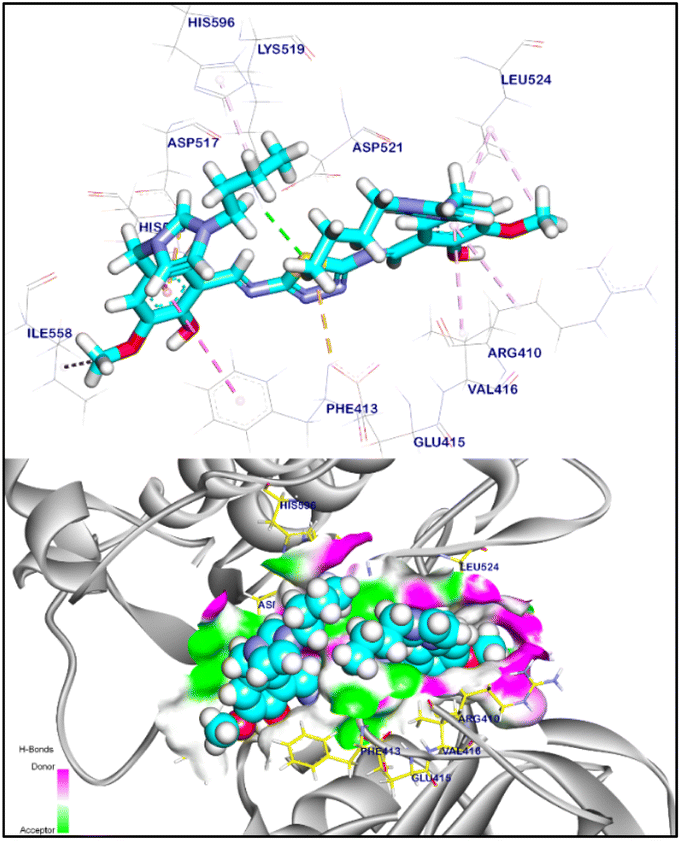
The binding mode of TISSB2 exhibited an affinity score of −11.79 kcal mol−1. Four hydrogen bonds was observed with Cys919, Glu917, Asp1046, and Ile1044 with distances of 2.01, 2.86, 2.66, and 2.97 Å, respectively. Moreover, TISSB2 interacted via seventeen hydrophobic π-interactions and two ionic bonds with Ile892, Leu1019, Val898, Leu889, Cys1045, Lys868, Phe1047, Val916, Val899, Leu1035, Ala866, Val848, Leu840, Phe918, Cys919, Asp1046, and Glu885 (Fig. 9).
 |
| | Fig. 9 3D and surface mapping of TISSB2 against VEGFR-2. | |
According to the molecular docking study withVEGFR-2 and NF-κB, TISSB2 demonstrated a significant ability to bind to the target sites of these proteins, as indicated by the affinity scores −11.79 and −9.25 kcal mol−1, respectively (see Table 3). Compared with the reference compound, TISSB2 exhibited a higher affinity value with both VEGFR-2 and NF-κB, suggesting a better potential for inhibiting these proteins. Therefore, TISSB2 appears to have anti-cancer properties. This reduces the angiogenesis of malignant cells, which in turn reduces the creation of new blood vessels and the supply of nutrition and oxygen to tumour masses. As a result, blocking VEGF is thought to be a successful cancer treatment method. Additionally, TISSB2 appears to have anti-cancer properties through NF-κB inhibition, nucleus localisation, decreased DNA transcription, and increased apoptosis.
Table 3 Docking results of TISSB2 against VEGFR-2 and NF-κB
| Target |
Tested compounds |
RMSD value (Å) |
Docking (affinity) score (kcal mol−1) |
Interactions |
| H-bonding |
π-interaction |
| VEGFR-2 |
Sorafenib |
1.03 |
−9.90 |
4 |
14 |
| TISSB2 |
1.41 |
−11.79 |
4 |
17 |
| NF-κB |
Reference ligand |
1.07 |
−7.80 |
2 |
12 |
| TISSB2 |
1.78 |
−9.25 |
1 |
10 |
3.11. Assay for detecting apoptosis
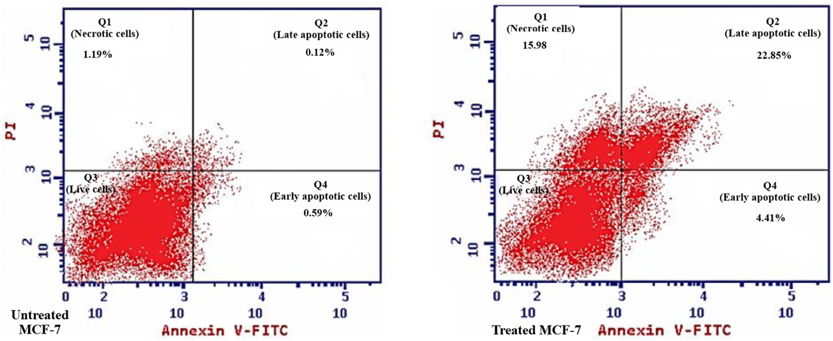
After the MCF-7 cells were stained with Annexin V-FITC/PI, flow cytometry was performed to examine the impact of 48 hours of incubation with TISSB2 (IC50 dosage) on the distribution of cells in four distinct quadrants (Q1, Q2, Q3, and Q4) (Fig. 10). Untreated MCF-7 cells had the largest cell dispersion in Q3 (Ann V/PI; viable cells), whereas Q1, Q2, and Q4 had the lowest cell distribution (indicating a very low number of necrotic, early and late apoptotic cells). However, in Q1, Q2, and Q4, TISSB2-treated MCF-7 cells showed the greatest dispersion (indicating a very high number of necrotic, early and late apoptotic cells), whereas Q3 had the lowest cell distribution.
 |
| | Fig. 10 Apoptosis in untreated and treated MCF-7 cells. | |
4 Conclusion
It has been demonstrated that EAC treatment causes liver toxification, however the new hepatoprotective agent bis(butylimidazolium tetrafluoroborate) thiadiazole Schiff base (TISSB2) has been shown to shield mice liver from the inflammatory and fibrotic damage caused by EAC. According to the experimental results, TISSB2 restores functional indices and pathological liver injury by downregulating the high TGF-β1, TNF-α, and VEGF levels to nearly normal levels. The in vitro anti-breast cancer study revealed that TISSB2 (IC50, 3.68 ± 0.31 μg mL−1) exhibited good ability to inhibit the proliferation of MCF-7 cells by inhibiting EAC-induced tumour angiogenesis, which is a critical characteristic of several malignancies, including HCC. This finding agreed with the molecular docking results. In the molecular docking study, TISSB2 exhibited strong interactions with VEGFR-2 and NF-κB proteins with binding affinity scores of −11.79 and −9.25 kcal mol−1, respectively. These strong interactions involved H-bonding, ionic bonds, and hydrophobic π-interactions.
Data availability
All data related to the new compounds reported in this manuscript are available in the ESI.†
Author contributions
N. F. A.: data curation, software, validation, visualization, writing – original draft. M. Y. A: software, validation, visualization, writing – original draft, writing – review & editing. A. A. S.: software, validation, visualization, writing – original draft, writing – review & editing. S. I. E.: data curation, methodology, software, validation, writing – original draft. A. M. S.: data curation, software, validation, visualization, writing – original draft. E. S. K.: data curation, methodology, validation, visualization, writing – original draft, writing – review & editing. W. M. S.: data curation, methodology, validation, visualization, writing – original draft, writing – review & editing. R. F. M. E.: conceptualization, data curation, methodology, validation, visualization, writing – original draft, writing – review & editing. H. W. A.: data curation, software, validation, visualization, writing – original draft. Y. A. H.: data curation, methodology, software, validation, visualization.
Conflicts of interest
The authors declare no conflict of interest.
Acknowledgements
The authors thank the Deanship of Scientific Research at King Khalid University for funding this work through large Groups (Project under grant numbers R.G.P. 2/ 217 /44).
References
- K. G. Wakim, Physiology of the liver, Am. J. Med., 1954, 16(2), 256–271 CrossRef CAS.
- F. C. Schmitt, A. Longatto Filho and J. M. Lopes, Angiogenesis and breast cancer, J. Oncol., 2010, 2010, 576384 Search PubMed.
- A. H. A. Moustafa, E. M. M. Ali, S. S. Moselhey, E. Tousson and K. S. El-Said, Effect of coriander on thioacetamide-induced hepatotoxicity in rats, Toxicol. Ind. Health, 2014, 30(7), 621–629 CrossRef CAS.
- N. Nishida, H. Yano, T. Nishida, T. Kamura and M. Kojiro, Angiogenesis in cancer, Vasc. Health Risk Manage., 2006, 2(3), 213–219 CrossRef CAS.
- S. Ziyad and M. L. Iruela-Arispe, Molecular mechanisms of tumor angiogenesis, Genes Cancer, 2011, 2(12), 1085–1096 CrossRef PubMed.
- J. Novotný and M. Zikán, Tumor angiogenesis, Klinická Farmakologie a Farmacie, 2010, 24(3), 124–126 Search PubMed.
- Z. Momenimovahed and H. Salehiniya, Epidemiological characteristics of and risk factors for breast cancer in the world, Breast Cancer: Targets Ther., 2019, 151–164 Search PubMed.
- M. L. Salem, N. M. Shoukry, W. K. Teleb, M. M. Abdel-Daim and M. A. Abdel-Rahman, In vitro and in vivo antitumor effects of the Egyptian scorpion Androctonus amoreuxi venom in an Ehrlich ascites tumor model, Springerplus, 2016, 5, 1–12 CrossRef PubMed.
- S. Mishra, A. K. Tamta, M. Sarikhani, P. A. Desingu, S. M. Kizkekra, A. S. Pandit, S. Kumar, D. Khan, S. C. Raghavan and N. R. Sundaresan, Subcutaneous Ehrlich Ascites Carcinoma mice model for studying cancer-induced cardiomyopathy, Sci. Rep., 2018, 8(1), 5599 CrossRef.
- M. Hegde, S. S. Karki, E. Thomas, S. Kumar, K. Panjamurthy, S. R. Ranganatha, K. S. Rangappa, B. Choudhary and S. C. Raghavan, Novel levamisole derivative induces extrinsic pathway of apoptosis in cancer cells and inhibits tumor progression in mice, PLoS One, 2012, e43632 CrossRef CAS.
- P. de Sena Murteira Pinheiro, L. S. Franco, T. L. Montagnoli and C. A. M. Fraga, Molecular hybridization: a powerful tool for multitarget drug discovery, Expert Opin. Drug Discovery, 2024, 19(4), 451–470 CrossRef CAS.
- R. F. M. Elshaarawy and C. Janiak, Antibacterial susceptibility of new copper (II) N-pyruvoyl anthranilate complexes against marine bacterial strains–In search of new antibiofouling candidate, Arabian J. Chem., 2016, 9(6), 825–834 CrossRef CAS.
- R. F. M. Elshaarawy, T. B. Mostafa, A. A. Refaee and E. A. El-Sawi, Ionic Sal-SG Schiff bases as new synergetic chemotherapeutic candidates: synthesis, metalation with Pd (II) and in vitro pharmacological evaluation, RSC Adv., 2015, 5(84), 68260–68269 RSC.
- M. Bala, P. Piplani, A. Ankalgi, A. Jain and L. Chandel, 1, 3, 4-Thiadiazole: A Versatile Pharmacophore of Medicinal Significance, Med. Chem., 2023, 19(8), 730–756 CrossRef CAS PubMed.
- S. S. Kadam, R. P. Kabra and A. L. Ganure, Thiadiazoles As Anti-Inflammatory Agents: A Review, Int. J. Curr. Pharm. Sci., 2015, 1(3), 207–215 Search PubMed.
- S. Ahmad, M. Z. Alam, U. Salma, M. Mohasin, P. F. Rahaman, H. Parveen and S. A. Khan, A Review on Recent Progress in Synthesis and Biological Activities of Thiadiazole and its Derivatives, J. Mol. Struct., 2024, 138438 CrossRef CAS.
- G. Serban, Synthetic compounds with 2-amino-1, 3, 4-thiadiazole moiety against viral infections, Molecules, 2020, 25(4), 942 CrossRef CAS PubMed.
- K. M. Dawood and T. A. Farghaly, Thiadiazole inhibitors: a patent review, Expert Opin. Ther. Pat., 2017, 27(4), 477–505 CrossRef CAS PubMed.
- R. F. M. Elshaarawy, H. R. Z. Tadros, R. M. Abd El-Aal, F. H. A. Mustafa, Y. A. Soliman and M. A. Hamed, Hybrid molecules comprising 1, 2, 4-triazole or diaminothiadiazole Schiff-bases and ionic liquid moieties as potent antibacterial and marine antibiofouling nominees, J. Environ. Chem. Eng., 2016, 4(3), 2754–2764 CrossRef CAS.
- B. Altuğ-Tasa, B. Kaya-Çavuşoğlu, A. T. Koparal, G. Turan, A. S. Koparal and Z. A. Kaplancıklı, Design, Synthesis and Biological Evaluation of a Novel Series of Thiadiazole-Based Anticancer Agents as Potent Angiogenesis Inhibitors, Anti-Cancer Agents Med. Chem., 2021, 21(15), 2041–2049 CrossRef.
- M. H. Hekal, P. S. Farag, M. M. Hemdan and W. M. El-Sayed, New N-(1, 3, 4-thiadiazol-2-yl) furan-2-carboxamide derivatives as potential inhibitors of the VEGFR-2, Bioorg. Chem., 2021, 115, 105176 CrossRef CAS PubMed.
- S. R. Atta-Allah, A. M. AboulMagd and P. S. Farag, Design, microwave assisted synthesis, and molecular modeling study of some new 1, 3, 4-thiadiazole derivatives as potent anticancer agents and potential VEGFR-2 inhibitors, Bioorg. Chem., 2021, 112, 104923 CrossRef CAS PubMed.
- S. Agarwal, Imidazole-Based Drug Discovery, Elsevier, 2021 Search PubMed.
- E. Vessally, S. Soleimani-Amiri, A. Hosseinian, L. Edjlali and A. Bekhradnia, New protocols to access imidazoles and their ring fused analogues: synthesis from N-propargylamines, RSC Adv., 2017, 7(12), 7079–7091 RSC.
- I. Ali, M. N. Lone and H. Y. Aboul-Enein, Imidazoles as potential anticancer agents, MedChemComm, 2017, 8(9), 1742–1773 RSC.
- O. A. A. Ali, W. Abd El-Fattah, M. Y. Alfaifi, A. A. Shati, S. E. I. Elbehairi, A. H. A. Almaaty, R. F. M. Elshaarawy and E. Fayad, New Mn (III)/Fe (III) complexes with thiohydantoin-supported imidazolium ionic liquids for breast cancer therapy, Inorg. Chim. Acta, 2023, 551, 121460 CrossRef.
- H. K. Ibrahim, S. H. El-Tamany, R. F. El-Shaarawy and I. M. El-Deen, Synthesis and investigation of mass spectra of some novel benzimidazole derivatives, Maced. J. Chem. Chem. Eng., 2008, 27(1), 65–79 CrossRef CAS.
- R. Gujjarappa, A. K. Kabi, S. Sravani, A. Garg, N. Vodnala, U. Tyagi, D. Kaldhi, R. Velayutham, V. Singh and S. Gupta, Overview on biological activities of imidazole derivatives, Nanostructured Biomaterials: Basic Structures and Applications, Springer, 2022, pp. 135–227 Search PubMed.
- D. Tiglani, M. Salahuddin, A. Mazumder, R. Kumar, M. S. Yar, M. J. Ahsan and K. Shabana, Synthesis Anticonvulsant and Cytotoxic Evaluation of Benzimidazole-Quinoline Hybrids Schiff Base Analogs, Polycyclic Aromat. Compd., 2024, 44(2), 960–980 CrossRef CAS.
- V. Malhotra, S. R. Pathak, R. Nath, D. Mukherjee and K. Shanker, Substituted imidazole derivatives as novel cardiovascular agents, Bioorg. Med. Chem. Lett., 2011, 21(3), 936–939 CrossRef CAS.
- G. Ş. Andrei, B. F. Andrei and P. R. Roxana, Imidazole derivatives and their antibacterial activity-a mini-review, Mini-Rev. Med. Chem., 2021, 21(11), 1380–1392 CrossRef CAS PubMed.
- N. S. Alahmadi and R. F. M. Elshaarawy, Novel aminothiazolyl-functionalized phosphonium ionic liquid as a scavenger for toxic metal ions from aqueous media; mining to useful antibiotic candidates, J. Mol. Liq., 2019, 281, 451–460 CrossRef CAS.
- R. F. M. Elshaarawy and C. Janiak, Ionic liquid-supported chiral saldach with tunable hydrogen bonding: synthesis, metalation with Fe (III) and in vitro antimicrobial susceptibility, Tetrahedron, 2014, 70(43), 8023–8032 CrossRef CAS.
- R. F. M. Elshaarawy, Z. H. Kheiralla, A. A. Rushdy and C. Janiak, New water soluble bis-imidazolium salts with a saldach scaffold: Synthesis, characterization and in vitro cytotoxicity/bactericidal studies, Inorg. Chim. Acta, 2014, 421, 110–122 CrossRef CAS.
- K. S. Egorova, E. G. Gordeev and V. P. Ananikov, Biological activity of ionic liquids and their application in pharmaceutics and medicine, Chem. Rev., 2017, 117(10), 7132–7189 CrossRef CAS.
- B. H. Asghar, R. K. A. Hassan, L. A. A. Barakat, A. Alharbi, M. El Behery, R. F. M. Elshaarawy and Y. A. Hassan, Cross-linked quaternized chitosan nanoparticles for effective delivery and controllable release of O. europaea phenolic extract targeting cancer therapy, J. Drug Delivery Sci. Technol., 2023, 83, 104388 CrossRef CAS.
- R. Lj, A simple method of estimating fifty per cent endpoints, Am. J. Hyg., 1938, 27, 493–495 Search PubMed.
- N. Percie du Sert, V. Hurst, A. Ahluwalia, S. Alam, M. T. Avey, M. Baker, W. J. Browne, A. Clark, I. C. Cuthill and U. Dirnagl, The ARRIVE guidelines 2.0: Updated guidelines for reporting animal research, J. Cereb. Blood Flow Metab., 2020, 40(9), 1769–1777 CrossRef PubMed.
- E. Noaman, N. K. B. El-Din, M. A. Bibars, A. A. Abou Mossallam and M. Ghoneum, Antioxidant potential by arabinoxylan rice bran, MGN-3/biobran, represents a mechanism for its oncostatic effect against murine solid Ehrlich carcinoma, Cancer Lett., 2008, 268(2), 348–359 CrossRef CAS.
- T. Alibakhshi, M. J. Khodayar, L. Khorsandi, M. Rashno and L. Zeidooni, Protective effects of zingerone on oxidative stress and inflammation in cisplatin-induced rat nephrotoxicity, Biomed. Pharmacother., 2018, 105, 225–232 CrossRef CAS PubMed.
- R. E. Mostafa, D. O. Saleh and D. F. Mansour, Cisplatin-Induced nephrotoxicity in rats: modulatory role of simvastatin and rosuvastatin against apoptosis and inflammation, J. Appl. Pharm. Sci., 2018, 8(4), 043–050 CAS.
- E. Frankenberg, Influence of light and temperature on daily activity patterns of three Israeli forms of Ptyodactylus (Reptilia: Gekkoninae), J. Zool., 1979, 189(1), 21–30 CrossRef.
- J. Hussein, D. A. El-matty, Z. El-Khayat and Y. Abdel-Latif, Therapeutic role of coenzyme Q10 in brain injury during experimental diabetes, J. Appl. Pharm. Sci., 2013, 3(6), 213–217 Search PubMed.
- S.-W. Kim, J. Roh and C.-S. Park, Immunohistochemistry for pathologists: protocols, pitfalls, and tips, J. Pathol. Transl. Med., 2016, 50(6), 411 Search PubMed.
- I. R. Ilić, N. M. Stojanović, N. S. Radulović, V. V. Živković, P. J. Randjelović, A. S. Petrović, M. Božić and R. S. Ilić, The quantitative ER immunohistochemical analysis in breast cancer: Detecting the 3+ 0, 4+ 0, and 5+ 0 allred score cases, Medicina, 2019, 55(8), 461 CrossRef.
- O. Ogunsuyi, O. Ogunsuyi, O. Akanni, O. Alabi, C. Alimba, O. Adaramoye, S. Cambier, S. Eswara, A. C. Gutleb and A. Bakare, Physiological and histopathological alterations in male Swiss mice after exposure to titanium dioxide (anatase) and zinc oxide nanoparticles and their binary mixture, Drug Chem. Toxicol., 2022, 45(3), 1188–1213 CrossRef CAS PubMed.
- International Consensus Group for Hepatocellular, Pathologic diagnosis of early hepatocellular carcinoma: a report of the international consensus group for hepatocellular neoplasia, Hepatology, 2009, 49(2), 658–664 CrossRef PubMed.
- K. Ishak, A. Baptista, L. Bianchi, F. Callea, J. De Groote, F. Gudat, H. Denk, V. Desmet, G. Korb and R. N. M. MacSween, Histological grading and staging of chronic hepatitis, J. Hepatol., 1995, 22(6), 696–699 CrossRef CAS.
- F. Tayel, M. E. Mahfouz, A. F. Salama and M. A. Mansour, Ethoxyquin inhibits the progression of Murine Ehrlich Ascites Carcinoma through the inhibition of autophagy and LDH, Biomedicines, 2021, 9(11), 1526 CrossRef CAS.
- G. Bazmandegan, Z. Kamiab, A. Shafiei, M. Khademalhosseini and A. Kaeidi, Calcium Dobesilate Ameliorates Cisplatin-induced Hepatotoxicity by Inhibiting Liver Oxidative Stress in Mice, Iran. J. Pharm. Res., 2023, 22(1), e126613 CAS.
- D. Doğan, I. Meydan and A. U. Kömüroğlu, Protective effect of silymarin and gallic acid against cisplatin-induced nephrotoxicity and hepatotoxicity, Int. J. Clin. Pract., 2022, 2022, 6541026 Search PubMed.
- M. Kukla, Angiogenesis: a Phenomenon Which Aggravates Chronic Liver Disease Progression, Springer, 2013, pp. 4–12 Search PubMed.
- A. Saha, S. Mohapatra, P. Kurkute, B. Jana, P. Mondal, D. Bhunia, S. Ghosh and S. Ghosh, Chem. Commun., 2015, 51, 2249–2252 RSC.
- X.-D. Zhu, Z.-Y. Tang and H.-C. Sun, Targeting angiogenesis for liver cancer: past, present, and future, Genes Dis., 2020, 7(3), 328–335 CrossRef CAS PubMed.
- M. Mossenta, D. Busato, L. Baboci, F. Di Cintio, G. Toffoli and M. Dal Bo, New insight into therapies targeting angiogenesis in hepatocellular carcinoma, Cancers, 2019, 11(8), 1086 CrossRef CAS PubMed.
- R. Ma, Y. Feng, S. Lin, J. Chen, H. Lin, X. Liang, H. Zheng and X. Cai, Mechanisms involved in breast cancer liver metastasis, J. Transl. Med., 2015, 13, 1–10 CrossRef CAS PubMed.
- R. M. Hazem, A. A. Mohamed, N. Ghareb, E. T. Mehanna, N. M. Mesbah, D. M. Abo-Elmatty and M. S. Elgawish, Anti-cancer activity of two novel heterocyclic compounds through modulation of VEGFR and miR-122 in mice bearing Ehrlich ascites carcinoma, Eur. J. Pharmacol., 2021, 892, 173747 CrossRef CAS PubMed.
- T. Tsuchida and S. L. Friedman, Mechanisms of hepatic stellate cell activation, Nat. Rev. Gastroenterol. Hepatol., 2017, 14(7), 397–411 CrossRef CAS.
- P. K. Santhekadur, M. Akiel, L. Emdad, R. Gredler, J. Srivastava, D. Rajasekaran, C. L. Robertson, N. D. Mukhopadhyay, P. B. Fisher and D. Sarkar, Staphylococcal nuclease domain containing-1 (SND1) promotes migration and invasion via angiotensin II type 1 receptor (AT1R) and TGFβ signaling, FEBS Open Bio, 2014, 4, 353–361 CrossRef CAS PubMed.
- H. Un, R. A. Ugan, M. A. Gurbuz, Y. Bayir, A. Kahramanlar, G. Kaya, E. Cadirci and Z. Halici, Phloretin and phloridzin guard against cisplatin-induced nephrotoxicity in mice through inhibiting oxidative stress and inflammation, Life Sci., 2021, 266, 118869 CrossRef CAS.
- J. Dambacher, T. Staudinger, J. Seiderer, Z. Sisic, F. Schnitzler, S. Pfennig, K. Hofbauer, A. Konrad, C. Tillack and J.-M. Otte, Macrophage migration inhibitory factor (MIF)− 173G/C promoter polymorphism influences upper gastrointestinal tract involvement and disease activity in patients with Crohn's disease, Inflammatory Bowel Dis., 2007, 13(1), 71–82 CrossRef.
- M. Zajkowska, E. K. Głażewska, G. E. Będkowska, P. Chorąży, M. Szmitkowski and S. Ławicki, Diagnostic power of vascular endothelial growth factor and macrophage colony-stimulating factor in breast cancer patients based on ROC analysis, Mediators Inflammation, 2016, 2016, 5962946 Search PubMed.
- A. Saha, S. Mohapatra, P. Kurkute, B. Jana, J. Sarkar, P. Mondal and S. Ghosh, RSC Adv., 2015, 5, 92596–92601 RSC.
- J. Wang, Q. Zhang, D. Wang, S. Yang, S. Zhou, H. Xu, H. Zhang, S. Zhong and J. Feng, Microenvironment-induced TIMP2 loss by cancer-secreted exosomal miR-4443 promotes liver metastasis of breast cancer, J. Cell. Physiol., 2020, 235(7–8), 5722–5735 CrossRef CAS.
- S. M. Abou-Seri, W. M. Eldehna, M. M. Ali and D. A. Abou El Ella, 1-Piperazinylphthalazines as potential VEGFR-2 inhibitors and anticancer agents: synthesis and in vitro biological evaluation, Eur. J. Med. Chem., 2016, 107, 165–179 CrossRef CAS.
- D. Bhunia, A. Saha, A. Adak, G. Das and S. Ghosh, RSC Adv., 2016, 6, 113487–113491 RSC.
- N. G. S. Gowda, V. D. Shiragannavar, S. C. Prabhuswamimath, S. Tuladhar, S. B. Chidambaram and P. K. Santhekadur, Ehrlich Ascites carcinoma mice model for studying liver inflammation and fibrosis, Adv. Cancer Biol.:Metastasis, 2022, 4, 100029 Search PubMed.
- L. Niu, X. Cui, Y. Qi, D. Xie, Q. Wu, X. Chen, J. Ge and Z. Liu, Involvement of TGF-β1/Smad3 signaling in carbon tetrachloride-induced acute liver injury in mice, PLoS One, 2016, 11(5), e0156090 CrossRef.
Footnote |
| † Electronic supplementary information (ESI) available: Fig. S1: ESI-MS spectrum of TISSB1; Fig. S2: ESI-MS spectrum of TISSB2; Fig. S3: FTIR spectrum of 2,5-H2N-TDA; Fig. S4: FTIR spectrum of VBIIL1; Fig. S5: FTIR spectrum of VBIIL2; Fig. S6: FTIR spectrum of TISSB1; Fig. S7: FTIR spectrum of TISSB2; Fig. S8: 1H NMR spectrum (500 MHz) of VBIIL1; Fig. S9: 13C NMR spectrum (126 MHz) of VBIIL1; Fig. S10: 1H NMR spectrum (500 MHz) of VBIIL2; Fig. S11: 13C NMR spectrum (126 MHz) of VBIIL2; Fig. S12: 1H NMR spectrum (500 MHz) of TISSB1; Fig. S13: 13C NMR spectrum (126 MHz) of TISSB1; Fig. S14: Impacts of serial doses of TISSBs on the viability of MCF-7 cell line, in comparison to a clinical drug 5-Fluorouracil (5-Fu); Fig. S15: Impacts of serial doses of TISSBs on the viability of HSF cell line. Table S1: Values of IC50 (μg mL−1) of newly developed anticancer agents against MCF-7 and HSF cell lines, as compared to a clinical anticancer drug (5-FU). See DOI: https://doi.org/10.1039/d4ra06398h |
|
| This journal is © The Royal Society of Chemistry 2024 |
Click here to see how this site uses Cookies. View our privacy policy here. 
 Open Access Article
 This Open Access Article is licensed under a Creative Commons Attribution-Non Commercial 3.0 Unported Licence
 *g,
Reda F. M. Elshaarawy
*g,
Reda F. M. Elshaarawy