DOI:
10.1039/D4QI00275J
(Research Article)
Inorg. Chem. Front., 2024,
11, 2039-2048
Chiral lanthanide hexaazamacrocycles for circularly polarized luminescence, high relaxivity and magnetic resonance imaging†
Received
29th January 2024
, Accepted 23rd February 2024
First published on 24th February 2024
Abstract
Multifunctional lanthanide complexes have been extensively studied in recent years owing to their widespread applications in physics, chemistry and biology, including quantum information processing, molecular spintronics and theranostics. The multifunctionality includes chirality, luminescence, magnetism, ferroelectricity, fluorescence/magnetic resonance imaging etc. Although various N- and O-donor ligands have shown the ability to synthesize these complexes, Schiff bases are still the most widely used in constructing air-stable species. Herein, we report the facile gram-level synthesis of three pairs of lanthanide macrocycle enantiomers via an in situ [2 + 2] imine condensation with a trivalent lanthanide ion as a template. Eu(III)-based compounds, R/S-Eu and R/S-Eu-Ph3PO, both show efficient circularly polarized luminescence (CPL) with maximum dissymmetry factors (glum) of 0.098 and 0.110, respectively. The latter exhibited stronger emission intensity and a longer luminescence lifetime than the former due to the lower vibrational coordination structures around the Eu(III) ion. Gd(III)-based species, R/S-Gd, possess a relatively high relaxivity of up to 35.04 and 34.09 mM−1 s−1 for R- and S-enantiomers, benefitting from the presence of one coordinated water molecule and abundant intermolecular H-bonds. In addition, the results of the MTT assay and in vitro experiments demonstrated the low toxicity and efficient MRI of Gd(III)-chelates in A549 cells.
Introduction
Lanthanide macrocyclic compounds have exhibited versatile applications across various fields, including display devices, information storage, magnetic resonance imaging (MRI) and molecular theranostics by virtue of their excellent luminescence properties, enormous magnetic anisotropy, high thermostability/air-stability and facile modification.1–5 They can be mainly divided into the following families according to the type of organic ligand (Fig. 1): DOTA derivates,6 porphyrin,7,8 phthalocyanine,9 crown ether,10 Schiff-base,11 metallacrown12 and heteroatom-assisted macrocycles.13 Among them, Schiff-base lanthanide macrocycles are probably the easiest to obtain via a metal-template reaction between a primary amine and a carbonyl. In particular, the commercially available chiral primary amines, e.g. (1R,2R)-1,2-diphenylethylenediamine, (1R,2R)-1,2-diaminocyclohexane and (R)-1-phenylethan-1-amine (Fig. 1), facilitate the construction of multifunctional lanthanide compounds.14,15 Comparatively, chiral DOTA,16 porphyrin17 and phthalocyanine18 usually involve more complicated synthesis and purification. In addition, Schiff-base macrocycles offer a simpler method to construct multiple-decker lanthanide sandwich complexes for magnetic interaction studies compared to phthalocyanine derivates.19,20
 |
| | Fig. 1 Representative macrocyclic ligands for lanthanide ions and commercially available chiral primary amines. | |
For circularly polarized luminescence (CPL), chiral lanthanide compounds with suitable sensitizing ligands usually exhibit a much higher dissymmetry factor (glum) than organic molecules by virtue of most magnetically allowed f–f transitions,21 which expands the application of lanthanide probes with CPL detection to biological assays.22 Recently, a more comprehensive evaluating tool, i.e. CPL brightness (BCPL), involving two additional parameters, absorption extinction coefficient, ε and quantum yield, φ, besides glum, was proposed to quantify the performance of CPL emitters.23 Among all chiral lanthanide compounds, Eu(III)-based compounds are the most studied giving the largest glum value to date, up to 1.38.24 One of the most efficient strategies to enhance CPL activity is increasing the rigidity of the complex,25e.g. the recently reported heterobimetallic binolate Er(III) complex26 and spinolate Tb(III) complex.27 In addition, substituents, auxiliary ligands and external stimuli can also influence CPL properties, which has been clearly demonstrated by a series of enantiopure Eu(III) macrocycle complexes.28 Both the chiral substituents on the macrocycle scaffolds and external static magnetic fields enabled an evident increase in glum. It is worth noting that apart from improving CPL performance, some macrocyclic ligands, e.g. DOTA derivates, also enable the exceptional water solubility of complexes, which is advantageous for their biological applications.
In terms of the size of the macrocycle cavity, penta- and hexa-azamacrocycles exhibited the ability to encapsulate trivalent lanthanide ions in the equatorial plane, giving rise to complexes with high symmetry.11,29 Recently, such lanthanide macrocycles have received increasing attention in the field of single-molecule magnets (SMMs), making some impressive achievements,30e.g. the current record-holder of the anisotropy barrier for air-stable SMMs, RRRR/SSSS-Dy-D6hF12.31 In 2022, Shanmugam et al. reported a discrete bifunctional macrocyclic Schiff-base Ce(III) complex that combines ferroelectricity with field-induced SMM behavior.32 It is noteworthy that such encapsulations can also prevent the proximity of high-energy oscillators to lanthanide ions to some extent, therefore enhancing the luminescence properties.33 In addition, since the Gd-DOTA compound was clinically approved as a contrast agent for human use in 1989, macrocyclic Gd(III) chelates have been greatly studied in the MRI contrast agent field due to their improved safety (more kinetically inert than acyclic species) and satisfactory relaxivity.2 From a molecular structural point of view, excellent luminescence properties and high relaxivity seem to be contradictory as the latter usually needs inner-sphere coordinated water (vide infra) while the former does not, with the presence of an O–H oscillator accelerating the non-radiative quenching pathways. Herein, we report the one-pot synthesis of lanthanide hexaazamacrocycle enantiomers, [Ln(LN6)Cl2CH3OH][Ln(LN6)Cl2H2O]Cl2 (Ln = Eu and Gd, LN6 is the chiral Schiff-base macrocycle derived from the imine condensation between 2,6-diformylpyridine and (1R,2R)/(1S,2S)-1,2-diphenylethylenediamine; R/S-Eu and R/S-Gd). Eu(III) complexes show strong chiroptical properties (|glum| = 0.1, BCPL = 71.45 M−1 cm−1), while Gd(III) analogues possess relatively high relaxivity up to 35.04 and 34.09 mM−1 s−1 for the R- and S-enantiomer, respectively, and exhibit efficient MRI at low concentrations. In addition, another pair of Eu(III) enantiomers, [Eu(LN6)(Ph3PO)2(ClO4)][ClO4]2 (R/S-Eu-Ph3PO), with the same equatorial macrocycle as R/S-Eu, was also prepared, showing enhanced luminescence and chiroptical properties by virtue of lower-vibrational coordination structures around Eu(III) ions. To the best of our knowledge, this is the first study of chiral hexaazamacrocycles that support both strong circularly polarized luminescence (CPL) and high relaxivity as well as in vitro MRI.
Experimental
Chemicals and synthesis
EuCl3·6H2O, GdCl3·6H2O, (1R,2R)-1,2-diphenylethylenediamine and (1S,2S)-1,2-diphenylethylenediamine were purchased from Sigma-Aldrich. 2,6-Diformylpyridine was synthesized as reported previously.34 All solvents were commercially available and used as received without any further purification. All experiments were performed under aerobic conditions. R/S-Eu and R/S-Gd were synthesized by the following procedure: LnCl3·6H2O (4 mmol) was dissolved in 80 ml of CH3OH at room temperature and then homochiral diphenylethylenediamine (8 mmol) and 2,6-diformylpyridine (8 mmol) were added. The mixture was heated under reflux for 8 h. After cooling to room temperature, the solvent was removed on a rotary evaporator, affording the target compound as a white solid in quantitative yield. The single crystals suitable for X-ray measurement can be obtained by the slow diffusion of Et2O into CH3OH solution at room temperature for several days. R/S-Eu-Ph3PO was prepared by two steps: firstly, the precursor was synthesized using a similar method to R/S-Eu except that Eu(ClO4)3·6H2O was used; then, 1 mmol (ca. 1.07 g) precursor and 2 mmol Ph3PO (0.56 g) were dissolved in 25 ml of CH3OH and then heated at 65 °C for 10 h. After that, the system was allowed to cool to room temperature within 24 h, giving light yellow crystals at the bottom of the vial (yield: 51% based Eu).
Instruments and measurements
Single-crystal X-ray data for R/S-Ln and R/S-Eu-Ph3PO were collected at 180 K on a Bruker SMART APEX diffractometer (Bruker AXS, Ettlingen, Germany) equipped with graphite-monochromatized Mo Kα radiation (λ = 0.71073 Å). The structures were solved and refined in Olex2 by intrinsic phasing (SHELXT) and full-matrix least-squares on F2 (SHELXL), respectively.35–37 All non-hydrogen atoms were refined with anisotropic thermal parameters. All hydrogen atom positions were calculated geometrically and refined using the riding model. The crystal data have been deposited at the Cambridge Structural Database (CCDC 2313857, 2313859, 2313860, 2313872, 2326392, 2326394, and 2328816†). UV-vis spectra were recorded in moderate rate mode on a SHIMADZU UV-1750 UV-vis spectrophotometer. The photoluminescence excitation and emission spectra and luminescence decay lifetimes were collected on an Edinburgh FLSP-920 at room temperature. The overall luminescence quantum yields were determined using an integrating sphere on a fluorescence spectrophotometer (C9920-2, Hamamatsu Photonics K. K., Japan). CPL and total luminescence measurements were performed on a JASCO CPL-300 with a bandpass of 10 nm. The 1H-NMR and 13C-NMR spectra were recorded at 500 and 126 MHz, respectively, on a Bruker AV. Chemical shifts are given relative to TMS. High-resolution mass spectra (HRMS) were obtained on an IonSpec Ultima 7.0 T FT-ICR-MS (IonSpec, USA) using ESI and MALDI as an ionization method. The static magnetic properties were explored by variable-temperature direct current (dc) magnetic susceptibility measurement in the temperature range of 1.9–300 K and under a 1000 Oe dc field on a Quantum Design MPMS XL-7 SQUID magnetometer. Thermogravimetric analysis (TGA) spectra were recorded on a NETZSCH STA 449F5 instrument in the range of 30–800 °C at a heating rate of 10 K min−1 under N2 conditions. The relaxation rates of R/S-Gd aqueous solutions with different concentrations were determined on a 1.2 T MRI scanner (Shanghai, China). The cytotoxicity of gadodiamide hydrate and R/S-Gd was assessed using a conventional 3-(4,5-dimethylthiazol-2-yl)-2,5-diphenyltetrazolium bromide (MTT) assay. Briefly, the lung cancer cells (A549 cells) were seeded at 10![[thin space (1/6-em)]](https://www.rsc.org/images/entities/char_2009.gif) 000 cells per well in a 96-well plate and cultured in Dulbecco's modified Eagle's medium (DMEM) supplemented with 10% fetal bovine serum (FBS) and 100 U mL−1 penicillin–streptomycin for 24 h. After washing with PBS three times, the fresh culture medium containing various concentrations of gadodiamide hydrate and R/S-Gd was added to the cells and continually incubated for 24 h. Finally, the cells were washed with PBS three times and cultured in 100 μL of fresh medium containing 10 μL of 0.5 mg mL−1 MTT. After 4 h of incubation, the supernatant was replaced with 100 μL of dimethyl sulfoxide (DMSO) and shaken for 10 min. The optical intensity (OD) at 490 nm was read using a PowerWave XS2 microplate reader. The untreated cells served as the control samples, and the cell viability was calculated by dividing the OD of drug-treated samples by the OD of control samples. For in vitro T1-weighted MRI studies, A549 cells were seeded in a 6-well plate for 24 h and incubated with various concentrations of gadodiamide hydrate and R/S-Gd for another 24 h. After being washed with PBS three times, the cells were detached using trypsin and concentrated via centrifugation (1000 rpm, 5 min). The cells were immobilized using 1% agarose for MRI testing.
000 cells per well in a 96-well plate and cultured in Dulbecco's modified Eagle's medium (DMEM) supplemented with 10% fetal bovine serum (FBS) and 100 U mL−1 penicillin–streptomycin for 24 h. After washing with PBS three times, the fresh culture medium containing various concentrations of gadodiamide hydrate and R/S-Gd was added to the cells and continually incubated for 24 h. Finally, the cells were washed with PBS three times and cultured in 100 μL of fresh medium containing 10 μL of 0.5 mg mL−1 MTT. After 4 h of incubation, the supernatant was replaced with 100 μL of dimethyl sulfoxide (DMSO) and shaken for 10 min. The optical intensity (OD) at 490 nm was read using a PowerWave XS2 microplate reader. The untreated cells served as the control samples, and the cell viability was calculated by dividing the OD of drug-treated samples by the OD of control samples. For in vitro T1-weighted MRI studies, A549 cells were seeded in a 6-well plate for 24 h and incubated with various concentrations of gadodiamide hydrate and R/S-Gd for another 24 h. After being washed with PBS three times, the cells were detached using trypsin and concentrated via centrifugation (1000 rpm, 5 min). The cells were immobilized using 1% agarose for MRI testing.
Results and discussion
Syntheses, crystal structures and magnetism
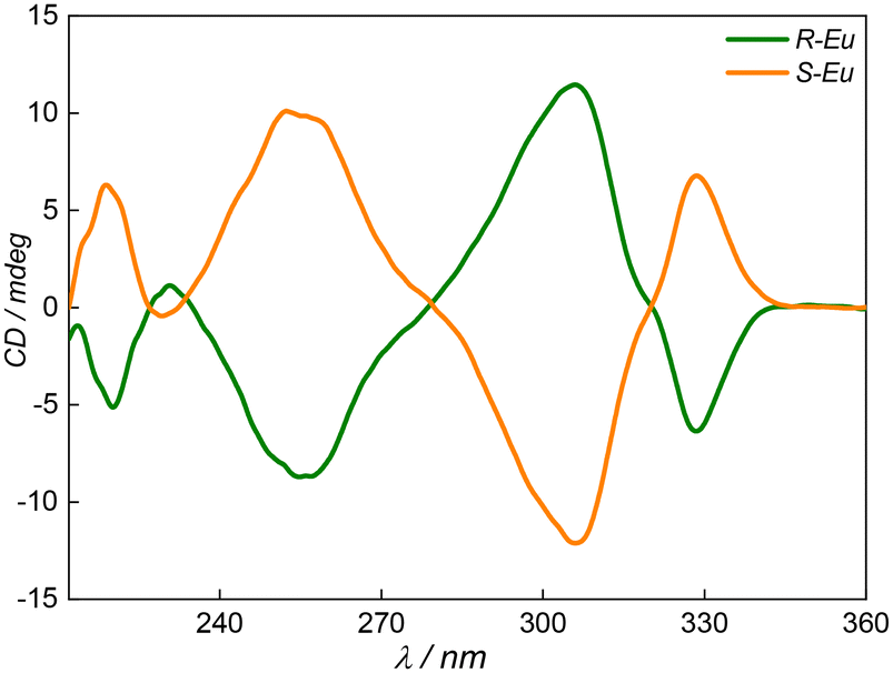
R/S-Ln were in situ synthesized via a [2 + 2] imine condensation reaction between (1R,2R)/(1S,2S)-1,2-diphenylethylenediamine and 2,6-diformylpyridine with LnCl3·6H2O (Ln = Eu, Gd) as a template in CH3OH at 65 °C, while R/S-Eu-Ph3PO were prepared using two steps: (i) Eu(III)-macrocycle precursors were synthesized using Eu(ClO4)3·6H2O as a template; (ii) the coordinated solvent molecules at the axial positions in the precursors were replaced by Ph3PO (Scheme 1). The crystalline samples suitable for single-crystal X-ray diffraction (SCXRD) were obtained by slow vapor diffusion and slow cooling approaches for R/S-Ln and R/S-Eu-Ph3PO, respectively. The SCXRD structural analysis suggested that R/S-Ln and R/S-Eu-Ph3PO crystallized in the chiral polar space group, P21, and the chiral non-polar space group, P212121, respectively (Tables S1–S3†). The asymmetric unit of R/S-Ln contains two independent cationic lanthanide macrocycles while for R/S-Eu-Ph3PO, only one cationic Eu(III) macrocycle, [Eu(LN6)(Ph3PO)2(ClO4)]2+, was observed. As shown in Fig. 2, Eu(III) and Gd(III) ions are both nine-coordinate and encapsulated in the Schiff-base macrocycle, giving a hula-hoop local coordination geometry determined by continuous shape measurement (Tables S11–S13†). Therefore, all complexes have the same equatorial ligand, but for axial positions, they are occupied by two chlorides and one H2O or CH3OH molecule for R/S-Ln and three oxygen atoms from two Ph3PO and one ClO4− for R/S-Eu-Ph3PO, respectively. Given that enantiomeric lanthanide complexes generally show little difference in the first coordination environment, here, we only described the structural characteristics of S-Ln and S-Eu-Ph3PO. The important structural parameters of these six compounds can be found in Tables S5–S10.† For S-Eu, the average Eu–N bond length is respectively 2.623 and 2.620 Å for Eu1 and Eu2. The Eu–Cl distances are 2.725(3) and 2.678(3) Å for Eu1 and 2.686(3) and 2.716(3) Å for Eu2, respectively. The axial Cl–Eu–Cl bond angles for Eu1 and Eu2 are similar, with a difference of <3° (Table S8†). It is evident that the largest difference in the coordination environment between Eu1 and Eu2 is the axially coordinated oxygen atoms which come from H2O and CH3OH molecules with Eu–O distances of 2.434(6) and 2.518(7) Å, respectively. The Eu1⋯Eu2 distance is found to be 10.862 Å. We found that S-Eu-Ph3PO possesses slightly longer Eu–N bond lengths, averaged at 2.630 Å, compared to S-Eu, suggesting that the replacement of axial ligands can also influence the equatorial crystal field. The average Dy–O distance from Ph3PO, 2.309 Å, is shorter than the length of Dy–OClO4− of 2.469 Å, due to the strong affinity of Ph3PO to lanthanide ions, which has also been demonstrated by other luminescent lanthanide compounds with Ph3PO as the antenna.33,38 The axial OPh3PO–Dy–OPh3PO angle is 145.27°, which is comparable to Cl–Eu–Cl angles in S-Eu. For S-Gd, axial Gd–Cl distances are very similar for Gd1 and Gd2, averaging 2.690 and 2.691 Å, respectively. The equatorial Dy–N bond lengths range from 2.578(6) to 2.679(6) Å for Gd1 and 2.571(7) to 2.688(6) Å for Gd2. The axial Gd–O distances, 2.416(5) Å for Gd1–OH2O and 2.501(6) Å for Gd2–OCH3OH, are little shorter than those for S-Eu. The Gd1⋯Gd2 distance is 9.669 Å, which is obviously shorter than that of Eu1⋯Eu2. In addition, the molecular packing of the complexes revealed the presence of abundant hydrogen bonds in the lattice, which is beneficial for relaxivity and luminescence (vide infra, Fig. S1†). The static magnetic properties of R/S-Gd were determined (Fig. S2 and S3†), giving the respective χMT values at 300 K of 15.41 and 15.20 cm3 K mol−1, which are close to the theoretical value of 15.76 cm3 K mol−1 for two isolated spin-only Gd(III) ions. Fig. 3 and S4, S5† display the perfect mirror-image CD spectra of these three enantiomeric pairs of R/S-Eu, R/S-Gd and R/S-Eu-Ph3PO in CH3OH with intense Cotton effects at 255 and 305 nm deriving from the equatorial macrocyclic Schiff-base, demonstrating their chiral nature. It should be noted that these macrocyclic compounds are stable up to 240 °C, as revealed by TGA analysis (Fig. S6 and S7†).
 |
| | Scheme 1 Synthetic route of R/S-Ln (Ln = Eu and Gd) and R/S-Eu-Ph3PO. | |
 |
| | Fig. 2 The ball-and-stick representation of the crystal structures of S-Eu (a), S-Gd (b) and R/S-Eu-Ph3PO (c). Color code: orange, Dy; red, O; blue, N; green, Cl; purple, P; white, H. | |
 |
| | Fig. 3 CD spectra of R/S-Eu in CH3OH (c = 1.0 × 10⁻⁵ mol L−1). | |
Photophysical properties
The excitation and luminescence spectra of S-Eu were collected in CH3OH solution at 1.0 × 10⁻⁵ mol L−1 at room temperature. Fig. 4 shows the excitation spectrum of S-Eu with a ligand-centered band between 275 and 350 nm when monitoring the transition 5D0 → 7F2 of the Eu(III) ion at 617 nm. It can be clearly seen that two strong bands in the 275–350 nm range are located at the same wavelength in the absorption spectrum (Fig. 4 and S8†). The energy of the triplet state (T1) located on the ligands was determined to be ca. 20367 cm−1 by the zero-phonon component in the phosphorescence spectrum of the Gd(III) species (Fig. S8†). This value is 3117 cm−1 higher than the acceptance level of the Eu(III) ion. All these indicated efficient energy transfer from the ligand to the Eu(III) ion and suppressed back energy transfer from the Eu(III) ion to the ligand as the above energy gap is in the optimum range of 2000–5000 cm−1.8,39–41 Upon exciting the solution at 306 nm, all characteristic emission bands of the Eu(III) ion associated with 5D0 → 7FJ (J = 0–5) transitions were clearly observed (Fig. 4). The spectrum was dominated by three bands which correspond to the transitions from the excited state 5D0 to 7F1 (591 nm), 7F2 (612 nm), and 7F4 (702 nm). In addition, it is evident that the emission intensities in H2O are strongly weaker than those in CH3OH, which can be expected because more O–H oscillators exist in the former that induce more serious nonradiative quenching processes. Quantum yields and luminescence lifetimes in both solutions were determined, up to 18.5% and 0.65 ms in CH3OH and 7.2% and 0.33 ms in H2O (Fig. S9 and S10†). As shown in Fig. 4, S-Eu-Ph3PO showed similar optical properties and comparable quantum yields (Fig. S11 and S12†) to S-Eu but stronger emission intensity and longer luminescence lifetimes, i.e. 1.37 ms in CH3OH and 0.38 ms in H2O, under the same measurement conditions, as non-radiative quenching by O–H vibrations from coordinated water molecules in S-Eu was eradicated via replacing H2O with Ph3PO.
 |
| | Fig. 4 Absorption and excitation spectra (left), emission spectra (middle) and decay curves (right) of S-Eu (top) and S-Eu-Ph3PO (right) in CH3OH and H2O at room temperature, c = 1.0 × 10⁻⁵ mol L−1. | |
Circularly polarized luminescence spectra of two Eu(III) macrocycle enantiomers were also recorded in CH3OH solution, which showed near-perfect mirror image signals (Fig. 5). It can be clearly seen that a strong CPL signal was observed at 597 nm, which is common for most chiral Eu(III) compounds and due to the special electronic and magnetic 5D0 → 7F1 transitions.23 The luminescence dissymmetry factor, glum, defined by 2(IL − IR)/(IL + IR) (where IL and IR are the emission intensities of left- and right-circularly polarized light, respectively), and BCPL, were used to evaluate the CPL properties. As shown in Fig. S13 and S14† for the glum plots, S-Eu-Ph3PO exhibited larger |glum| values of the 5D0 → 7F1 and 5D0 → 7F3 transitions than S-Eu. The |glum| values of the above two transitions of S-Eu-Ph3PO increased by 10% compared to those of S-Eu, indicating that (i) the homochiral equatorial macrocycle is mainly responsible for the CPL activity; (ii) the replacement of solvent molecules enhances the molecular rigidity to some extent, therefore improving the CPL properties. The |glum| values for the transition of 5D0 → 7F1 are respectively 0.098 and 0.110 for S-Eu and S-Eu-Ph3PO. The CPL brightness for these two compounds was also calculated and the median BCPL values, 71.45 and 30.89 M−1 cm−1, for the 5D0 → 7F1 transition (λexc 329 nm, λem 597 nm) were obtained (Table S15†), which are comparable to Eu(PyBox)TTA3,42 CsEu(hfbc)443 and Eu(BnMeH22IAM).44
 |
| | Fig. 5 (Left) CPL spectra and total luminescence traced in the background of R/S-Eu (top) and R/S-Eu-Ph3PO (bottom) at room temperature in CH3OH solution (c = 1.0 × 10⁻⁵ mol L−1). Excitation at 329 nm. Bandpass: 10 nm; (middle) HR-ESI-MS of R/S-Gd in CH3OH/H2O solution, confirming the presence of the Gd(III) macrocycle; (right) 1H NMR (top) and HMQC (bottom) of the diamagnetic Y(III) macrocycle, R-Y, in CD3OD. | |
Relaxivity and in vitro MRI of Gd(III) chelates
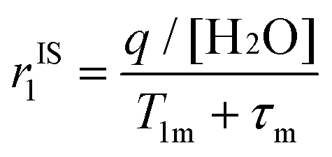
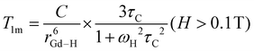
Hydrophilic gadolinium(III) contrast agents, often affectionately called “gado” by medical workers,45 have greatly expanded the utility of MRI which is a noninvasive clinical tool without ionizing radiation.46,47 They can shorten the longitudinal relaxation time (T1) of water protons in their vicinity by virtue of the slow electron relaxations and fast water exchange kinetics of gadolinium(III) ions in the S = 7/2 ground state, therefore affording brighter MR signals.48 In the design of novel Gd(III)-based MR contrast agents (GBCAs), there have been several long recognized design principles (vide infra) at the molecular scale for high relaxivity (r1), mainly involving three intrinsic physical parameters: the hydration number (q), the rotational correlation time (τR) and the water exchange rate (1/τm).49 Other molecular factors, e.g. electron relaxation and the Gd–H(water) distance (rGd–H), are usually not considered, as the former contributes little to r1 in high magnetic fields while the latter is relatively difficult to control via chemical synthesis although it exhibits the inverse six power to T1m (T1 of the coordinated water molecule).2 According to the following eqn (1)–(4), if a GBCA has q = 0, its relaxivity mainly comes from outer-sphere water molecules that are hydrogen bonded to the GBCA. Under this circumstance, polar groups will play a vital role in transmitting the relaxation.50 For GBCAs with q > 0, the contribution of inner-sphere relaxation to r1 should be considered. Theoretically, the greater the q value, the higher the inner-sphere relaxivity; however, more coordinated water molecules will greatly destroy the stability of chelates, increasing the risk of releasing toxic Gd(III) ions.2,51 At present, almost all commercially approved GBCAs are macrocyclic chelates with a nine-coordinate Gd(III) ion and q = 1. For τR and τm, it should be noted that (i) τR dominates τc and further determines T1; (ii) for small GBCAs, τm ≪ T1m. Therefore, increasing r1 can be achieved by slowing down the rotation of the compound. A straightforward and effective way is to increase the molecular weight of the compound by constructing a polynuclear structure or binding to macromolecules, e.g. serum albumin.3,52,53
As mentioned above, the Gd–OH2O distances are respectively 2.416(5) Å and 2.424(5) for S-Gd and R-Gd (Table S3†), which are shorter than the values in Gd-DOTA (2.463 Å) and Gd-DPTA (2.448 Å).6,54,55 In addition, from the packing modes of solid structures shown in Fig. S4,† it was clearly observed that the Gd1-macrocycle interacts with the Gd2-macrocycle via an O–H⋯O–H⋯O–H⋯Cl hydrogen-bond bridge, forming a dimeric structure, and the coordinated water molecule also produces O–H⋯O–H hydrogen bonding interactions with two free CH3OH molecules. The presence of abundant hydrogen bonds not only promotes second-sphere relaxation but also reduces the rate of molecular rotation.2
| |  | (2) |
| |  | (3) |
| |  | (4) |
Where rIS1 and rOS1 are inner-sphere and outer-sphere relaxivity, respectively; C is a physical constant; τC is a correlation time for magnetic fluctuation and is composed of three parts: τR, the water residency time, τm and the electron relaxation time, T1e.
One should be cautioned that although single-crystal X-ray diffraction confirmed the presence of a single aqua ligand that directly bonds to the Gd(III) ion (Fig. 3), the q value should be determined in solution by other physical methods, e.g. electron-nuclear double resonance spectroscopy and luminescence lifetime measurements of isostructural Eu(III) or Tb(III) complexes.56,57 The luminescence lifetimes of the 5D0 → 7F2 transition for the S-Eu complex in H2O and D2O are 0.33 ms and 1.7340 ms, respectively (Fig. 4 and S15†). Therefore, the hydration state for the Gd(III) complex was estimated from the equation proposed by Horrocks et al.58 The obtained q value was 2.22, suggesting that the CH3OH molecule coordinated to Gd2 was replaced by the water molecule.
The above structural characteristics imply a high r1 for Gd(III) compounds. Before attempting the relaxivity rate characterization, the stability of R/S-Ln in solution was examined by NMR measurements. Given that the chemical shifts of paramagnetic lanthanide compounds span a wide range and are not even observed,59 we prepared the analogous Yb(III) complex, R-Y (Tables S4 and S14†). As shown in Fig. 5, the 1H and 13C NMR spectra of R-Y in CD3OD clearly showed 7 proton resonances and 9 carbon signals, indicating a single species with high symmetry in solution. Integration of the 1H spectrum gives the formula of the species, [Y(LN6)Cl2]+. In addition, all 1H and 13C signals were accurately assigned combined with the HMQC spectrum. The HR-ESI-MS of S-Gd showed sharp peaks with +1 charge states, corresponding to Gd(III) macrocycles with different axial ligands (Fig. 5, middle). The MALDI-TOF-MS of the same compound exhibited one set of isotope peaks, indicating the species [Gd(LN6)Cl2]+ (Fig. S16†). The HR-ESI-MS spectrum of S-Eu-Ph3PO and the 1H spectrum of its isostructural Y(III) compound confirmed the presence of only one species of [Eu(LN6)(Ph3PO)2(ClO4)]2+ in solution (Fig. S17†), which is consistent with the solid structure determined by SCXRD. All results and analysis revealed that such macrocyclic compounds possess good solution stability. Fig. 6 and S18† illustrate the concentration-dependent behavior of the longitudinal relaxation rate (1/T1) of Gd(III) compounds at 25 °C and 1.2 T. A linear fit of the data gives an r1 value of 35.04 mM−1 s−1 for R-Gd and 34.09 mM−1 s−1 for S-Gd, respectively, i.e. each Gd(III) ion possesses a relatively high r1, ca. 17 mM−1 s−1. This value is much larger than that of commercially approved Gd(III)-based small molecules and comparable to that of some macromolecular compounds. In order to explore the potential of Gd(III)-based species in T1-weighted MRI, we selected the commercially available Gd(III) MRI contrast agent, gadodiaminde hydrate, for comparison and first evaluated their cytotoxicities by the MTT assay. The results presented in Fig. S19† clearly show that the viabilities of A549 cells exceed 90% after incubation with various concentrations of R/S-Gd and gadodiaminde hydrate for 24 h, demonstrating that R/S-Gd do not exhibit obvious cell toxicity. Then, in vitro T1-weighted MRI at 1.2 tesla was performed with A549 cells after incubation with the above three Gd(III) compounds for another 24 h and the results suggest that R/S-Gd possess comparable MR signal values to gadodiaminde hydrate (Fig. 6).
 |
| | Fig. 6 (a) Relaxivity determined from concentration-dependent T1-weighted MR measurements at 1.2 T (bottom) of S-Gd at room temperature; (b) T1-weighted MRI of gadodiaminde hydrate (grey), S-Gd (soft red) and R-Gd (light blue) after incubation with A549 cells at various concentrations of Gd(III). The error bars mean the standard derivation (n = 3). | |
Conclusions
In conclusion, three pairs of mononuclear lanthanide macrocyclic enantiomers, R/S-Eu, R/S-Gd and R/S-Eu-Ph3PO, were prepared via a facile [2 + 2] imine condensation with lanthanide ions as the template. Their enantiomeric relationship was verified by the mirror-symmetric solid structure determined from SCXRD and CD spectra. S-Eu exhibited brilliant photoluminescence and efficient CPL with an emission quantum yield, luminescence lifetime and luminescence dissymmetry factor of 18.5%, 0.65 ms and 0.098, respectively, in CH3OH solution at room temperature, while isostructural Gd(III) chelates displayed one of the highest relaxivities to date, ca. 17 mM−1 s−1 per Gd(III) ion, low cell toxicity and efficient MRI at low concentrations. By employing Ph3PO to replace H2O and CH3OH molecules located at the axial positions of S-Eu, a longer luminescence lifetime was achieved in S-Eu-Ph3PO, i.e. 1.37 ms in CH3OH solution, benefitting from the eradication of non-radiative quenching by high energy O–H vibrations in the above solvent molecules. Biodistribution studies and in vivo MRI of Gd(III) species are underway.
Conflicts of interest
The authors declare no conflict of interest.
Acknowledgements
We thank the National Natural Science Foundation of China (22201279 and 22171100) for financial support.
References
- W.-L. Zhou, Y. Chen, W. Lin and Y. Liu, Luminescent lanthanide–macrocycle supramolecular assembly, Chem. Commun., 2021, 57, 11443–11456 RSC.
- J. Wahsner, E. M. Gale, A. Rodríguez-Rodríguez and P. Caravan, Chemistry of MRI Contrast Agents: Current Challenges and New Frontiers, Chem. Rev., 2019, 119, 957–1057 CrossRef CAS.
- A. J. L. Villaraza, A. Bumb and M. W. Brechbiel, Macromolecules, Dendrimers, and Nanomaterials in Magnetic Resonance Imaging: The Interplay between Size, Function, and Pharmacokinetics, Chem. Rev., 2010, 110, 2921–2959 CrossRef CAS.
- G.-Q. Jin, C. V. Chau, J. F. Arambula, S. Gao, J. L. Sessler and J.-L. Zhang, Lanthanide porphyrinoids as molecular theranostics, Chem. Soc. Rev., 2022, 51, 6177–6209 RSC.
- R. Jankowski, M. Wyczesany and S. Chorazy, Multifunctionality of luminescent molecular nanomagnets based on lanthanide complexes, Chem. Commun., 2023, 59, 5961–5986 RSC.
- C. A. Chang, L. C. Francesconi, M. F. Malley, K. Kumar, J. Z. Gougoutas, M. F. Tweedle, D. W. Lee and L. J. Wilson, Synthesis, characterization, and crystal structures of M(DO3A) (M = iron, gadolinium) and Na[M(DOTA)] (M = Fe, yttrium, Gd), Inorg. Chem., 1993, 32, 3501–3508 CrossRef CAS.
- J.-Y. Hu, Y. Ning, Y.-S. Meng, J. Zhang, Z.-Y. Wu, S. Gao and J.-L. Zhang, Highly near-IR emissive ytterbium(III) complexes with unprecedented quantum yields, Chem. Sci., 2017, 8, 2702–2709 RSC.
- J.-X. Zhang, W.-L. Chan, C. Xie, Y. Zhou, H.-F. Chau, P. Maity, G. T. Harrison, A. Amassian, O. F. Mohammed, P. A. Tanner, W.-K. Wong and K.-L. Wong, Impressive near-infrared brightness and singlet oxygen generation from strategic lanthanide–porphyrin double-decker complexes in aqueous solution, Light: Sci. Appl., 2019, 8, 46 CrossRef.
- N. Ishikawa, M. Sugita, T. Ishikawa, S.-Y. Koshihara and Y. Kaizu, Lanthanide Double-Decker Complexes Functioning as Magnets at the Single-Molecular Level, J. Am. Chem. Soc., 2003, 125, 8694–8695 CrossRef CAS.
- H. Wada, S. Ooka, T. Yamamura and T. Kajiwara, Light Lanthanide Complexes with Crown Ether and Its Aza Derivative Which Show Slow Magnetic Relaxation Behaviors, Inorg. Chem., 2017, 56, 147–155 CrossRef CAS.
- W. Radecka-Paryzek, Template synthesis and characterization of 18-membered hexaaza macrocyclic complex of lanthanum(III) perchlorate, Inorg. Chim. Acta, 1980, 45, L147–L148 CrossRef CAS.
- S. Akine, S. Sunaga, T. Taniguchi, H. Miyazaki and T. Nabeshima, Core/Shell Oligometallic Template Synthesis of Macrocyclic Hexaoxime, Inorg. Chem., 2007, 46, 2959–2961 CrossRef CAS.
- R. K. Das, E. Barnea, T. Andrea, M. Kapon, N. Fridman, M. Botoshansky and M. S. Eisen, Group 4 Lanthanide and Actinide Organometallic Inclusion Complexes, Organometallics, 2015, 34, 742–752 CrossRef CAS.
- J. Long, J. Rouquette, J.-M. Thibaud, R. A. S. Ferreira, L. D. Carlos, B. Donnadieu, V. Vieru, L. F. Chibotaru, L. Konczewicz, J. Haines, Y. Guari and J. Larionova, A High-Temperature Molecular Ferroelectric Zn/Dy Complex Exhibiting Single-Ion-Magnet Behavior and Lanthanide Luminescence, Angew. Chem., Int. Ed., 2015, 54, 2236–2240 CrossRef CAS.
- J. Long, M. S. Ivanov, V. A. Khomchenko, E. Mamontova, J.-M. Thibaud, J. Rouquette, M. Beaudhuin, D. Granier, R. A. S. Ferreira, L. D. Carlos, B. Donnadieu, M. S. C. Henriques, J. A. Paixão, Y. Guari and J. Larionova, Room temperature magnetoelectric coupling in a molecular ferroelectric ytterbium(III) complex, Science, 2020, 367, 671–676 CrossRef CAS.
- L. Dai, C. M. Jones, W. T. K. Chan, T. A. Pham, X. Ling, E. M. Gale, N. J. Rotile, W. C.-S. Tai, C. J. Anderson, P. Caravan and G.-L. Law, Chiral DOTA chelators as an improved platform for biomedical imaging and therapy applications, Nat. Commun., 2018, 9, 857 CrossRef.
- J. M. Van Raden, D. I. Alexandropoulos, M. Slota, S. Sopp, T. Matsuno, A. L. Thompson, H. Isobe, H. L. Anderson and L. Bogani, Singly and Triply Linked Magnetic Porphyrin Lanthanide Arrays, J. Am. Chem. Soc., 2022, 144, 8693–8706 CrossRef CAS.
- K. Wang, F. Ma, D. Qi, X. Chen, Y. Chen, Y.-C. Chen, H.-L. Sun, M.-L. Tong and J. Jiang, Chiral bis(phthalocyaninato) terbium double-decker compounds with enhanced single-ion magnetic behavior, Inorg. Chem. Front., 2018, 5, 939–943 RSC.
- C. Zhao, Z. Zhu, X.-L. Li and J. Tang, Air-stable chiral mono- and dinuclear dysprosium single-molecule magnets: steric hindrance of hexaazamacrocycles, Inorg. Chem. Front., 2022, 9, 4049–4055 RSC.
- T. Morita, M. Damjanović, K. Katoh, Y. Kitagawa, N. Yasuda, Y. Lan, W. Wernsdorfer, B. K. Breedlove, M. Enders and M. Yamashita, Comparison of the Magnetic Anisotropy and Spin Relaxation Phenomenon of Dinuclear Terbium(III) Phthalocyaninato Single-Molecule Magnets Using the Geometric Spin Arrangement, J. Am. Chem. Soc., 2018, 140, 2995–3007 CrossRef CAS.
- H.-Y. Wong, W.-S. Lo, K.-H. Yim and G.-L. Law, Chirality and Chiroptics of Lanthanide Molecular and Supramolecular Assemblies, Chem, 2019, 5, 3058–3095 CAS.
- R. Carr, N. H. Evans and D. Parker, Lanthanide complexes as chiral probes exploiting circularly polarized luminescence, Chem. Soc. Rev., 2012, 41, 7673–7686 RSC.
- L. Arrico, L. Di Bari and F. Zinna, Quantifying the Overall Efficiency of Circularly Polarized Emitters, Chem. – Eur. J., 2021, 27, 2920–2934 CrossRef CAS PubMed.
- J. L. Lunkley, D. Shirotani, K. Yamanari, S. Kaizaki and G. Muller, Extraordinary Circularly Polarized Luminescence Activity Exhibited by Cesium Tetrakis(3-heptafluoro-butylryl-(+)-camphorato) Eu(III) Complexes in EtOH and CHCl3 Solutions, J. Am. Chem. Soc., 2008, 130, 13814–13815 CrossRef CAS PubMed.
- M. C. Heffern, L. M. Matosziuk and T. J. Meade, Lanthanide Probes for Bioresponsive Imaging, Chem. Rev., 2014, 114, 4496–4539 CrossRef CAS PubMed.
- N. F. M. Mukthar, N. D. Schley and G. Ung, Strong Circularly Polarized Luminescence at 1550 nm from Enantiopure Molecular Erbium Complexes, J. Am. Chem. Soc., 2022, 144, 6148–6153 CrossRef CAS PubMed.
- B.-A. N. Willis, D. Schnable, N. D. Schley and G. Ung, Spinolate Lanthanide Complexes for High Circularly Polarized Luminescence Metrics in the Visible and Near-Infrared, J. Am. Chem. Soc., 2022, 144, 22421–22425 CrossRef CAS PubMed.
- J. Zhang, L. Dai, A. M. Webster, W. T. K. Chan, L. E. Mackenzie, R. Pal, S. L. Cobb and G.-L. Law, Unusual Magnetic Field Responsive Circularly Polarized Luminescence Probes with Highly Emissive Chiral Europium(III) Complexes, Angew. Chem., Int. Ed., 2021, 60, 1004–1010 CrossRef CAS PubMed.
- W. Radecka-Paryzek, V. Patroniak and M. Kubicki, The template synthesis and characterization of pentaaza macrocyclic complexes of rare earth elements. The crystal structure of the 2,14-dimethyl-3,6,10,13,19-pentaazabicyclo[13.3.1]nonadeca-1(19),2,13,15,17-pentaene-dichlorolutetium(III)perchlorate, Polyhedron, 2003, 22, 2773–2779 CrossRef CAS.
- Y. Gil, A. Castro-Alvarez, P. Fuentealba, E. Spodine and D. Aravena, Lanthanide SMMs Based on Belt Macrocycles: Recent Advances and General Trends, Chem. – Eur. J., 2022, 28, e202200336 CrossRef CAS PubMed.
- Z. Zhu, C. Zhao, T. Feng, X. Liu, X. Ying, X.-L. Li, Y.-Q. Zhang and J. Tang, Air-Stable Chiral Single-Molecule Magnets with Record Anisotropy Barrier Exceeding 1800 K, J. Am. Chem. Soc., 2021, 143, 10077–10082 CrossRef CAS.
- M. Wasim, K. U. Ansari, P. Kumar, B. Mallick and M. Shanmugam, A unique and discrete Ce(III) macrocyclic complex exhibits ferroelectric, dielectric, and slow relaxation of magnetization properties, Inorg. Chem. Front., 2022, 9, 2284–2289 RSC.
- Z. Zhu, G.-Q. Jin, J. Wu, X. Ying, C. Zhao, J.-L. Zhang and J. Tang, Highly symmetric Ln(III) boron-containing macrocycles as bright fluorophores for living cell imaging, Inorg. Chem. Front., 2022, 9, 5048–5054 RSC.
- F. Hamon, E. Largy, A. Guédin-Beaurepaire, M. Rouchon-Dagois, A. Sidibe, D. Monchaud, J.-L. Mergny, J.-F. Riou, C.-H. Nguyen and M.-P. Teulade-Fichou, An Acyclic Oligoheteroaryle That Discriminates Strongly between Diverse G-Quadruplex Topologies, Angew. Chem., Int. Ed., 2011, 50, 8745–8749 CrossRef CAS PubMed.
- O. V. Dolomanov, L. J. Bourhis, R. J. Gildea, J. A. K. Howard and H. Puschmann, OLEX2: a complete structure solution, refinement and analysis program, J. Appl. Crystallogr., 2009, 42, 339–341 CrossRef CAS.
- G. Sheldrick, SHELXT - Integrated space-group and crystal-structure determination, Acta Crystallogr., Sect. A: Found. Adv., 2015, 71, 3–8 CrossRef PubMed.
- G. Sheldrick, Crystal structure refinement with SHELXL, Acta Crystallogr., Sect. C: Struct. Chem., 2015, 71, 3–8 Search PubMed.
- S. Biju, N. Gopakumar, J. C. G. Bünzli, R. Scopelliti, H. K. Kim and M. L. P. Reddy, Brilliant Photoluminescence and Triboluminescence from Ternary Complexes of DyIII and TbIII with 3-Phenyl-4-propanoyl-5-isoxazolonate and a Bidentate Phosphine Oxide Coligand, Inorg. Chem., 2013, 52, 8750–8758 CrossRef CAS PubMed.
- J.-C. G. Bünzli, On the design of highly luminescent lanthanide complexes, Coord. Chem. Rev., 2015, 293–294, 19–47 CrossRef.
- O. L. Malta, Mechanisms of non-radiative energy transfer involving lanthanide ions revisited, J. Non-Cryst. Solids, 2008, 354, 4770–4776 CrossRef CAS.
- T. N. Nguyen, C. Y. Chow, S. V. Eliseeva, E. R. Trivedi, J. W. Kampf, I. Martinić, S. Petoud and V. L. Pecoraro, One-Step Assembly of Visible and Near-Infrared Emitting Metallacrown Dimers Using a Bifunctional Linker, Chem. – Eur. J., 2018, 24, 1031–1035 CrossRef CAS PubMed.
- M. Górecki, L. Carpita, L. Arrico, F. Zinna and L. Di Bari, Chiroptical methods in a wide wavelength range for obtaining Ln3+ complexes with circularly polarized luminescence of practical interest, Dalton Trans., 2018, 47, 7166–7177 RSC.
- F. Zinna, U. Giovanella and L. D. Bari, Highly Circularly Polarized Electroluminescence from a Chiral Europium Complex, Adv. Mater., 2015, 27, 1791–1795 CrossRef CAS.
- S. Petoud, G. Muller, E. G. Moore, J. Xu, J. Sokolnicki, J. P. Riehl, U. N. Le, S. M. Cohen and K. N. Raymond, Brilliant Sm, Eu, Tb, and Dy Chiral Lanthanide Complexes with Strong Circularly Polarized Luminescence, J. Am. Chem. Soc., 2007, 129, 77–83 CrossRef CAS.
- P. Caravan, J. J. Ellison, T. J. McMurry and R. B. Lauffer, Gadolinium(III) Chelates as MRI Contrast Agents: Structure, Dynamics, and Applications, Chem. Rev., 1999, 99, 2293–2352 CrossRef CAS.
- J.-H. Tang, H. Li, C. Yuan, G. Parigi, C. Luchinat and T. J. Meade, Molecular Engineering of Self-Immolative Bioresponsive MR Probes, J. Am. Chem. Soc., 2023, 145, 10045–10050 CrossRef CAS.
- P. Caravan, Protein-Targeted Gadolinium-Based Magnetic Resonance Imaging (MRI) Contrast Agents: Design and Mechanism of Action, Acc. Chem. Res., 2009, 42, 851–862 CrossRef CAS.
- E. M. Gale, I. P. Atanasova, F. Blasi, I. Ay and P. Caravan, A Manganese Alternative to Gadolinium for MRI Contrast, J. Am. Chem. Soc., 2015, 137, 15548–15557 CrossRef CAS.
- H. Li and T. J. Meade, Molecular Magnetic Resonance Imaging with Gd(III)-Based Contrast Agents: Challenges and Key Advances, J. Am. Chem. Soc., 2019, 141, 17025–17041 CrossRef CAS.
- G.-Q. Jin, H. Lai, Z.-S. Yang, Y. Ning, L. Duan, J. Zhang, T. Chen, S. Gao and J.-L. Zhang, Gadolinium(III) Porphyrinoid Phototheranostics, Chem. – Asian J., 2022, 17, e202200181 CrossRef CAS.
- M. Polasek and P. Caravan, Is Macrocycle a Synonym for Kinetic Inertness in Gd(III) Complexes? Effect of Coordinating and Noncoordinating Substituents on Inertness and Relaxivity of Gd(III) Chelates with DO3A-like Ligands, Inorg. Chem., 2013, 52, 4084–4096 CrossRef CAS.
- Z. Wang, L. He, B. Liu, L.-P. Zhou, L.-X. Cai, S.-J. Hu, X.-Z. Li, Z. Li, T. Chen, X. Li and Q.-F. Sun, Coordination-Assembled Water-Soluble Anionic Lanthanide Organic Polyhedra for Luminescent Labeling and Magnetic Resonance Imaging, J. Am. Chem. Soc., 2020, 142, 16409–16419 CrossRef CAS.
- Z. Zhang, M. T. Greenfield, M. Spiller, T. J. McMurry, R. B. Lauffer and P. Caravan, Multilocus Binding Increases the Relaxivity of Protein-Bound MRI Contrast Agents, Angew. Chem., Int. Ed., 2005, 44, 6766–6769 CrossRef CAS.
- M. Briganti, E. Lucaccini, L. Chelazzi, S. Ciattini, L. Sorace, R. Sessoli, F. Totti and M. Perfetti, Magnetic Anisotropy Trends along a Full 4f-Series: The fn+7 Effect, J. Am. Chem. Soc., 2021, 143, 8108–8115 CrossRef CAS.
- S. Gao, S. J. George and Z.-H. Zhou, Interaction of Gd-DTPA with phosphate and phosphite: toward the reaction intermediate in nephrogenic systemic fibrosis, Dalton Trans., 2016, 45, 5388–5394 RSC.
- S. G. Zech, W.-C. Sun, V. Jacques, P. Caravan, A. V. Astashkin and A. M. Raitsimring, Probing the Water Coordination of Protein-Targeted MRI Contrast Agents by Pulsed ENDOR Spectroscopy, ChemPhysChem, 2005, 6, 2570–2577 CrossRef CAS.
- W. D. Horrocks Jr. and D. R. Sudnick, Lanthanide ion probes of structure in biology. Laser-induced luminescence decay constants provide a direct measure of the number of metal-coordinated water molecules, J. Am. Chem. Soc., 1979, 101, 334–340 CrossRef.
- R. M. Supkowski and W. D. Horrocks, On the determination of the number of water molecules, q, coordinated to europium(III) ions in solution from luminescence decay lifetimes, Inorg. Chim. Acta, 2002, 340, 44–48 CrossRef CAS.
- A. Gerus, K. Ślepokura and J. Lisowski, Anion and Solvent Induced Chirality Inversion in Macrocyclic Lanthanide Complexes, Inorg. Chem., 2013, 52, 12450–12460 CrossRef CAS.
|
| This journal is © the Partner Organisations 2024 |
Click here to see how this site uses Cookies. View our privacy policy here. 
 d,
Zhenxin
Wang
d,
Zhenxin
Wang