
 Open Access Article
 This Open Access Article is licensed under a
 Creative Commons Attribution 3.0 Unported Licence
Recent advances in graphene-based electroanalytical devices for healthcare applications
Vinay
Kammarchedu
 abc,
Heshmat
Asgharian
ac,
Keren
Zhou
ac,
Pouya
Soltan Khamsi
ac and
Aida
Ebrahimi
*abcd
abc,
Heshmat
Asgharian
ac,
Keren
Zhou
ac,
Pouya
Soltan Khamsi
ac and
Aida
Ebrahimi
*abcd
aDepartment of Electrical Engineering, The Pennsylvania State University, University Park, Pennsylvania 16802, USA. E-mail: sue66@psu.edu
bCenter for Atomically Thin Multifunctional Coatings, The Pennsylvania State University, University Park, Pennsylvania 16802, USA
cMaterials Research Institute, The Pennsylvania State University, University Park, Pennsylvania 16802, USA
dDepartment of Biomedical Engineering, The Pennsylvania State University, University Park, Pennsylvania 16802, USA
First published on 29th May 2024
Abstract
Graphene, with its outstanding mechanical, electrical, and biocompatible properties, stands out as an emerging nanomaterial for healthcare applications, especially in building electroanalytical biodevices. With the rising prevalence of chronic diseases and infectious diseases, such as the COVID-19 pandemic, the demand for point-of-care testing and remote patient monitoring has never been greater. Owing to their portability, ease of manufacturing, scalability, and rapid and sensitive response, electroanalytical devices excel in these settings for improved healthcare accessibility, especially in resource-limited settings. The development of different synthesis methods yielding large-scale graphene and its derivatives with controllable properties, compatible with device manufacturing – from lithography to various printing methods – and tunable electrical, chemical, and electrochemical properties make it an attractive candidate for electroanalytical devices. This review article sheds light on how graphene-based devices can be transformative in addressing pressing healthcare needs, ranging from the fundamental understanding of biology in in vivo and ex vivo studies to early disease detection and management using in vitro assays and wearable devices. In particular, the article provides a special focus on (i) synthesis and functionalization techniques, emphasizing their suitability for scalable integration into devices, (ii) various transduction methods to design diverse electroanalytical device architectures, (iii) a myriad of applications using devices based on graphene, its derivatives, and hybrids with other nanomaterials, and (iv) emerging technologies at the intersection of device engineering and advanced data analytics. Finally, some of the major hurdles that graphene biodevices face for translation into clinical applications are discussed.
Introduction
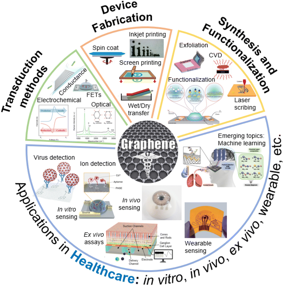
Graphene is a two-dimensional (2D) sheet of hexagonal sp2-bonded carbon,1 wherein each carbon atom is bonded to three neighbouring atoms, forming a highly stable lattice.2 This arrangement grants graphene exceptional mechanical strength.3 Moreover, its 2D nature and high surface area-to-volume ratio offer an excellent platform for chemical interactions, making graphene an excellent material for sensing applications.4 The hallmark of graphene's electrical properties lies in its remarkable electron mobility, surpassing that of traditional semiconductors.5 This property, coupled with its high (and tunable) electrical conductivity, positions graphene as an ideal candidate for constructing advanced electrical and electrochemical devices. Interestingly, drastic changes to the electronic band structure are observed when the thickness of graphene increases from monolayer to a few layers, and further to thick graphitic crystals.6–8 Thus, it is critical to elucidate these differences when comparing the sensing performance of devices. In this review article, we use ISO standard terminology9 to refer to graphene, i.e., the monolayer as graphene, and the subsequent thicknesses with appended layer numbers (e.g., 2LG, 3LG, and FLG for 4–10 layers). Furthermore, for brevity in this paper, the abbreviation GR is used to refer to graphene up to 10 layers in thickness.Fig. 1 provides a summary of the scope of this review article. The first section discusses various synthesis methods and their suitability for building biodevices in healthcare applications. Central to the successful integration of GR into electroanalytical devices is the development of precise and scalable synthesis techniques.10 Various top-down and bottom-up methods enable the synthesis of GR with vast differences in defect densities, surface functional groups, hetero-atom presence, etc. The synthesis techniques also produce GR of various dimensions.11 For example, quantum dots are zero dimensional, whereas graphene nanoribbons are one dimensional. A highly three dimensional and porous GR can also be produced using techniques such as laser scribing.12 Furthermore, incorporating functional groups onto the GR surface is a pivotal step in tailoring its interaction with target bioanalytes.13 GR's basal plane offers a plethora of π-conjugated electrons,14 while its edges and defects can act as active sites for chemical interactions.15
 | ||
| Fig. 1 A snapshot of the scope of this review. Different ways to synthesize graphene, functionalization, device fabrication, and sensor transduction mechanisms are discussed. Adapted with permission from ref. 16 and 17. Copyright 2018–2022 American Chemical Society. Adapted with permission from ref. 18–20. Copyright 2020–2021 John Wiley and Sons. Adapted with permission from ref. 21–23. Copyright 2020 Springer Nature. Adapted with permission from ref. 24. Copyright 2020 MDPI. Adapted with permission from ref. 25–27. Copyright 2022 The Royal Society of Chemistry. | ||
The versatility of the physiochemical properties of GR enables the design and manufacturing of diverse electroanalytical device architectures, as highlighted in the Device Manufacturing section. In this section, various device fabrication methods along with specific examples of devices are described. The following section focuses on the transduction mechanisms, including field-effect transistors, electrochemical methods, and electro-optical devices, and their respective uses in healthcare settings. The next section of this article focuses on emerging healthcare applications. GR-based devices, characterized by their ability to precisely analyze biological samples, detect biomarkers, and monitor physiological parameters, hold the potential to revolutionize disease diagnosis, personalized medicine, remote patient monitoring, and life sciences research. As technology advances, the integration of electroanalytical devices with digital platforms holds the promise of ushering in an era of preventive and precision medicine, ultimately improving patient outcomes and healthcare management on a global scale.
Finally, the current progress and the existing challenges involved in translating GR-based biodevices into practical setting are discussed. While the potential of GR-based electroanalytical devices in healthcare is undeniable, several challenges remain to be addressed.28 Achieving reproducibility in GR synthesis and scalability of device preparation (with high yield) are crucial for the widespread adoption of these devices. The functionalization processes require careful consideration to maintain GR's properties while enhancing the device performance.29 Moreover, the integration of GR-based sensors into complex biological matrices, especially for in vivo analysis and long-term readout demands robustness, stability, and biocompatibility. As the global healthcare industry faces hurdles ranging from the early detection of diseases to the management of chronic conditions, we hope that this article sheds some light on the transformative role that GR-based devices can play in addressing such critical needs.
Synthesis and functionalization
Synthesis techniques and further processing steps greatly influence the properties and quality of the GR that is produced.30,31 In this section, we first provide an overview of different dimensionalities of graphene that have been explored in biodevices. Next, we discuss the synthesis methods (Fig. 2A), which can be broadly categorized as top-down (Fig. 2A(i)) or bottom-up (Fig. 2A(ii)) approaches depending on whether GR is produced by removing nanostructures from the bulk solid or adding suitable atoms, respectively. We then summarize the various processing/functionalization methods that further transform the physiochemical properties of GR.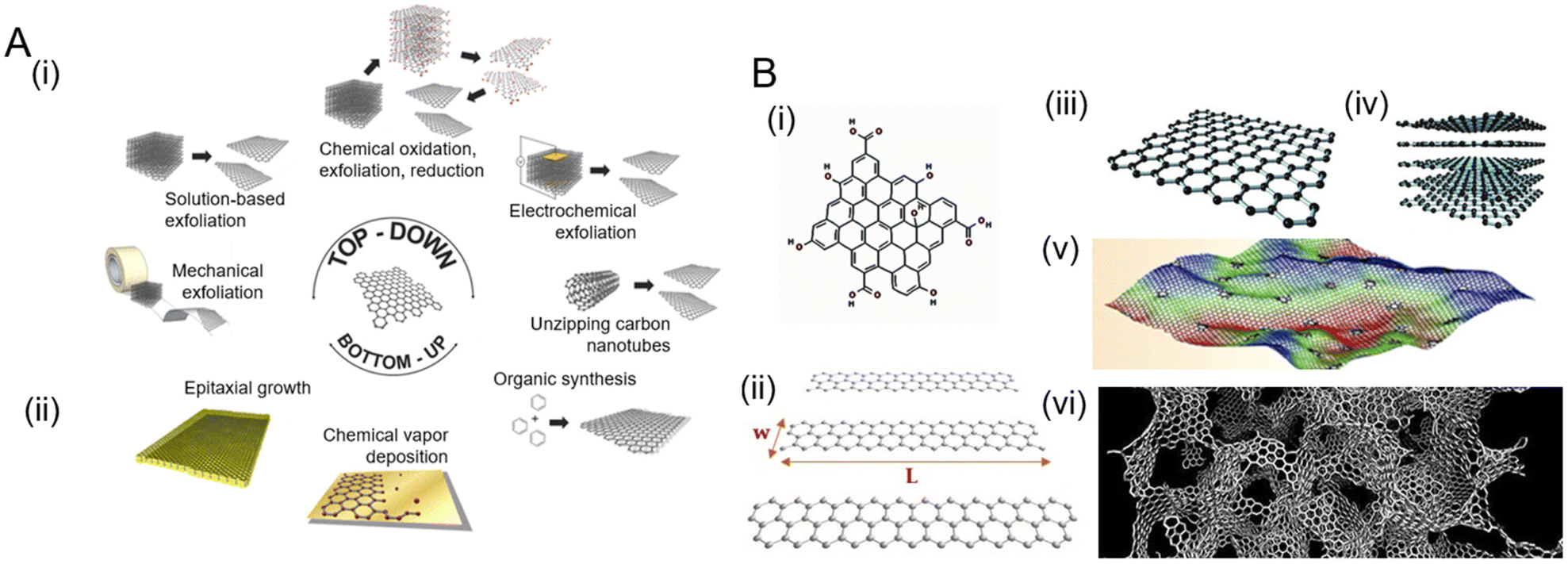 | ||
| Fig. 2 Various routes for graphene synthesis and the different dimensions of graphene that are produced. [A] Schematic showing common synthesis approaches: (i) top-down methods create graphene by removing material, whereas (ii) bottom-up methods produce graphene by sequential addition of suitable atoms. Adapted with permission from ref. 32. Copyright 2014 American Chemical Society. [B] Different dimensions of graphene: (i) quantum dots and (ii) nanoribbons. Adapted with permission from ref. 33. Copyright 2018 MDPI. (iii) Graphene sheets, (iv) few layer graphene (FLH), and (v) defective and corrugated graphene. Adapted with permission from ref. 34. Copyright 2021 American Chemical Society. (vi) 3D graphene obtained by the combination of graphene sheets of various thickness and defects. Adapted with permission from ref. 35. Copyright Qin et al. | ||
Dimensions of graphene
The basic two-dimensional graphene (2D-GR) building block can be used to visualize the creation of various carbon-based materials by tailoring, folding, and stacking graphene to create materials from zero to three dimensions (Fig. 2B). Zero-dimensional graphene quantum dots (0D-GR) are nanometer-sized particles (Fig. 2B(i)), such as nanocrystals and buckyballs. They are usually rich in functional groups such as hydroxyl, epoxy, and carboxyl on their edges.36 0D-GR have interesting electrical and chemical properties that can be tuned by the modification of their dimensions and surface chemistry, especially for the observation of optical phenomena.37 Common routes for the synthesis of 0D-GR38 include plasma etching, liquid exfoliation, hydrothermal, and solvothermal methods.39 One-dimensional graphene (1D-GR) is produced by the confinement of GR in a certain direction (Fig. 2B(ii)).40 Nanoribbons of graphene can be produced by top-down methods such as patterning and etching, catalytic cutting, and in situ lithography.41 The electrical and optical properties of 1D-GR can be tuned by doping, strain engineering, and modifying chemical functional groups at the edge.42 2D-GR (Fig. 2B(iii)) can be stacked to form FLG (Fig. 2B(iv)). Three-dimensional graphene (3D-GR) such as graphene foams, hydrogels, and aerogels comprised of defective and corrugated graphene (Fig. 2B(v)) enables a porous and high specific surface area (Fig. 2B(vi)). 3D-GR is especially promising for energy storage, conversion, catalysis, and sensing applications. Synthesis methods for 3D-GR include hydrothermal, chemical vapor deposition, and on-site polymerization techniques.43 The properties of 3D-GR can be tuned by doping, surface chemical functionalization, defect engineering, and porosity engineering.43Top-down synthesis strategies
The top-down strategy for GR production generally entails the separation of material from carbon sources such as graphite crystals and powders (Fig. 2A(i)). These methods destructively attempt to separate the existing stacked layers of carbon to obtain monolayer graphene to FLG. One of the very first top-down methods to isolate graphene from graphite was based on micromechanical exfoliation using a tape technique.44 Even though it is capable of producing high quality graphene, this method does not yield very large area graphene and is unsuitable for large scale production and commercialization. In contrast, liquid phase exfoliation (LPE) is used to exfoliate graphite using thermal, chemical, or mechanical processes on a large scale.45 Electrochemical exfoliation can also be used to exfoliate graphite by intercalating ionic species into graphite where they form gaseous molecules, expanding and exfoliating graphite into GR sheets.46 Graphite can also be oxidized chemically and subsequently reduced to form GR such as in Hummers’ method47 and the Brodie method.31 For a thorough review of different top-down methods for the synthesis of GR, we refer the readers to a review article by Kumar et al.48Bottom-up synthesis strategies
Bottom-up strategies focus on building layers of graphene using suitable precursors (Fig. 2A(ii)). These methods may include chemical vapor deposition (CVD),49 epitaxial growth,50 and thermal pyrolysis, among others.51 CVD is a popular method to produce large area 2D monolayer to FLG films. Many hydrocarbon precursors and a variety of substrates (e.g., copper) have been used to grow these graphene films.52 The main drawback of the CVD process is its sensitivity to the growth parameters and transfer methods that limit its overall quality in devices as well as requiring high temperatures. Epitaxial growth of graphene is also possible on silicon carbide substrates.53 Thermal pyrolysis54 or laser-based scribing of various carbon-containing sources (which are discussed later) also yield GR with diverse structures and properties.12 These methods generally result in GR that has a higher density of defects due to the chaotic formation process during manufacturing.16 For a thorough review of different top-down methods for the synthesis of GR, we refer the readers to a review article by Gutiérrez-Cruz et al.55Functionalization of graphene
GR can be functionalized through various methods to enhance its response to bioanalytes (Fig. 3).13,56–60 Some of the strategies include doping, defect engineering, surface modification, porosity engineering, and annealing, which are summarized below.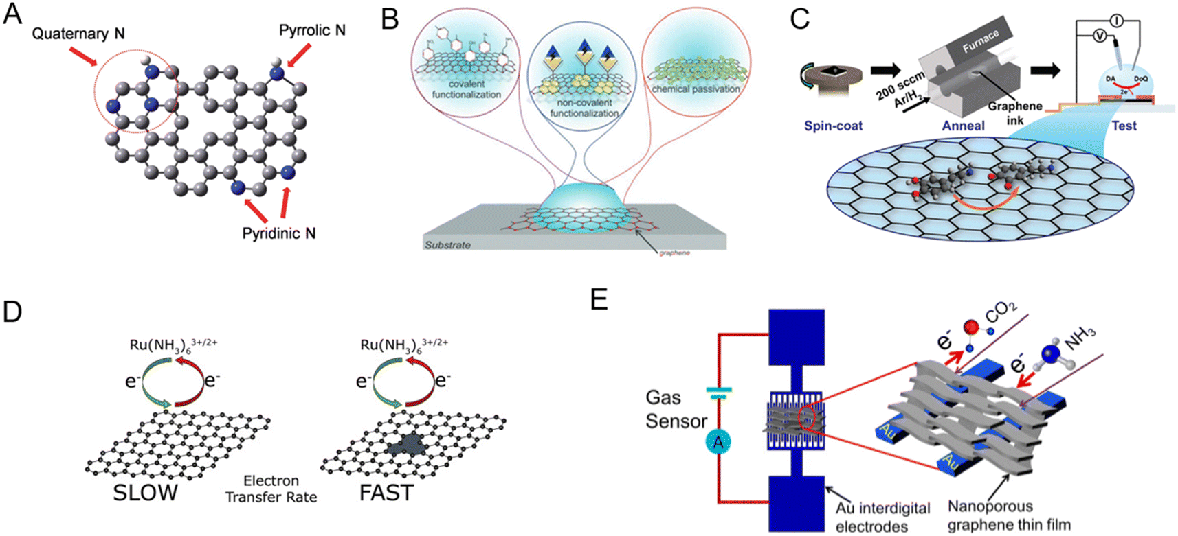 | ||
| Fig. 3 Different modification methods to tune the sensitivity and specificity of graphene to target analytes. [A] Doping with heteroatoms (example: nitrogen-doped graphene was shown to enhance the electrocatalytic activity of hydrogen peroxide reduction, which was then utilized to detect glucose). Adapted with permission from ref. 61. Copyright 2014 American Chemical Society. [B] Surface chemical modification via covalent, non-covalent, and chemical passivation. Adapted with permission from ref. 18. Copyright 2020 John Wiley and Sons. [C] Annealing (example: ink-based electrochemical sensors; tuning the annealing conditions enhanced the sensitivity to dopamine). Adapted with permission from ref. 62. Copyright 2021 American Chemical Society. [D] Defect engineering to enhance surface reactions (example: charge transfer kinetics are enhanced by introducing defects). Adapted with permission from ref. 63. Copyright 2020 Elsevier. [E] Porosity engineering (example: compared to planar GR, porous GR had 4.2 times higher response to CO2 and 10.4 times higher response to NH3). Adapted with permission from ref. 64. Copyright 2020 Elsevier. | ||
Graphene derivatives
Apart from graphene itself, different derivatives have been synthesized and incorporated into bioanalytical devices. This section provides a brief overview of important GR derivatives.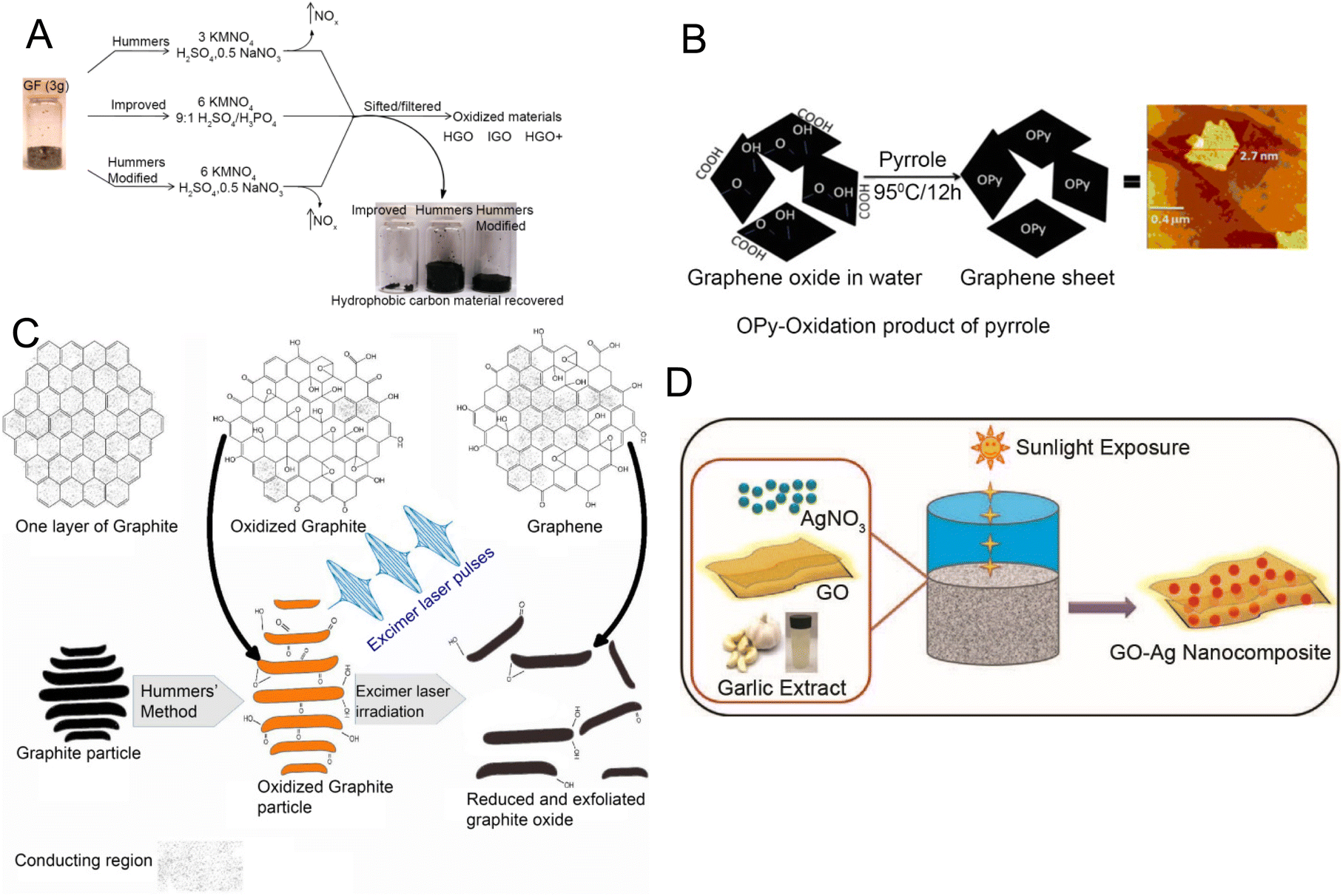 | ||
| Fig. 4 Synthesis methods for examples of graphene derivatives used in electroanalytical devices. [A] Schematic illustration of graphene oxide (GO) synthesis by chemical oxidation. Reprinted with permission from ref. 96. Copyright 2010 American Chemical Society. [B] Synthesis of reduced graphene oxide (rGO) via pyrrole process. Reprinted with permission from ref. 97. Copyright 2011 Elsevier. [C] rGO synthesis via excimer laser processing. Reprinted with permission from ref. 98. Copyright 2018 Elsevier. [D] Synthesis of GO–Ag (silver) nanocomposite using garlic extract and sunlight. Reprinted with permission from ref. 99. Copyright 2015 Elsevier. | ||
rGO is produced by reducing GO. It has a lower oxidation state and reduced surface reactivity in comparison with its precursor material, which makes it suitable for biomedical applications due to minimal cellular damage.100 Factors such as production efficiency, biocompatibility, and low toxicity should be considered when synthesizing rGO that is suitable for biomedical applications.101 Hybrid methods that combine photothermal and photochemical treatments using ultraviolet light102 or laser techniques103,104 introduce a time-efficient and cleaner production process. Another potential way to obtain relatively clean and efficient rGO is the electrochemical reduction method in which rGO can be precipitated from a graphite electrode through changes in voltage.105 Thermal reduction relies on heat combined with organic solvents like dimethyl sulfoxide,106 dimethylformamide,107 or N-methyl-2-pyrrolidone108 offering an alternative way to reduce GO to rGO. Another approach is the chemical reduction of GO to rGO using reductants such as hydrazine monohydrate,109 hydrazine hydrate,110 sodium borohydride in high excess,111 pyrrole97 (Fig. 4B), and others. Besides, green alternatives such as Delphinium root extract have been explored for their potential in biocompatible applications.112 Photothermal and photochemical treatments using an excimer laser are also developed as faster and relatively cleaner processes (Fig. 4C) to create rGO.98
Device manufacturing
The preparation of GR-based biosensors has been the subject of various research endeavors, especially for healthcare applications. Typical manufacturing methods include graphene transfer (Fig. 5A), printing (e.g., inkjet printing and screen printing, Fig. 5B), photo/electron lithography (Fig. 5C), and direct writing methods (Fig. 5D).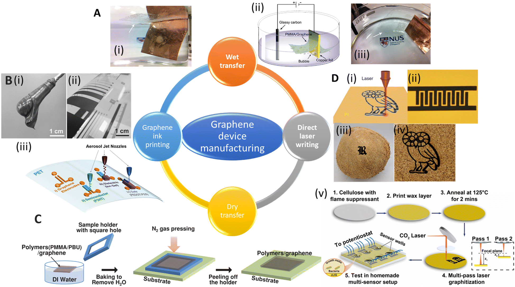 | ||
| Fig. 5 Schematic showing various manufacturing methods for graphene devices. [A] (i) Images of electrochemical exfoliation of graphene from Cu foil. (ii) Schematic diagram of an electrochemical cell used for electrochemical exfoliation. (iii) Another side view of (i). Reprinted with permission from ref. 133. Copyright 2011 American Chemical Society. [B] (i) Image of graphene paste. (ii) Bent PET foil with printed graphene film. Reprinted with permission from ref. 134. Copyright 2015 Wiley-VCH GmbH. (iii) Schematic and optical image of aerosol-jet printed devices using graphene ink. Reprinted with permission from ref. 140. Copyright 2017 American Chemical Society. [C] Illustration of the dry transfer method for CVD-grown graphene. Reprinted with permission from ref. 135. Copyright 2014 Wiley-VCH GmbH. [D] (i) Direct laser scribing to produce laser induced graphene (LIG) on different substrates, such as (ii) polyimide (example: an image of the as-printed LIG interdigitated electrodes). Reprinted with permission from ref. 136. Copyright 2014 Springer Nature. (iii) Coconut shell and (iv) cork. Reprinted with permission from ref. 137. Copyright 2018 American Chemical Society. (v) A schematic overview of the fabrication process for sensors based on cLIG (cellulose-based laser induced graphene). The sensors were used for real time monitoring of the viability and phenazine production by P. aeruginosa cells. Reprinted with permission from ref. 138. Copyright 2023 Elsevier. | ||
Lithography of transferred graphene films
To fabricate GR devices, especially GR-based field-effect transistors (GFETs), GR (usually synthesized using the CVD method) is first transferred from the original substrate to the target substrate. Then lithography is used to transfer patterns of a mask onto graphene by means of etching.139 The graphene transfer is done either using wet or dry processes, which are summarized below. The target substrate is usually a silicon wafer for the nanopreparation process. Recently, GR has also been successfully transferred onto flexible substrates to create flexible/wearable devices. Some common flexible substrates used as supports for GR include polydimethylsiloxane (PDMS),140,141 polymethylmethacrylate (PMMA),142 and polyethylene terephthalate (PET).143 For a focused review of transfer processes, we refer the readers to a review article by Ullah et al.144Traditional wet transfer usually etches the metal layer during transfer. However, this approach produces lots of waste, takes a long time, is challenging to reproduce, and is usually costly for mass production of GR. The electrochemical bubbling transfer method was developed to reuse the metal substrate. In this approach, graphene/Cu was employed as the electrode for water electrolysis (Fig. 5A). The produced gas (O2 and H2) bubbles generate a peeling force between graphene and the metal.133 The electrochemical bubbling transfer method provides a fast, economical way compared to the traditional etching method, which makes this method a better fit for the large-scale production of GR.
While the wet transfer process is the most common method used in laboratories to obtain GR, it usually suffers from contamination by the etchant(s) and/or the supporting polymer, or the metal residues on GR may degrade the electron mobility and may lead to unnecessary doping of GR.147 In addition, the etching chemicals are not environmentally friendly. To address these challenges, dry transfer techniques have been developed as summarized below.
For large-scale and fast transfer to meet market demands, a roll-to-roll manufacturing method was developed. Bae et al. reported a way to transfer large-scale CVD-grown graphene using the roll-to-roll method in 2010.143 In this process, the graphene films are detached from the tapes and released to the substrates by thermal treatment. The transfer rate was 150–200 mm min−1 and a wet-chemical doping process could also be added to the process. In 2015, Liu's group reported a modified roll-to-roll clean transfer of CVD-grown graphene from copper to the ethylene vinyl acetate/poly(ethylene terephthalate) (EVA/PET) plastic substrate (Fig. 6A).149 This process was achieved by water penetrating between graphene and the copper oxide layer. They also used the transferred graphene to manufacture a wearable transparent and flexible triboelectric nanogenerator to show good conductivity and high transparency of the transferred graphene. The roll-to-roll method is compatible with the current industrial set-up for low-cost and large-scale transfer, but it can only transfer to flexible substrates, preventing its extensive application.
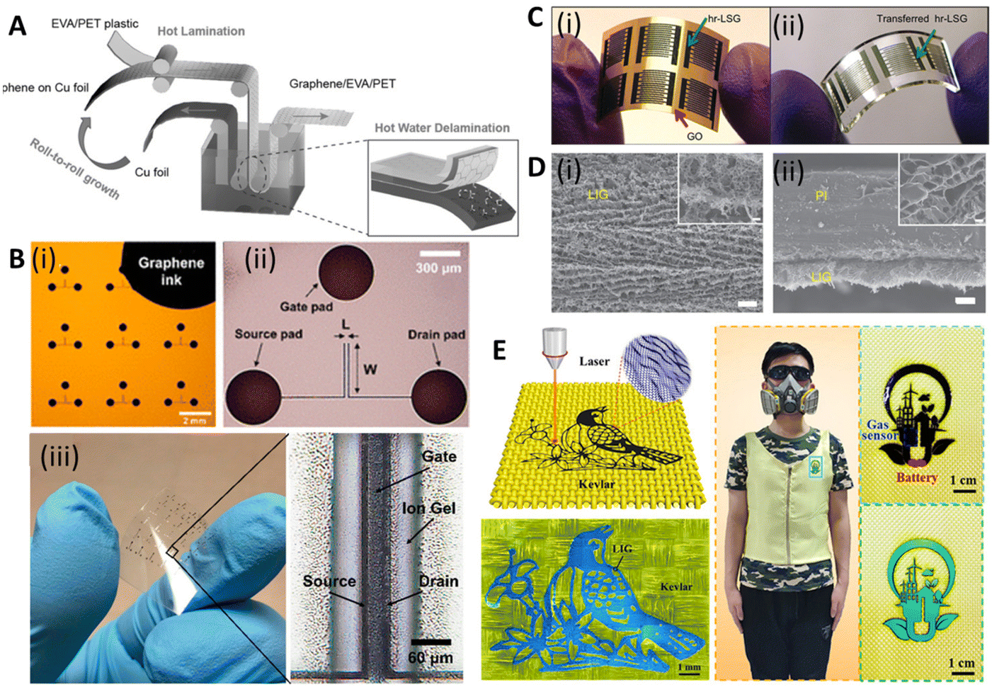 | ||
| Fig. 6 Scalable and printable graphene-based electroanalytical devices. [A] Illustration of roll-to-roll delamination of copper and graphene onto EVA/PET substrate. Reprinted with permission from ref. 149. Copyright 2015 Wiley-VCH GmbH. [B] (i) Image of an electrolyte-gated transistor (EGT), selectively printed with graphene ink, with L = 20 and W = 400 μm. (ii) Microscope image of a single device in (i). (iii) Photograph of flexible EGTs printed on PET and optical image showing printed layers of source, drain, ion gel, and gate. Reprinted with permission from ref. 150. Copyright 2017 American Chemical Society. [C] (i) Image of interdigitated electrodes fabricated using highly reduced laser scribed graphene (hr-LSG) (ii) Image of hr-LSG after transfer to PDMS. Reprinted with permission from ref. 151. Copyright 2012 American Chemical Society. [D] SEM images of (i) LIG film and (ii) its cross-section. Scale bar: 10 μm and 1 μm in the inset. Reprinted with permission from ref. 136. Copyright 2014 Springer Nature. [E] (i) Schematic of the preparation of graphene/Kevlar textile. (ii) Intelligent clothing integrated with the NO2 gas sensor based on the graphene/Kevlar. Reprinted with permission from ref. 152. Copyright 2020 American Chemical Society. | ||
Printing techniques
Although lithography with transferred GR films has shown its reproducibility and processability in manufacturing GR-based devices, there are some disadvantages that hinder their practical applications, e.g., they need complex and expensive tools and highly trained personnel. In addition, lithography mask residues often result in contamination of the devices and the high-energy etching processes may introduce defects into GR.139 In contrast, printing techniques offer a suite of desirable properties for printed electronics, including easier preparation, high electrical conductivity, flexibility, and robust mechanical, chemical, and environmental stability.153,154Despite their widespread use, the resolution of traditional inkjet printers is poor, about 30–50 μm, limiting their application in printing high-resolution electronic devices (e.g., flexible transistors). Song et al. demonstrated a method to fabricate high-resolution patterns using the transfer printing of GR ink with silicon molds150 (Fig. 5C(iii)). The smallest line width and spacing of the patterns were 3.2 μm and 2.7 μm, respectively. They further transferred the pattern onto PET and fabricated flexible electrolyte-gated transistors to show their great mechanical robustness and electrical properties (Fig. 6B). GR ink can also be spin-coated on flexible substrates. Muralidharan et al. reported a flexible electrochemical dopamine sensor by spin-coating GR ink onto polyimide followed by an annealing process.161 The sensor showed a limit of detection (LOD) of 100 nM of dopamine in phosphate buffered saline (PBS) and a linear range of up to 1 mM. Further study showed that the LOD was improved to 5 nM when the sensor was treated with copper sulfate solution to dope GR. Interfacing the sensor with a wireless system with an on-chip integrated potentiostat also made the sensors suitable for point of care monitoring. For a more focused review of printable GR devices, we refer readers to Htwe et al. and Fisher et al. Htwe et al. summarized recent progress on using GR ink in flexible electronics applications and compared different flexible substrates used in wearable devices.162 In addition, Fisher et al. provided a survey of state-of-the-art GR aerosol-jet printing for sensing applications.163
In 2014, Tour group reported a method that used CO2 infrared laser DLW to convert various flexible substrates, e.g., polyimide (PI), into porous 3D LIG films (Fig. 5D(i) and (ii)).136 The gas produced by the elevated temperature due to the laser source forms porous structures with more readily accessible surfaces (Fig. 6D). It is challenging to achieve precise high-resolution thermal treatments with conventional thermal treatments, but they can be easily accomplished with a focused laser beam by inducing photothermal or photochemical reactions with the substrate. These localized reactions strongly depend on the laser power, processing parameters, and material properties. Multiple lasing can create the LIG pattern on naturally occurring substrates such as cloth, paper, potato skins, coconut shells, and cork (Fig. 5D(iii) and (iv)).137 DLW is a maskless, eco-friendly, and cost-effective method compared to conventional GR functionalization methods.165 It also overcomes the delamination issues that usually cause failures in traditional flexible GR devices made via the transfer method.
Recently, LIG-based devices have become attractive alternative materials in the field of electrochemical biosensing. For example, Butler et al. developed a paper-based electrochemical sensor using cellulose-based laser-induced graphene using a multi-setup laser writing process (Fig. 5D(v)). The 3-D structure of paper closely replicates the native cellular environment, which is favourable for the growth of cells.138 In other work, Gao's group developed a wireless COVID-19 immunoassay called RapidPlex using LIG.166 Cheng's group successfully developed a series of LIG-based sensors for multiple applications including on-body glucose sensing,167 temperature and motion detection,168 NO2 detection,169 and self-powered sensing.170 Wang et al. implemented the LIG writing process on Kevlar textile in air and designed smart protective clothing based on the graphene/Kevlar NO2 sensor (Fig. 6E).152 Directly written LIG on cloth enables facile preparation of smart textile electronics.
It should be noted that various laser sources have been used to prepare carbon-based materials. While polyimide is optically transparent at the CO2 laser wavelength that is commonly used in developing LIG-based devices (10.6 μm), it is not transparent in the UV region. Thus, to create devices with a smaller feature size, lasers in the UV range are used. For example, Carvalho et al. demonstrated writing LIG using a UV pulsed laser (355 nm) to develop pulse wave sensors.171 Morosawa et al. showed laser graphitization of cellulose nanofiber using a high-repetition femtosecond laser with a central wavelength of 522 nm, achieving a conductivity of 6.9 S cm−1, which was about 100 times higher than that previously reported.172
Transduction mechanisms of GR-based electroanalytical devices
Sensor transduction mechanisms are pivotal in their functionality, as they convert the material-analyte interaction into a measurable signal.173 In the context of GR-based electroanalytical sensors, the main transduction mechanisms include electrochemical impedance spectroscopy (EIS), field-effect transistors (FETs), various voltammetry techniques, electrical conductivity-based sensors, and multimodal methods.174The EIS method measures the impedance of a system across a range of frequencies, reflecting changes in the electrical properties of the system (interface and bulk) upon analyte interaction. Methods based on FET utilize changes in the electric field to modulate conductivity within a transistor channel in the presence of an analyte. Voltammetry techniques (steady state and pulsed methods) involve potential sweeps and measuring the subsequent current to deduce the concentration of an analyte. Electrical conductivity-based sensors detect alterations in conductivity when an analyte interacts with the sensor's surface. Lastly, multimodal methods such as photoelectrochemical and Raman–electrochemical signals combine several techniques to improve the analytical accuracy.174 In the following, a brief overview of each method is provided.
Electrochemical impedance spectroscopy (EIS)
The EIS technique investigates the behaviour of electrochemical systems by imposing a sinusoidal voltage or current and observing the system's response across a spectrum of frequencies. EIS measures the complex impedance of a system (Z), which includes both faradaic and non-faradaic components. The impedance is depicted graphically using Nyquist or Bode plots (i.e. imaginary vs. real components or impedance magnitude and phase against frequency, respectively).175 One of the hallmarks of EIS is its capacity to correlate the measured total impedance to equivalent circuit models comprised of multiple impedance components, such as interfacial charge transfer resistance due to faradaic processes (Rct), double layer capacitance (Cdl), and Warburg impedance (Zw, which is associated with mass transport/diffusion) – each providing important insights into the system under study. By modelling and fitting EIS data to these circuit components, a multitude of biosensors have been developed for a variety of applications including in vitro detection of analytes,176–178 activation of ionic pumps in bacterial cells,179 and mechanisms involved in the interaction of cells with environmental triggers such as cancer drugs,180–183 antibiotics,184,185 and heat shock,186 among others. A thorough review of EIS-based biosensors can be found elsewhere.187–189GR's high electrical conductivity makes it a promising material for the development of EIS-based devices. Different electrode materials based on GR have been investigated for developing EIS-based sensors, including GR nanoplatelets,190 LIG,191 and GR ink.192 Moreira et al. demonstrated an aptasensor based on EIS.193 The proposed aptasensor for the detection of SARS-CoV-2 in human saliva was fabricated using LIG. A comprehensive analysis of EIS signals identified the optimal response along with suitable cutoff frequencies. Their findings highlighted the significance of variables such as the negative phase (−ϕ), total impedance (Z), real and imaginary impedance (Z′ and Z′′), as well as the real and imaginary capacitance (C′ and C′′) in establishing the GR-aptasensor baseline and selecting cutoff frequencies. In other work, Akbari et al. developed EIS-based sensors for the detection of microRNA (miR-223), which are one group of pertinent molecular biomarkers for the early detection of cancers. The sensor electrode was comprised of a layer of gold nanoparticles deposited on GO on fluorine doped tin oxide (FTO)-coated glass substrates. The sensors achieved an LOD of 0.012 aM. The sensor was utilized in practical applications for miR-223 detection in human serum and demonstrated an extraordinary capacity for accurate detection, as evidenced by a minimal percentage of relative standard deviation (RSD = 5.7%). The linear detection range of miR-223 using this sensor was from zM to nM. Additionally, the biosensor's selectivity was thoroughly evaluated using alternative miRNAs, particularly miR-486, as mismatch targets to discern the selectivity performance.194 In another work, Anghel et al.195 utilized vertically oriented graphene (VGR)-based electrochemical sensors to distinguish between two types of human colon adenocarcinoma cells, SW403 and HT29, with high and low invasiveness. The sensors were tested on cell concentrations ranging from 104 to 106 cells per mL. Through EIS, they discovered significant differences in electrical properties between the cell types. HT29 cells demonstrated lower electrical charge transfer resistance and higher permittivity and conductivity, attributed to their highly folded membrane surface. The study particularly noted that SW403 cells, at a concentration of 104 cells per mL, showed the highest charge transfer resistance (Rct = 3250 Ω) and a lower capacitance (Cdl = 6.38 μF) compared to higher concentrations of the same cells and HT29 cells. This observation was linked to SW403 cells forming large conglomerates, impeding electrical current flow. The EIS analysis effectively revealed how tumor cells captured at the electrode–molecule interface obstructed electron transfer, enabling impedance-based measurement of tumor cells. In another work, Tukimin et al. developed an electrochemical sensor using poly(3,4-ethylenedioxythiophene)/rGO (PrGO) demonstrating superior sensitivity and selectivity for uric acid (UA) detection in the presence of ascorbic acid (AA).196 Ehsan et al.197 developed a graphene-based EIS sensor for the detection of SARS-CoV-2. The sensor utilized IgG anti-SARS-CoV-2 spike antibodies to quantify viral antigens in various media. They used high conductivity GR/carbon ink, which reduced background impedance, thereby extending the dynamic detection range. Key performance metrics for this sensor included an impressively low limit of quantification of 0.25 fg mL−1. The sensor's linear detection range was significantly enhanced through biological entity-based antibody immobilization, spanning from 0.25 fg mL−1 to 1 ng mL−1. This range is particularly noteworthy as it marks a substantial improvement over conventional methods such as ELISA platforms. The sensor's selectivity was rigorously tested against H1N1 flu antigens, where it showed no significant response, indicating a high specificity for COVID-19 antigens (Fig. 7A(i) and (ii)).
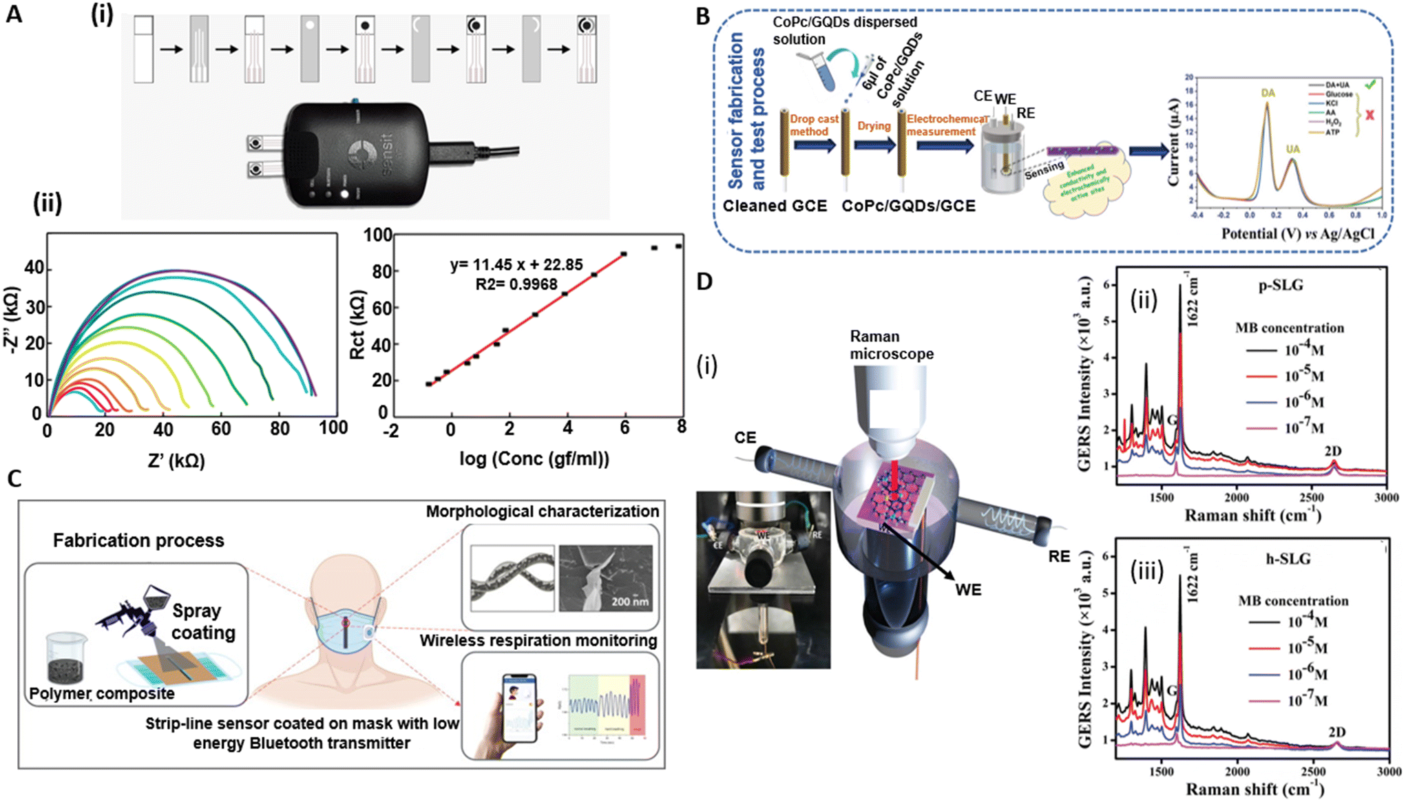 | ||
| Fig. 7 Electroanalytical devices based on electrochemical impedance spectroscopy (EIS), voltammetry, electrical conductivity, and multimodal methods. [A] (i) Preparation process to create graphene-based electrochemical test strips for SARS-CoV-2 detection. (ii) EIS data (Nyquist curves) and the extracted charge transfer resistance, Rct, for different analyte concentrations. Reprinted with permission from ref. 34. Copyright 2023 MDPI Diagnostics. [B] Performance of CoPc/GQDs/GCE electrodes for simultaneous dopamine (DA) and uric acid (UA) detection, showing low detection limits and high selectivity. Reprinted with permission from ref. 198. Copyright 2022 Wiley Advanced Materials Interfaces. [C] Graphene nanoplatelets (GR-NPs) utilized for enhancing the electrical and piezoresistive properties of surgical mask sensors, with an emphasis on the inverse relationship of GR-NP content to gauge factor. Reprinted with permission from ref. 199. Copyright 2023 Elsevier Materials and Design. [D] (i)–(iii) Application of pristine and hydrogenated single-layer graphene in graphene-enhanced Raman spectroscopy (GERS), demonstrating the modulation of GERS signals and sensitivity through doping with holes. Reprinted with permission from ref. 200. Copyright 2022 Wiley Advanced Materials Interfaces. | ||
Voltammetry techniques
Voltammetry methods include cyclic voltammetry (CV, where the potential between electrodes is cyclically varied to study redox reaction kinetics), linear sweep voltammetry (LSV, involving a linear potential sweep to analyze electron transfer reactions), and pulse-based methods such as differential pulse voltammetry (DPV, which uses periodic pulses superimposed on a linear potential sweep for enhanced resolution of closely spaced redox species), among others. In these techniques, a three-electrode system is usually used: a working electrode (WE, often modified with GR-based material for increased sensitivity and stability), a reference electrode (RE, typically Ag/AgCl ink in on-chip systems), and a counter electrode (CE, commonly platinum or gold).175 Among various steady state voltammetry methods, CV is used extensively to decipher the underlying electrochemical mechanisms, electron transfer kinetics, and diffusion processes, allowing researchers to elucidate the properties of sensing materials. On the other hand, pulsed voltammetry, like DPV, and square wave voltammetry (SWV) offer enhanced sensitivity and analytical capabilities. These methods achieve improved performance by applying pulses that decrease background noise and increase signal clarity, allowing for the detection of trace-level substances. These techniques also provide faster, high-resolution analysis, making them suitable for complex mixtures and a wide range of applications, from environmental to clinical diagnostics.201 A thorough overview of voltammetry-based biosensors can be found in review articles by Yuwen et al., Majer-Baranyi et al., and Bano et al.202–204Several voltammetry-based sensors utilizing GR and its derivatives/hybrids have been reported. For example, Saisahas et al. developed electrochemical paper-based analytical devices (ePAD) enriched with graphene ink and modified with polyaniline (PANI) for the detection of xylazine, a veterinary sedative, using DPV. Enhanced with graphene and polyaniline, the ePAD ensures efficient charge transfer with a larger surface area. It showcases a low detection limit of 0.06 μg mL−1 and a reliable performance with less than 5% deviation. Practical testing confirmed its accuracy, with 84–105% recovery in beverage samples.205
Another study used GR quantum dots (GQDs) coupled with cobalt phthalocyanine (CoPc) to modify glassy carbon electrodes (GCEs).198 The resulting CoPc/GQDs/GCEs showed superior electrocatalytic activity for the simultaneous detection of dopamine (DA) and uric acid (UA) in PBS, outperforming traditional CoPc electrodes. The CoPc/GQDs/GCE showed reversible DA redox peaks and an irreversible UA oxidation peak on CV curves displaying heightened sensitivity and selectivity for simultaneous DA and UA detection. Notably, the sensor achieved low detection limits of 21 nM for DA and 145 nM for UA, operating within distinct linear ranges: 2.91–33.38 μM and 45.39–164.2 μM for DA (with an R2 = 0.9809 and 0.9922, respectively) and 10.76–3003 μM for UA (R2 = 0.9901). Additionally, selectivity tests showed minimal interference from common substances, maintaining a relative error within ±5.5%, confirming its analytical precision. Furthermore, stability testing revealed impressive durability, with response retention of 87.9% for DA and 92.3% for UA over 13 days, underscoring the sensor's potential for long-term biomedical and clinical usage (Fig. 7B).
In another work, a LIG-based sensor for the detection of 4-nitrophenol (4-NP, a water pollutant) was developed. 4-NP was analyzed in a 0.1 M PBS solution using the LSV method. The sensors exhibited linear behavior in two concentration ranges (0.15 to 1 μM and 2.5 to 100 μM) with a LOD of 95 nM. The sensor displayed good selectivity for 4-NP, even in the presence of isomers and other phenolic compounds and in sewage samples with different 4-NP concentrations.206
Electrical conductivity
Electrical conductivity represents a material's capacity to conduct electric current. Upon occurrence of the event of interest (such as humidity change, biomolecule capture, or change of strain), the conductance (inverse of resistance) changes. For example, Wu et al.207 utilized crumpled GR, derived from heat-induced transformation of polystyrene-coated CVD-grown GR, for the sensitive detection of the cancer biomarker miRNA-21. This unique structure, enhanced through low-damage plasma treatment, increases the surface area and creates an optimized microenvironment for target capture, notably improving the sensor's sensitivity and selectivity. The biosensor achieved an impressive LOD of 1.74 pM and a high degree of linearity from 10 nM to 1 pM. Practical application assessments conducted in complex biological media, specifically undiluted human serum albumin (HSA) and PBS, confirmed the sensor's robust performance and low interference, maintaining effective biomarker detection under real-world conditions. Stability tests further revealed that the sensor's performance remained above 95% over a one-week period, indicating its suitability for sustained use in clinical settings.In another work, graphene nanoplatelets (GR-NPs) were used to enhance the capabilities of surgical mask sensors, specifically focusing on electrical conductivity and piezoresistive behavior.199 GR-NPs, due to their excellent electrical properties, were used to create conductive pathways on the mask's surface. Three types of masks, each with varying GR-NP concentrations, were subjected to electromechanical tests to examine changes in resistance under structural deformation. The increase in resistance under applied strain, particularly evident in the low GR-NP sample, was due to two primary mechanisms: (i) the disconnection mechanism involved a reduction in overlapped areas between GR flakes under strain, and (ii) the tunneling effect allowed electrons to cross the nonconductive polymer matrix, with tunneling resistance increasing proportionally to the GR-NP aggregation distance. The gauge factor, GR = (ΔR/R0/ε), representing the sensitivity of the sensors, was inversely related to the GR-NP content. The GR-NP-based strip lines demonstrated a fast response time (∼42 ms) and exceptional reproducibility and stability when used as a respiration sensor. Despite changes in temperature and humidity, the sensor's performance remained consistent, thereby validating its potential in healthcare applications (Fig. 7C).
Beniwal et al. used graphene–conductive carbon (GR-C) to construct humidity sensors via a screen-printing method.208 The screen-printing process enabled the creation of sensors with different layer numbers. The layers’ composition significantly influenced the sensors’ electrical conductivity, impacting the sensors’ response to humidity. Researchers observed that sensor resistance increased proportionally to relative humidity (RH) levels, ranging from 35%RH to 91%RH. Each sensor configuration showed distinct baseline resistances and resistance change rates due to their layer differences, with the single-layer sensor exhibiting superior electrical conductivity changes in response to varying humidity levels. The sensor's response and recovery times were the fastest for a one-layer sensor, underscoring its enhanced performance.
Multimodal methods
Apart from the methods summarized above, there are a variety of multimodal methods based on GR for biosensing. Two of the most studied ones include photoelectrochemical (PEC) and Raman–electrochemical methods. The PEC technique involves the generation of photocurrents when a semiconductor absorbs light, leading to electron–hole pair creation. In contrast, Raman–electrochemical methods provide insights into molecular vibrations in electrochemical environments, capturing changes in the Raman spectra due to alterations in the oxidation state or molecular structure. Recent studies have reported the combination of these techniques using GR-based biosensors. These sensors exploit the unique electronic properties of GR and its enhanced Raman and PEC responsiveness. When combined, they offer sensitive, rapid, and multiplexed detection of biomolecules, holding great promise for non-invasive diagnostics and real-time health monitoring.209In one of the notable contributions, a GR-based PEC immunosensor was developed, featuring green synthesized rGO-Au as the substrate.200 This study illuminated the sensor's promising capability to detect the S100β biomarker, an indicator of neurological disorders. rGO-Au was employed to create a biocompatible microenvironment for the immobilized antibodies, maintaining their activity and significantly enhancing the sensor's long-term stability. The immunosensor exhibited a “signal-on” response trend when subjected to different S100β concentrations. This trend denoted that the photocurrent intensity rose proportionately with increased S100β biomarker concentration during incubation with labelled anti-S100β. The immunosensor demonstrated a robust linear response over a wide dynamic linear range (DLR) of 0.25 to 10![[thin space (1/6-em)]](https://www.rsc.org/images/entities/char_2009.gif) 000 pg mL−1 S100β, with an impressively low LOD of 0.15 pg mL−1. Furthermore, this LOD surpassed the performance of prior GR-based immunosensors, and the DLR was higher than that of a poly(ethyleneimine)-based sensor; this was attributed to the effectiveness of the rGO-Au platform. Notably, the sensor was resilient to interference from common antigens such as HSA and human immunoglobulin G (HIgG), emphasizing its selectivity in S100β detection.
000 pg mL−1 S100β, with an impressively low LOD of 0.15 pg mL−1. Furthermore, this LOD surpassed the performance of prior GR-based immunosensors, and the DLR was higher than that of a poly(ethyleneimine)-based sensor; this was attributed to the effectiveness of the rGO-Au platform. Notably, the sensor was resilient to interference from common antigens such as HSA and human immunoglobulin G (HIgG), emphasizing its selectivity in S100β detection.
One of GR's distinctive applications is in GR-enhanced Raman spectroscopy (GERS), which is rooted in a charge transfer mechanism that fosters interaction between adsorbed molecules and the GR substrate. Contrary to traditional surface-enhanced Raman spectroscopy (SERS), GERS offers a more stable and reproducible Raman signal through a chemical mechanism, highlighting its potential over other methodologies. In this context, Kaushik et al. recently showcased the use of pristine (p-) (Fig. 7D (ii)) and hydrogenated (h-) (Fig. 7D (iii)) graphene in devising GR-enhanced spectro-electrochemical sensors (GE-SPECSs).210 Their study revealed that hole-doped h-graphene exhibited superior GERS signals compared to p-graphene, achieving a LOD of around 10–7 M. By capitalizing on the adjustable work function of graphene, they demonstrated the ability to modulate the GERS signal and probe various oxidation states of molecules through the application of appropriate external potentials (Fig. 7D (i)).
Biosensors based on graphene field effect transistors (GFETs)
Field effect transistors, particularly electrolyte-gated transistors (EGTs) are central to bioelectronic devices. They can transduce and amplify biological signals into electronic ones at low voltages making them suitable candidates for low power biosensing. With three terminals (source: s, drain: d, and gate: g), the current between the source and the drain (Ids) is modulated by applying a voltage to the gate electrode (Vgs), which directly contacts both the electrolyte and the transistor channel. By applying voltage, the ions drift from the electrolyte toward the channel material, leading to alterations in the electronic charges within the channel influencing its conductivity. This results in modulation of the electronic current flowing through the transistor channel. The unique design of EGTs facilitates their operation at low voltages compared to conventional FETs, making them suitable for cell monitoring,211 electrophysiology,212 and in vitro biosensing,213 among others.The operating characteristics of GFETs are commonly described through three foundational curves: transfer characteristics (Ids–Vgs), output curves (Ids–Vds), and time-series measurements. In GR-based devices, the transfer characteristic – often depicted as a V-shaped graph – is generated by changing the gate voltage, Vgs, while maintaining a constant source–drain voltage, Vds. This curve is crucial for understanding key parameters, mainly the transconductance (gm = (W/L)μCgVds, where W and L are the width and length of the GR channel, μ is the mobility of charge carriers and Cg is the gate capacitance). Transconductance itself is influenced by various factors, such as the dimensions of the channel and the gate capacitance. The choice of the gate configuration also impacts the performance of a GFET sensor. For applications involving liquid samples, an electrolyte gate is frequently preferred due to its ability to facilitate efficient electrical measurements directly within the sample medium. On the other hand, back-gate configurations are better suited for sensing volatile compounds in gaseous media.214
Most GFET biosensors operate mainly based on the charge transfer principles and the shift of the Dirac point/voltage (Vdirac), which is the Vgs at which the minimum conductivity point (σmin) occurs in GR.215 In a pristine state, this point occurs at Vgs = 0; however, it can shift upon the functionalization/doping of GR or its interaction with other molecules.216,217 One mechanism that causes shift in the Dirac point involves direct charge transfers between redox probes and targets, resulting in either n-doping or p-doping effects on GR. This alteration affects GR's Fermi level, leading to changes in conductivity and subsequently shifting the Dirac point.218 Another factor influencing this shift is the electrostatic gating effect, where probe–target binding induces a local external voltage drop across the GR channel due to the accumulated charges. This effect shifts the Dirac point similarly to the doping effect, contributing a significant role to the sensing mechanism by GFET.219,220
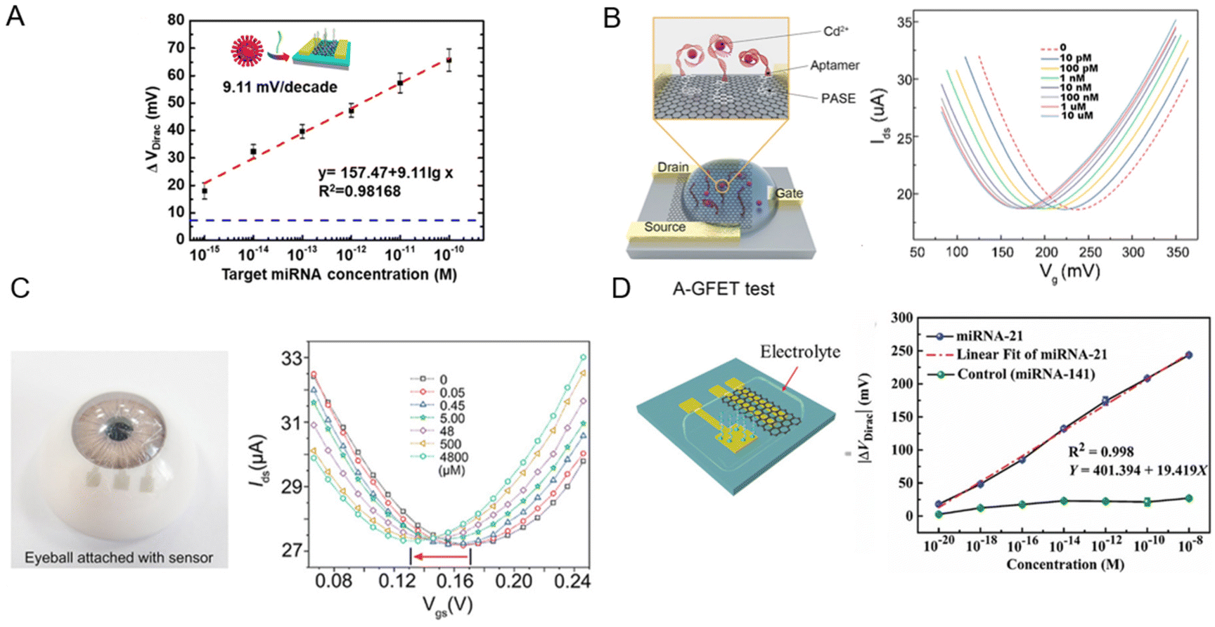
In one example, Gao et al. conducted a study where they synthesized monolayer graphene via chemical vapor deposition to fabricate poly-L-lysine (PLL)-functionalized graphene field-effect transistor biosensors (Fig. 8A).221 These devices exhibited the ability to detect specific miRNA sequences associated with breast cancer (miR-4732, miR-191, miR-21, miR-125) and SARS-CoV-2 virus sequences. The biosensor showed a LOD of 1 fM, demonstrating its potential for physiological applications. The readout method for the system was based on changes to the Vdirac of the biosensors, which served as a crucial indicator of successful detection. The Dirac voltage shifted up to ∼60 mV to the left when exposed to 100 pM of fully complementary SARS-CoV-2 RNA. This pronounced shift implied a robust interaction between the graphene surface and SARS-CoV-2 RNA, which led to change of the electronic properties of graphene and as a result, shift of the Dirac point. The shift toward the left indicated that the RNA interaction was inducing electron-like (n-type) behaviour in graphene.221
 | ||
| Fig. 8 Graphene-based field effect transistors (GFETs) for biosensing. [A] A GFET functionalized with poly-L-lysine (PLL) for detection of miRNA and SARS-CoV-2 RNA. ΔVDirac as a function of analyte concentration is plotted. Reprinted with permission from ref. 221. Copyright 2022 American Chemical Society. [B] A comprehensive three-stage process showing efficient oil–water separation and Cd2+ detection via an aptamer-functionalized GFET. Reproduced with permission from ref. 222. Copyright 2022 the Royal Society of Chemistry. [C] A wearable nanosensor, utilizing a single-layer GR channel, leverages the Dirac point shift to detect variations in L-cysteine concentrations in artificial tear. Used with permission from ref. 223. Copyright 2022 Wiley. [D] Solution-gated graphene transistors functionalized with ssDNA probes offer a label-free approach for miRNA-21 detection, using Dirac voltage shifts for highly sensitive prostate cancer diagnosis. Used with permission from ref. 224. Copyright 2022 Wiley. | ||
Zhang et al. used single-crystal GR in an electrolyte-gated GFET biosensor.225 The sensing was based on a dual detection mechanism employing ultra-high-frequency (UHF) interferometry and monitoring the variation in graphene resistance. The binding of biotin to streptavidin and the interaction between complementary peptides (E and K) were explored in the two model systems in this study. The experiments were conducted in PBS solution at a neutral pH, with a non-covalent labelling technique being used to attach biotin and peptide E onto the surface of the graphene layer. The LOD for streptavidin was remarkably low at 1 × 10⁻⁹ M, enhancing its physiological relevancy. To enhance sensitivity, the impedance-matching circuits were integrated into the system setup. The GR surface was functionalized with biotin or peptide E, linked via a 4 nm long pyrene–polyethylene glycol linker. This functionalization led to a negative (leftward) shift in the Dirac point by around 100 mV, which indicated a change in carrier concentration. Notably, the transconductance and carrier mobility decreased in the electron-dominant region post-functionalization; however, they remained stable in the hole-dominant region. This implied that functionalization introduced more scattering for electrons than holes. Additionally, the UHF signal showed abrupt changes due to its sensitivity towards GR's electrical properties and local capacitance fluctuations upon molecular interactions.225
In another work, Wang et al. developed a graphene FET functionalized with aptamers (A-GFET) to detect cadmium ions (Cd2+) in oily wastewater (Fig. 8B).222 This A-GFET showed a remarkable detection limit of 0.125 picomolar (pM) significantly surpassing the World Health Organization's permissible concentration of Cd2+ in drinking water. The readout system was based on changes to the A-GFET's electrical signal upon Cd2+ ion interaction. As the concentration of Cd2+ increased, the graphene transfer characteristic curve consistently shifted to the negative direction along the x-axis of the characteristic curve of A-GFET. Besides, an integrated photoacoustic alarm system alerted when the Cd2+ concentration exceeded a predetermined threshold.222
Huang et al. fabricated an innovative transparent wearable GFET biosensor (Fig. 8C).223 They employed a PET substrate with a single-layer GR channel and transparent WO3/Au/WO3 electrodes as drain, source, and gate in order to detect L-cysteine at mildly acidic to neutral pH. To detect changes in biomarker concertation, a shift in the Dirac point is exploited. As the L-cysteine concentrations increase from 0 to 4800 × 10–6 M, the ΔVDirac of the biosensor increases by 22 mV to the left. By accurate tracking of the Dirac point through the optimized procedure, LODs as low as 0.043 × 10⁻⁶ M in artificial tears and 0.022 × 10⁻⁶ M in undiluted sweat were achieved.223
Xue et al. introduced a graphene-based bioelectronic sensing platform employing the Dirac point for detecting multiple ions such as potassium (K+), sodium (Na+), and calcium ions (Ca2+).226 The sensor array featured a 30 × 30 μm graphene channel and two titanium/gold (Ti/Au) source/drain electrodes, functionalized with ion-selective membranes (ISMs) for K+, Na+, and Ca2+.226 Machine learning algorithms were employed for data analysis, resulting in excellent sensitivity and reversibility. Interestingly, Nernstian slopes of −54.7 ± 2.90 mV per decade for K+, −56.8 ± 5.87 mV per decade for Na+, and −30.1 ± 1.90 mV per decade for Ca2+ were achieved through this approach. Furthermore, with negligible sensitivity drifting over six months, the sensor exhibited excellent reversibility and long-term stability.
Deng et al. fabricated a solution-gated graphene transistor (SGGT) biosensor for rapid detection of miRNA-21, which is a significant biomarker for early prostate cancer diagnosis (Fig. 8D).224 High-quality CVD graphene was grown as the conductive channel of a SGGT for detecting different concentrations of miRNA-21. As the concentrations of the miRNA-21 target increased, the transfer curve moved toward a positive gate voltage due to the negative charge carried by miRNA in the electrolyte solution. By applying single-stranded DNA (ssDNA) probes immobilized on an Au gate electrode, the biosensor achieved a LOD of 10–20 M. Importantly, the device was able to distinguish cancer patients from the control group, surpassing the conventional prostate-specific antigen detection technique commonly used for this purpose.
In summary, FET-based biosensors, particularly GR variants, exhibit high sensitivity and adaptability in detecting a wide range of biomolecules. Despite their challenges such as complex manufacturing processes, ionic screening interference, pre-treatment requirements, and shelf-life, these devices offer promising potential in advancing point-of-care sensing. A thorough overview of GFET-based biosensors can be found in review articles by Krishnan et al. and Dai et al.227,228
Applications
In vitro assays
GR-based sensors have been developed for in vitro detection of various important small biomolecules (such as AA, DA, and UA, which are frequently found together in body fluids229), hormones (e.g., cortisol, ghrelin, and peptide YY),230,231 proteins (e.g., antibodies, spike protein),232,233 and pathogens (bacteria, virus). In the following, we provide a summary of examples of in vitro assays based on GR for various target analytes.GR-based devices have shown great success in DA detection. An abnormal level of DA in the brain may indicate Parkinson's disease and schizophrenia, among other neurological conditions.234 Li et al. prepared a 3D nanostructured composite of the MoS2 nanospheres and polyaniline on the rGO framework, using a one-pot hydrothermal approach.229 The MoS2-PANI/rGO suspension was then dropped on a GCE and dried to make the WE. The sensor exhibits high sensitivity for the simultaneous detection of AA, DA, and UA with three distinguishable oxidation peaks (EAA peak = 20 mV, ΔEDA peak = 196 mV, ΔEUA peak = 320 mV) in DPV measurements. The responses toward AA, DA, and UA were in the linear ranges of 0.05–8.0 mM, 5–500 μM, and 1–500 μM, respectively, with LOD values of 22.20, 0.70, and 0.36 μM (Fig. 9A). Human serum and urine samples were also tested to show the sensor's selectivity and stability.
 | ||
| Fig. 9 Graphene-based devices for in vitro and in vivo detection of biomarkers. [A] Schematic diagram of a MoS2-PANI/rGO-based electrochemical biosensor for simultaneous detection of ascorbic acid (AA), dopamine (DA), and uric acid (UA). Reprinted with permission from ref. 229. Copyright 2019 The Royal Society of Chemistry. [B] Schematic illustration of immobilization and hybridization steps of a paper-based electrochemical DNA biosensor. Reprinted with permission from ref. 235. Copyright 2016 Elsevier B.V. [C] Preparation, functionalization, and sensing scheme of a LIG-based immunosensor for bacterial detection. Reprinted with permission from ref. 232. Copyright 2020 American Chemical Society. [D] Aptamer–(Ru) probes based on graphene oxide (GO) relate inflammation duration to IFN-γ concentration in enteritis mice, advancing in vivo sensing. Reprinted with permission from ref. 236. Copyright 2018 American Chemical Society. [E] An in vivo corneal biosensor responds to varying dopamine levels in (i) rabbit's eye and (ii) human tear, opening avenues for real-time biological monitoring. Used with permission from ref. 237. Copyright 2020 Wiley. | ||
In a work by Jain et al., they developed an electrochemical sensor using the AuNPs/N2-doped graphene nanosheet/FTO electrode. The sensor was used to detect the level of glycated hemoglobin (HbA1c) and was accordingly examined to check for diabetes mellitus. GR nanosheets were prepared using the modified Hummers’ method and then doped with nitrogen and gold NPs by solvothermal methods. The mixture was drop cast onto the cleaned FTO electrode to make the working electrode to measure the blood samples in buffer using the CV technique.238
Weng et al. reported a GFET device integrated with clustered regularly interspaced short palindromic repeats (CRISPR) assay. The sensor was used to detect both ssDNA and double-stranded DNA (dsDNA) targets. The LOD was 1 aM for the ssDNA human papillomavirus 16 synthetic target and 10 aM for the dsDNA Escherichia coli (E. coli) plasmid target without preamplification. Each chip contained a 48 GFET array to reduce the effects of measurement outliers from devices.239 In another work, Teengam et al. developed a paper-based electrochemical sensor for the detection of the human papillomavirus. The graphene–polyaniline composite was inkjet printed on paper and then functionalized with anthraquinone-labelled pyrrolidinyl peptide nucleic acid.235 Probe immobilization on the WE was first characterized by the EIS technique, and then the probe was investigated using SWV before and after hybridization with the target DNA, resulting in an LOD of 2.3 nM (Fig. 9B). The sensor was used to monitor the amount of HPV-DNA type 16 to identify the primary stages of cervical cancer.
Soares et al. reported label-free immunosensors based on the LIG to electrochemically quantify the food-borne pathogen Salmonella enterica serovar Typhimurium.232 LIG was functionalized with polyclonal anti-Salmonella using N-hydroxysuccinimide and 1-ethyl-3-(3-dimethylaminopropyl) carbodiimide as an activating agent. The immunosensor enabled the detection of the pathogen at 13 ± 7 CFU mL−1 in a complex medium (chicken broth), with a response time of 22 min via the EIS method (Fig. 9C). This work showed a low-cost preparation and fast detection process compared to CVD-graphene devices. In a work by Tan et al., a GFET was functionalized with phage tail spike proteins to measure E. coli concentration, achieving a Dirac point shift of 35 mV when about 50 bacteria were bound to GR.240
GR has also been used to develop in vitro sensors for detecting viral infections, such as SARS-CoV-2. Virus sensing is usually achieved using impedimetric methods or GFETs. Gao et al. developed a GFET-based sensor with a poly-L-lysine functionalized channel to specifically detect breast cancer miRNA and SARS-CoV-2 RNA within 20 min. The LOD was as low as 1 fM using 2 μL samples.241 Walters et al. reported an Au nanoparticle-functionalized graphene resistor sensor to detect the hepatitis C virus core antigen (HCVcAg) in real time.242 The sensor demonstrated good sensitivity to HCVcAg of higher than 100 pg mL−1. In another work, LIG was functionalized with pyrenebutyric acid (PBA) as a linker to immobilize antibodies to specifically detect biomarkers including SARS-CoV-2 nucleocapsid protein (NP), immunoglobulins (Igs) against SARS-CoV-2 spike protein and C-reactive protein. The authors noted that since the SARS-CoV-2 NP structure was 90% similar to SARS-CoV NP, high interference was observed.166
In vivo monitoring and real-time study of biological cells
In this part, we discuss how GR-based biosensors have been applied to study living organisms. The real-time monitoring capability is critical for immediate medical interventions, serving as a cornerstone for advancing personalized healthcare. By providing insights into individual health dynamics, GR-based biosensors offer the potential to tailor medical interventions and treatments to the specific needs and conditions of patients. GR is well suited for this application owing to its biocompatibility, chemical inertness, and feasibility of various functionalization routes.In this context, Jin et al. developed a biosensor to monitor hydrogen peroxide (H2O2, a crucial oxidative stress and inflammation biomarker) in real time and in vivo.243 A rGO/platinum nanoparticle nanohybrid-based microneedle array was applied to facilitate seamless skin penetration, resulting in highly sensitive detection. The real-time in vivo testing performed on mice demonstrated the potential for this biosensor to provide continuous, non-invasive H2O2 monitoring in humans, representing an important step forward in chronic disease management. In another study, Cao et al. introduced an aptamer-based biosensor employing GO for signal amplification (Fig. 9D).236 This sensor was specifically targeted to detect interferon-γ (IFN-γ), a crucial biomarker in immune responses. The sensor achieved a low LOD of 1.3 pg mL−1, within the physiological range of IFN-γ in human blood. It demonstrated an exceptional ability to accurately detect IFN-γ over a period of 48 h, with no requirement for physical barriers or active drift correction algorithms, making it a reliable detection methodology.236
Taking a different approach, Zhang et al. developed a sensor for real-time dopamine detection in tear fluids, a potentially transformative approach for diagnosing and monitoring neurological conditions (Fig. 9E).237 They introduced novel functionalized sulfur-doped graphene, significantly enhancing the LOD down to 101 × 10–9 M. Their sensor exhibited remarkable stability with a one-month shelf life at 4 °C, retaining 85% sensitivity. The in vivo testing performed on New Zealand white rabbits confirmed its potential and suitability for tear fluid-based dopamine detection.237
Similarly, Wu et al. fabricated a microtransistor probe using GR-tethered dopamine-specific aptamers for in vivo dopamine monitoring in brain.244 With a LOD of 10 pM in artificial cerebrospinal fluid, the biosensor can detect physiologically relevant dopamine levels. Ex vivo and in vivo tests were conducted on mice to validate its potential for real time neurotransmitter tracking, which is a critical asset for understanding and managing neurological disorders.244
In addition to studies with animals, bacterial cells have also been directly investigated using GR-based devices. For example, Zhou et al. introduced a flexible MoSx/LIG-based electrochemical sensor for real-time monitoring of phenazine produced by Pseudomonas aeruginosa bacteria. In this work, different concentrations (100 nM – 100 μM) of pyocyanin (PYO) and phenazine-1-carboxylic acid (PCA) for the production of phenazine molecules were detected via the SWV method. For detecting PYO and PCA, LIG was functionalized with MoSx on the WE via electrodeposition. The LIG/MoSx electrode, with a deposition time of 60 min, showed an LOD of 0.19 μM and a sensitivity of 0.97 μA μM−1 for PYO in brain heart infusion (BHI). Additionally, for PCA, the LOD was reported to be 1.2 μM in BHI. Also, real-time monitoring of phenazines produced by P. aeruginosa in wound simulating medium showed that LIG/MoSx could detect PYO with a LOD of 1.3 μM.245
Wearable devices
Wearable devices are extremely useful for personalized health monitoring by continuously collecting and analyzing physiological signals from patients. GR is well suited for these applications due to its mechanical properties, allowing the sensors to be flexible and stretchable. It is envisioned that in the future, non-invasive wearable devices will be an affordable and convenient alternative to conventional bulky and expensive instruments.In 2016, Lee et al. developed a GR–gold hybrid electrochemical device for sweat-based diabetes monitoring and therapy.246 The soft substrate enables conformal contact with human skin under deformation, enabling stable sensing with body movements. The glucose sensor measured the electrochemical signal of the Prussian Blue functionalized GR by the reduction of H2O2 generated from glucose oxidase. Kwon et al. reported an all-printed wireless biosensor using biocompatible GR ink for electromyogram (EMG) recording.247 The high-aspect-ratio GR material offers excellent conformal lamination on human skin for EMG recording. The EMG data of specific muscle motion during flexion of each digit were recorded and analysed with a deep-learning algorithm to enable real-time classification of individual digit movement for robotic hand control. Yang et al. showed a wearable sensor using LIG as the multiplexing electrode to detect UA and tyrosine (TYR) from sweat as well as body temperature and the respiration rate (Fig. 10A).248 Laser engraving was used to fabricate the LIG electrode and pattern the double-sided medical adhesives. Microfluidic channels improved the rate of collecting sweat from the skin. Sensor validation was performed by applying the integrated system on different body parts and measuring the UA and TYR levels in real time. The results showed its reliability for the non-invasive monitoring of physiological signals and the potential for disease diagnosis such as hyperuricemia.
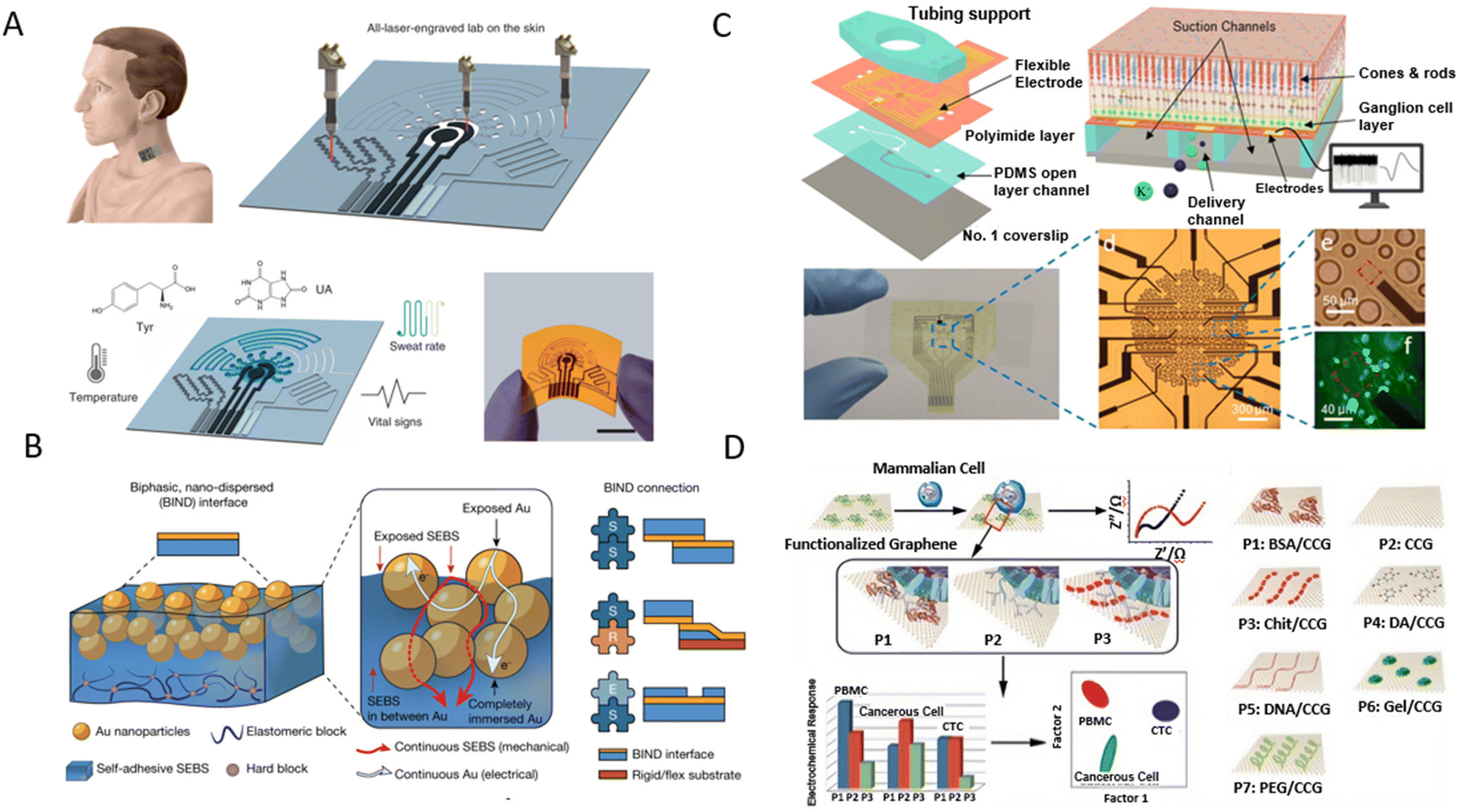 | ||
| Fig. 10 Graphene electroanalytical devices used for wearable and ex vivo applications. [A] An entirely laser-engraved sensing system for detection of tyrosine (TYR) and uric acid (UA) in sweat. Reproduced with permission from ref. 248. Copyright 2020 Springer Nature Limited. [B] Schematic showing that stretchable hybrid devices are typically assembled from three elementary modules (encapsulation and soft and rigid modules) and illustration of a BIND interface. Reproduced from with permission from ref. 249. Copyright 2023 Springer Nature Limited. [C] THY1.2-YFP retina analysis using graphene-based microelectrode array integrated with an Olympus IX-81 microscope for detailed retinal studies, employing advanced temperature and pressure control, high-frequency voltage sampling, and complex data processing for comprehensive anatomical and electrophysiological insights. Reprinted with permission from ref. 250. Copyright 2023 The Royal Society of Chemistry. [D] A graphene-based sensor where unique electrochemical signatures of different cell types, identified through impedance measurements and linear discriminant analysis, reveal distinct cellular properties. Reprinted with permission from ref. 251. Copyright 2017 NPG Asia Materials. | ||
Cai et al. took advantage of the biocompatibility and flexibility of graphene fiber fabric, modified with glucose oxidase and chitosan, to fabricate a wearable glucose monitoring patch.252 The sensor was able to detect glucose concentrations from 2 μM to 650 μM, aligned with the human interstitial fluid's physiological glucose range. They carried out tests to compare the sensor output to traditional finger-prick blood samples, showing a strong correlation between them, confirming its efficacy and potential for continuous, non-invasive glucose monitoring.252 Recently, Kireev et al. developed GR bioimpedance tattoos using CVD grown GR for continuous monitoring of arterial blood pressure.253 The atomically thin, self-adhesive, light weight, and unobtrusive GR electronic tattoos as human bioelectronic interfaces enabled a longer monitoring time than that previously reported. Combined with the cycling-trained machine learning regression model, the accuracies were 0.06 ± 2.5 mm Hg (diastolic) and 0.2 ± 3.6 mm Hg (systolic).
It is worth highlighting that on-body measurements need good interconnects between soft tissue and Si-based devices. Stress at the interface may result in debonding failure. In addition, the encapsulation of devices is also essential since any contact on the interface can result biological fouling and device failure. To overcome these issues, Jiang et al. developed a universal interface that connected soft, rigid, and encapsulation modules through simply pressing (Fig. 10B). The connection between soft modules shows 3× electrical stretchability and 10× mechanical stretchability compared to commercial pastes. A stretchable device was also assembled using these connection modules for tests as a proof of concept.249
Ex vivo studies
GR-based electroanalytical devices are being increasingly employed in ex vivo studies, which enable the testing of drug effects outside a living organism (such as in organ-on-chip devices), often in a controlled laboratory environment.254,255 Such assays are critical for drug screening, especially in understanding drug interactions with target cells like cancer cells or neurons. The exceptional electronic properties of graphene enable sensitive detection and quantification of cellular responses, providing crucial insights into drug efficacy and potential side effects. Moreover, the integration of sensors that mimic the microenvironment of human organs has paved the way for more realistic and high-throughput drug testing. These advances highlight the transformative potential of GR-based devices in accelerating drug discovery and improving patient-specific therapeutic strategies.256Recent research has advanced the application of GR-based microelectrode array (MEA) platforms for ex vivo analysis.250 In this work, the GR-based MEAs, paired with an Olympus IX-81 inverted epifluorescence microscope, operated in oxygenated media at a temperature of 36 °C. The use of two syringe pumps created a controllable negative pressure. The readout methodology from these GR electrodes utilized a 16-channel amplifier and a lab-made printed circuit board zero-insertion-force connector, allowing for the recording of voltage at a 20 kHz sampling rate. Data processing incorporated spike sorting with the Plexon offline sorter and a Butterworth filter for further analysis. There has been considerable progress in detecting THY1.2-YFP retinas using GR-based MEAs, which is instrumental in creating a detailed anatomical and electrophysiological analysis (Fig. 10C).
In another work, Wu et al. used a modified Hummers’ method of preparing GR to develop an array-based system for cell sensing using a chemical nose/tongue approach that exploits subtle changes in the physicochemical nature of different cell surfaces.251 The chitosan-mediated graphene suspension was added dropwise onto the pretreated GCE and the EIS signals were recorded. The interaction of the cells with the functionalized GR array depended on the surface properties of the cell. Each type of cell had its colour-change fingerprint. To deconvolute the electrochemical reactions that provided discriminating signatures, the electrochemical data set was classified for all seven GR probes using linear discriminant analysis (Fig. 10D).
Lin et al. leveraged electronic GR tattoos as soft, tissue-imperceptible, and transparent bioelectronic interfaces.257 They meticulously characterized GFETs and employed them for monitoring cardiac activity in the well-established ex vivo Lutgendorf-perfused mouse heart model. In this setting, the electrogram recorded by graphene (gEG) was contrasted with the traditional far-field ECG, captured simultaneously using commercial Ag/AgCl needle electrodes in the perfusion bath. Notably, when comparing the signal-to-noise ratio (SNR) across varying gEG electrode sizes, the 1 mm electrode was found to be very effective, consistent with earlier observations. The capability of these GFET electrodes in cardiac actuation was also explored, highlighting their performance in both unipolar and bipolar modes. A pivotal pacing strength–duration curve, essential for understanding the efficiency of various GFET electrodes was studied, positioning the 1 mm unipolar GFET electrode favourably against a reference platinum electrode (Fig. 11A).
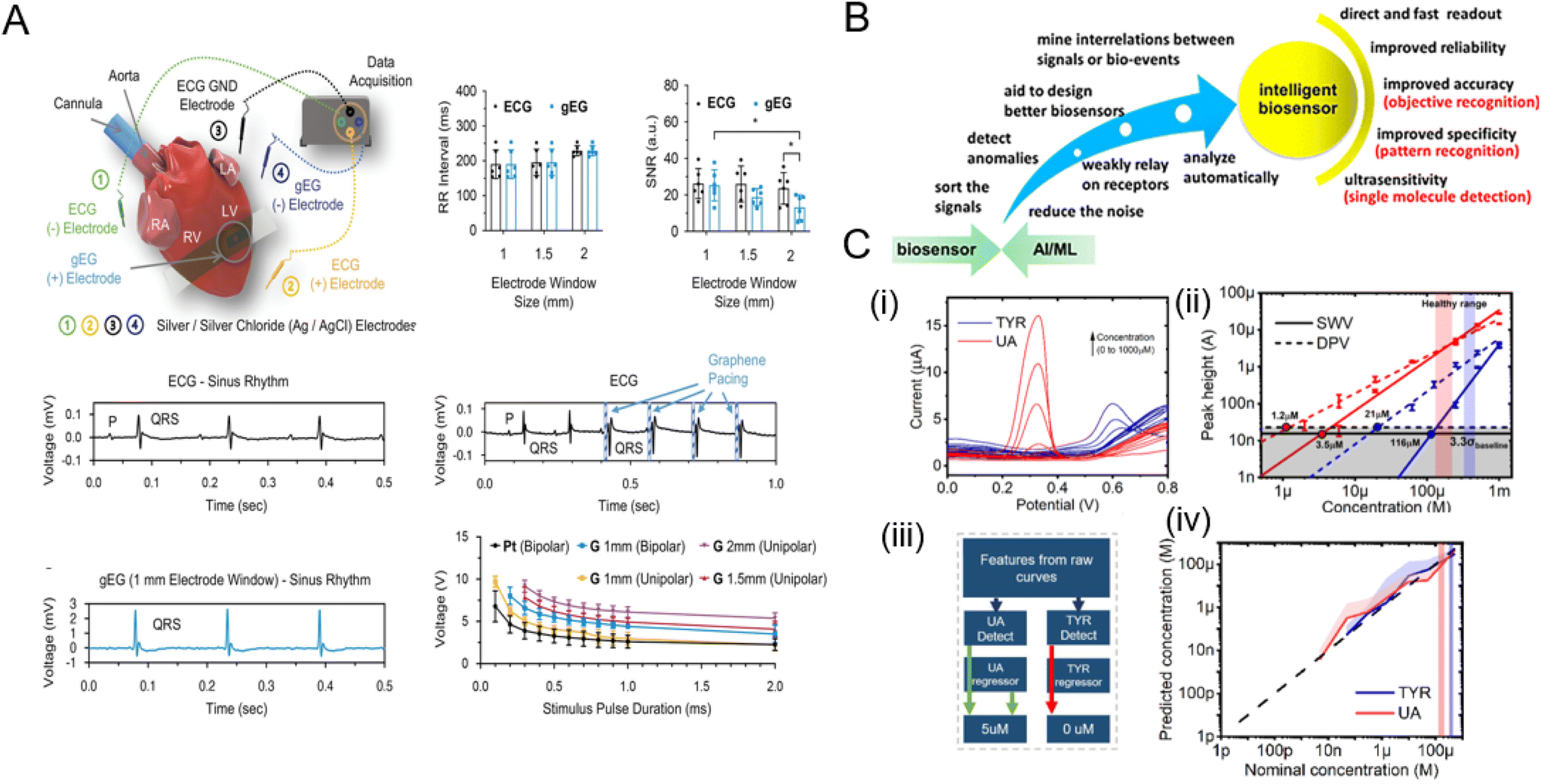 | ||
| Fig. 11 Ex vivo studies and emerging fields based on graphene-based biodevices. [A] Ex vivo cardiac electrophysiology sensing and pacing with GFETs. Reprinted with permission from ref. 257. Copyright 2023 Wiley-VCH GmbH. [B] Schematic highlighting the benefits of using machine learning (ML) in conjunction with biosensors. Adapted with permission from ref. 258. Copyright 2014 American Chemical Society. [C] Enhancement in limit of detection (LOD) using data processing techniques: (i) Raw DPV curves showing peaks for tyrosine (TYR) and uric acid (UA), (ii) extracted LODs using single peak analysis, (iii) the ML architecture that was developed to boost LOD by at least two orders of magnitude compared to conventional voltammetry methods (iv). Adapted with permission from ref. 259. Copyright 2022 Elsevier. | ||
Emerging topics
In recent years, there has been growing interest in the incorporation of machine learning (ML) algorithms into electrochemical biosensors (Fig. 11B). The recent trend in electrochemical sensor research has been to apply complex potential waveforms to the sensor in order to extract more information than just a linear potential sweep as in CV or pulse voltammetry (such as SWV, DPV).260 These complex waveforms may consist of using sinusoid superimposed ramps that form large amplitude alternating current voltammetry (LAACV),261 semicircular sweep voltammetry that employs a varying scan rate,262 or designer sinusoidal waveforms.263 Conventionally, data processing in the form of manual baseline subtraction and single peak-based trend analysis is used to quantify and detect analytes. One approach for data processing is to use ML to learn the subtle and complex signals associated with sensing.258 One of the key advantages of using ML in electrochemical biosensors is to facilitate pattern recognition and multiplex correlative data processing of the complex sensor output signals. It is common for other analytical/diagnostic methods such as spectroscopy or imaging techniques to use ML; however, the use of ML in electroanalytical biosensors has been slow to implement due to the lack of data corpus and complex signal variations in biological media.258 Using CV, ML-based support vector machine (SVM) models have been used to estimate the nitrate concentration.264 In other works,265,266 fast scan cyclic voltammetry (FSCV) was used along with ML models to selectively and accurately predict the neurotransmitter levels.The power of ML is in the vast data processing it can handle along with the added advantages of combining multiple sources of data. Multiple electrochemical methods, such as CV, SWV, DPV, and LAACV, are known to have varying sensitivities267 and varying background current rejection,268 and hence are expected to behave differently with different sensor materials, analytes, and media. Kammarchedu et al. demonstrated the effect of the multimodal effects and data fusion techniques and their effects on the sensing performance of the sensors by improving the LOD of TYR and UA in artificial saliva on LIG-based sensors 100 times by using ML (Fig. 11C).259
Furthermore, ML can also be used to predict new materials and material modifications for optimizing the sensing performance.269 One of the limiting factors for rapid sensor development is the slow process of material synthesis and subsequent benchmarking to find ideal material–biomolecule pairs. Towards this goal, density functional theory (DFT) studies as well as the establishment of other theoretical frameworks are crucial. One way to do this is to couple experimental and theoretical results in published studies in order to build a database that can be used to train an ML model.270
Discussion and conclusion
The remarkable properties of GR, precise synthesis techniques, and its diverse applications in biosensing and bioelectronics present a promising avenue for addressing the evolving challenges in the healthcare landscape. GR can be synthesized at scale using various top-down and bottom-up techniques. Furthermore, GR properties can be altered/tuned using doping, molecular/chemical functionalization, defect engineering, etc. Electrical, electrochemical, and electro-optical transduction methods can be utilized to fabricate devices on both rigid and flexible substrates. These GR-based electroanalytical devices offer the potential to revolutionize disease diagnosis, personalized medicine, and remote patient monitoring, particularly in the context of chronic diseases and infectious outbreaks, as well as bioelectronics and life science research when interfacing with biological cells. GR's exceptional electronic attributes augment the sensitive detection of cellular responses, thus enriching our understanding of drug efficacies and probable adverse effects. Recent innovations, including GR-based microelectrode array platforms and integrations that mirror human organ microenvironments, have markedly enhanced the realism and throughput of drug testing. These advancements signify a transformative era, potentially catalysing drug discovery and refining patient-tailored therapeutic strategies.Although significant progress has been made in the past years in GR-based wearable devices, there are still bottlenecks, such as power management, system integration, and commercializing high-performance sensors.255 In addition, achieving reproducibility in synthesis to maintain GR's properties during functionalization and ensuring biocompatibility for in vivo applications, demand careful consideration. Furthermore, the issue of sensor calibration and drift add to the challenges of prolonged storage and usage of these electroanalytical devices. Several drift correction algorithms have been developed271–273 to offset sensor drift and calibration-free sensors have been proposed274,275 to mitigate the need for recalibration. Still, further research and novel techniques are needed to solve these problems. Lack of unified standards for synthesis and fabrication of biosensor materials and devices, including those based on graphene, is also another major challenge which needs to be addressed for utilization of GR-based biosensors in the healthcare market. Overcoming these hurdles is crucial to realizing the full transformative potential of GR-based biodevices in public health and life sciences.
In the face of these challenges, this review article underscores the transformative role that GR-based devices can play in addressing critical healthcare needs. It is envisioned that smart devices enabling continuous and non-invasive monitoring of human health could be combined with machine learning and the Internet of Things (IoT), to generate predictive algorithms for early disease diagnosis. Especially, emerging technologies such as machine learning can help in enhancing the sensing performance of sensors as well as predict novel materials for sensing applications. As technology continues to advance, the integration of these devices with digital platforms holds the promise of ushering in an era of preventive and precision medicine, ultimately improving patient outcomes and healthcare management on a global scale.
Conflicts of interest
There are no conflicts to declare.Acknowledgements
This work was partially supported by NSF:I/UCRC Phase II: Center for Atomically Thin Multifunctional Coatings (ATOMIC; Award #2113864 ), NSF award #2236997, and the National Institutes of Health (awards R21EB031354 and R21GM132793). The content of this report is solely the responsibility of the authors and does not necessarily represent the official views of the National Science Foundation (NSF) or National Institutes of Health (NIH).References
- G. Ashraf, A. Aziz, T. Iftikhar, Z. T. Zhong, M. Asif and W. Chen, Biosensors, 2022, 12, 1183 CrossRef CAS PubMed.
- Y. Qiao, X. Li, T. Hirtz, G. Deng, Y. Wei, M. Li, S. Ji, Q. Wu, J. Jian, F. Wu, Y. Shen, H. Tian, Y. Yang and T. L. Ren, Nanoscale, 2019, 11, 18923–18945 RSC.
- D. G. Papageorgiou, I. A. Kinloch and R. J. Young, Prog. Mater. Sci., 2017, 90, 75–127 CrossRef CAS.
- A. Bolotsky, D. Butler, C. Dong, K. Gerace, N. R. Glavin, C. Muratore, J. A. Robinson and A. Ebrahimi, ACS Nano, 2019, 13, 9781–9810 CrossRef CAS PubMed.
- W. Zhang, Z. Huang, W. Zhang and Y. Li, Nano Res., 2014, 7, 1731–1737 CrossRef CAS.
- A. K. Geim and K. S. Novoselov, Nat. Mater., 2007, 6, 183–191 CrossRef CAS PubMed.
- X. Ling, J. Wu, L. Xie and J. Zhang, J. Phys. Chem. C, 2013, 117, 2369–2376 CrossRef CAS.
- R. Mas-Ballesté, C. Gómez-Navarro, J. Gómez-Herrero and F. Zamora, Nanoscale, 2011, 3, 20–30 RSC.
- ISO/TS 80004-13:2017(en), Nanotechnologies—Vocabulary—Part 13: Graphene and related two-dimensional (2D) materials, https://www.iso.org/obp/ui/en/#iso:std:iso:ts:80004:-13:ed-1:v1:en, (accessed 8 July 2023).
- A. R. Urade, I. Lahiri and K. S. Suresh, JOM, 2022, 75, 614–630 CrossRef PubMed.
- H. Wang, X. Yuan, G. Zeng, Y. Wu, Y. Liu, Q. Jiang and S. Gu, Adv. Colloid Interface Sci., 2015, 221, 41–59 CrossRef CAS PubMed.
- K. Y. Lau and J. Qiu, Light: Sci. Appl., 2023, 12, 1–2 CrossRef PubMed.
- T. Kuila, S. Bose, A. K. Mishra, P. Khanra, N. H. Kim and J. H. Lee, Prog. Mater. Sci., 2012, 57, 1061–1105 CrossRef CAS.
- S. Alwarappan, S. Boyapalle, A. Kumar, C. Z. Li and S. Mohapatra, J. Phys. Chem. C, 2012, 116, 6556–6559 CrossRef CAS.
- A. Bellunato, H. Arjmandi Tash, Y. Cesa and G. F. Schneider, ChemPhysChem, 2016, 17, 785–801 CrossRef CAS PubMed.
- R. Ye, D. K. James and J. M. Tour, DOI:10.1021/acs.accounts.8b00084.
- W. Wang, L. Lu, Z. Li, L. Lin, Z. Liang, X. Lu and Y. Xie, ACS Appl. Mater. Interfaces, 2022, 14, 1315–1325 CrossRef CAS PubMed.
- X. Zhang, Q. Jing, S. Ao, G. F. Schneider, D. Kireev, Z. Zhang and W. Fu, Small, 2020, 16, 1902820 CrossRef CAS PubMed.
- B. Sun, J. Pang, Q. Cheng, S. Zhang, Y. Li, C. Zhang, D. Sun, B. Ibarlucea, Y. Li, D. Chen, H. Fan, Q. Han, M. Chao, H. Liu, J. Wang, G. Cuniberti, L. Han and W. Zhou, Adv. Mater. Technol., 2021, 6, 2000744 CrossRef CAS.
- C. Huang, Z. Hao, Z. Wang, H. Wang, X. Zhao and Y. Pan, Adv. Mater. Technol., 2022, 7, 2101131 CrossRef CAS.
- M. T. Hwang, M. Heiranian, Y. Kim, S. You, J. Leem, A. Taqieddin, V. Faramarzi, Y. Jing, I. Park, A. M. van der Zande, S. Nam, N. R. Aluru and R. Bashir, Nat. Commun., 2020, 11, 1–11 CrossRef PubMed.
- Y. J. Hwang, H. Yu, G. Lee, I. Shackery, J. Seong, Y. Jung, S. H. Sung, J. Choi and S. C. Jun, Microsyst. Nanoeng., 2023, 9, 1–13 CrossRef PubMed.
- Y. Yang, Y. Song, X. Bo, J. Min, O. S. Pak, L. Zhu, M. Wang, J. Tu, A. Kogan, H. Zhang, T. K. Hsiai, Z. Li and W. Gao, Nat. Biotechnol., 2019, 38, 217–224 CrossRef PubMed.
- M. A. Yoon, C. Kim, J. H. Kim, H. J. Lee and K. S. Kim, Sensors, 2022, 22, 3944 CrossRef CAS PubMed.
- L. Xu, S. Ramadan, B. G. Rosa, Y. Zhang, T. Yin, E. Torres, O. Shaforost, A. Panagiotopoulos, B. Li, G. Kerherve, D. K. Kim, C. Mattevi, L. R. Jiao, P. K. Petrov and N. Klein, Sens. Diagn., 2022, 1, 719–730 RSC.
- H. Wang, Z. Hao, C. Huang, F. Li and Y. Pan, Nanoscale Adv., 2022, 5, 1416–1424 RSC.
- A. Esteban-Linares, X. Zhang, H. H. Lee, M. L. Risner, S. M. Weiss, Y. Q. Xu, E. Levine and D. Li, Lab Chip, 2023, 23, 2193–2205 RSC.
- D. Bitounis, H. Ali-Boucetta, B. H. Hong, D. H. Min and K. Kostarelos, Adv. Mater., 2013, 25, 2258–2268 CrossRef CAS PubMed.
- G. Reina, J. M. González-Domínguez, A. Criado, E. Vázquez, A. Bianco and M. Prato, Chem. Soc. Rev., 2017, 46, 4400–4416 RSC.
- N. Kumar, R. Salehiyan, V. Chauke, O. Joseph Botlhoko, K. Setshedi, M. Scriba, M. Masukume and S. Sinha Ray, FlatChem, 2021, 27, 100224 CrossRef CAS.
- C. Botas, P. Álvarez, P. Blanco, M. Granda, C. Blanco, R. Santamaría, L. J. Romasanta, R. Verdejo, M. A. López-Manchado and R. Menéndez, Carbon, 2013, 65, 156–164 CrossRef CAS.
- A. Ambrosi, C. K. Chua, A. Bonanni and M. Pumera, Chem. Rev., 2014, 114, 7150–7188 CrossRef CAS PubMed.
- A. Hazra and S. Basu, C, 2018, 4, 49 Search PubMed.
- F. L. Thiemann, P. Rowe, A. Zen, E. A. Müller and A. Michaelides, Nano Lett., 2021, 21, 8143–8150 CrossRef CAS PubMed.
- Z. Qin, G. S. Jung, M. J. Kang and M. J. Buehler, Sci. Adv., 2017, 3, e1601536 CrossRef PubMed.
- P. Tian, L. Tang, K. S. Teng and S. P. Lau, Mater. Today Chem., 2018, 10, 221–258 CrossRef CAS.
- P. Zheng and N. Wu, Chem. – Asian J., 2017, 12, 2343–2353 CrossRef CAS PubMed.
- M. R. Mahajan and P. O. Patil, Inorg. Chem. Commun., 2022, 144, 109883 CrossRef CAS.
- C. Han, N. Zhang and Y. J. Xu, Nano Today, 2016, 11, 351–372 CrossRef CAS.
- A. Celis, M. N. Nair, A. Taleb-Ibrahimi, E. H. Conrad, C. Berger, W. A. De Heer and A. Tejeda, J. Phys. D: Appl. Phys., 2016, 49, 143001 CrossRef.
- S. Dutta and S. K. Pati, J. Mater. Chem., 2010, 20, 8207–8223 RSC.
- M. C. F. Costa, P. R. Ng, S. Grebenchuk, J. Y. Tan, G. K. W. Koon, H. L. Tan, C. R. Woods, R. K. Donato, K. S. Novoselov and A. H. C. Neto, Carbon, 2023, 208, 140–147 CrossRef CAS.
- Z. Sun, S. Fang and Y. H. Hu, Chem. Rev., 2020, 120, 10336–10453 CrossRef CAS PubMed.
- K. S. Novoselov, A. K. Geim, S. V. Morozov, D. Jiang, Y. Zhang, S. V. Dubonos, I. V. Grigorieva and A. A. Firsov, Science, 2004, 306, 666–669 CrossRef CAS PubMed.
- Z. Li, R. J. Young, C. Backes, W. Zhao, X. Zhang, A. A. Zhukov, E. Tillotson, A. P. Conlan, F. Ding, S. J. Haigh, K. S. Novoselov and J. N. Coleman, ACS Nano, 2020, 14, 10976–10985 CrossRef CAS PubMed.
- T. C. Achee, W. Sun, J. T. Hope, S. G. Quitzau, C. B. Sweeney, S. A. Shah, T. Habib and M. J. Green, Sci. Rep., 2018, 8, 14525 CrossRef PubMed.
- W. S. Hummers and R. E. Offeman, J. Am. Chem. Soc., 1958, 80, 1339 CrossRef CAS.
- N. Kumar, R. Salehiyan, V. Chauke, O. Joseph Botlhoko, K. Setshedi, M. Scriba, M. Masukume and S. Sinha Ray, FlatChem, 2021, 27, 100224 CrossRef CAS.
- M. Bahri, S. H. Gebre, M. A. Elaguech, F. T. Dajan, M. G. Sendeku, C. Tlili and D. Wang, Coord. Chem. Rev., 2023, 475, 214910 CrossRef CAS.
- H. Tetlow, J. Posthuma de Boer, I. J. Ford, D. D. Vvedensky, J. Coraux and L. Kantorovich, Phys. Rep., 2014, 542, 195–295 CrossRef CAS.
- H. A. Alhazmi, W. Ahsan, B. Mangla, S. Javed, M. Z. Hassan, M. Asmari, M. Al Bratty and A. Najmi, Nanotechnol. Rev., 2021, 11, 96–116 CrossRef.
- M. Bahri, S. H. Gebre, M. A. Elaguech, F. T. Dajan, M. G. Sendeku, C. Tlili and D. Wang, Coord. Chem. Rev., 2023, 475, 214910 CrossRef CAS.
- U. Okoroanyanwu, A. Bhardwaj and J. J. Watkins, ACS Appl. Mater. Interfaces, 2022, 15, 13495–13507 CrossRef PubMed.
- I. B. Khadka, K. B. Rai, M. M. Alsardia, B. U. Haq and S. H. Kim, Opt. Mater., 2023, 140, 113836 CrossRef CAS.
- A. Gutiérrez-Cruz, A. R. Ruiz-Hernández, J. F. Vega-Clemente, D. G. Luna-Gazcón and J. Campos-Delgado, J. Mater. Sci., 2022, 57, 14543–14578 CrossRef.
- T. Terse-Thakoor, S. Badhulika and A. Mulchandani, J. Mater. Res., 2017, 32, 2905–2929 CrossRef CAS.
- A. Criado, M. Melchionna, S. Marchesan and M. Prato, Angew. Chem., Int. Ed., 2015, 54, 10734–10750 CrossRef CAS PubMed.
- Y. H. Zhang, K. G. Zhou, K. F. Xie, J. Zeng, H. L. Zhang and Y. Peng, Nanotechnology, 2010, 21, 065201 CrossRef PubMed.
- G. Yang, D. Bao, H. Liu, D. Zhang, N. Wang and H. Li, J. Inorg. Organomet. Polym. Mater., 2017, 27, 1129–1141 CrossRef CAS.
- X. Wang, X. Liu, H. Yuan, H. Liu, C. Liu, T. Li, C. Yan, X. Yan, C. Shen and Z. Guo, Mater. Des., 2018, 139, 372–379 CrossRef CAS.
- Y. Wang, Y. Shao, D. W. Matson, J. Li and Y. Lin, DOI:10.1021/nn100315s.
- D. Butler, D. Moore, N. R. Glavin, J. A. Robinson and A. Ebrahimi, ACS Appl. Mater. Interfaces, 2021, 13, 11185–11194 CrossRef CAS PubMed.
- V. A. Kislenko, S. V. Pavlov and S. A. Kislenko, Electrochim. Acta, 2020, 341, 136011 CrossRef CAS.
- J. Wu, S. Feng, Z. Li, K. Tao, J. Chu, J. Miao and L. K. Norford, Sens. Actuators, B, 2018, 255, 1805–1813 CrossRef CAS.
- H. Lee, K. Paeng and I. S. Kim, Synth. Met., 2018, 244, 36–47 CrossRef CAS.
- H. L. Chia, C. C. Mayorga-Martinez and M. Pumera, Adv. Funct. Mater., 2021, 31, 2102555 CrossRef CAS.
- M. Li, C. Liu, H. Zhao, H. An, H. Cao, Y. Zhang and Z. Fan, Carbon, 2015, 86, 197–206 CrossRef CAS.
- N. Sandhyarani, Electrochem. Biosens., 2019, 45–75 CAS.
- C. F. Wang, X. Y. Sun, M. Su, Y. P. Wang and Y. K. Lv, Analyst, 2020, 145, 1550–1562 RSC.
- M. S. Mannoor, H. Tao, J. D. Clayton, A. Sengupta, D. L. Kaplan, R. R. Naik, N. Verma, F. G. Omenetto and M. C. McAlpine, Nat. Commun., 2012, 3, 1–9 Search PubMed.
- Y. C. Lin, C. C. Lu, C. H. Yeh, C. Jin, K. Suenaga and P. W. Chiu, Nano Lett., 2012, 12, 414–419 CrossRef CAS PubMed.
- J. H. Choi, J. Lee, M. Byeon, T. E. Hong, H. Park and C. Y. Lee, ACS Appl. Nano Mater., 2020, 3, 2257–2265 CrossRef CAS.
- B. Kumar, K. Min, M. Bashirzadeh, A. B. Farimani, M. H. Bae, D. Estrada, Y. D. Kim, P. Yasaei, Y. D. Park, E. Pop, N. R. Aluru and A. Salehi-Khojin, Nano Lett., 2013, 13, 1962–1968 CrossRef CAS PubMed.
- L. Liu, M. Qing, Y. Wang and S. Chen, J. Mater. Sci. Technol., 2015, 31, 599–606 CrossRef CAS.
- L. G. Cançado, A. Jorio, E. H. M. Ferreira, F. Stavale, C. A. Achete, R. B. Capaz, M. V. O. Moutinho, A. Lombardo, T. S. Kulmala and A. C. Ferrari, Nano Lett., 2011, 11, 3190–3196 CrossRef PubMed.
- T. Wu, A. Alharbi, R. Kiani and D. Shahrjerdi, Adv. Mater., 2019, 31, 1805752 CrossRef PubMed.
- E. Cuniberto, A. Alharbi, T. Wu, Z. Huang, K. Sardashti, K. D. You, K. Kisslinger, T. Taniguchi, K. Watanabe, R. Kiani and D. Shahrjerdi, Sci. Rep., 2020, 10, 1–11 CrossRef PubMed.
- C. Punckt, M. A. Pope, J. Liu, Y. Lin and I. A. Aksay, Electroanalysis, 2010, 22, 2834–2841 CrossRef CAS.
- L. Jiang and Z. Fan, Nanoscale, 2014, 6, 1922–1945 RSC.
- Y. Huang, Q. Wei, Y. Wang and L. Dai, Carbon, 2018, 136, 150–159 CrossRef CAS.
- Y. Liang, D. Wu, X. Feng and K. Müllen, Adv. Mater., 2009, 21, 1679–1683 CrossRef CAS.
- D. A. Dikin, S. Stankovich, E. J. Zimney, R. D. Piner, G. H. B. Dommett, G. Evmenenko, S. T. Nguyen and R. S. Ruoff, Nature, 2007, 448, 457–460 CrossRef CAS PubMed.
- V. V. Neklyudov, N. R. Khafizov, I. A. Sedov and A. M. Dimiev, Phys. Chem. Chem. Phys., 2017, 19, 17000–17008 RSC.
- D. Konios, M. M. Stylianakis, E. Stratakis and E. Kymakis, J. Colloid Interface Sci., 2014, 430, 108–112 CrossRef CAS PubMed.
- Z. Liu, J. T. Robinson, X. Sun and H. Dai, J. Am. Chem. Soc., 2008, 130, 10876–10877 CrossRef CAS PubMed.
- J. Lee, J. Kim, S. Kim and D. H. Min, Adv. Drug Delivery Rev., 2016, 105, 275–287 CrossRef CAS PubMed.
- H. A. Becerril, J. Mao, Z. Liu, R. M. Stoltenberg, Z. Bao and Y. Chen, ACS Nano, 2008, 2, 463–470 CrossRef CAS PubMed.
- M. Gwiazda, A. Kaushik, A. Chlanda, E. Kijeńska-Gawrońska, J. Jagiełło, K. Kowiorski, L. Lipińska, W. Święszkowski and S. K. Bhardwaj, Appl.Surf. Sci. Adv., 2022, 9, 100258 CrossRef.
- K. Nakama, M. Sedki and A. Mulchandani, Anal. Chim. Acta, 2021, 1150, 338232 CrossRef CAS PubMed.
- D. Yan, J. Liu, Z. Zhang, Y. Wang, M. Zhang, D. Song, T. Zhang, J. Liu, F. He and J. Wang, Composites, Part B, 2021, 222, 109075 CrossRef CAS.
- R. R. May, R. R. May, X. Zhang, B. Gao, A. E. Creamer, C. Cao, Y. Li, W. Xuan, C. Zhu, Y. Liu and Y. Cui, J. Hazard. Mater., 2012, 338, 1677–1695 Search PubMed.
- C. Botas, P. Álvarez, P. Blanco, M. Granda, C. Blanco, R. Santamaría, L. J. Romasanta, R. Verdejo, M. A. López-Manchado and R. Menéndez, Carbon, 2013, 65, 156–164 CrossRef CAS.
- L. Staudenmaier, Ber. Dtsch. Chem. Ges., 1898, 31, 1481–1487 CrossRef CAS.
- S. Sali, H. R. Mackey and A. A. Abdala, Nanomaterials, 2019, 9, 769 CrossRef PubMed.
- W. S. Hummers and R. E. Offeman, J. Am. Chem. Soc., 1958, 80, 1339 CrossRef CAS.
- D. C. Marcano, D. V. Kosynkin, J. M. Berlin, A. Sinitskii, Z. Sun, A. Slesarev, L. B. Alemany, W. Lu and J. M. Tour, ACS Nano, 2010, 4, 4806–4814 CrossRef CAS PubMed.
- C. A. Amarnath, C. E. Hong, N. H. Kim, B. C. Ku, T. Kuila and J. H. Lee, Carbon, 2011, 49, 3497–3502 CrossRef CAS.
- S. M. B. Malek Hosseini, S. M. Baizaee, H. R. Naderi and A. Dare Kordi, Appl. Surf. Sci., 2018, 427, 507–516 CrossRef CAS.
- N. I. Ikhsan, P. Rameshkumar, A. Pandikumar, M. M. Shahid, N. M. Huang, S. V. Kumar and H. N. Lim, Talanta, 2015, 144, 908–914 CrossRef CAS PubMed.
- C. C. Barrera, H. Groot, W. L. Vargas and D. M. Narváez, Int. J. Nanomed., 2020, 15, 6421–6432 CrossRef PubMed.
- Y. Xiao, Y. X. Pang, Y. Yan, P. Qian, H. Zhao, S. Manickam, T. Wu and C. H. Pang, Adv. Sci., 2023, 10, 1–46 Search PubMed.
- L. J. Cote, R. Cruz-Silva and J. Huang, J. Am. Chem. Soc., 2009, 131, 11027–11032 CrossRef CAS PubMed.
- S. M. B. Malek Hosseini, S. M. Baizaee, H. R. Naderi and A. Dare Kordi, Appl. Surf. Sci., 2018, 427, 507–516 CrossRef CAS.
- D. A. Sokolov, C. M. Rouleau, D. B. Geohegan and T. M. Orlando, Carbon, 2013, 53, 81–89 CrossRef CAS.
- J. A. Quezada-Renteria, C. O. Ania, L. F. Chazaro-Ruiz and J. R. Rangel-Mendez, Carbon, 2019, 149, 722–732 CrossRef CAS.
- X. Wang, H. Tian, Y. Yang, H. Wang, S. Wang, W. Zheng and Y. Liu, J. Alloys Compd., 2012, 524, 5–12 CrossRef CAS.
- J. H. Zhang, T. Sun, A. Niu, Y. M. Tang, S. Deng, W. Luo, Q. Xu, D. Wei and D. S. Pei, Biomaterials, 2017, 133, 49–59 CrossRef CAS PubMed.
- O. C. Compton, B. Jain, D. A. Dikin, A. Abouimrane, K. Amine and S. T. Nguyen, ACS Nano, 2011, 5, 4380–4391 CrossRef CAS PubMed.
- R. M. N. M. Rathnayake, H. W. M. A. C. Wijayasinghe, H. M. T. G. A. Pitawala, M. Yoshimura and H. H. Huang, Appl. Surf. Sci., 2017, 393, 309–315 CrossRef CAS.
- S. Bai, J. Han, J. C. Meng, L. Sun, J. Sun, Y. Zhao, P. Tang, R. Luo, D. Li and A. Chen, Sens. Actuators, B, 2021, 339, 129720 CrossRef CAS.
- S. Zhang, K. Yang, L. Feng and Z. Liu, Carbon, 2011, 49, 4040–4049 CrossRef CAS.
- S. Manchala, V. Tandava, D. Jampaiah, S. K. Bhargava and V. Shanker, ChemistrySelect, 2020, 5, 2701–2709 CrossRef CAS.
- D. Zhang, H. Chang, P. Li, R. Liu and Q. Xue, Sens. Actuators, B, 2016, 225, 233–240 CrossRef CAS.
- A. S. M. I. Uddin, D.-T. Phan and G.-S. Chung, Sens. Actuators, B, 2015, 207, 362–369 CrossRef.
- A. A. Maarouf, A. Kasry, B. Chandra and G. J. Martyna, Carbon, 2016, 102, 74–80 CrossRef CAS.
- C. Tang, Q. Zhang, M.-Q. Zhao, G.-L. Tian and F. Wei, Nano Energy, 2014, 7, 161–169 CrossRef CAS.
- D. H. Lee, J. E. Kim, T. H. Han, J. W. Hwang, S. Jeon, S. Choi, S. H. Hong, W. J. Lee, R. S. Ruoff and S. O. Kim, Adv. Mater., 2010, 22, 1247–1252 CrossRef CAS PubMed.
- N. Van Chuc, C. T. Thanh, N. Van Tu, V. T. Q. Phuong, P. V. Thang and N. T. T. Tam, J. Mater. Sci. Technol., 2015, 31, 479–483 CrossRef.
- J. G. Zhao, B. Y. Xing, H. Yang, Q. L. Pan, Z. P. Li and Z. J. Liu, New Carbon Mater., 2016, 31, 31–36 CrossRef CAS.
- X. Zang, Y. Jiang, M. Sanghadasa and L. Lin, Sens. Actuators, A, 2020, 304, 111886 CrossRef CAS.
- J. Zhang, Z. Chen, X. Xu, W. Liao and L. Yang, RSC Adv., 2017, 7, 52555–52560 RSC.
- G. Decher, Science, 1997, 277, 1232–1237 CrossRef CAS.
- S. Pansri and S. Noothongkaew, J. Energy Storage, 2019, 22, 153–156 CrossRef.
- I. Shakir, Electrochim. Acta, 2014, 129, 396–400 CrossRef CAS.
- J. Yoon, T. Lee, J. Jo, B.-K. Oh and J.-W. Choi, Biosens. Bioelectron., 2017, 93, 14–20 CrossRef CAS PubMed.
- Y. Han, R. Zhang, C. Dong, F. Cheng and Y. Guo, Biosens. Bioelectron., 2019, 142, 111529 CrossRef CAS PubMed.
- M. Alamri, R. Sakidja, R. Goul, S. Ghopry and J. Z. Wu, ACS Appl. Nano Mater., 2019, 2, 1412–1420 CrossRef CAS.
- A. Ebrahimi, K. Zhang, C. Dong, S. Subramanian, D. Butler, A. Bolotsky, L. Goodnight, Y. Cheng and J. A. Robinson, Sens. Actuators, B, 2019, 285, 631–638 CrossRef CAS.
- K.-J. Huang, Y.-J. Liu, H.-B. Wang, T. Gan, Y.-M. Liu and L.-L. Wang, Sens. Actuators, B, 2014, 191, 828–836 CrossRef CAS.
- H. Wang, D. Tran, J. Qian, F. Ding and D. Losic, Adv. Mater. Interfaces, 2019, 6, 1900915 CrossRef CAS.
- R. Jerome and A. K. Sundramoorthy, Anal. Chim. Acta, 2020, 1132, 110–120 CrossRef CAS PubMed.
- K. Selvarani, A. Prabhakaran, P. Arumugam, S. Berchmans and P. Nayak, Microchim. Acta, 2018, 185, 1–8 CrossRef CAS PubMed.
- Y. Wang, Y. Zheng, X. Xu, E. Dubuisson, Q. Bao, J. Lu and K. P. Loh, ACS Nano, 2011, 5, 9927–9933 CrossRef CAS PubMed.
- K. Arapov, J. Laven, G. de With, H. Friedrich, E. Rubingh, R. Abbel, K. Arapov, E. Rubingh, R. Abbel, J. Laven, G. de With and H. Friedrich, Adv. Funct. Mater., 2016, 26, 586–593 CrossRef CAS.
- H. Ho Kim, Y. Chung, E. Lee, S. Kyu Lee, K. Cho, H. H. Kim, E. Lee, S. K. Lee, K. Cho and Y. Chung, Adv. Mater., 2014, 26, 3213–3217 CrossRef PubMed.
- J. Lin, Z. Peng, Y. Liu, F. Ruiz-Zepeda, R. Ye, E. L. G. Samuel, M. J. Yacaman, B. I. Yakobson and J. M. Tour, Nat. Commun., 2014, 5, 5714 CrossRef CAS PubMed.
- Y. Chyan, R. Ye, Y. Li, S. P. Singh, C. J. Arnusch and J. M. Tour, ACS Nano, 2018, 12, 2176–2183 CrossRef CAS PubMed.
- D. Butler, V. Kammarchedu, K. Zhou, L. Peeke, L. Lyle, D. W. Snyder and A. Ebrahimi, Sens. Actuators, B, 2023, 378, 133090 CrossRef CAS PubMed.
- T. Wei, F. Hauke, A. Hirsch, T. Wei, F. Hauke and A. Hirsch, Adv. Mater., 2021, 33, 2104060 CrossRef CAS PubMed.
- A. George, S. Mathew, R. van Gastel, M. Nijland, K. Gopinadhan, P. Brinks, T. Venkatesan and J. E. ten Elshof, Small, 2013, 9, 711–715 CrossRef CAS PubMed.
- H. Kim, M. W. Jung, S. Myung, D. Jung, S. S. Lee, K. J. Kong, J. Lim, J. H. Lee, C. Y. Park and K. S. An, J. Mater. Chem. C, 2013, 1, 1076–1079 RSC.
- G. Liu, Y. Wu, Y. M. Lin, D. B. Farmer, J. A. Ott, J. Bruley, A. Grill, P. Avouris, D. Pfeiffer, A. A. Balandin and C. Dimitrakopoulos, ACS Nano, 2012, 6, 6786–6792 CrossRef CAS PubMed.
- S. Bae, H. Kim, Y. Lee, X. Xu, J. S. Park, Y. Zheng, J. Balakrishnan, T. Lei, H. Ri Kim, Y. Il Song, Y. J. Kim, K. S. Kim, B. Özyilmaz, J. H. Ahn, B. H. Hong and S. Iijima, Nat. Nanotechnol., 2010, 5, 574–578 CrossRef CAS PubMed.
- S. Ullah, X. Yang, H. Q. Ta, M. Hasan, A. Bachmatiuk, K. Tokarska, B. Trzebicka, L. Fu and M. H. Rummeli, Nano Res., 2021, 14, 3756–3772 CrossRef CAS.
- S. Kabiri Ameri, R. Ho, H. Jang, L. Tao, Y. Wang, L. Wang, D. M. Schnyer, D. Akinwande and N. Lu, ACS Nano, 2017, 11, 7634–7641 CrossRef CAS PubMed.
- J. D. Wood, G. P. Doidge, E. A. Carrion, J. C. Koepke, J. A. Kaitz, I. Datye, A. Behnam, J. Hewaparakrama, B. Aruin, Y. Chen, H. Dong, R. T. Haasch, J. W. Lyding and E. Pop, Nanotechnology, 2015, 26, 055302 CrossRef CAS PubMed.
- Y. Y. Wang and P. J. Burke, Appl. Phys. Lett., 2013, 103, 52103 CrossRef.
- J. D. Caldwell, T. J. Anderson, J. C. Culbertson, G. G. Jernigan, K. D. Hobart, F. J. Kub, M. J. Tadjer, J. L. Tedesco, J. K. Hite, M. A. Mastro, R. L. Myers-Ward, C. R. Eddy, P. M. Campbell and D. K. Gaskill, ACS Nano, 2010, 4, 1108–1114 CrossRef CAS PubMed.
- B. Nanjegowda Chandrashekar, B. Deng, A. Shankaregowda Smitha, Y. Chen, C. Tan, H. Zhang, H. Peng, Z. Liu, B. N. Chandrashekar, B. Deng, Y. Chen, C. Tan, H. Peng, Z. Liu, A. S. Smitha and H. Zhang, Adv. Mater., 2015, 27, 5210–5216 CrossRef PubMed.
- D. Song, A. Mahajan, E. B. Secor, M. C. Hersam, L. F. Francis and C. D. Frisbie, ACS Nano, 2017, 11, 7431–7439 CrossRef CAS PubMed.
- V. Strong, S. Dubin, M. F. El-Kady, A. Lech, Y. Wang, B. H. Weiller and R. B. Kaner, ACS Nano, 2012, 6, 1395–1403 CrossRef CAS PubMed.
- H. Wang, H. Wang, Y. Wang, X. Su, C. Wang, M. Zhang, M. Jian, K. Xia, X. Liang, H. Lu, S. Li and Y. Zhang, ACS Nano, 2020, 14, 3219–3226 CrossRef CAS PubMed.
- T. S. Tran, N. K. Dutta and N. R. Choudhury, Adv. Colloid Interface Sci., 2018, 261, 41–61 CrossRef CAS PubMed.
- S. R. Das, Q. Nian, A. A. Cargill, J. A. Hondred, S. Ding, M. Saei, G. J. Cheng and J. C. Claussen, Nanoscale, 2016, 8, 15870–15879 RSC.
- Y. Hernandez, V. Nicolosi, M. Lotya, F. M. Blighe, Z. Sun, S. De, I. T. McGovern, B. Holland, M. Byrne, Y. K. Gun'ko, J. J. Boland, P. Niraj, G. Duesberg, S. Krishnamurthy, R. Goodhue, J. Hutchison, V. Scardaci, A. C. Ferrari and J. N. Coleman, Nat. Nanotechnol., 2008, 3, 563–568 CrossRef CAS PubMed.
- A. O'Neill, U. Khan, P. N. Nirmalraj, J. Boland and J. N. Coleman, J. Phys. Chem. C, 2011, 115, 5422–5428 CrossRef.
- F. Torrisi, T. Hasan, W. Wu, Z. Sun, A. Lombardo, T. S. Kulmala, G. W. Hsieh, S. Jung, F. Bonaccorso, P. J. Paul, D. Chu and A. C. Ferrari, ACS Nano, 2012, 6, 2992–3006 CrossRef CAS PubMed.
- E. B. Secor, P. L. Prabhumirashi, K. Puntambekar, M. L. Geier and M. C. Hersam, J. Phys. Chem. Lett., 2013, 4, 1347–1351 CrossRef CAS PubMed.
- H. A. Alhazmi, W. Ahsan, B. Mangla, S. Javed, M. Z. Hassan, M. Asmari, M. Al Bratty and A. Najmi, Nanotechnol. Rev., 2021, 11, 96–116 CrossRef.
- E. Jabari and E. Toyserkani, Mater. Lett., 2016, 174, 40–43 CrossRef CAS.
- R. Muralidharan, V. Chandrashekhar, D. Butler and A. Ebrahimi, IEEE Sens. J., 2020, 20, 13204–13211 CAS.
- Y. Z. N. Htwe and M. Mariatti, J. Sci.: Adv. Mater. Devices, 2022, 7, 100435 CAS.
- C. Fisher, L. N. Skolrood, K. Li, P. C. Joshi and T. Aytug, Adv. Mater. Technol., 2023, 8, 2300030 CrossRef CAS.
- X. Chen and M. Gu, Ultrafast Science, 2022, 2022, 0001 CrossRef.
- T. S. D. Le, H. P. Phan, S. Kwon, S. Park, Y. Jung, J. Min, B. J. Chun, H. Yoon, S. H. Ko, S. W. Kim and Y. J. Kim, Adv. Funct. Mater., 2022, 32, 2205158 CrossRef CAS.
- R. M. Torrente-Rodríguez, H. Lukas, J. Tu, J. Min, Y. Yang, C. Xu, H. B. Rossiter and W. Gao, Matter, 2020, 3, 1981–1998 CrossRef PubMed.
- J. Zhu, S. Liu, Z. Hu, X. Zhang, N. Yi, K. Tang, M. G. Dexheimer, X. Lian, Q. Wang, J. Yang, J. Gray and H. Cheng, Biosens. Bioelectron., 2021, 193, 113606 CrossRef CAS PubMed.
- X. Chen, R. Li, G. Niu, M. Xin, G. Xu, H. Cheng and L. Yang, Chem. Eng. J., 2022, 444, 136631 CrossRef CAS.
- L. Yang, H. Ji, C. Meng, Y. Li, G. Zheng, X. Chen, G. Niu, J. Yan, Y. Xue, S. Guo and H. Cheng, ACS Appl. Mater. Interfaces, 2022, 14, 17818–17825 CrossRef CAS PubMed.
- C. Zhang, H. Chen, X. Ding, F. Lorestani, C. Huang, B. Zhang, B. Zheng, J. Wang, H. Cheng and Y. Xu, Appl. Phys. Rev., 2022, 9, 011413 CAS.
- A. F. Carvalho, A. J. S. Fernandes, C. Leitão, J. Deuermeier, A. C. Marques, R. Martins, E. Fortunato, F. M. Costa, A. F. Carvalho, A. J. S. Fernandes, C. Leitão, F. M. Costa, J. Deuermeier, A. C. Marques, R. Martins and E. Fortunato, Adv. Funct. Mater., 2018, 28, 1805271 CrossRef.
- F. Morosawa, S. Hayashi and M. Terakawa, ACS Sustainable Chem. Eng., 2021, 9, 2955–2961 CrossRef CAS.
- L. Xu, Y. Wen, S. Pandit, V. R. S. S. Mokkapati, I. Mijakovic, Y. Li, M. Ding, S. Ren, W. Li and G. Liu, BMC Chem., 2019, 13, 1–12 CrossRef CAS PubMed.
- D. Zhang and Q. Liu, Biosens. Bioelectron., 2016, 75, 273–284 CrossRef CAS PubMed.
- A. J. Bard and L. R. Faulkner, Electrochemical Methods: Fundamentals and Applications, 2nd Edition, 2001 Search PubMed.
- M. Mehrban, T. Madrakian, A. Afkhami and N. R. Jalal, Sci. Rep., 2023, 13, 11381 CrossRef CAS PubMed.
- A. Ebrahimi, P. Dak, E. Salm, S. Dash, S. V. Garimella, R. Bashir and M. A. Alam, Lab Chip, 2013, 13, 4248–4256 RSC.
- P. Dak, A. Ebrahimi and M. A. Alam, Lab Chip, 2014, 14, 2469–2479 RSC.
- A. Ebrahimi and M. A. Alam, Proc. Natl. Acad. Sci., 2016, 113, 7059–7064 CrossRef CAS PubMed.
- P. K. Kalambate, N. Larpant, R. P. Kalambate, W. Niamsi, V. Primpray, C. Karuwan and W. Laiwattanapaisal, Sens. Actuators, B, 2023, 378, 133103 CrossRef CAS.
- F. Chen, Q. Guo, Q. Yang, Y. Meng, M. Jiang, J. Wang and D. W. Zhang, Anal. Chem., 2023, 95, 8898–8905 CrossRef CAS PubMed.
- M. A. Khayamian, S. Shalileh, S. Vanaei, M. Salemizadeh Parizi, S. Ansaryan, M. Saghafi, F. Abbasvandi, A. Ebadi, P. Soltan Khamsi and M. Abdolahad, Drug Delivery, 2019, 26, 928–934 CrossRef CAS PubMed.
- M. Shojaee, P. Soltan Khamsi, M. Hosseini, M. Khodayari, H. Moghtaderi, M. R. Mousavi, M. Farajpour and S. Mohajerzadeh, Microelectron. Eng., 2022, 257, 111738 CrossRef CAS.
- A. W. Simonson, A. S. Mongia, M. R. Aronson, J. N. Alumasa, D. C. Chan, A. Lawanprasert, M. D. Howe, A. Bolotsky, T. K. Mal, C. George, A. Ebrahimi, A. D. Baughn, E. A. Proctor, K. C. Keiler and S. H. Medina, Nat. Biomed. Eng., 2021, 5, 467–480 CrossRef CAS PubMed.
- D. Butler, N. Goel, L. Goodnight, S. Tadigadapa and A. Ebrahimi, Biosens. Bioelectron., 2019, 129, 269–276 CrossRef CAS PubMed.
- A. Ebrahimi, L. N. Csonka and M. A. Alam, Biophys. J., 2018, 114, 609–618 CrossRef CAS PubMed.
- A. Nourizad, S. Golmohammadi, A. Aghanejad and M. R. Tohidkia, Environ. Res., 2023, 236, 116726 CrossRef CAS PubMed.
- S. Ameer, H. Ibrahim, M. U. Yaseen, F. Kulsoom, S. Cinti and M. Sher, Biosensors, 2023, 13, 777 CrossRef PubMed.
- Y. Liu, L. Wang, D. Li and K. Wang, Prot. Control Mod. Power Syst., 2023, 8, 1–17 CrossRef.
- A. del Bosque, X. F. Sánchez-Romate, A. Gómez, M. Sánchez and A. Ureña, Sens. Actuators, A, 2023, 353, 114249 CrossRef CAS.
- F. Cui, H. Sun, X. Yang, H. Zhou, Y. Wu, J. Li, H. Li, J. Liu, C. Zeng, B. Qu, J. Zhang and Q. Zhou, Chem. Eng. J., 2023, 457, 141303 CrossRef CAS.
- S. Demuru, C. H. Huang, K. Parvez, R. Worsley, G. Mattana, B. Piro, V. Noël, C. Casiraghi and D. Briand, ACS Appl. Nano Mater., 2022, 5, 1664–1673 CrossRef CAS.
- G. Moreira, H. Qian, S. P. A. Datta, N. Bliznyuk, J. Carpenter, D. Dean, E. McLamore and D. Vanegas, PLoS One, 2023, 18, e0290256 CrossRef CAS PubMed.
- A. Akbari, H. Hashemzadeh, Z. S. Eshkiki, M. Masoodi, S. P. Tabaeian, H. Naderi-Manesh, A. A. Zare and S. Agah, Biosens. Bioelectron.: X, 2023, 100331 CAS.
- E. Anghel, B. Adiaconita, A. Avram and I. Demetrescu, Appl. Sci., 2023, 13, 11833 CrossRef CAS.
- N. Tukimin, J. Abdullah and Y. Sulaiman, Sensors, 2017, 17, 1539 CrossRef PubMed.
- M. A. Ehsan, S. A. Khan and A. Rehman, Diagnostics, 2021, 11, 1030 CrossRef CAS PubMed.
- B. Wu, M. Li, R. Ramachandran, G. Niu, M. Zhang, C. Zhao, Z. Xu and F. Wang, Adv. Mater. Interfaces, 2023, 10, 2200738 CrossRef CAS.
- H. Cheraghi Bidsorkhi, N. Faramarzi, B. Ali, L. R. Ballam, A. G. D’Aloia, A. Tamburrano and M. S. Sarto, Mater. Des., 2023, 230, 111970 CrossRef CAS PubMed.
- M. A. Tabrizi, J. Ferré-Borrull, P. Kapruwan and L. F. Marsal, Microchim. Acta, 2019, 186, 1–9 CrossRef CAS PubMed.
- B. A. Patel, in Electrochemistry for Bioanalysis, 2021 Search PubMed.
- L. Yuwen, S. Zhang and J. Chao, Biosensors, 2023, 13, 822 CrossRef CAS PubMed.
- K. Majer-Baranyi, A. Székács and N. Adányi, Biosensors, 2023, 13, 456 CrossRef CAS PubMed.
- H. Bano, S. Suleman, N. Anzar, S. Parvez and J. Narang, Int. J. Environ. Anal. Chem., 2023, 1–29 CrossRef.
- K. Saisahas, A. Soleh, K. Promsuwan, J. Saichanapan, A. Phonchai, N. S. M. Sadiq, W. K. Teoh, K. H. Chang, A. F. L. Abdullah and W. Limbut, ACS Omega, 2022, 7, 13913–13924 CrossRef CAS PubMed.
- V. P. Wanjari, S. P. Duttagupta and S. P. Singh, ACS Appl. Nano Mater., 2023, 6, 11351–11360 CrossRef CAS.
- T. Z. Wu, C. R. Jian, M. Govindasamy, Y. C. Li, Y. T. Lin, C. Y. Su, S. Samukawa and C. H. Huang, Microchem. J., 2023, 195, 109469 CrossRef CAS.
- A. Beniwal, P. Ganguly, A. K. Aliyana, G. Khandelwal and R. Dahiya, Sens. Actuators, B, 2023, 374, 132731 CrossRef CAS.
- Y. AlSalka, S. Schwabe, J. Geweke, G. Ctistis and H. Wackerbarth, Energy Technology, 2023, 11, 2200788 CrossRef CAS.
- P. Kaushik, F. J. Sonia, G. Haider, M. K. Thakur, V. Valeš, J. Kong and M. Kalbáč, Adv. Mater. Interfaces, 2022, 9, 2200478 CrossRef CAS.
- A. Spanu, L. Martines and A. Bonfiglio, Lab Chip, 2021, 21, 795–820 RSC.
- A. Kyndiah, M. Dipalo, A. Molazemhosseini, F. Viola, F. Modena, G. Iachetta, N. F. Zorn, F. J. Berger, J. Zaumseil, M. Caironi and F. De Angelis, Sens. Actuators, B, 2023, 393, 134227 CrossRef CAS.
- H. Asgharian, M. Khodayari, M. Rajabali and S. Mohajerzadeh, Electrochim. Acta, 2023, 469, 143228 CrossRef CAS.
- A. Béraud, M. Sauvage, C. M. Bazán, M. Tie, A. Bencherif and D. Bouilly, Analyst, 2021, 146, 403–428 RSC.
- A. K. Singh, V. Chaudhary, A. K. Singh and S. R. P. Sinha, RSC Adv., 2021, 11, 3096–3103 RSC.
- A. R. Jang, E. K. Jeon, D. Kang, G. Kim, B. S. Kim, D. J. Kang and H. S. Shin, ACS Nano, 2012, 6, 9207–9213 CrossRef CAS PubMed.
- N. I. Khan, M. Mousazadehkasin, S. Ghosh, J. G. Tsavalas and E. Song, Analyst, 2020, 145, 4494–4503 RSC.
- J. Yu, M. Zhang, J. He, C. Zhang, W. Cui, N. Wang and C. Huang, Appl. Surf. Sci., 2019, 463, 900–906 CrossRef CAS.
- X. Wang, Y. Zhu, T. R. Olsen, N. Sun, W. Zhang, R. Pei and Q. Lin, Electrochim. Acta, 2018, 290, 356–363 CrossRef CAS PubMed.
- E. Danielson, V. A. Sontakke, A. J. Porkovich, Z. Wang, P. Kumar, Z. Ziadi, Y. Yokobayashi and M. Sowwan, Sens. Actuators, B, 2020, 320, 128432 CrossRef CAS.
- J. Gao, C. Wang, C. Wang, Y. Chu, S. Wang, M. Y. Sun, H. Ji, Y. Gao, Y. Wang, Y. Han, F. Song, H. Liu, Y. Zhang and L. Han, Anal. Chem., 2022, 94, 1626–1636 CrossRef CAS PubMed.
- H. Wang, Z. Hao, C. Huang, F. Li and Y. Pan, Nanoscale Adv., 2022, 5, 1416–1424 RSC.
- C. Huang, Z. Hao, Z. Wang, H. Wang, X. Zhao and Y. Pan, Adv. Mater. Technol., 2022, 7, 2101131 CrossRef CAS.
- M. Deng, Z. Ren, H. Zhang, Z. Li, C. Xue, J. Wang, D. Zhang, H. Yang, X. Wang and J. Li, Adv. Sci., 2023, 10, 2205886 CrossRef CAS PubMed.
- X. Zhang, T. Liu, A. Boyle, A. Bahreman, L. Bao, Q. Jing, H. Xue, R. Kieltyka, A. Kros, G. F. Schneider and W. Fu, Adv. Mater., 2022, 34, 8–15 Search PubMed.
- M. Xue, C. Mackin, W. H. Weng, J. Zhu, Y. Luo, S. X. L. Luo, A. Y. Lu, M. Hempel, E. McVay, J. Kong and T. Palacios, Nat. Commun., 2022, 13, 1–11 Search PubMed.
- S. K. Krishnan, N. Nataraj, M. Meyyappan and U. Pal, Anal. Chem., 2023, 95, 2590–2622 CrossRef CAS PubMed.
- C. Dai, D. Kong, C. Chen, Y. Liu and D. Wei, Adv. Funct. Mater., 2023, 33, 1–26 Search PubMed.
- S. Li, Y. Ma, Y. Liu, G. Xin, M. Wang, Z. Zhang and Z. Liu, RSC Adv., 2019, 9, 2997–3003 RSC.
- E. Santiago, S. S. Poudyal, S. Y. Shin and H. J. Yoon, Sensors, 2022, 22, 558 CrossRef CAS PubMed.
- G. Martínez-García, L. Agüí, P. Yáñez-Sedeño and J. M. Pingarrón, Electrochim. Acta, 2016, 202, 209–215 CrossRef.
- R. R. A. Soares, R. G. Hjort, C. C. Pola, K. Parate, E. L. Reis, N. F. F. Soares, E. S. Mclamore, J. C. Claussen and C. L. Gomes, ACS Sens., 2020, 5, 1900–1911 CrossRef CAS PubMed.
- J. Gao, C. Wang, Y. Chu, Y. Han, Y. Gao, Y. Wang, C. Wang, H. Liu, L. Han and Y. Zhang, Talanta, 2022, 240, 123197 CrossRef CAS PubMed.
- E. Dragicevic, J. Schiemann and B. Liss, Neuroscience, 2015, 284, 798–814 CrossRef CAS PubMed.
- P. Teengam, W. Siangproh, A. Tuantranont, C. S. Henry, T. Vilaivan and O. Chailapakul, Anal. Chim. Acta, 2017, 952, 32–40 CrossRef CAS PubMed.
- C. Cao, R. Jin, H. Wei, W. Yang, E. M. Goldys, M. R. Hutchinson, S. Liu, X. Chen, G. Yang and G. Liu, ACS Appl. Mater. Interfaces, 2018, 10, 33078–33087 CrossRef CAS PubMed.
- W. Zhang, G. Dong, H. Feng, S. Shan, L. Huang, F. Yuan, B. Bao, L. Yan, Z. Xia and T. Lawson, Adv. Mater. Technol., 2020, 5, 2000682 CrossRef CAS.
- U. Jain and N. Chauhan, Biosens. Bioelectron., 2017, 89, 578–584 CrossRef CAS PubMed.
- Z. Weng, Z. You, H. Li, G. Wu, Y. Song, H. Sun, A. Fradlin, C. Neal-Harris, M. Lin, X. Gao and Y. Zhang, ACS Sens., 2023, 8, 1489–1499 CrossRef CAS PubMed.
- X. Tan, M. Yang, L. Zhu, G. Gunathilaka, Z. Zhou, P. Y. Chen, Y. Zhang and M. M. C. Cheng, IEEE Sens. J., 2022, 22, 5514–5520 CAS.
- J. Gao, C. Wang, C. Wang, Y. Chu, S. Wang, M. Y. Sun, H. Ji, Y. Gao, Y. Wang, Y. Han, F. Song, H. Liu, Y. Zhang and L. Han, Anal. Chem., 2022, 94, 1626–1636 CrossRef CAS PubMed.
- F. Walters, G. Burwell, J. J. Mitchell, M. M. Ali, E. D. Ahmadi, A. B. Mostert, C. A. Jenkins, S. Rozhko, O. Kazakova and O. J. Guy, Adv. NanoBiomed Res., 2022, 2, 2100140 CrossRef CAS.
- Q. Jin, H. Chen, X. Li, X. Huang, Q. Wu, G. He, T. Hang, C. Yang, Z. Jiang and E. Li, Small, 2019, 15, 1804298 CrossRef PubMed.
- G. Wu, N. Zhang, A. Matarasso, I. Heck, H. Li, W. Lu, J. G. Phaup, M. J. Schneider, Y. Wu, Z. Weng, H. Sun, Z. Gao, X. Zhang, S. G. Sandberg, D. Parvin, E. Seaholm, S. K. Islam, X. Wang, P. E. M. Phillips, D. C. Castro, S. Ding, D. P. Li, M. R. Bruchas and Y. Zhang, Nano Lett., 2022, 22, 3668–3677 CrossRef CAS PubMed.
- K. Zhou, V. Kammarchedu, D. Butler, P. Soltan Khamsi and A. Ebrahimi, Adv. Healthcare Mater., 2022, 11, 2200773 CrossRef CAS PubMed.
- H. Lee, T. K. Choi, Y. B. Lee, H. R. Cho, R. Ghaffari, L. Wang, H. J. Choi, T. D. Chung, N. Lu, T. Hyeon, S. H. Choi and D. H. Kim, Nat. Nanotechnol., 2016, 11, 566–572 CrossRef CAS PubMed.
- Y. T. Kwon, Y. S. Kim, S. Kwon, M. Mahmood, H. R. Lim, S. W. Park, S. O. Kang, J. J. Choi, R. Herbert, Y. C. Jang, Y. H. Choa and W. H. Yeo, Nat. Commun., 2020, 11, 1–11 CrossRef PubMed.
- Y. Yang, Y. Song, X. Bo, J. Min, O. S. Pak, L. Zhu, M. Wang, J. Tu, A. Kogan, H. Zhang, T. K. Hsiai, Z. Li and W. Gao, Nat. Biotechnol., 2020, 38, 217–224 CrossRef CAS PubMed.
- Y. Jiang, S. Ji, J. Sun, J. Huang, Y. Li, G. Zou, T. Salim, C. Wang, W. Li, H. Jin, J. Xu, S. Wang, T. Lei, X. Yan, W. Y. X. Peh, S. C. Yen, Z. Liu, M. Yu, H. Zhao, Z. Lu, G. Li, H. Gao, Z. Liu, Z. Bao and X. Chen, Nature, 2023, 614, 456–462 CrossRef CAS PubMed.
- A. Esteban-Linares, X. Zhang, H. H. Lee, M. L. Risner, S. M. Weiss, Y. Q. Xu, E. Levine and D. Li, Lab Chip, 2023, 23, 2193–2205 RSC.
- L. Wu, H. Ji, Y. Guan, X. Ran, J. Ren and X. Qu, NPG Asia Mater., 2017, 9, e356 CrossRef CAS.
- S. Cai, C. Xu, D. Jiang, M. Yuan, Q. Zhang, Z. Li and Y. Wang, Nano Energy, 2022, 93, 106904 CrossRef CAS.
- D. Kireev, K. Sel, B. Ibrahim, N. Kumar, A. Akbari, R. Jafari and D. Akinwande, Nat. Nanotechnol., 2022, 17, 864–870 CrossRef CAS PubMed.
- M. Hoseini-Ghahfarokhi, S. Mirkiani, N. Mozaffari, M. A. Abdolahi Sadatlu, A. Ghasemi, S. Abbaspour, M. Akbarian, F. Farjadian and M. Karimi, Int. J. Nanomed., 2020, 15, 9469–9496 CrossRef CAS PubMed.
- D. Butler, C. Sankhe, P. Soltan Khamsi, E. Gomez and A. Ebrahimi, 2D Mater., 2023, 11, 015021 CrossRef.
- J. Sengupta and C. M. Hussain, TrAC, Trends Anal. Chem., 2019, 114, 326–337 CrossRef CAS.
- Z. Lin, D. Kireev, N. Liu, S. Gupta, J. LaPiano, S. N. Obaid, Z. Chen, D. Akinwande and I. R. Efimov, Adv. Mater., 2023, 35, 2212190 CrossRef CAS PubMed.
- F. Cui, Y. Yue, Y. Zhang, Z. Zhang and H. S. Zhou, ACS Sens., 2020, 5, 3364 Search PubMed.
- V. Kammarchedu, D. Butler and A. Ebrahimi, Anal. Chim. Acta, 2022, 1232, 340447 CrossRef CAS PubMed.
- J. G. Osteryoung and M. M. Schreiner, CRC Crit. Rev. Anal. Chem., 1988, 19, S1–S27 CrossRef.
- J. Zhang, S. X. Guo and A. M. Bond, Anal. Chem., 2007, 79, 2276–2288 CrossRef CAS PubMed.
- Y. Wang and R. G. Compton, Biosens. Bioelectron., 2021, 179, 113083 CrossRef CAS PubMed.
- Y. Tan, G. P. Stevenson, R. E. Baker, D. Elton, K. Gillow, J. Zhang, A. M. Bond and D. J. Gavaghan, J. Electroanal. Chem., 2009, 634, 11–21 CrossRef CAS.
- J. Massah and K. Asefpour Vakilian, Biosyst. Eng., 2019, 177, 49–58 CrossRef.
- K. T. Kishida, I. Saez, T. Lohrenz, M. R. Witcher, A. W. Laxton, S. B. Tatter, J. P. White, T. L. Ellis, P. E. M. Phillips and P. R. Montague, Proc. Natl. Acad. Sci. U. S. A., 2016, 113, 200–205 CrossRef CAS PubMed.
- R. J. Moran, K. T. Kishida, T. Lohrenz, I. Saez, A. W. Laxton, M. R. Witcher, S. B. Tatter, T. L. Ellis, P. E. Phillips, P. Dayan and P. R. Montague, Neuropsychopharmacology, 2018, 43, 1425–1435 CrossRef CAS PubMed.
- G. V. Guerreiro, A. J. Zaitouna and R. Y. Lai, Anal. Chim. Acta, 2014, 810, 79–85 CrossRef CAS PubMed.
- Electroanalytical Chemistry: A Series of Advances: - Google Books, https://books.google.com/books?hl=en&lr=&id=sUSFshvHgfYC&oi=fnd&pg=PA209&dq=square+wave+voltammetry+background+rejection&ots=s1rVWy4Gmz&sig=DNpzMBBPgXGCntopOJzn4lE-MH0#v=onepage&q=square%20wave%20voltammetry%20background%20rejection&f=false, (accessed 14 March 2022).
- B. Ryu, L. Wang, H. Pu, M. K. Y. Chan and J. Chen, Chem. Soc. Rev., 2022, 51, 1899 RSC.
- Y. Lei, D. Butler, M. C. Lucking, F. Zhang, T. Xia, K. Fujisawa, T. Granzier-Nakajima, R. Cruz-Silva, M. Endo, H. Terrones, M. Terrones and A. Ebrahimi, Sci. Adv., 2020, 6, 4250–4257 CrossRef PubMed.
- K. K. Leung, A. M. Downs, G. Ortega, M. Kurnik and K. W. Plaxco, ACS Sens., 2021, 6, 3340–3347 CrossRef CAS PubMed.
- Y. Liang, C. Wu, S. Jiang, Y. J. Li, D. Wu, M. Li, P. Cheng, W. Yang, C. Cheng, L. Li, T. Deng, J. Yin Sun, G. He, B. Liu, T. Yao, M. Wu and Z. Zhou, Sens. Actuators, B, 2021, 327, 128897 CrossRef CAS.
- O. A. M. Popoola, G. B. Stewart, M. I. Mead and R. L. Jones, DOI:10.1016/j.atmosenv.2016.10.024.
- A. Rudnitskaya, Front. Chem., 2018, 6, 375258 Search PubMed.
- H. Li, P. Dauphin-Ducharme, G. Ortega and K. W. Plaxco, J. Am. Chem. Soc., 2017, 139, 11207–11213 CrossRef CAS PubMed.
| This journal is © The Royal Society of Chemistry 2024 |