Nanomedicine as a multimodal therapeutic paradigm against cancer: on the way forward in advancing precision therapy

Puja Sandbhor *a, Pranoti Palkar b, Sakshi Bhat b, Geofrey John b and Jayant S. Goda b
aInstitute for NanoBioTechnology, Department of Chemical and Biomolecular Engineering, Johns Hopkins University, Baltimore, MD, 21218, USA. E-mail: p00jasandbhor@gmail.com
bRadiobiology, Department of Radiation Oncology & Homi Bhabha National Institute, Mumbai, 400012, India

Received 1st December 2023 , Accepted 20th February 2024

First published on 12th March 2024


Abstract

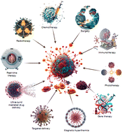
Recent years have witnessed dramatic improvements in nanotechnology-based cancer therapeutics, and it continues to evolve from the use of conventional therapies (chemotherapy, surgery, and radiotherapy) to increasingly multi-complex approaches incorporating thermal energy-based tumor ablation (e.g. magnetic hyperthermia and photothermal therapy), dynamic therapy (e.g. photodynamic therapy), gene therapy, sonodynamic therapy (e.g. ultrasound), immunotherapy, and more recently real-time treatment efficacy monitoring (e.g. theranostic MRI-sensitive nanoparticles). Unlike monotherapy, these multimodal therapies (bimodal, i.e., a combination of two therapies, and trimodal, i.e., a combination of more than two therapies) incorporating nanoplatforms have tremendous potential to improve the tumor tissue penetration and retention of therapeutic agents through selective active/passive targeting effects. These combinatorial therapies can correspondingly alleviate drug response against hypoxic/acidic and immunosuppressive tumor microenvironments and promote/induce tumor cell death through various multi-mechanisms such as apoptosis, autophagy, and reactive oxygen-based cytotoxicity, e.g., ferroptosis, etc. These multi-faced approaches such as targeting the tumor vasculature, neoangiogenic vessels, drug-resistant cancer stem cells (CSCs), preventing intra/extravasation to reduce metastatic growth, and modulation of antitumor immune responses work complementary to each other, enhancing treatment efficacy. In this review, we discuss recent advances in different nanotechnology-mediated synergistic/additive combination therapies, emphasizing their underlying mechanisms for improving cancer prognosis and survival outcomes. Additionally, significant challenges such as CSCs, hypoxia, immunosuppression, and distant/local metastasis associated with therapy resistance and tumor recurrences are reviewed. Furthermore, to improve the clinical precision of these multimodal nanoplatforms in cancer treatment, their successful bench-to-clinic translation with controlled and localized drug-release kinetics, maximizing the therapeutic window while addressing safety and regulatory concerns are discussed. As we advance further, exploiting these strategies in clinically more relevant models such as patient-derived xenografts and 3D organoids will pave the way for the application of precision therapy.


image file: d3nr06131k-p1.tif

Puja Sandbhor

Puja obtained her Doctorate's degree from the Indian Institute of Technology Bombay, India in 2020. She continued working with the Late Prof. Rinti Banerjee as Institute Postdoctoral Fellow for a year to translate her patented technology for different biomedical applications. Currently, she is working as a Postdoctoral fellow at Johns Hopkins University, USA with Prof. Efie Kokkoli on nucleic acid nanoplatforms targeting tumor-associated macrophages for brain cancer. She is now keenly interested in designing 3D tumor organoids to understand and exploit the anti-invasive characteristics of developed nanoplatforms. Her research is focused on the development of personalized and translational nanoplatforms against brain cancers. Pertaining to her doctoral and postdoctoral research on developing next-generation biomimetic nanoplatforms and non-invasive implantable systems for the loco-regional chemo-immunotherapy of incurable brain cancers, she has been granted patents and published several peer-reviewed research and review papers in esteemed international journals. She strongly believes that nanotechnology and immunotherapy together can bring a revolutionary improvement in brain cancer research along with treatments with non-invasiveness and affordability for better patient compliance.

image file: d3nr06131k-p2.tif

Pranoti Palkar

Pranoti Palkar pursued her Master's Degree in Pharmaceutical Biotechnology at DY. Patil University, India, in 2023. Her master's dissertation research focused on nanomaterial-based dual-drug-targeted delivery for effective glioblastoma therapy. In addition to her work in nanotechnology, Pranoti has a keen interest in the field of radiation biology. After completing her Master's, Pranoti joined ACTREC, Tata Memorial Centre, as a Research Fellow in the Department of Radiation Oncology. Driven by a desire to make a real-world impact, Pranoti's research interest has evolved to encompass the field of immunology. In the research lab, she leads the in vivo experiments on CAR-T cell therapy for treating multiple myeloma.

image file: d3nr06131k-p3.tif

Sakshi Bhat

Sakshi Bhat completed her Master's in Microbiology from the Central University of Punjab, India, in 2023. She has worked on evaluating the cytotoxic effects of green-synthesized silver nanoparticles in human breast cancer cells. Currently, she is working as a research trainee at the Advanced Centre for Treatment, Research, and Education in Cancer (ACTREC) under the guidance of Dr Jayant S Goda. She is keenly interested in understanding the intricate mechanisms underlying cancer immunology. Her research focuses on developing novel therapeutic approaches using nanotechnology with immunotherapy to enhance the efficacy and specificity of cancer treatment while minimizing adverse effects.

image file: d3nr06131k-p4.tif

Geofrey John

Geofrey John is a final year Master of Pharmacy student in Pharmaceutical Biotechnology at the JSS College of Pharmacy, Ooty. He has a keen interest in the application of oncology for developing novel and effective treatments for cancer. His dissertation project involves conducting an in vivo study on the efficacy of a liposomal nanoplatform containing dual chemotherapeutic agents in an orthotopic glioblastoma model at ACTREC, Tata Memorial Center. His aspiration is to emerge as a distinguished researcher and innovator in the realm of oncology, striving to advance human health and well-being.

image file: d3nr06131k-p5.tif

Jayant S. Goda

Dr Jayant S Goda received his MD in Radiation Oncology. As a translational researcher, his current focus is on testing novel compounds as radio-sensitizers and radio-protectors in mouse tumour xenografts, orthotopic, and PDX models. He is actively pursuing research on novel drug-delivery platforms using nanotechnology, which is exemplified by his publications and patents in this area of research. As an academician and researcher, he has published more than 100 scientific articles in the field of clinical oncology and translational oncology in peer-reviewed high-impact journals.


1. Introduction

Cancer is a spectrum of diseases in which body cells acquire immortality and grow uncontrollably at the expense of normal cells. The more worrying aspect of cancer is its ability to spread to neighboring and distant tissues by metastasis cascade. According to the World Health Organization (WHO), cancer is a major contributor to morbidity and mortality worldwide, with the most common form being breast cancer, followed by lung cancer, prostate cancer, blood cancer, and brain tumors.1 The International Agency for Research on Cancer (IARC) foresees a substantial upswing in cancer incidence, increasing to 29.5 million new cases each year by 2040.2 Currently, surgery, radiation therapy (RT), and chemotherapy are widely used as standard modalities for prolonging patient survival. Nevertheless, their clinical utility is challenged by major barriers such as the immunosuppressive and hypoxic tumor microenvironment, abnormal vasculature, immature neovessels, non-accessible anatomical location, cancer stem cells, and tumor heterogenicity.3,4 For instance, surgery aims at removing the locally invasive tumor but is also associated with uncontrolled bleeding and thrombosis that may have implications in damaging nearby tissues with a possible loss of organ function.5 Additionally, infections increase morbidity, reduce the pace of recovery, and increase hospital admission. Although, RT has a cytotoxic effect on tumor cells, resistance due to the hypoxic tumor microenvironment and infiltrating residual tumor cell invasion restrict its clinical utility.6 The clinical effectiveness of chemotherapy is frequently associated with off-target toxicity, which is responsible for its ineffectiveness when multidrug resistance7 and non-specific drug delivery are induced.

Interestingly, in the past, cancer research and treatment have witnessed substantial advancements as researchers have been actively working on developing various therapies designed to target cancerous tissue precisely while minimizing any harm to normal tissue.8 However, in the mid-20th century, chemotherapy and radiotherapy emerged as primary treatments for cancer.9 In the late 20th and early 21st centuries, various “Trojan horse”-based targeted therapies and immunotherapies were developed10,11 to overcome the challenges encountered by conventional anticancer treatment modalities. However, it has been found that a single treatment modality is incapable of eliminating gross and microscopic tumors (e.g., surgery can remove only the primary tumor) and is ineffective at preventing tumor metastasis.12 To overcome the limitations of monotherapy, combinatorial approaches have been introduced to offset the limitations of monotherapy, resulting in an improvement in long-term survival. This shift from monotherapy to combinatorial therapy has improved therapeutic efficacy significantly. For instance, surgery alone in breast cancer therapy resulted in more than 30% relapses, which was reduced to 11% by using a combination approach of surgery and chemotherapy, which also led to a 15% reduction in mortality.13 In lung cancer, surgery alone resulted in a 1-year overall survival (OS) of only 15%, which was improved to 36% at 5 years when chemotherapy was added as an adjuvant to surgery.14 Contemporary and modern treatment modalities, like immunotherapy, gene therapy, photothermal therapy (PTT), photodynamic therapy (PDT), magnetic hyperthermia (MHT), and high-intensity focused ultrasound therapy (HIFU), in combination with each other have shown substantial promise for future clinical translation15,16 as a result of the spatial cooperation between two or more monotherapies that produce synergistic or super-additive (1 + 1 > 2) effects that are better than those of any single therapy.17 However, combining several monotherapies to enhance their synergistic effect can be cumbersome; hence using a single platform to combine various therapeutic modalities could be easier to administer.18

Within the realms of combinatorial approaches to cancer therapy, several nanoplatforms are being developed for anticancer therapies to enhance their selectivity and efficacy.19 Nanotechnology offers new therapeutic and diagnostic methods with high efficacy and fewer adverse effects compared to conventional therapies.20 Various types of nanoparticles have been developed for cancer therapy, such as liposomes, solid-lipid nanoparticles, polymeric nanoparticles, extracellular vesicles, gold nanoparticles, quantum dots, and magnetic nanoparticles21 (Fig. 1). These nanoplatforms carry a combination of drugs and have shown promising results in targeted drug delivery, a reduction in off-target toxicity, and improved bioavailability at the target tissue.22 Recently, Gao et al. meticulously reviewed the potential of combining nanomedicines with radiotherapy, chemotherapy, immunotherapy, phototherapy, etc., in various cancers to improve the treatment efficacy.23 Using these nanoparticles as drug-delivery carriers can show several advantages, such as (1) the active targeting of cancer cells, which could improve treatment accuracy and, thereby, efficacy;24 (2) an enhanced permeability and retention (EPR) effect, which allows nanoparticles to accumulate passively in tumor tissues, thereby increasing the drug concentration within the targeted site;25,26 (3) a stimuli-responsive delivery system, which reduces off-target toxicity by preventing drug release to normal tissues;27 and (4) a delayed release of nanomedicine and an increase in its half-life (e.g., PEGylation), further enhancing the therapeutic efficacy.28 Furthermore, nanomaterials, when used in combination with other therapies mentioned above, can potentially reduce drug resistance and non-specific toxicity, thus augmenting the therapeutic gain. Additionally, drug(s) incorporated into nanoplatforms allow sustained payload delivery within the tumor tissue and optimization of their pharmacokinetics profile, resulting in a longer retention time within the tumor.29


image file: d3nr06131k-f1.tif
Fig. 1 Schematic illustration of different nanoparticle-based monotherapies and combination therapies. An illustrative overview of the individual and combined advantages of various cancer therapies. The figure delineates the benefits of chemotherapy, radiation therapy, phototherapy, and immunotherapy. It further explores the synergistic potential of bimodal and trimodal approaches, highlighting combinations such as photodynamic therapy (PDT) with immunotherapy, high-intensity focused ultrasound (HIFU) with micro/nanobubbles, radiotherapy with chemotherapy, and more complex trimodal therapies. This comprehensive figure emphasizes the role of nanomedicine in augmenting the effectiveness of multimodal cancer therapies.

2. Nanomedicine-augmented combination therapy outcomes

Multimodal cancer therapies involve the combination of two (bimodal) or more than two (e.g., trimodal) different modalities in a single nanoplatform to expand treatment success. Adjuvant cancer therapy, like hyperthermia, artificially raises tissue temperature to 42 ± 2 °C,30 which can directly ablate tumor cells, thereby reducing the side effects on healthy tissue. When thermal ablation is combined with chemotherapy and RT, it allows for a dose reduction of RT and chemotherapy.31 Moreover, the combinatorial approach helps to overcome drug efflux and resistance, which is a major challenge leading to poor prognosis. Multidrug resistance (MDR) is caused by an overexpression of ATP-binding cassette (ABC) efflux transporters, dysfunctional apoptotic pathways, and the hypoxic tumor microenvironment (TME). The correlation of dysfunctional apoptotic pathways and drug resistance results in impaired apoptosis, causing tumorigenesis and uncontrolled cell proliferation. Multiple proteins, including Bcl-2 superfamily proteins, inhibitors of apoptosis (IAPs), p53 (tumor suppressor protein), and the hyperactive PI3/AKT pathway proteins, are linked to dysfunctional apoptosis, which contributes to the development of MDR tumors. Additionally, factors released from cancer-associated fibroblasts prevent the intrinsic pathway of apoptosis and may have a role to play in impairing the efficacy of anticancer drugs. Potential therapeutic strategies targeting the Bcl-2 family members Venetoclax (ABT-199),32 inhibitors against IAPs (LCL161)33 or the AKT pathway inhibitor (MK-2206),34 epigenetic alterations decitabine (5-aza-2′-deoxycytidin),35 and natural drugs (curcumin)36 should be explored to combat drug resistance.37 Therefore, using nanotechnology in drug delivery helps to overcome drug resistance.38 In this review, we aimed to discuss the essential synergistic/additive mechanisms behind various nanotechnology-based bi/tri-modalities.

2.1. Nanoplatforms for bimodal therapies

2.1.1. Chemotherapy with immunotherapy. In the past, immunotherapy emerged as a ground-breaking development in anticancer research that uses the host immune system to combat cancer. However, it is frequently hindered by poor patient response rates due to immune-related side effects during clinical treatment.39 Chemotherapy promotes tumor cells to undergo an apoptotic pathway known as immunogenic cell death (ICD) through the release of danger-associated signals, such as high mobility binding box 1 (HMGB1) protein and adenosine triphosphate (ATP) emitted by dying/dead cells that strengthen the immunocompetent host's defense against cancer.40,41 The tumor-related ICD further promotes the expression of essential protein markers for the exposure of additional tumor-associated antigens (TAAs) that help dendritic cells (DCs) develop and expose the TAAs to T cells. Pro-inflammatory cytokines released by tumor cells promote as well as favor the M2 macrophages for their growth and proliferation. To enhance the synergistically tumor-suppressive effects of nanomedicine, the initiation of an intense anticancer immune response triggered by ICD represents a highly promising and feasible therapeutic approach.42 Chemotherapy drugs have a dual influence on tumor-associated macrophages (TAMs); whereby they can either promote or prevent an immunosuppressive microenvironment. For example, a misdirected macrophage-orchestrated tissue-repair response can cause chemoresistance, showing an immunosuppressive effect. However, some drugs, like doxorubicin, can induce immunogenic cell death, activating adaptive immune reactions and reducing immunosuppression. A balance between these effects is essential for ideal potential therapeutic outcomes. It is crucial to gain more insights into the interaction between chemotherapy and innate immunity, specifically TAMs, which could lead to novel combinations of immunotherapy with conventional chemotherapy, resulting in an enhanced therapeutic ant-tumor efficacy.43 A wide spectrum of antineoplastic agents, like doxorubicin (DOX), irinotecan, etc., trigger ICD, which stimulates the antitumor immune responses during chemotherapy.44 For chemotherapy-sensitized immunotherapy, a self-assembled nanoscale oxidative stress amplifier, Cu-DON, made by the self-assembly of DOX, copper ions (Cu2+), and NLG919 (a potent indoleamine 2,3-dioxygenase (IDO) pathway inhibitor) through π–π stacking and coordination effects was designed. Cu-DON NPs can suppress a tumor's initial growth, enhance DOX-induced oxidative stress, and inhibit indoleamine 2,3-dioxygenase 1 (IDO-1, an immunosuppressive enzyme within the tumor microenvironment), hence restoring the immune response to combat both primary as well as metastatic tumors. When it comes to deep tumor treatment, Cu-DON NPs have better tumor penetration than free DOX,45 which can simultaneously stimulate an effective immune response. Arginine-modified camptothecin (CPT) prodrug nanofibers have been designed to improve tumor penetration through their fiber-like structure. These nanofibers can co-deliver DNA plasmids, i.e., pshPD-L1 and pSpam1, expressing programmed cell death ligand 1 (PD-L1) and hyaluronidase (Haase) gene carriers. Hyaluronidase destroys the extracellular matrix (ECM), which inhibits the growth and recurrence of cancer by improving tumor penetration and therapeutic drug delivery. PD-L1, a tumor cell surface protein, binds with PD-1 receptors that cause T-cell exhaustion and immune evasion. The tumor-specific T-cell responses can be improved by suppressing the expression of PD-L1, which converts anergic T cells into tumor-reactive T cells. Therefore, combining these two carriers can achieve a combinatorial effect.46 This new synergistic chemo-immunotherapy strategy has the potential to improve efficacy and offers novel perspectives for developing self-delivering nanoplatforms for targeted cancer therapy.
2.1.2. Photothermal therapy (PTT) with chemotherapy. PTT is a near-infrared (NIR) laser-based treatment for tumors that transforms light energy into heat.47 This heat promotes drug release and photothermal conversion (conversion of absorbed light energy into heat), which can rapidly destroy tumor cells and prevent tumor regrowth. Consequently, the combination of PTT and chemotherapy can maximize their benefits and offset their drawbacks, significantly enhancing the antitumor efficacy.48 Fascinatingly, the last several years have seen tremendous attempts to create new nanotherapy antitumor responses by combining the synergistic action of chemotherapy and PTT, and notable success has been achieved in this regard. Hollow mesoporous Prussian blue (HMPB)-based therapeutic nanoplatforms have been designed by encapsulating the drug disulfiram (DSF) into copper-enriched and polyvinylpyrrolidone (PVP)-decorated HMPB nanoparticles, known as DSF@PVP/Cu-HMPB. When DSF@PVP/Cu-HMPB accumulates in the tumor, the acidic tumor microenvironment causes the HMPB nanoparticles to biodegrade and release DSF and Cu2+ simultaneously; this results in the formation of cytotoxic bis(N,N-diethyl dithiocarbamate) copper(II) complexes (CuL2) through the DSF-Cu2+ chelating reaction.49 Wu et al. determined a photothermal conversion efficiency of up to 35% after the doping, meaning that DSF@PVP/HMPB could effectively and continuously produce a large amount of heat under NIR irradiation, suggesting that the ability to convert light energy into PVP/HMPB heat was not damaged. Due to its extended blood-circulation half-time, DSF@PVP/Cu-HMPB exhibited improved anticancer effects both in vitro and in vivo, with a passive-targeting efficiency reaching up to 7% 12 h post-injection. Laser irradiation enhanced the anticancer effects of DSF@PVP/Cu-HMPB nanoparticles, generating highly toxic CuL2.50 Furthermore, Liu et al. developed DSF-loaded hollow copper sulfide nanoparticles (DSF@PEG-HCuSNPs) to achieve photonic hyperthermia-amplified DSF chemotherapy.51 Additionally, for non-invasiveness and site-specific controlled drug release, Yin et al. synthesized an NIR-responsive nanoparticle using a 2D mesoporous core/shell multifunctional theranostic nanoplatform (Silicene@Silica) to accomplish simultaneous thermo-chemotherapy and the NIR laser-controlled prodrug release of banoxantrone dihydrochloride (AQ4N).52 Zhao et al. developed a biomimetic pH-sensitive nano-system using a zeolitic imidazolate framework 8 (ZIF8), loaded with doxorubicin hydrochloride (DOX·HCl) and copper sulphide (CuS) and then coated with the red cell membrane and catalase components. ZIF8 rapidly broke down in the acidic TME, which allowed for the targeted co-delivery of PTT and chemotherapeutics.53 The findings of this study further validate the remarkable synergistic effects between PTT and trigger-responsive nano-based chemotherapy.
2.1.3. Chemotherapy with photodynamic therapy (PDT). PDT is a non-invasive, promising treatment approach for cancer therapy that uses light to stimulate photosensitizers (PSs) to generate cytotoxic reactive oxygen species (ROS) that kill tumor cells by peroxiding proteins, lipids, RNA, and DNA.54 Additionally, ROS help to initiate controlled drug release by breaking down the covalent bioconjugation of chemotherapeutics and photosensitizers, thus establishing a backing for synergistic chemotherapy and PDT.55 Huang et al. developed a photoactivatable self-assembling prodrug cocktail (PSPC) nano-assembly for melanoma treatment, which was made from the LNA-conjugated photosensitizer chlorine e6 (L-Ce6) and α-linolenic acid (LNA)-thioketal-cabazitaxel (LTK-CTX) prodrug. When L-Ce6 is absorbed by tumor cells, an excessive amount of ROS was produced under NIR irradiation, activating cabazitaxel's cytotoxic effects.56 Furthermore, Wu et al. confirmed with the help of TEM that the monomer conjugate of LTK-CTX and L-Ce6 could self-assemble into spherical nanostructures in water, and their miscibility with LTK-CTX resulted in PSPC nano-assemblies. The carboxyl groups of free Ce6 restrict ROS production in tumor cells, while the L-Ce6 self-assembled nanoparticles can be quickly absorbed by tumor cells for effective PDT. PSPC NAs may enhance oxidative DNA damage when combined with ROS-activated CTX, which would eventually trigger tumor cells to undergo cell death when excited by a laser.42 Moreover, Wang et al. developed a nano-catalytic medicine using DOX-PEG-PS@MIL-100 nanoparticles for efficient photo-chemotherapy in deep tumors. These nanomedicines were developed using pH-sensitive DOX-PEG, which breaks down into fragments and releases PSs at the tumor sites, producing ROS when exposed to laser irradiation. Furthermore, the exogenous DOX-PEG could self-assemble into ultrasmall DOX NPs, which might prove useful for chemotherapy and deep tumor penetration.57 Collectively, these findings suggest that nanomedicines can deliver effective on-demand photo-chemotherapy for deep-seated tumors with reduced adverse effects.
2.1.4. PTT with PDT. In this bimodal therapy, PTT uses NIR light to accomplish thermal destruction, while PDT destroys cells by photosensitization and cytotoxic ROS production.58 The main difference between them is in terms of the oxygen concentration, i.e., PTT is more effective in hypoxic conditions since it does not depend on oxygen to cause cell death, whereas PDT is less effective due to its oxygen-dependent ROS production mechanism, which restricts its efficacy in hypoxic conditions.59 Zhu et al. developed an injectable agarose hydrogel containing Cu-Hemin and indocyanine green (ICG), a photothermal agent, which could absorb and convert NIR light energy provided by an 808 nm laser into thermal energy. This results in Cu-Hemin mediating antioxidant glutathione consumption and allows it to function as a photosensitizer, enabling tumor cells to produce cytotoxic ROS. Hence, the GSH level inside the TME can be successfully modulated by PTT/PDT therapy to enhance PDT's antineoplastic effects.60 In addition, in 2015 physiologically stable NIR plasmonic copper sulfide nanocrystals (NCs) as a PTT material were developed by Wang et al. These NCs had high efficacy and NIR-triggered PDT activity that led to the production of ROS.61 Moreover, Fan et al. designed a methylene blue (MB)-bound A9-aptamer-attached nanoplatform for PSMA-positive LNCaP prostate cancer cells, which offered targeted treatment. This nanoplatform was triggered by 785 nm NIR light, activating PTT/PDT therapy.62 Collectively, this study defines the potential role of NIR-based systems for inducing cytotoxic effects and may lead to the development of extremely effective bi-modality nanotherapeutics.
2.1.5. Gene therapy with PTT. Gene therapy is a promising approach for cancer treatment with enhanced targeting capabilities and minimal adverse events. However, it often fails to eradicate tumors and their recurrence.63 Combining gene therapy with other therapies, like PTT/PDT using antitumor genes and PS together, can enhance the therapeutic effects. Nucleic acids like siRNAs/miRNAs are used in combined PTT/PDT for achieving a synergistic effect. Hyun et al. designed a Zn(II)-dipicolylamine (Zn-DPA) molecule for siRNA delivery. Zn-DPA-conjugated gold nanorods (GNRs) were fabricated that could deliver PTT and PLK1-siRNA at the same time, effectively treating PC-3 tumors via apoptosis.64 When the Zn-DPA molecule was combined with ICG and PVP polymers, it formed nano-ICG with RGD, a peptide motif that could target angiogenesis with prolonged tumor accumulation. The combined PTT/gene therapy showed a reduced amount of CD31, indicating that the combined therapy was more effective at killing tumor blood vessels.65 Furthermore, Zhang et al. developed a PTT/gene therapy platform using cell-penetrating membrane peptides (CPP)-functionalized gold nanorods to load transcription factor EB (TFEB)-siRNA, which enhanced the cell permeability and targeting of autolysosomes under NIR irradiation. Due to this, drug resistance was found to be reduced in osteosarcoma cells, resulting in an increased efficacy of PTT. Moreover, they showed that TFEB silencing using this platform could effectively prevent osteosarcoma cells from metastasizing to the lungs, which was important because TFEB is linked to autophagy and encourages the spread of cancer.66 Thus, these studies further validated the remarkable synergistic effects between phototherapy and nucleic acid-mediated gene delivery. Collectively, the aforementioned bimodal therapies showed improved efficacy over monotherapy. However, it is also known that further cooperative enhancement among three different treatment approaches can lead to even more optimal treatment efficacy at substantially lower doses of therapeutic agents. In the subsequent section, we attempt to review a few trimodal treatment options within a single nanostructure.

2.2. Nanoplatforms for trimodal therapies

2.2.1. Chemotherapy/gene therapy/PTT. Photothermal therapy (PTT) is limited in its efficacy by the short valid range of the laser, being 0.33 W cm−2 for the 808 nm NIR laser and 0.72 W cm−2 for the 980 nm NIR laser;67 albeit chemotherapy can be considered to complement it. Alternatively, gene therapy is another effective method for treating cancer as it delivers dysregulated genes into target cells. Further, a combination of these three methods could improve the therapeutic effects against tumor progression. Shun-Duan monitored the intracellular trafficking behavior of glioma C6 cells treated with ASQ-DOX-PGEA2/p53 (Au NRs@SiO2-QD [Au nanorods, mesoporous silica, quantum dots] denoted as ASQ, cyclodextrin-ethanolamine [EA]-modified poly [glycidyl methacrylate] denoted as CD-PGEA, at 300 nm size), with and without NIR irradiation.68 ASQ-DOX-PGEA2 with NIR irradiation exhibited nearly 20% cell viability, implying the potential of this combinatorial nanotherapy to inhibit the proliferation of tumor cells.69 Further, when the team studied the in vivo antitumor performance of ASQ-DOX-PGEA2, they found that the tumor volume in the single modal PTT group was larger than that in the dual-modal PTT/Chemotherapy group, as the bimodal group showed combined effects of the NIR-induced release of the drug (DOX) and PTT, which was more effective for reducing the tumor volume.68 However, it was further observed that the tumor grew after 4 days, indicating recurrence after dual-modal PTT/chemotherapy. However, the tumors treated with ASQ-DOX-PGEA2/p53 and irradiation by NIR laser showed remarkable inhibition, with the smallest tumor volume being in this group, which indicated the greatest effects from trimodal PTT/chemotherapy/gene therapy. Mechanistically, PTT increased the temperature of the tumor tissues, and the induced heat then triggered the detachment of CD-PGEA to facilitate the release of doxorubicin, while gene therapy was responsible for the escaped p53 from the cascading processes.
2.2.2. Chemotherapy/immunotherapy/PDT. Chemotherapy and PDT, in combination with immunotherapy, can be achieved by compiling drugs and pyrolipids (photosensitizers) into a single nanocarrier. Lin et al. developed a nanoscale coordination polymer (NCP) to deliver multiple agents, namely oxaliplatin (chemotherapy agent), pyrolipid (PDT), and immunotherapy using anti-PD-L1 (PD-L1-programmed cell death ligand-1) to achieve superior anticancer efficacy in colorectal cancer.70 NCP@pyrolipid could simultaneously kill cancer cells and stimulate the immune system to activate immune responses utilizing PDT/chemotherapy. NCP@pyrolipid, in combination with anti-PD-L1 treatment, enhanced antitumor immunity for the effective treatment of colorectal cancer.71 Also, multiple preclinical studies have witnessed a blockade of the interaction between PD-1 and PD-L1 using anti-PD-L1, which can revive T-cell activity against the tumor cells, thereby further preventing its metastasis.72 The anti-tumor immunity induced by NCP@pyrolipid in combination with anti-PD-L1 has been demonstrated in in vivo models.73 The findings elucidated that PD-L1 checkpoint blockade therapy with chemo-immunotherapy with PDT not only enabled regression in the primary tumor but also at distant sites, which strongly confirmed the superiority of the trimodal treatment approach against metastatic colorectal cancer. Jingjing Ding and team members reported a recent advancement in the synergism of chemotherapy, immunotherapy, and PDT, resulting in potent anticancer efficacy. They constructed a self-activatable and self-luminous extracellular vesicles (EVs) delivery system that exhibited these three modalities against cancer. No nanoplatforms were involved in this treatment approach, and further optimization is needed before it can be put into clinical practice.74
2.2.3. Chemotherapy/PDT/PTT. It has been very well established that almost all chemotherapy drugs must be aimed at particular sites of action to attain optimal effects according to their mechanisms of action. Therefore, in recent years, combination approaches for enhancing chemotherapy have been explored with photodynamic therapy and photothermal therapy by targeting different pathways in cancer cells and with different regimens to lower drug doses. In 2016, Lin's group performed preclinical studies for bladder cancer with a single PLZ4-nanoporphyrin (PNP) platform.75 PNPs are small, approximately 25 nm in diameter, which increases their permeability at the tumor site. These PNPs showed a low uptake in normal tissues but longer retention at the tumor site as they have the bladder cancer-specific ligand PLZ4 on the surface, which prevents the back-flow of PNP into the circulation.76 Their findings demonstrated that the superior anti-tumor efficacy of PNPs could be linked to the fusion of three modalities, i.e., chemotherapy/PDT/PTT, in a single nano-formulation. Additionally, the traditional photosensitizer prodrug 5-ALA (5-aminolevulinic acid) has clinical limitations because of its poor selectivity between cancerous and non-cancerous tissues and longer incubation time needed to metabolize into an active photosensitizer.77 An in vivo comparison between the drug delivery of PNPs, PNP-DOX, and 5-ALA was carried out, with free 5-ALA considered as a control group. Whereas the group that was treated with PNP-DOX activated with light showed inhibitions of the tumors in comparison with the control 5-ALA group, PNP-DOX-mediated phototherapy combined with chemotherapy showed higher efficacy.78 Altogether, these findings suggested the potential of monitoring drug delivery in real-time by combining PDT, PTT, and chemotherapy in a single procedure and confirmed the easy translation of the PNP platform-based triple combination in clinical application.
2.2.4. Gene therapy/immunotherapy/PDT. The combination of gene therapy with other treatments utilizing nanocarriers, such as in PDT and immunotherapy, could result in synergetic effects via multifunctional mechanisms. For instance, Wang's team demonstrated that PDT-mediated cancer immunotherapy could be multiplied by PD-L1 knockdown (KD) in tumor cells, for which they designed an adaptable micelleplex by integrating an acid-activatable cationic micelle, photosensitizer, and small interfering RNA (siRNA).79 It has been reported that particular categories of siRNA can block PD-1-PD-L1 interaction to augment PDT-induced immune activity, wherein gene therapy can amplify the efficacy of PDT-mediated immunotherapy, subsequently leading to a superior therapeutic outcome.80 In cancer cells, siRNA targeted at PDL1 was found to remarkably suppress its expression in melanoma tumor cells through a dosage-dependent mechanism. The complete eradication of B16-F10 (murine melanoma) tumors and effective inhibition of tumor recurrence have been achieved by combining PDT-induced immunotherapies with siRNA-mediated PD-L1 KD.81
2.2.5. Radiation therapy/chemotherapy/immunotherapy. Radiotherapy and chemotherapy, being traditional cancer treatments, have certain disadvantages, such as non-specific targeting, killing of normal cells (including immune cells), and producing toxic side effects. At present, chemotherapy and radiotherapy cause an immunological response by encouraging the death of immunogenic tumor cells or destroying the tumor microenvironment (TME).82 In this way, it is possible to promote the reconstitution of antitumor immunity with a combination of immunotherapies and chemoradiotherapy that minimizes side effects. Scott Antonia and co-workers in their study hypothesized that durvalumab, an immunotherapy agent with chemoradiotherapy, could provide clinical benefits for treating non-small-cell lung cancer (NSCLC). They designed a phase III PACIFIC study for comparing the IgG1 PD-L1 inhibitor durvalumab with a placebo in stage III NSCLC patients.83 A total 713 number of patients were randomized in a 2[thin space (1/6-em)]:[thin space (1/6-em)]1 ratio in durvalumab and placebo groups for 42 days after chemoradiotherapy. They found that in the placebo group, a greater number of patients had distant relapses, whereas in the durvalumab group, brain recurrences were reduced. It was reported that 24.2% of the patients in the durvalumab group and 8.1% of the patients in the placebo group experienced immune-mediated adverse events, due to which 15.4% of the patients from the durvalumab group and 9.8% of the patients from the placebo group discontinued the study.84 Also, the patients who received durvalumab had 11 months longer survival than those who received the placebo. In this study, the majority of participants had less than 25% PD-L1 expression on tumor cells than participants with 25% or more PD-L1 positive cells.83 These findings suggested that chemoradiotherapy with durvalumab-based immunotherapy has acceptable side effects. Thus, the potential interaction between immunotherapy and chemoradiotherapy demands further detailed investigation.

3. Challenges appended by multimodal nanotherapy

It is now a well-established fact that the growth and dissemination of cancer is regulated by the complex TME,85,86 which is composed of two major environments, i.e., the cellular and physiological microenvironments.87 The cellular microenvironment involves bone marrow-derived antigen-presenting cells (APCs, e.g., DCs), tumor-associated macrophages (TAMs), cancer-associated fibroblasts (CAFs), and cancer stem cells (CSCs), which can self-renew and promote tumor growth, and therapy resistance.88,89 Whereas the physiological microenvironment consists of chronic/cyclic hypoxia (due to abnormal blood vessels and uncontrolled tumor cell proliferation leading to altered oxygen tension within the tumor vasculature), an acidic environment (due to insufficient blood supply and tumor cells glycolytic metabolism), and an alteration in various metabolic pathways (such as amino acid metabolism, increase in glucose uptake/lactate fermentation due to activated mitochondria under aerobic environment) etc.90,91 Within the TME, the co-existence and dynamic communication of tumor cells with the extracellular matrix (ECM), stromal cells recruited from the bone marrow/surrounding tissues (e.g., fibroblasts), immune cells (e.g., macrophages, resident microglia), lymphatic and vascular endothelial cells, pericytes, inflammatory mediators, such as soluble factors (e.g., cytokines, chemokines), and ECM-degrading enzymes (e.g., matrix metalloproteases [MMPs]) etc. drive the progression, recurrence, drug resistance, and immunosuppression tumor milieu.92–95

Moreover, these complex interactions further promote neovascularization to meet the oxygen and nutrient demands, and to provide optimal growth conditions for cancer cells to grow and metastasize. Increasing evidence has shown that the abnormal blood vessels, high interstitial fluid pressure (IFP) within the tumor tissue, leaky vasculature, disorganized endothelial cells and peri-cellular cells with loose gap junctions, hypoxia, and acidic microenvironment, etc., can limit the effectiveness of conventional therapy.96 Thus, the TME is considered a “viable target” because it largely determines the fate of anticancer treatment (Fig. 2). For instance, a limited penetration and retention of the therapeutic agent into the tumor tissue due to increased IFP, the hypoxic environment reducing the sensitivity of tumor cells to chemo-drugs, and the impaired cytotoxic functions of immune cells due to the acidic microenvironment, etc. determine the impact of the TME on therapeutic outcomes.97 In later sections, we briefly discuss how nanotechnology-based multimodal strategies are addressing these challenges to improve the anticancer effect of treatments.


image file: d3nr06131k-f2.tif
Fig. 2 Schematic illustration showing different challenges imposed by the tumor microenvironment for effective anticancer treatment and how the multifunctionality of nanotechnology-based combined therapy aims to resolve them. This figure encapsulates the complex challenges posed by the tumor microenvironment (TME) and the multifunctional approach of nanotechnology-based combined therapy to address them. The left segment depicts the TME, divided into a normal and a hypoxic TME. The hypoxic TME, characterized by leaky blood vasculature, is actively targeted by gold nanoparticles (AuNPs). It shows hypoxia-induced epithelial–mesenchymal transition (EMT), where hypoxia-inducible factor 1-alpha (HIF1alpha) activates factors like TWIST, SIP1 (SMAD-interacting protein 1), and ZEB1 (zinc finger E-box-binding homeobox), inhibiting EMT. Additionally, AuNPs inhibit factors like VEGF (vascular endothelial growth factor), FGF2 (fibroblast growth factor 2), and APLN (apelin), thereby inhibiting angiogenesis. The central segment illustrates the dual strategy of nanoparticle targeting, enhancing the precision and efficacy of the therapy. The extreme right segment shows nanoparticles and photodynamic therapy (PDT) using near-Infrared (NIR) light, working in tandem for more effective tumor cell killing.

3.1. Multimodal nanoplatforms targeting tumor vasculature and neovessels

To reach the rapidly proliferating cancer cells in solid tumors, therapeutic agents must enter the tumor neovessels, permeate the vessel wall, and ultimately migrate through the tumor interstitium.98 The past decades have witnessed the rapid development of anticancer nanomedicines targeting the tumor vasculature to penetrate deeper into the tumor tissue for survival improvement and to enhance the treatment efficacy. In 2019, Li and co-workers reviewed various TME-responsive nanotherapeutics smartly manipulating the tumor vasculature in the context of improving therapeutic efficacy.99 TME-sensitive nanotherapeutic strategies are based on an inhibition of vascular endothelial growth factor and its receptors (VEGF/VEGFR), the use of antiangiogenic compounds to regulate the structural and functional abnormalities of tumor blood vessels, the destruction of tumor endothelial cells, and the local depletion of tumor-associated platelets (TAPs), etc.100 These strategies induce and trigger intratumoral thrombosis to block the blood supply to the tumor and suppress tumor progression in various cancers, like melanoma, lung, and ovarian cancers. Novel strategies, such as DNA origami-based thrombin-loaded nanorobots (nanorobot-Th) shielded from circulating platelets/plasma fibrinogen with ligand functionalization (e.g., AS1411 aptamer) for the selective targeting of nucleolin receptors overexpressed tumor endothelial cells inducing thrombosis and the occlusion of tumor-associated blood vessels for precise anticancer therapy, have been discovered recently.101,102 Similarly, surface-modified nanotherapeutics recognizing specific receptors, such as transferrin (TfR), low-density lipoprotein (LDR), lactoferrin, αvβ3-integrin, have been used to improve tumor retention and accumulation in different cancers. For instance, various tumors have shown 100–300 times high folate compared to endogenous levels along with overexpression of the folic acid receptor in the tumor vasculature; therefore, strategies involving folate-functionalized nanotherapeutic targeting of the TME have been widely investigated to improve antitumor effects. However, the existence of various anatomical and physiological barriers, such as the blood–brain barrier (BBB) and phagocytic clearance (reticuloendothelial system [RES] uptake), still present tremendous challenges in successful systemic delivery into tumors of the brain and central nervous system. Likewise, although the surface modification/functionalization strategies of nanotherapeutics have shown enormous advantages, the efficient delivery of chemotherapeutics to the tumor tissue is still restricted due to the intrinsic characteristics of the TME. For instance, the constitutive expression of targeted ligands (e.g., EGFR, TfR) in normal cells/tissue leads to the uptake of functionalized nanocarriers by non-malignant cells, thereby inducing potential off-target toxicities. Therefore, technological improvement and novel multifunctional anticancer treatment approaches that improve the drug's therapeutic index with minimal off-target events are truly required. Recently, various combinatorial approaches, such as selective and localized temperature-induced cancer cell killing with chemo/radio-therapy, have been studied widely. Herein, we attempted to review these approaches with a special emphasis on targeting the tumor vasculature and neovessels below.
3.1.1. Thermal ablation-based targeting vasculature niche. As discussed previously, the combination of nanoparticles with thermal therapy (e.g., hyperthermia) and chemotherapy offers promising advantages, like target selectivity, specificity, and non-invasive theranostic modalities. The FDA has approved various antibodies/small molecules functionalized with hyperthermic nanoparticles for cancer therapy. For instance, Avastin® (bevacizumab antibody targeting VEGFR in the colon, NSCLCs, breast cancers) and Herceptin® (trastuzumab antibody targeting human epidermal growth factor-2 [HER2] in breast cancer).103 Moreover, these multimodal nanoplatform-based chemotherapies intrude deeply into the tumor tissue without affecting normal cells/tissue and enhance their cytotoxicity to cancer cells. However, the destruction of tumors near large neovessels is not possible due to the limited thermal dose delivered to the target site because of the heat dissipation caused by blood perfusion. Therefore, a promising solution to augment this thermal effect is to alter the blood flow without increasing the energy deposition before heating, and this can be achieved by using hyperthermia with adjuvant targeting of the tumor vasculature (e.g., with anti-vascular agents like interleukin-α, tumor necrosis factor-α [TNF-α], prostaglandin-E, etc.). For instance, in 2006, Visaria et al. demonstrated the augmentation of breast cancer thermal therapy using systemic PEGylated gold nanoparticles-assisted tumor necrosis factor-alpha (TNF-α) delivery. Their findings suggested the combined effect of hyperthermia and the anti-vascular agent could prevent tumor regrowth through vascular-mediated cancer cell death compared to hyperthermia alone.104 Interestingly, recent findings have suggested that vascular targeting alters the neovessels architecture (increasing neovascular permeability) through remodeling/maturation of TME, thereby reducing its hypoxic environment via correcting pressure gradient disparity and improving the permeability and retention of the active agent in the tumor interstitium. Various agents that can selectively improve tumor neovascular permeability and increase intravascular coagulation have been investigated to enhance the anticancer effects, such as TNF-α, an inflammatory cytokine with a potent anticancer effect. Despite the significant potential of TNF-α in the selective targeting of tumor neovascular permeability, its clinical use has been limited due to severe off-target toxicities after systemic administration (maximum tolerated dose is 8–10 μg kg−1, which is 10–50 times less than the effective dose needed to exert cytotoxicity).105 However, in 2007, Farma and co-workers demonstrated direct evidence of the selective induction of tumor neovascular permeabilization using a colloidal gold-bound TNF-α nanoplatform with similar antitumor efficacy but at lower doses compared to that of conventional TNF-α therapies. Thus, by augmenting the neovascular permeability, the system has the potential to improve the selective delivery of chemotherapeutics into the tumor interstitium followed by tumor cell necrosis through intravascular coagulation with acidosis/ischemia.106 Furthermore, combining vascular disrupting agents (VDAs) with nano-based thermal ablation was reported to be more effective in multimodal prostate cancer therapy, suggesting a potential role of VDAs-conjugated nanotherapy in preconditioning the tumor vasculature for further clinical translation.107
3.1.2. Near-IR selective thermal ablation. Targeting tumor vasculature using NIR (650–900 nm)-based phototherapy (PTT/PDT) cancer nanotherapy (composed of photosensitive agents, e.g., porphyrin, phthalocyanine, chlorin, anthraquinone, light source, and/or oxygen) is an emerging class of anticancer treatment. The technique involves the formation of singlet oxygen and/or the generation of ROS through intersystem crossing upon photoirradiation (i.e., transition of the activated excited singlet state of a photosensitive molecule to the excited triplet state). Although tumor accumulation of the nano-transducer (i.e., PTT/PDT-based carrier of therapeutic agents) is mediated by the EPR effect, often the compromised tumor vasculature and leakiness promote the toxicity on luminal/endothelial target cells, such as RBCs, platelets, causing collapsed blood vessels and thrombosis. Additionally, the penetration of nano-transducers is significantly hindered by the impaired blood supply and IFPs during tumor progression, leading to an incomplete therapeutic response. To overcome these challenges, phototherapy was preconditioned with various strategies, such as selective receptor-mediated targeting of the tumor vasculature, increasing the vessel permeability without inducing vessel occlusion, and modulation of the vessels by antiangiogenic and hyperthermic (42 ± 1 °C) effects. These co-therapies were found to augment the nanotherapeutic extravasation and retention through selective endothelium permeabilization. For instance, the co-therapy of PTT and hyperthermia not only caused the focal and efficient conversion of photon energy to thermal energy for the ablation of adjacent tumor cells but also triggered the necrosis of hypoxic cancer cells. Furthermore, this unique co-therapy approach stimulates the generation of various nanomaterials to improve treatment efficiency through multi-mechanisms, such as gold nanoparticles (nanorods, nanocubes, nanoshells), graphene oxides nanoparticles, palladium nanosheets, single/multi-wall carbon nanotubes, transition metal ion-based nanostructures (sulfide, oxide nanocrystals), and porphyrin-based nanostructures (porphysomes, nanoporphyrins). For instance, Zhen et al. in 2014 demonstrated that RGD-mediated targeted delivery coupled with photosensitizer (ferritin)-based different nanoparticles (albumin, iron oxide, and quantum dots) could augment the EPR effect linked with endothelium selectivity, leading to increased vessel leakiness without inducing occlusion against various tumor xenograft models such as 4T1, human U87MG, PC-3, and MDA-MD-435S.108 Another preclinical study on VEGF-functionalized PEGylated Au nanoshells reported the potential of PTT-based VEGF targeting in vascular disruption as a valuable adjuvant therapy with chemotherapy and radiation and also as an alternative to conventional antiangiogenic therapies for brain tumors.109 However, the impact of these anti-vasculature PTT-based tumor ablation treatments are affected by other crucial factors, such as the tissue oxygen levels, nutrients transport into the tumor interstitium, long-term survival outcomes, and the influence of nanoparticles localization on the temperature difference between the tumor and normal brain tissues. Therefore, these challenges demand future investigations for the development of more effective PTT therapy for cancer of the brain. Furthermore, Li et al. in 2018 discovered the combined anticancer effect of mild-hyperthermia and low-dose antiangiogenic agent (sunitinib, an FDA-approved tyrosine kinase inhibitor)-loaded PEGylated reduced graphene oxide nanoparticles (RGO-PEG) in a 4T1 breast tumor mouse model. Surprisingly, they found that the anti-vascular PTT therapy decreased the tumor distribution of RGO-PEG, probably due to the variation in the response of different tumors to antiangiogenic therapy. These findings, however, contradicted the previous findings on the potential effect of vasculature-modulating co-therapy in tumor inhibition. Conclusively, they suggested that vasculature-modulating therapy does not necessarily improve the efficacy of PTT, probably as the intratumoral distribution of nanoparticles and spatiotemporal heat deposition during combination treatment are largely affected by the tumor heterogenicity and extent of PTT thermal energy penetration (periphery vs. center). Thus, this study highlights the need for a detailed understanding of nanoparticles and light intervals, doses, fluence/irradiance rates, and the variation in tumor responses to anti-angiogenic therapy to improve PTT-based antitumor therapeutic efficacy.110

In 2008, the first PTT nanoparticles (AuroShell® Particles) entered into clinical trials. Moreover, two different clinical trials (NCT01679470 and NCT00848042) of AuroLase Therapy (composed of PEGylated silica-cored Au nanoshells) for efficacy evaluation in primary and metastasis lung cancer, and refractory head and neck cancer revealed the potentially improved tumor accumulation of the PTT-based nanoparticles. Besides, the recent development of novel photothermal nanoplatforms, such as copper selenide (Cu2−xSe) nanocrystals (strong NIR optical absorption, high molar extinction coefficient, i.e. 7.7 × 107 cm−1 M−1 @ 980 nm), and radioisotopic doping of CuS nanoparticles with copper-64 (half-life of ∼12 h) enhanced the photothermal stability and succeeded in the integration of PTT with other imaging modalities, i.e., positron emission tomography (PET), magnetic resonance imaging (MRI), to improve real-time treatment monitoring111–114

3.1.3. Ultrasound-based tiny bubbles promoting vasculature targeting. Recent years have witnessed the emergence of combinatorial anti-vasculature/antiangiogenic therapy as non-invasive multimodalities to monitor tumor vasculature changes (e.g., abnormal perfusion, vascular permeability, microvascular blood volume), accumulation of the nanotherapeutics during vessel wall interactions, inflammation, primary/secondary metastasis (because vascular density is correlated with metastasis), and to access the tumor response to the therapy. In this regard, contrast-enhanced ultrasound (CEU) functionalized with tiny bubbles (e.g., micro/nanobubbles) has been recently investigated for the non-invasive assessment of tumor vascular phenotypes to predict the susceptibility of novel anti-vascular/antiangiogenic therapies.115–117 For instance, in 2003 Ellegala and co-workers developed endothelial integrins (αvβ3) receptor-targeted echistatin (ligand with an RGD binding motif that has affinity for integrin receptors)-conjugated microbubbles as a non-invasive antiangiogenic ultrasound therapy against glioma. The intravital microscopy, histology, molecular, and perfusion CEU imaging used in this study reported increased interstitial pressure, spatial variations in oxygen and pH influence on tumor growth and necrosis, and changes in microvascular blood volume that were further correlated with tumor progression. In addition, the study revealed upregulated angiogenic activity at the outer margin of the tumor, microvascular sprouting from adjacent normal brain parenchyma (due to signal enhancement from microbubbles in non-neoplastic parenchyma improving the sensitivity to detect small/early-stage tumor and metastases), and a radial gradient in RBC velocity with the tumor growth (due to decreased glioma microcirculation blood flow). Taken together, these findings suggested the potential of ultrasound-based microbubble targeted therapy for the assessment of angiogenic responses in the early development of malignant brain tumors. This platform can be further used in monitoring angiogenic phenotypes in different tumor stages and types as theranostic modalities.118 Subsequently, several generations of microbubbles targeting commonly overexpressed vasculature receptors/molecules/peptides were investigated to improve microbubble binding, interaction, and microvasculature/angiogenesis visualization. For instance, first-generation (single targeted), second-generation (dual targeted, e.g., VEGFR2 and αvβ3-integrins), and third-generation (triple targeted, e.g., surface molecules found in activated platelets, endothelial cells, like P-selectin, vascular adhesion molecule-1, and overexpressed receptors, like VEGFR2 and αvβ3-integrins) etc. have been shown to enhance tumor vasculature contrast relative to the surrounding tissue, thereby improving the assessment of vessels for predicting treatment responses against various cancers.119,120

More recently, anti-vascular ultrasound therapy (AVUS) was implemented by several research teams for potential loco-regional targeting of the tumor vasculature using low-intensity unfocused ultrasound (1–3 MHz) in combination with microbubbles. In this approach, US stimulates microbubbles at their resonance frequency, resulting in transient disruption of adjacent endothelial cells and tumor vasculature vessel supply (slowing down the blood flow due to the disorganized and tortuous vasculature) and ultimately leading to the ischemia/necrosis of neoplastic cells. However, achieving a uniform treatment response is challenged by heterogeneity in the tumor vasculature, the mixture of early and late mature tumor blood vessels that varies between tumors and the stages of progression, and the variation in vessel functionality (i.e., ∼50% of disorganized and tortuous vessels do not support the blood flow).121–123 To overcome these challenges, Sultan et al. in 2021 reported repetitive subsequent treatment targeting hepatocellular carcinoma (HCC) vessels at different intervals so that tumors with initial incomplete/poor responses due to the angiogenesis/formation of neovessels during different stages of tumor growth could be targeted in subsequent treatment sessions. Their study showed that although AVUS efficacy could be improved significantly when applied in repetitive regimes, after several days, the tumor ages and started losing its vascularity, making the treatment less effective together with possible long-term treatment-associated side effects.124

3.2. Multimodal nanoplatforms in therapy resistance

Although technological refinements have brought great improvements in conventional anticancer therapy, tumor recurrence and treatment resistance have remained a challenge, especially in the advanced stage of cancer. Treatment resistance CSCs (alternatively known as tumor-initiating cells [TICs]), although their origin is not yet completely understood, are believed to have evolved from adult stem/progenitor cells that have mutated or from differentiated tumor cells that have obtained stemness characteristics through dedifferentiation due to acquired genetic/epigenetic mutations. Singh and Settleman, in their review, highlighted the important mechanisms regulating CSC functions in therapy resistance.125 Collectively, various mechanisms, such as epithelial–mesenchymal transition (EMT), a high expression of MDR/detoxification proteins (e.g., aldehyde dehydrogenase [ALDH]), a dormancy of CSCs (e.g., residual endogenous tumor cells are a relatively quiescent subset of the population with stemness that can lead to tumor recurrence through highly proliferating a transient population generation), resistance to DNA damage-induced cell death (e.g., ROS-mediated oxidative DNA damage, promoting DNA repair via anti-apoptotic signaling cascades, like P13K/Akt, Wnt/b-catechin, Notch), hypoxia and hypoxia-inducing factor signaling pathways (HIFs) via the upregulated expression of HIF-1α, IL-6, CSC marker genes (Oct4, EXH2, Nanog, VEGF), epigenetics histone modification (e.g., H3K27 in pancreatic cancer), and DNA methylation (e.g., MGMT methylation in brain cancers, loss of DNA mismatch repair gene [hMLH1] via hypermethylation of its promoter gene, e.g., in breast and ovarian cancer) etc. regulate the functions of CSCs in promoting drug resistance and tumor recurrence.126 Interestingly, the two most popular theories, i.e., “cancer stem cell theory” (CSC theory) and “seed–soil theory”, provide the basis of the “CSC niche”, with the CSC in tumor tissue termed as the “seed” and specific microenvironment that helps to flourish CSCs acting as the “soil”.127,128 In 2007, Dalerba et al. reviewed the potential of the CSC niche for promoting the initiation, progression, invasion, metastasis, recurrence, and therapy resistance. The CSC niche was revealed to dominate self-renewal, the proliferation/differentiation of CSCs through signals received from the TME, overexpression of the MDR transporter, dysregulation of multiple signaling pathways, modulation of inflammatory cytokines and growth factors secretion profile for ECM remodeling, and various types of supportive cells (CAFs, macrophages). Moreover, this conductive TME further promotes tumor heterogeneity (a histological hallmark of cancer), which in turn confers different responses to treatment.129 Thus, conventional chemotherapy is not sufficient for the complete eradication of CSCs since they differentiate into new cancer cells, promoting the development of metastasis or recurrence through tumor cell dissemination/invasion in the flourishing microenvironment nature through the CSC niche.130 Recently, novel strategies, such as nanoplatforms targeting CSCs organelles (mitochondria), CSCs proto-oncogenes, and tumor suppressor genes (e.g., SOX2, NANOG, c-MYC) were investigated to kill CSCs and destroy the CSC niche for the effective management of cancer.131–133 Duan et al., in 2021, meticulously reviewed the recent advances and developments in smart drug-delivery strategies against CSCs.134 The combination of more than two drugs (e.g., thioridazine, doxorubicin, polyphenolic compounds [e.g., curcumin], all-tarns-retinoic acid [ATRA], salinomycin, siRNAs (target NOTCH-1), etc.), and combination of two or more ligands (targeting surface biomarkers [CD33, CD44, CD133], ligand-interacting domain through nuclear receptor [NF-κb], multifunctional nanoparticles targeting more than two ligands (hyaluronic acid, peptide, antibodies) in response to different internal/external stimuli) have been studied for the elimination of the bulk tumor, as well as CSCs through different mechanisms. These include, the inhibition of ATP-binding cassette transporters, oncogenes (RNA interference) silencing, and modulation of the CSC niche by targeting key signaling pathways, etc. to induce the differentiation/apoptosis of CSCs.135–138

Emerging evidence suggests that integrating nanomaterials with multimodal strategies to track and target CSCs is an important step forward to improve the efficacy of anticancer therapy.139 We reviewed the role of CSCs in tumor recurrence and progression with a special emphasis on novel nanoplatforms targeting CSCs and their microenvironment, as shown in Fig. 3. Magnetic hyperthermia has been studied since 1957 to treat tumors. Several magnetic materials have been investigated for inducing hyperthermia, in which magnetic iron oxide, magnetite (Fe3O4), and maghemite (γ-Fe2O3) have been reported to be well tolerated in clinical studies.140 In this context, Sadhukha and co-workers in 2013 demonstrated the localized killing of CSCs by Pluronic F127 stabilized super-paramagnetic iron oxide nanoparticles (SPIO-NPs, 185 nm size) under exposure to an AMF (alternating magnetic field commonly used to generate heat via Néel relaxation or Brownian relaxation-based mechanism) in breast tumor models. Their findings suggested that magnetic hyperthermia reduced/eliminated the CSC subpopulation in A459 and MDA-MB-231 treated cells through the induction of acute necrosis via ROS generation. The SPIO-NPs induced localized hyperthermia through various mechanisms such as the inhibition of overexpressed efflux transporters (P-gp and breast cancer resistance protein [BCRP]), reduction in mammosphere formation (quantitative indicator of CSC subpopulation), decreased ALDH activity (marker for colony formation), delayed tumorigenicity (ability to initiate a tumor in xenotransplantation in nude mice), and induction of late apoptosis/necrosis (LDH measurement). They also demonstrated that the generation of heat by SPOI-NPs could decrease the ROS scavenging ability of CSCs, thereby increasing their susceptibility to ROS, and this could be a possible mechanism for CSC killing.141 Furthermore, a step-forward receptor-mediated targeting approach was used to target overexpressed CD44 receptors on CSCs in head and neck cancer using CD44-SPIO-NPs. The program cell death in CSCs and a significant inhibition of tumor growth with necrotic areas around the magnetic fluid within the tumor tissue were found after nanoparticle treatment in various in vitro/in vivo studies.142 Likewise, these magnetic nanoparticle platforms were further expanded for more than one functionalized targeting (e.g., for anticancer agents, like doxorubicin, and tumor-targeting aptamers, like AS1411) and for high magnetization structures using diisopropylamine (reducing agent with surface stabilization properties)-based polymeric magnetic nanocarriers as a promising theranostic modality against image-guided breast cancers.143,144


image file: d3nr06131k-f3.tif
Fig. 3 Schematic representation showing cancer stem cells (CSCs) and nanocarriers targeting the CSCs in combination with other antineoplastic agents. The figure depicts the process of the metastasis of cancer cells from a primary organ to a secondary organ, represented in the top left. Adjacent to this, we see a cancer stem cell from the primary organ, its differentiation, and the ineffectiveness of conventional therapies, leading to tumor growth and metastasis. In contrast, CSC-specific therapies lead to tumor regression. The bottom section of the figure showcases various nanodrug-delivery systems, their targeting strategies, and therapeutic applications, with a focus on nanoparticles targeting CSCs.

Interestingly, in 2013, Burke et al. developed nanoparticles (carbon nanotubes)-mediated localized PTT against “triple negative” (HER2, estrogen, and progesterone receptors) breast cancer stem cells (CD44high/CD24low) that are recognized as the most difficult to treat. The beauty of this carbon nanotube (CNT) system is its efficient antenna behavior, i.e., absorbance under electromagnetic radiation and thermal conductance. The study showed that the CNTs generated localized and homogenous heating at the target tumor site upon tissue-specific-transparent NIR/radiofrequency (RF) energy stimulation. The thermal energy of the nanotubes promoted membrane permeabilization and the necrosis of CSCs, simultaneously eliminating the bulk tumor as studied in the in vitro/in vivo breast cancer model.145 Furthermore, Mayer et al. in 2006 systematically assessed the effect of combinations of drugs, such as floxuridine + irinotecan (1[thin space (1/6-em)]:[thin space (1/6-em)]1), cytarabine + daunorubicin (5[thin space (1/6-em)]:[thin space (1/6-em)]1), and daunorubicin + cisplatin (1[thin space (1/6-em)]:[thin space (1/6-em)]10), and found that these combinations were synergistic only at their molar ratios mentioned above used in the in vitro studies; however, the other ratios were noted to be antagonistic/additive. The study highlighted the importance of a judicious consideration of the optimized synergistic molar ratio of combination treatment agents and their delivery sequence, i.e., simultaneously or sequentially at the target site, to achieve the maximum therapeutic benefits.146 However, differences in the physicochemical and kinetics properties of two drugs (e.g., solubility, half-life) and variation and slow degradation of the polymer matrix (e.g., PLGA-NP, PLLA-NP, mPEG-PCL micelles, poly[alkyl cyanoacrylate] NP) can cause non-specific release of the loading cargo and affect the ratiometric delivery of the co-loaded drugs through nanocarriers.147 In this regard, various on-demand and controlled-release nanoparticles and adjuvant multimodal therapies have been investigated in the past few years to improve the synergistic ratiometric delivery of combinatorial therapy.148–150 For instance, pH-responsive core–shell nanoparticles (PDCP-NPs, 160 nm size and −30 mV zeta potential) co-loaded with a 3[thin space (1/6-em)]:[thin space (1/6-em)]1 molar ratio of curcumin (loaded into an inner core composed of vitamin-E succinate-grafted polylysine, i.e., VES-g-ε-PLL) and DOX (loaded into the outer shell composed of poly-γ-glutamic acid-DOPA, i.e., γ-PGA-DOPA) were formed by a Michael addition/Schiff-base crosslinking as a mussel-inspired surface modification to target CSCs and the bulk tumor simultaneously. The NPs were pH sensitive; that is, at physiological pH (pH 7.4), the core–shell structure of γ-PGA-DOPA shielded the cationic-curcumin core cargo, whereas, at the acidic pH (pH 5) of the glioma, they cast their shed off and were re-exposed to release the cargo in a controlled manner for better tumor tissue penetration. The study carefully investigated the optimal molar ratio of curcumin and doxorubicin for their synergistic effect against CSCs-enriched glioma spheres for their antitumor effect. Furthermore, to improve the BBB targeting ability of the core–shell NPs, they were coupled with an ultrasound-targeted microbubble destruction (UTMD) technique. Overall, the optimal combination of dual drugs-loaded trigger-responsive polymeric materials-based NPs with ultrasound-mediated BBB destruction technology exhibited a significant inhibition of CSCs, delayed glioma spheroids growth, and suppressed invasion of the glioma in both in vivo and in vitro studies.151

Recently, various inorganic nanoplatforms with high absorption cross-sections with the ability to convert external energy, like radiofrequency, ultrasound, magnetic field, laser, microwave, etc., into heat to improve thermo-chemotherapy have been studied by various research groups.152,153 NIR-sensitive photothermal nanoparticles (e.g., gold nanoparticles/nanorods/nanocages/nanoshells [AuNP/AuNR/AuNC/AuNS], carbon nanocrystals [NCs], fullerenes [e.g., polyhydroxy-fullerenes, carboxy-fullerenes], palladium nanosheets [PdNS], and copper-based nanostructures) have been extensively investigated due to their unique spatiotemporal properties. Upon laser irradiation, the dissipation of light energy is not only used as a heat source for the thermal ablation of tumors but also used to mediate controlled cargo release coupled with added technological advantages, such as high spatiotemporal resolution, high light-scattering intensity in the NIR region, and photostability for optical and photothermal applications in cancer treatment.154,155 Lately, localized surface plasmon resonance-based gold nanoplatforms (LSPR-AuNPs) have been used as anticancer therapy. The interactions between photons–electrons and electrons–electrons generate heat due to the electromagnetic mode associated with the collective oscillation of free electrons in conduction bands. This heat can be further used to improve the field/contrast in image-guided anticancer therapies, such as in hyperthermia, photoacoustic tomography, optical coherence tomography, and two-photon luminescence imaging.156 For instance, Xu et al. in 2014 reported that non-covalent methods, such as polyelectrolyte adsorption, are more suitable for photothermally triggered cargo release due to the relatively weaker interactions between nanocarriers and encapsulated drugs compared to other methods for chemical functionalization (e.g., thiol modification, mesoporous silica coating) for loading anticancer drugs into the gold nanostructure. This study investigated the combined effect of hyperthermia and chemotherapy against breast CSCs, wherein they adopted a layer-by-layer technique to modify the salinomycin-loaded AuNRs with a polyelectrolyte bearing anionic poly (acrylic acid) and cationic quaternary ammonium groups (e.g., polydiallyldimehylammonium chloride [PDC]) to reduce the cytotoxicity of CTAB and to facilitate the higher and selective uptake of the cargo by breast cancer stem cells in MCF7 cells. Their findings reported that suitable tuning of the gold nanostructure provides a new strategy for the synergistic and multimodal targeting of CSCs selectively and is encouraging in the battle against therapy resistance and cancer recurrence.157 Taken together, hyperthermia therapy, ultrasound-mediated microbubble therapy, and laser irradiation-based controlled drug delivery have attracted intensive research attention as combinatorial therapies with chemotherapy and radiation. The integration of thermal ablation and chemotherapy in a single photothermal nanoplatform has been proven to be very effective in optimizing the anticancer treatment efficacy, with minimal off-target effects. Multimodal nanoplatforms, compared to the conventional hyperthermia and chemotherapy, have several potential advantages, such as enabling site-specific, on-demand, and trigger-responsive controllable drug release for theranostic applications, and offer innovative prospects to address the current “one remedy fits all” issue. However, the safety and toxicity of these multimodal approaches (discussed in a subsequent section) could undoubtedly be a major concern that needs to be addressed to make these experimental investigations more translational in the future.

3.3. Multimodal nanoplatforms targeting intra/extravasation, invasion, and metastasis

The ability of primary cancer cells to spread to the neighboring tissue and/or distant sites to seed secondary or tertiary tumors cells is known as “metastasis”. More than 90% of tumor-associated mortality is correlated with its degree of metastasis. The process of tumor cell metastasis involves multidirectional and multi-step cellular/biological events, known as the “metastasis cascade”. As shown in Fig. 4, first, the cancer cells lose their adhesion to the primary tumor cells, leading to detachment and local invasion, followed by intravasation, wherein they adhere to the vascular walls and disseminate through the circulatory and/or lymphatic system. Subsequently, they extravasate by squeezing the endothelial barrier at the distant sites. Thereafter, they start invading and colonizing from pre-metastatic niches to form a new micro/macro-metastasis tumor outgrowth and deposition. Various mechanisms, such as cell polarity changes, cytoskeleton remodeling, dysregulation of membrane protein expression, post-translation modifications, acquired mutation, and the activation of pro-tumorigenic immune phenotypes (M2), etc., participate in this metastasis cascade. Also, the type of cancer, its primary site of development, and its surrounding vasculature play important roles in the degree and extent of metastasis events.158,159 For instance, there exists differences in the pattern of breast and lung cancer metastasis wherein, breast cancer remains silent for many years, while metastasis from lung cancer can be seen quite early on at diagnosis; moreover breast cancer commonly metastasizes to bone but lung cancer metastasizes to visceral organs.160 The characteristics differences between the metastasized tumor and its primary tumor (e.g., breast, renal cancer), tumor heterogeneity, unique tumor microenvironment, the plasticity of tumor cells, and the contribution from immunosuppressive players to flourish the tumor growth play important roles in the failure of current anti-metastasis treatment.161–163 Conventional monotherapy approaches have certain pitfalls (non-specificity, undesirable toxicity to normal cells, short half-lives, poor bioavailability, MDR, etc.) that can lead to a poor rate of success in managing metastatic tumors. In this regard, the combination of multifunction nanoplatforms with other modalities may present possible solutions to prevent the systemic dissemination of malignant cells to control the metastasis cascades with minimal off-target events. To combat tumor metastasis, multifunctional nanoparticles (Fig. 4) have been designed to target the primary tumor site by different approaches, mainly the induction of apoptosis of tumor cells, preventing EMT, modulation of the tumor vasculature and angiogenesis, inducing necrosis of drug-resistant CSCs, and immunomodulation.164,165 Harrison and co-workers identified circular RNA (circRNA), cerebellar degeneration-related protein-1 (CDR1as, involved in the promotion of tumor cells migration and metastases) and its axis (CDR1 axis)-mediated regulation of Golgi trafficking as responsible for lung squamous carcinoma (LUSC) metastasis. In their study, they developed nanoparticles loaded with miR-671-5p to silence the CDR1 axis and its antisense transcript, CDR1. Their findings suggested the integration of circular RNA-based immuno-nanoplatforms could promote the inhibition of the lymphatic dissemination of metastasis cells in mice models of LUSC metastasis to improve disease prognosis.166 Furthermore, the FDA-approved cetuximab monoclonal antibody (against EGFR in colon, head, and neck cancer) was investigated in combination with MRI-sensitive magnetic iron oxide nanoparticles (IONPs) in convection-enhanced delivery (CED, a technique involving the direct infusion of a therapeutic agent into the target site of the brain) for reducing the CNS metastasis and recurrence of glioblastoma (GBM). The study demonstrated that antibody modification on the IONPs could improve their internalization by GBM cells expressing EGFR/EGFRvIII-expressing CSCs, thereby preventing metastasis and the recurrence of brain cancer.167
image file: d3nr06131k-f4.tif
Fig. 4 Schematic illustration of multifunctional nanotherapy targeting metastasis cascades to prevent tumor progression. The diagram illustrates the complex process of metastasis, beginning with the tumor microenvironment on a blood vessel. It depicts how the tumor invades and evades the blood vessel, forming a pre-metastatic niche that leads to micro-metastasis, metastatic colonization, and invasion of a new metastatic site. The extended blood vessel connects to various organs, demonstrating the spread of metastatic cells and the targeted action of nanoparticles on different organs affected by cancer. A specific example is a light-responsive azobenzene nanocapsule system, activated at 254 nm, that targets metastatic prostate cancer, showcasing the potential of nanotherapy for preventing tumor progression.

Multistage-responsive tumor homing peptide (cRGD) decorated nanoparticles co-loaded with small-size dendrimers (dendri-graft-L-lysine conjugated doxorubicin and NIR dye ICG) and further modified with a stimuli-responsive nitric oxide (NO)-donor hyaluronic acid shell are being developed to improve tumor targeting and penetration against metastasis in a breast cancer model. The significant suppression of tumor growth suggested the potential of combination therapy, with laser irradiation-induced NO-triggered release and cRGD-mediated active targeting of the TME and hyperthermia-based tumor tissue penetration in breast cancer management.168 Furthermore, CSCs have the potential to home far from tumor neovessels, leading to difficulties for anticancer drugs to reach them, which can further contribute to the chemo/radio-therapy resistance of both the primary and metastasis tumors, promoting their progression and recurrence. In this regard, various nanotherapeutic strategies targeting and diagnosing the molecular markers expressed by these CSCs can enhance tumor penetration in stem cell-based therapy. Drug-carrying nanocarriers that can induce the necrosis of CSCs have been developed in recent years to improve anticancer and anti-metastasis effects. For instance, silica-based multifunctional magnetic nanoparticles containing a chemo-drug (heat shock protein inhibitor [HSPI]) with a surface decoration of a specific antibody (CD20) against lung CSCs (i.e., CD20-HSPI and Fe3O4@SiNP, size: 50 nm, charge: −22 mV) simultaneously activated the combination of thermotherapy and targeted chemotherapy. This work demonstrated the feasibility of the development of nanoplatforms targeting CSCs for the effective combined treatment of primary as well as metastasis cancers.169 Another interesting combination of chemotherapy, nanotechnology (triggered responsive, targeted-nanocarriers), and immunotherapy with PTT/PDT/hyperthermia, etc., was recently studied by several research groups as anti-metastasis and antitumor therapies. Immune checkpoint blocker (ICB), e.g., indoleamine 2,3-dioxygenase, anti-PD1/PD-L1, anti-cytotoxic T-lymphocytes antigen-4 (CTLA4), etc. based immunotherapies have emerged as a promising therapeutic approach. The ICB works by activating cytotoxic/helper-T cells (e.g., CD8+) with the suppression of regulatory T-cells (Treg helps in promoting the secretion of pro-inflammatory cytokines in the TME) and inhibition of upregulated tumor markers, like PD-1/PD-L1, to reduce the tumor growth. Yang et al. in 2019 demonstrated the application of combination and targeted stimuli-responsive chemotherapy and immunotherapy (PD-1/PD-L1 antibody and cRGD-functionalized ROS-responsive PEGylated bilirubin nanoparticles [BRNP] encapsulated with glutathione sensitive drugs, i.e., dimer-7-ethyl-10-hydroxycamptothececin [d-SN38] and dimer-lonidamine [d-LND]) for the inhibition of breast cancer growth and prevention of its lung metastasis. The improvement in CD8+ T cell/Treg cell ratio in tumor cells showed a higher immune memory effect and prevented the lung metastasis of breast cancer through the ROS-mediated triggered release of activatable drug dimers. Their findings pave the way for nanotechnology-based chemotherapy in combination with immunotherapy using a checkpoint blockade to prevent metastasis and primary tumor growth.170

Moreover, MRI visible folate-conjugated PEGylated (PEG)-grafted-polyetherimide (PEI)-SPION nanocarriers were developed by Guo and co-workers to target oncomiRs (e.g., miR-125b-5p reported to be associated with clinicopathological analysis in tumor recurrence and metastasis) that regulate the EMT transition, and CSCs-mediated invasion and metastasis through activation of the STAT3 and Wnt/β-Catenin signaling pathways in hepatocellular carcinoma (HCC). The orthotopic liver HCC and subcutaneous xenograft model of HCC showed the efficient and selective target gene transfection of hepatoma tumor cells due to folate modification and MRI-guided in vivo tracing for inhibiting EMT and CSCs. This study suggested the potential of an image-guided microRNAs-based non-invasive nanoplatform for the effective monitoring and adjustment of treatment efficacy and a timely personalized treatment course, respectively.171 Several studies have reported image-guided combinatorial nanocarriers for the disruption and prevention of tumor cell migration and the binding of hetero-aggregates and the endothelium, respectively, as a targeting strategy to cease metastasis cascade.172,173 For instance, a microfluidics system coupled with a nanotechnology-based platform was developed to study circulatory tumor cells (CTCs, biomarkers of early-stage cancer detection in lymphatic/circulatory system) in early metastasis, wherein the nanoparticles binding to the CTCs resulted in a reduction and/or arrest in their migration, thereby slowing down the metastasis spread.174 Subsequently, various combination therapies, such as platelet-modified biomimetic PLGA nanoparticles co-loaded with the FDA-approved photothermal ICG dye, were used to improve lymphatic CTC adhesion and suppress lung metastasis in breast cancer mice models.175

Another research group reported the targeting of “micro-thrombi” and the “pre-metastatic niche” as promising therapeutic strategies to reduce tumor metastasis. For instance, integrin peptide-modified PEGylated gold nanoparticles (AuNP, size 22 nm) were combined with radionuclide (Technetuim-99 m) as a theranostic platform for targeting tumor vascular micro-metastasis in mouse breast cancer metastasis model.176 Additionally, to improve the reach of the therapeutic cargo to the target tumor site, various strategies, such as active targeting (e.g., folate, cRGD conjugation, PDGFR, mannose-6-phosphate [M6P]), trigger-responsive drug-release systems (e.g., light-assisted phototherapy, MRI, X-ray CT, PET), and gene therapy (e.g., promelittin-mediated CXCR4 targeting, which is a breast cancer brain metastatic marker), etc. were used in combination to reduce the metastasis.177–181 Recently published promising clinical study (phase I, n = 15) findings of the combination of a radiosensitizer with the single systemic administration of gadolinium (Gd) ultrasmall nanoparticle (AGuIX)-based radiotherapy in patients with brain metastases from breast, lung, colon, and melanoma cancer suggested the translation potential of theranostic multifunctional nanoplatforms for real-time tumor therapy.182 Collectively, nanotechnology-aided multimodal approaches in combination with conventional chemo/radiotherapy shed light on the development of translational efforts in combating tumor metastasis.

3.4. Multimodal nanoplatforms in the era of immunomodulation

The cold microenvironment of cancer cells is characteristic of malignant tumors, wherein they can evade immune cell recognition, escaping immune destruction due to complex and multifactorial mechanisms, e.g., immunosuppressive APCs, poor trafficking and the priming of T-cells or impaired T-cell trafficking, exhausted or dysfunctional cytotoxic T-lymphocytes, poor immunogenicity of tumor antigens, and abnormal-leaky neovascularization (Fig. 5). Ni et al. in 2022 systematically reviewed the principle of immunomodulation strategies attempting to convert the immunosuppressive cold tumor microenvironment into a hot tumor by, i.e., (1) radio/chemo-therapy, targeted therapy (HER2, c-MET, m-TOR, EGFR, MAPK, PARP) and oncolytic virotherapy (e.g. Talimogene laherparepvec, an attenuated HSV-1 expressing granulocytes macrophage colony stimulating factor) for targeting the release of a tumor antigen, (2) vaccines based on targeting tumor-specific antigen(s), Toll-like receptor agonists, cytokines-based therapy (IFN-α) acting on immunosuppressive APCs through a tumor antigen presentation strategy, (3) ICBs, dendritic cells-based vaccines targeting T-cells priming, (4) genetically modified strategies, like chimeric antigen receptor (CAR-T) cell therapy against impaired T-cells trafficking, (5) various peptide/cytokines modified stroma targeting, ant-VEGF/VEGFR therapies for T-cells infiltration in abnormal neovascularization, (6) improving T-cell recognition of cancer cells using bispecific T-cells engaging antibodies (e.g., Epkinly, Columvi), and (7) the inhibition of exhausted/dysfunctional CTLs using ant-PD-1/PDL-1 therapy, natural killer cells-CAR-T cells therapy, and therapy targeting CAFs and macrophages etc.183 However, various preclinical and clinical studies demonstrated that, due to the complexity of the tumor microenvironment, the immunotherapy response/treatment effectiveness faces huge patient-to-patient variation. Also, inflammatory reactions, tissue/organ toxicity, hematopoietic system dysfunction, etc., limit the clinical optimization of immunotherapy.184 Therefore, to improve the sensitivity and clinical responses, a combination of various other therapies, such as chemotherapy, radiation, oncolytic virus therapy, ICBs, CAFs, macrophages targeted therapy, antiangiogenic therapy, stroma-targeted therapy, biomarker-based targeting (like PD-1/PDL-1), cancer vaccines, therapy targeting pathways connecting innate and adaptive immunity (like cGAG-STING), etc. with immunotherapy are under continuous investigation. For instance, chemo-drugs, such as cyclophosphamide, platinum, etoposide, topotecan, paclitaxel, cisplatin, and doxorubicin, can strengthen the antitumor immune response via various mechanisms, like improving the sensitivity of tumor cells to cytotoxicity mediated by NK cells, CTLs through an upregulated expression of surface antigen (MHC-I), and production of an antigen-specific immune response via ICB induction, and inhibited immunosuppressive cells, such as Treg, myeloid-derived stem cells, and alternatively activated pro-tumorigenic M2 macrophages, etc.183
image file: d3nr06131k-f5.tif
Fig. 5 Schematic representation of the immunosuppressive tumor microenvironment and nanoplatform-based combination therapy for training the immune system to promote an antitumor immune response. This figure presents a dual-sided view of the tumor microenvironment and the transition from a cold to a hot tumor. On the left, the immunosuppressive tumor microenvironment is depicted, illustrating the complex interplay between cancer-associated inflammation, myeloid cells, and suppressive cells, such as Tregs, immature dendritic cells (iDCs), myeloid-derived suppressor cells (MDSCs), and M2 macrophages. The mechanisms through which these cells inhibit T-cell responses and contribute to T-cell energy and exhaustion are also detailed. On the right, the transformation from a cold to a hot tumor is shown, highlighting how different cancer modalities target these tumors and induce cancer cell apoptosis.

Recent studies have mainly focused on the spatiotemporal immune response after nanoplatform-based combination therapies (Fig. 5), wherein drug(s) can be targeted to the tumor site with active or passive targeting followed by the localized killing of cancer cells through PTT, PDT, hyperthermia, and US, etc. The localized killing of tumor cells releases TAAs, pro-inflammatory cytokines, and non-immunogenic cell fragments/intracellular substrates, thereby promoting the immune responses and/or generating immune memory that could prevent proliferation, metastasis, and the regrowth of tumors.185 Various studies have reported that thermal therapies (e.g., PTT) induce the immunogenic cell death (ICD)-mediated killing of cancer cells, although the multifactorial mechanisms that are involved in promoting antitumor effects are yet to be explored. For instance, the depolarization of the mitochondrial membrane after PTT has activated the free radical flux and oxidation inside the cell, thereby regulating apoptosis-mediated cell damage.186 Additionally, other studies have reported an altered phenylalanine metabolism after PTT has activated various apoptosis signaling pathways (e.g., ACAT1, HADHA, p53, cytochrome-c, caspase, granzyme-B signal, etc.) by different spectroscopic techniques.187,188 Moreover, during PTT-based ICD, various molecules (DAMPs, calreticulin [CRT], ATP, HMGB-1, heat shock proteins [HSPs]) are released that act as a chemical attractor to promote APC to phagocytose dying cancer cells/their fragments via stimulating antigen presentation to T-cells.189,190 Furthermore, Sweeney et al. in 2018 explored the “thermal window” of ICD using Prussian blue nanocarriers-based PTT in a neuroblastoma animal model. Their study highlighted the importance of various parameters, such as laser exposure time, frequency, power, with optimal thermal windows affecting the ICD-associated necrosis/apoptosis.191 In the past few years, nano-based PTT therapy with an immunoadjuvant has been a widely used strategy to trigger the immune response to kill cancer cells.192 For instance, Zhoul et al. demonstrated the combination of PTT and imiquimod (R837), an FDA-approved immunoadjuvant, that acted on TRL-7 functionalized with bovine serum albumin bioinspired PEGylated gold nanorods (m-PEG-GNRs@BSA-R837) to trigger strong immune responses against metastasis melanoma and its lung evasion due to long-term antitumor immunity in a mice model. The trigger immune responses were associated with the increased secretion of antitumor inflammatory mediators (IL6, IL12, TNF-α), maturation of DC cells, and CD8+-T cells.193 Furthermore, localized hexapod-liked DNA, CpG oligodeoxynucleotide modified gold nanoparticles-based hydrogel therapy with PTT was reported to increase the expression of heat shock protein-70 (HSP70, helps to reduce the heat resistance of cancer cells and improve sensitivity to thermal therapy) along with TNF-α, IL6, IFN-γ, thereby inhibiting tumor growth and leading to improved survival outcomes in an animal model.194 Additionally, several studies have reported that combining PTT with two or more therapies, like chemotherapy (e.g., doxorubicin) and PDT (e.g., MoS2 nanosheet hydrogel co-loaded with a photodynamic agent, i.e., PC10A/DOX/MoS2 hydrogel), could induce a strong antitumor response to metastasis tumors, such as 4T1 breast and CT26 colon cancer, and long-term resistance to tumor re-challenges, suggesting establishment of immunological memory against tumor relapse.195,196

Interestingly, magnetic hyperthermia under AFM-based liposomes (modified with cell-penetrating peptide [TAT] for the targeted delivery of CSF1R inhibitor [e.g., BLZ945]) was developed by Fang et al. in 2021 to induce the ICD-mediated normalization of tumor neovessels, and promote T-lymphocytes infiltration (CD8+ T) to treat immunologically cold tumors (CT26 colon cancer). The developed system significantly remodeled the cold tumor microenvironment through M2 macrophages repolarization and increased CRT expression to transmit an “eat-me” signal to dendritic cells to relieve immunosuppression followed by activating immune responses/memory to prevent tumor growth and recurrences.197 Similarly, another research group reported a ferri-magnetic vortex-domain iron oxide nano-rings (FVIO) system with hyperthermia and anti-PD1 blockage therapy that could activate the host immune response through increased CRT expression, cytotoxic CD8+-T-cell infiltration and the downregulation of MDSCs against metastasis breast cancer.198

Stimulatingly, many studies have shown that radiotherapy (e.g., low dose of radiation <2 Gy per fraction) synergizes with immunotherapy. Radiation can improve the antitumor immune responses mediated by ICD through the release of TAAs, like tumor neoantigens [TNAs]. These TNAs were then taken up by APC when they move to lymph nodes, followed by the cross-priming of T cells via various pathways, such as the interaction between the MHC-I pathway, its co-stimulatory signals (e.g., CD80, CD86), and the PD-L1 of APC with T cells expressing T-cell receptors (TCRs). These activated T-cells (e.g., cytotoxic CD8+-T cells, cytotoxic natural killer-T cells, CD4+-T cells) were further directed toward the primary tumor and its distant metastatic sites via lymph nodes/lymphatic vessels/blood, wherein they induced immune-mediated anticancer responses to control metastasis outside of the local irradiation field. This effect is known as the “abscopal effect”. However, cancer cells overexpress immune-inhibitory receptors/molecules (e.g., PD-L1, PD-L2 on APCs, CTLA4-on Treg cell) that prevent T-cell mediated signal transduction. Therefore, various antibodies against these immune-inhibitory molecules/receptors have been developed in combination with nanotechnology to reduce the immunosuppressive effect and to augment the abscopal anticancer effect of radiotherapy. For instance, Erel-Akbaba et al. demonstrated a significantly improved anti-glioma effect and survival outcomes of radiotherapy followed by cRGD-functionalized solid-lipid nanoparticles (size 24 nm, zeta +14 mV) to deliver small interfering RNA (siRNA) against EGFR and PD-L1 as combined targeted and immunotherapy against glioblastoma.199 Furthermore, Li et al. used high Z-quantum dots (e.g., PbS/CdS QDs emitting in the NIR IIB window, i.e., 1500–1700 nm) modified with PEG, catalase, and RGD as theranostic nanoprobes to promote ICD by triggering the activation of DCs and T-cells mediated antitumor immune responses against lung metastasis. They found that these nanoprobes reduced intratumoral hypoxia-induced resistance to radiotherapy through catalase-mediated H2O2 decomposition and also inhibited the infiltration of immunosuppressive tumor cells (e.g., Treg, M2 macrophages). Additionally, the abscopal effect was further augmented when these QDs were combined with PD-1 antibody.200 Their findings suggested that these nanoplatforms could boost the anticancer effect of radiotherapy combined with immunotherapy.

Different cell death mechanisms, such as autophagy, apoptosis, necrosis, ferroptosis, pyroptosis, and necroptosis, and/or crosstalk between them modulate the anticancer immune responses. However, the crosstalk between these cell death mechanisms influencing the anticancer therapeutic efficacy is yet to be investigated. Interestingly, among various cell death pathways, autophagy in combination with different nanoplatforms and immunotherapy was reported to be crucial in promoting or inhibiting tumor cell death by TME modulation. The three major autophagy-based mechanisms, i.e., mTOR/PI3K/Akt signaling pathway (upregulated in cancer, which supports tumor cells proliferation, growth, survival), MAPK pathway (e.g., extracellular signal-regulated kinase [ERK], C-Jun N-terminal kinase [JNK], stress-activated protein kinase [SAPK], p38 kinase, etc., which induce an oncolytic effect or inhibit autophagy), and AMPK pathway (regulate energy metabolism in cancer cells) elicit dual roles of autophagy in immunotherapy. Different nanomaterials, such as AgNPs, HMSNs, gold nano spikes, gadolinium oxide (Gd2O3), have been reported to induce pro-death autophagy, which serves as an important mechanism for their anticancer effect. Moreover, they augment the antitumor effect when combined with radiation and immunotherapy. For instance, gold nano spikes (GNSs) used as radiosensitizers exhibited a protective role of autophagy inhibiting cancer cell proliferation during radiotherapy and boosted radiation-induced cell death by inhibiting pro-survival autophagy.201 Going forward, the application of different nano-based therapies combined with radiotherapy and immunotherapy-inducing autophagy could be a good therapeutic strategy to potentially augment the anticancer effect. However, the negative role of autophagy in promoting cancer cell survival should be carefully investigated when used in combination with radiotherapy and immunotherapy.

4. Viability of multimodal therapy in clinical trials

Despite the promise and encouraging outcomes shown by targeted nanoparticle delivery systems in preclinical trials, only a small percentage of nanomedicine-based approaches have been successfully translated into clinics. Several obstacles related to safety, efficacy, scalability, regulatory concerns, and the dearth of similarities between preclinical models and real tumors stand in the way of targeted nanoplatforms reaching clinical trials.202,203Table 1 reports some nanoparticle-mediated drugs in combination with monotherapies as well as multimodal therapies that have been completed, terminated, and are currently undergoing phase I and II clinical trials.
Table 1 Nanomedicine-based mono/combinatorial cancer therapies in clinical trials
Clinical trial number/study site Cancer type Interventions Phase of clinical study/sample size Study design and status Efficacy Toxicity/mortality (%) Remarks Ref.
A. Monotherapy
Nanoparticle-mediated chemotherapy
NCT00733408 Metastatic breast cancer • Paclitaxel albumin-stabilized nanoparticle formulation Phase-II • Intervention model: Single-arm study • PFS: 9.1 months AE: 94.55% Adverse effects were observed, such as neutropenia, lymphopenia, fatigue, and neuropathy 204
University of Washington • Bevacizumab, Erlotinib N: 59 • Primary endpoint: efficacy • OS: 18.1 months SAE: 9.09%
Status: completed • PR: 74% Mortality: 5.45%
NCT01646762 Refractory plasma cell myeloma Paclitaxel albumin-stabilized nanoparticle formulation Phase-II • Intervention model: single-arm study • PFS (3-month): 25.4% AE: 100.00% Due to high organ systemic toxicity, leukopenia (100%), and neutropenia (85%), the study was terminated prematurely 205
National Cancer Institute (NCI) N: 13 • Primary endpoint: efficacy • OS: 15% SAE: 84.62%
Status: terminated • MST: 3.7 months Mortality: 7.69%
NCT00616967 Breast cancer • Paclitaxel albumin-stabilized nanoparticle formulation Phase-II • Allocation: randomized pCR rate: Arm I: Adverse effects were observed, with a high incidence of gastrointestinal and hematological complications 206
Sidney Kimmel comprehensive cancer center • Vorinostat N:68 • Intervention model: parallel Arm I (29%) AE: 100.00%
• Carboplatin Arm I (carboplatin and Nab-paclitaxel) – N:31 • Primary endpoint: efficacy Arm II (25.8%) SAE: 9.68%
Arm I (carboplatin, Nab-paclitaxel and vorinostat) – N:31 Status: active, not recruiting cCR rate: Mortality: 0.00%
Arm I (51.6%) Arm II:
Arm II (48.4%) AE: 100.00%
SAE: 3.23%
Mortality: 12.94%
NCT01525966 Triple negative breast cancer • Paclitaxel albumin-stabilized nanoparticle formulation Phase-II • Intervention model: single-arm study • pCR: 47.8% AE: 100.00% Various adverse effects, with the most prevalent being fatigue, anemia, and decreased neutrophil count 207
City of Hope Medical Centre • Carboplatin N:67 • OS: 90.2% SAE: 7.46%
• Primary endpoint: efficacy • PFS: 87.3% Mortality: 7.46%
Status: active, not recruiting
NCT00407888 Breast cancer • Paclitaxel albumin-stabilized nanoparticle formulation Phase-II • Intervention model: single-arm study • DFS: 93.3% AE: 55.00% Adverse effects, such as neutropenia, mucositis, and hand-foot syndrome, were observed 208
University of Washington • Doxorubicin N:60 • OS:2 years (98.3%) SAE: 8.33%
• Cyclophosphamide • Primary endpoint: efficacy Mortality: —/—
• Filgrastim Status: completed 6 years (88.3%)
• Trastuzumab
NCT03566199 Diffuse intrinsic pontine glioma • Panobinostat nanoparticle formulation MTX110 Phase-I/II • Intervention model: single-arm study • OS: 85.7% AE: 100.00% Adverse effects in phase I were muscle weakness, vagus nerve disorder, and sinus bradycardia. Also, multiple adverse events in various organ systems, including the nervous system, musculoskeletal system, and gastrointestinal system 209
Midatech Pharma US Inc. • Convection-enhanced delivery (CED) N:7 • Primary endpoint: safety and efficacy SAE: 14.29%
Status: completed Mortality: 100.00%

Clinical trial number/study site Cancer type Interventions Phase of clinical study/sample size Study design and status Efficacy Toxicity/mortality (%) Remarks Ref.
B. Bimodal
Chemo-immunotherapy mediated by nanoparticles
NCT00466960 Fallopian tube cancer • Paclitaxel albumin-stabilized nanoparticle formulation Phase-II • Intervention model: single-arm study • MPT: 4.07 months AE: 95.24% Various adverse events included leukopenia, hemoglobinopathy, hypoalbuminemia, and vomiting 210
University of Washington N:21 • Primary endpoint: efficacy SAE: 9.52%
Advanced ovarian cancer Mortality: —/—
Primary peritoneal cancer • Sargramostim Status: completed • RR: 71.4%

Clinical trial number/study site Cancer type Interventions Phase of clinical study/sample size Study design and status Efficacy Toxicity/mortality (%) Remarks Ref.
Nanoparticle-mediated chemotherapy with surgery
NCT02562716 Pancreatic adenocarcinoma • Paclitaxel albumin-stabilized nanoparticle formulation Phase-II • Allocation: randomized OS rate: Arm I: Adverse effects included cardiac arrest, hepatobiliary complications, sepsis, colonic perforation, lung infection, hypomagnesemia, dyspnea, and pneumonitis 211
SWOG Cancer Research Network • Fluorouracil N:147 • Intervention model: parallel • Arm I- 23.2 months AE: 100.00%
• Gemcitabine Arm I (mFolfirinox-Surg-mFolfirinox) – N:55 • Primary endpoint: efficacy • Arm II- 23.6 months SAE: 5.66%
• Irinotecan Arm II (Gem/Nab-P-Surg-Gem/Nab-P) – N:47 Status: completed Mortality: 58.18%
• Oxaliplatin Arm II:
• Pancreatectomy AE: 100.00%
SAE: 11.11%
Mortality: 68.09%
NCT00618657 Breast cancer • Paclitaxel albumin-stabilized nanoparticle formulation Phase-II • Allocation: non-randomized PFS rate: Arm I: Some of the adverse effects included neutropenia, thrombocytopenia, anemia, fatigue, and hypertension 212
University of California, Irvine • Carboplatin N:127 • Intervention model: single-arm study Arm I (95%) AE: 45.24%
• Bevacizumab Arm I (HER-2 Positive) – N:42 Arm II (93%) SAE: 4.76%
• Trastuzumab Arm II (HER-2 Negative) – N:85 • Primary endpoint: efficacy pCR rate: Mortality: 11.90%
• Magnetic resonance imaging Arm I (50%) Arm II:
• Conventional surgery Status: completed Arm II (17.6%) AE: 58.82%
SAE: 1.18%
Mortality: 12.94%

Clinical trial number/ study site Cancer type Interventions Phase of clinical study/sample size Study design and status Efficacy Toxicity/mortality (%) Remarks Ref.
Nanoparticle-mediated chemotherapy with radiotherapy
NCT01566435 Head and neck squamous cell carcinoma (HNSCC) • Paclitaxel albumin-stabilized nanoparticle formulation Phase-II • Intervention model: single-arm study Arm I: Arm I: The most frequent adverse effects included weight loss, leukopenia, thrombocytopenia, neutropenia, and fatigue 213
Washington University School of Medicine • Cisplatin N:30 • Primary endpoint: efficacy • OS: 93% AE: 100%
• Fluorouracil Arm I (ACF Induction Therapy) – N: 30 Status: completed • DFS: 97% SAE: 50%
• Cetuximab Arm II (ACF definitive chemoradiation-cisplatin) – N:27 • PFS: 97% Mortality: —/—
• Radiation therapy • CR: 76.7% Arm II:
• PR: 16.7% AE: 100%
SAE: 14.81%
Mortality: —/—
Arm III:
Arm III (ACF definitive chemoradiation-cetuximab) – N:2 AE: 100%
SAE: 0.00%
Mortality: —/—
NCT00553462 Lung cancer • Paclitaxel albumin-stabilized nanoparticle formulation Phase-II • Intervention model: Single-arm study • OS: 57% AE: 94.67% Various organ system-related adverse events were reported, including blood and lymphatic, cardiac, gastrointestinal, respiratory, thoracic, and mediastinal systems 214
Alliance for clinical trials in oncology • Carboplatin N:78 • Primary endpoint: efficacy • PFS: 11 months SAE: 25.33%
• Erlotinib Status: completed • Response rate: Mortality: —/—
• Radiotherapy CR: 8%
PR: 59%
SD: 27%
PD: 7%
NCT02258659 HPV-related oropharyngeal cancer • Paclitaxel albumin-stabilized nanoparticle formulation Phase-II • Allocation: Non-randomized • OS: 87% AE: 96.77% Common side effects included fatigue, anemia, and organ system-related events, such as peripheral sensory neuropathy and dermatitis 215
University of Chicago • Carboplatin N:62 • Intervention model: single-arm study • PFS: 94.5% SAE: 8.06%
• Fluorouracil • Primary endpoint: Efficacy • pCR: 90% Mortality: 8.06%
• Hydroxyurea • Status: active, not recruiting • CSS of 95%
• Cisplatin
• Radiotherapy
NCT04881032 Glioblastoma • Polysiloxane Gd-chelates based nanoparticles (AGuIX) Phase-I/II • Intervention model: single-arm study NA NA NR 216
Ministry for Health and Solidarity, France • Temozolomide N:66 • Primary endpoint: Safety and efficacy
• Radiotherapy: 60 Gy Status: ongoing
NCT05157542 Stage III non-small cell lung cancer • Durvalumab nanoparticle albumin-bound paclitaxel Phase-I • Intervention model: single-arm study NA NA NR 217
Sichuan Cancer Hospital and Research Institute • Low-dose radiation therapy: N:9 • Primary endpoint: safety
10 Gy in 5 fractions, Status: ongoing
20 Gy in 10 fractions,
30 Gy in 15 fractions
NCT02626520 Pancreatic ductal adenocarcinoma (PDAC) • Gemcitabine and nanoparticle albumin-bound paclitaxel Phase-II • Allocation: Non-randomized NA Arm I: Due to the higher incidence of gastrointestinal toxicity, specifically diarrhea (30%) and neutropenia (10%) in Arm II, the study was terminated prematurely 218
Essentia Health • 5-Fluorouracil and irinotecan N:11 • Intervention model: single-arm study AE: 0.00%
Ampullary adenocarcinoma (AAC) • Preoperative chemoradiation: 40 Gy in 20 fractions Arm I (resectable, low risk) – N:1 • Primary endpoint: efficacy SAE: 0.00%
Arm II (locally advanced) – N:10 Status: terminated Mortality: 0.00%
Arm II:
AE: 20.00%
SAE: 30.00%
Mortality: 10.00%
NCT03550001 Rectal cancer Injection of CNP before NAT NA • Allocation: Randomized NA NA NR 219
YE Yingjiang, Peking University N:252 • Intervention model: single-arm study
• Primary endpoint: efficacy
Status: unknown

Clinical trial number/study site Cancer type Interventions Phase of clinical study/sample size Study design and status Efficacy Toxicity/mortality (%) Remarks Ref.
PFS – progression-free survival; OS – overall survival; RR – response rate; PR – partial response; CR – complete response; SD – stable disease; PD – progressive disease; cCR – clinical complete response; pCR – pathological complete response's – median survival time; MPT – median progression time; DFS – disease-free survival; CSS – cancer-specific survival; AE – adverse event; SAE – serious adverse event.
C. Trimodal
Nanoparticle-mediated chemotherapy with radiotherapy and surgery
NCT03308604 Locally advanced cervical cancer • Polysiloxane Gd-Chelates based nanoparticles (AGuIX) Phase-II • Intervention model: single-arm study NA NA NR 220
Gustave Roussy, Cancer Campus, Grand Paris • External beam radiotherapy (EBRT) with 45 Gy with the integrated boost to 55–57.5 Gy in the case of macroscopic lymph node metastases N:18 • Primary endpoint: efficacy
• Uterovaginal brachytherapy Status: recruiting
• Cisplatin
NCT01847326 Head and neck squamous cell carcinoma • Paclitaxel albumin-stabilized nanoparticle formulation Phase-I • Intervention model: single-arm study NA NA NR 221
University of Chicago • Carboplatin N:48 • Primary endpoint: safety
• Fluorouracil Status: recruiting
• Hydroxyurea
• Conventional surgery
• Hyper-fractionated radiation therapy


5. Toxicities and safety issues related to combinatorial nanotherapeutics

Nanotheranostics is an emerging and promising approach for cancer therapy that is growing rapidly with both beneficial and adverse effects. Although nanoplatforms are being developed to reduce toxicity and enhance efficacy through improved intracellular penetration and targeted delivery, clinically approved nano drugs still face limitations due to their potential to produce ROS, oxidative stress, fibrosis, inflammation, and DNA damage.222 Multiple factors, such as size, shape, physicochemical characteristics, surface properties, and immune response, can contribute to the toxicity of nanodrugs.223 The physical and chemical features of nanoparticles play an important role in defining their biocompatibility and cytotoxicity in the body. Thus, the production and characterization of nanoparticles for drug delivery need to be prevented from having unwanted side effects on normal cells. In addition, the interaction of nanocarriers with biological environments has the potential to accumulate and form a protein corona, which can disrupt the normal function of nanoformulations and make them useless in inhibiting tumor progression.224 Therefore, nanotoxicology is essential to evaluate the hazardous effects of nanoparticles on individuals.225 For instance, carbon-based NPs are showing toxicity in in vitro and in vivo studies, but the findings are contradictory. It has been reported that carbon nanotubes are causing mesothelioma that is associated with asbestos, and it is due to the shape of the carbon nanotube rather than the material.226 Some serious adverse events and toxicity of nanoformulations listed in Table 1 are pulmonary toxicities, like COPD, vascular dysfunction, immunotoxicity, and genotoxicity.227 Although nanoplatforms are being used to reduce toxicity, some of them have inherent harmful effects. For example, compared to conventional doxorubicin, PEGylated doxorubicin (Caelyx/Doxil®) reduces cardiotoxicity but causes hand-foot syndrome, pigmentation, and rash.228 Another nano-formulation, [albumin-bound paclitaxel (Abraxane®)], reduces neutropenia but worsens the neuropathy in treated patients229

The ever-evolving field of nanomedicine in targeted drug delivery is expected to completely transform the medical care system. Nevertheless, its transition to clinics has been challenging, and only a few of the developed nanocarriers have made it clinical trials so far due to safety concerns of long-term side effects despite their immediate impact on efficacy.230 The development of nanomedicines faces challenges in terms of safety and regulatory approvals. Recent advancements in drug delivery using various nanomaterials have revealed challenges in their use in cancer therapeutics.231 However, the surface modifications of NPs can lower the harmful effects, such as increasing cell viability, as shown by coating iron oxide NPs with a polymer.232 Liposome nanoformulations, like fremovist and feruglose, have FDA approval but are still withdrawn from the market because of safety concerns.233

6. Regulatory and commercialization challenges for drug delivery using nanoparticles in multimodal cancer therapeutics

In addition to the challenges discussed above, the most significant are regulatory approval and the commercialization of nanomedicines, as well as the lack of specific guidelines by the FDA.233 Presently, the FDA's approval process for nanodrugs is similar to that of any other drug, and nowadays, the criteria used are adapted from the guidelines for bulk materials.234 To evaluate the efficacy, safety, and tolerable dosage range, preclinical studies are done using animal models, and then clinical trials are carried out233 The regulatory procedure for nano drugs is a protracted process that includes Phase I trials focused on determining a safe dose, toxicity, and defecation in healthy individuals; Phase II extends the evaluation of both the efficacy and safety in the target population; and Phase III involves randomized, placebo-controlled, and multicenter trials.233 After the completion of these phases, nanodrugs can be filed for FDA approval, paving the way for subsequent Phase 4 trials that will be conducted after getting approval. This entire process shows that it is critical and time-consuming to ensure the safety and efficacy of nanodrugs.233 Delays in the commercialization of nano drugs are caused by regulatory decisions that depend on subjective evaluations of risks and benefits, which take a lot of time to complete. To reduce the issues related to nanomaterial-based therapeutic agents, design and development strategies must be used for cancer treatment, as regulatory approval for multifunctional nanoplatforms is becoming more challenging. To accelerate the assessment and approval of nanomedicines, a complete set of rules for regulatory approval is necessary.

Moreover, a major barrier to commercializing a nanodrug is its manufacturing, as mass production is very challenging, but at the same time, only very small numbers of nanodrugs are required for preclinical and clinical trial studies.234 However, because of their potential to differ in the physical and chemical properties of nanoformulations from batch to batch, scale-up is quite challenging. Furthermore, the production of nanotherapeutics is expensive due to their complex, multiple-phase production, and high raw material costs.234 To address this, well-planned manufacturing processes and significant clinical benefits are required to justify the manufacturing costs.

7. Future perspective toward precision therapy

In the era of anticancer therapy, nanotechnology-aided approaches with other therapies, such as radiotherapy, immunotherapy, chemotherapy, and thermal ablation-based phototherapy/hyperthermia, hold great promise for improving therapeutic outcomes in clinics. Going forward, it is important to perform detailed studies involving biomarkers-based monitoring of treatment efficacy to verify their efficacy (augmentation of tumor response) when given as part of combinatorial therapy. Moreover, there is much work needed to understand the actual impact of the nanoplatform-based bi/tri-modal therapies on triggering tumor-specific immune responses. These approaches should be assessed in well-characterized and physiologically relevant models (e.g., 3D organoids, patient-derived xenograft models) with appropriate safety and tolerability endpoints. Undoubtedly, nanoparticles have endowed systems with high specificity, selectivity, and controlled release of the drug at the target site, which allows for synergistic/additive effects when given in combination with other treatments; however, the potential safety issues, such as toxicity of inorganic nanoparticles (acute/chronic) and high phototoxicity, limit the clinical translational of these advanced technologies. Furthermore, personalized therapy is even more important since the genetic basis of the disease changes constantly (e.g., inter/intra tumor heterogeneity, i.e., cancer diversity), inducing heterogeneous responses in each patient. A more detailed investigation targeting the TME, tumor vasculature, neo-angiogenesis, tumor immune cells crosstalk, and clinical staging could offer better biomarker-specificity for improved outcomes in anti-metastasis treatment. More interestingly, combinatorial nanotherapies with diagnostic intervention and real-time therapeutic efficacy monitoring could pave the way for future precision-guided anticancer therapy to accurately identify the responders to balance efficacy with safety and cost burdens on targeted versus ineffective populations. Consequently, regulatory approval (e.g., nanoparticles containing heavy metals that have non-degradable and long-term toxicity concerns compared to biocompatible lipid-based nanoparticles) and production scale-up of these biomimetic, reproducible nanoparticles, etc., are great challenges in pushing combination nanotherapy from the lab to clinics that need more detailed investigation in the future.

Author contributions

P. S.: wrote, investigated, reviewed, and edited the work. P. P., S. B., and G. J. contributed equally to writing and reviewing the original draft, and J. G. reviewed and edited the work. All the authors discussed, reviewed, and edited the work. All authors have read and agreed to the published version of the review article.

Conflicts of interest

The authors declare no conflict of interest.

References

  1. National Cancer Institute. (n.d.). Retrieved November 30, 2023, from https://www.cancer.gov/about-cancer.
  2. M. J. Thun, J. O. DeLancey, M. M. Center, A. Jemal and E. M. Ward, The global burden of cancer: priorities for prevention, Carcinogenesis, 2010, 31(1), 100–110,  DOI:10.1093/carcin/bgp263.
  3. D. W. Siemann, The unique characteristics of tumor vasculature and preclinical evidence for its selective disruption by Tumor-Vascular Disrupting Agents, Cancer Treat. Rev., 2011, 37(1), 63–74,  DOI:10.1016/j.ctrv.2010.05.001.
  4. A. Emami Nejad, S. Najafgholian, A. Rostami, A. Sistani, S. Shojaeifar, M. Esparvarinha, R. Nedaeinia, S. Haghjooy Javanmard, M. Taherian, M. Ahmadlou, R. Salehi, B. Sadeghi and M. Manian, The role of hypoxia in the tumor microenvironment and development of cancer stem cell: a novel approach to developing treatment, Cancer Cell Int., 2021, 21(1), 62,  DOI:10.1186/s12935-020-01719-5.
  5. F. C. Hamdy, J. L. Donovan, J. A. Lane, C. Metcalfe, M. Davis, E. L. Turner, R. M. Martin, G. J. Young, E. I. Walsh, R. J. Bryant, P. Bollina and A. Doble, et al., for the ProtecT Study Group Fifteen-Year Outcomes after Monitoring, Surgery, or Radiotherapy for Prostate Cancer, N. Engl. J. Med., 2023, 388(17), 1547–1558,  DOI:10.1056/NEJMoa2214122.
  6. W. Fan, B. Yung, P. Huang and X. Chen, Nanotechnology for Multimodal Synergistic Cancer Therapy, Chem. Rev., 2017, 117(22), 13566–13638,  DOI:10.1021/acs.chemrev.7b00258.
  7. Y.-C. He, Z.-N. Hao, Z. Li and D.-W. Gao, Nanomedicine-based multimodal therapies: Recent progress and perspectives in colon cancer, World J. Gastroenterol., 2023, 29(4), 670–681,  DOI:10.3748/wjg.v29.i4.670.
  8. C. Pucci, C. Martinelli and G. Ciofani, Innovative approaches for cancer treatment: current perspectives and new challenges, Ecancermedicalscience, 2019, 13 DOI:10.3332/ecancer.2019.961.
  9. National Cancer Institute. (n.d.). Retrieved November 30, 2023, from https://www.cancer.gov/research/progress/250-years-milestones.
  10. L. Falzone, S. Salomone and M. Libra, Evolution of Cancer Pharmacological Treatments at the Turn of the Third Millennium, Front. Pharmacol., 2018, 9 DOI:10.3389/fphar.2018.01300.
  11. H. Chen, P. Timashev, Y. Zhang, X. Xue and X.-J. Liang, Nanotechnology-based combinatorial phototherapy for enhanced cancer treatment, RSC Adv., 2022, 12(16), 9725–9737,  10.1039/D1RA09067D.
  12. W. Fan, B. Yung, P. Huang and X. Chen, Nanotechnology for Multimodal Synergistic Cancer Therapy, Chem. Rev., 2017, 117(22), 13566–13638,  DOI:10.1021/acs.chemrev.7b00258.
  13. N. Harbeck, F. Penault-Llorca, J. Cortes, M. Gnant, N. Houssami, P. Poortmans, K. Ruddy, J. Tsang and F. Cardoso, Breast cancer, Nat. Rev. Dis. Primers, 2019, 5(1), 66,  DOI:10.1038/s41572-019-0111-2.
  14. M. P. Rivera, Multimodality Therapy in the Treatment of Lung Cancer, Semin. Respir. Crit. Care Med., 2004, 25, 3–10,  DOI:10.1055/s-2004-829639.
  15. J. Lopes, C. M. P. Rodrigues, M. M. Gaspar and C. P. Reis, How to Treat Melanoma? The Current Status of Innovative Nanotechnological Strategies and the Role of Minimally Invasive Approaches like PTT and PDT, Pharmaceutics, 2022, 14(9), 1817,  DOI:10.3390/pharmaceutics14091817.
  16. D. T. Debela, S. G. Muzazu, K. D. Heraro, M. T. Ndalama, B. W. Mesele, D. C. Haile, S. K. Kitui and T. Manyazewal, New approaches and procedures for cancer treatment: Current perspectives, SAGE Open Med., 2021, 9, 205031212110343,  DOI:10.1177/20503121211034366.
  17. G. Tian, X. Zhang, Z. Gu and Y. Zhao, Recent Advances in Upconversion Nanoparticles–Based Multifunctional Nanocomposites for Combined Cancer Therapy, Adv. Mater., 2015, 27(47), 7692–7712,  DOI:10.1002/adma.201503280.
  18. W. Fan, B. Yung, P. Huang and X. Chen, Nanotechnology for Multimodal Synergistic Cancer Therapy, Chem. Rev., 2017, 117(22), 13566–13638,  DOI:10.1021/acs.chemrev.7b00258.
  19. P. Yang, X. Zhi, Y. Xu, L. Qian and Z. Dai, Nanomedicines enhance minimally invasive therapy of pancreatic cancer, Nano Today, 2023, 51, 101891,  DOI:10.1016/j.nantod.2023.101891.
  20. Q. Liu, J. Zou, Z. Chen, W. He and W. Wu, Current research trends of nanomedicines, Acta Pharm. Sin. B, 2023, 13(11), 4391–4416,  DOI:10.1016/j.apsb.2023.05.018.
  21. M. J. Nirmala, U. Kizhuveetil, A. Johnson, G. Balaji, R. Nagarajan and V. Muthuvijayan, Cancer nanomedicine: a review of nano-therapeutics and challenges ahead, RSC Adv., 2023, 13(13), 8606–8629,  10.1039/D2RA07863E.
  22. D. M. Azagury, B. F. Gluck, Y. Harris, Y. Avrutin, D. Niezni, H. Sason and Y. Shamay, Prediction of cancer nanomedicines self-assembled from meta-synergistic drug pairs, J. Controlled Release, 2023, 360, 418–432,  DOI:10.1016/j.jconrel.2023.06.040.
  23. H. Gao, Z. Cao, H. Liu, L. Chen, Y. Bai, Q. Wu, X. Yu, W. Wei and M. Wang, Multifunctional nanomedicines-enabled chemodynamic-synergized multimodal tumor therapy via Fenton and Fenton-like reactions, Theranostics, 2023, 13(6), 1974–2014,  DOI:10.7150/thno.80887.
  24. I. E. Palamà and S. Leporatti, Nanomedicine in Cancer Targeting and Therapy, J. Pers. Med., 2022, 12(8), 1312,  DOI:10.3390/jpm12081312.
  25. S. G. Klochkov, M. E. Neganova, V. N. Nikolenko, K. Chen, S. G. Somasundaram, C. E. Kirkland and G. Aliev, Implications of nanotechnology for the treatment of cancer: Recent advances, Semin. Cancer Biol., 2021, 69, 190–199,  DOI:10.1016/j.semcancer.2019.08.028.
  26. X. Wang, S. Li, X. Liu, X. Wu, N. Ye, X. Yang and Z. Li, Boosting Nanomedicine Efficacy with Hyperbaric Oxygen Therapy, 2021, pp. 77–95.  DOI:10.1007/978-3-030-58174-9_4.
  27. A. Kermanizadeh, N. R. Jacobsen, F. Murphy, L. Powell, L. Parry, H. Zhang and P. Møller, A Review of the Current State of Nanomedicines for Targeting and Treatment of Cancers: Achievements and Future Challenges, Adv. Ther., 2021, 4(2) DOI:10.1002/adtp.202000186.
  28. M. Norouzi, M. Amerian, M. Amerian and F. Atyabi, Clinical applications of nanomedicine in cancer therapy, Drug Discovery Today, 2020, 25(1), 107–125,  DOI:10.1016/j.drudis.2019.09.017.
  29. Y. Yang, Q. Chen, Y. Qiu, Y. Wang, Q. Huang and K. Ai, Editorial: Nanomaterials and multimodal tumor therapy, Front. Oncol., 2022, 12 DOI:10.3389/fonc.2022.1081687.
  30. T. Refaat, S. Sachdev, V. Sathiaseelan, I. Helenowski, S. Abdelmoneim, M. C. Pierce, G. Woloschak, W. Small, B. Mittal and K. D. Kiel, Hyperthermia and radiation therapy for locally advanced or recurrent breast cancer, Breast, 2015, 24(4), 418–425,  DOI:10.1016/j.breast.2015.03.008.
  31. M. Hurwitz and P. Stauffer, Hyperthermia, Radiation and Chemotherapy: The Role of Heat in Multidisciplinary Cancer Care, Semin. Oncol., 2014, 41(6), 714–729,  DOI:10.1053/j.seminoncol.2014.09.014.
  32. E. Campos, V. do and R. Pinto, Targeted therapy with a selective BCL-2 inhibitor in older patients with acute myeloid leukemia, Hematol. Transfus. Cell Ther., 2019, 41(2), 169–177,  DOI:10.1016/j.htct.2018.09.001.
  33. Y.-C. Chang, S. K. Kondapuram, T.-H. Yang, S. B. Syed, S. M. Cheng, T.-Y. Lin, Y.-C. Lin, M. S. Coumar, J.-Y. Chang, E. Leung and C. H. A. Cheung, The SMAC mimetic LCL161 is a direct ABCB1/MDR1-ATPase activity modulator and BIRC5/Survivin expression down-regulator in cancer cells, Toxicol. Appl. Pharmacol., 2020, 401, 115080,  DOI:10.1016/j.taap.2020.115080.
  34. H.-L. Gao, Q. Cui, J.-Q. Wang, C. R. Ashby, Y. Chen, Z.-X. Shen and Z.-S. Chen, The AKT inhibitor, MK-2206, attenuates ABCG2-mediated drug resistance in lung and colon cancer cells, Front. Pharmacol., 2023, 14 DOI:10.3389/fphar.2023.1235285.
  35. G. Greville, E. Llop, J. Howard, S. F. Madden, A. S. Perry, R. Peracaula, P. M. Rudd, A. McCann and R. Saldova, 5-AZA-dC induces epigenetic changes associated with modified glycosylation of secreted glycoproteins and increased EMT and migration in chemo-sensitive cancer cells, Clin. Epigenet., 2021, 13(1), 34,  DOI:10.1186/s13148-021-01015-7.
  36. L. Xiao-ai, W. Bei, X. Xiao-hong, P. Lei, W. Bin, D. Xiao-xue, Z. Chen-hui and D. Qi-wei, Curcumin re-sensitizes multidrug resistant (MDR) breast cancer to cisplatin through inducing autophagy by decreasing CCAT1 expression, RSC Adv., 2017, 7(53), 33572–33579,  10.1039/C7RA02994B.
  37. C. M. Neophytou, I. P. Trougakos, N. Erin and P. Papageorgis, Apoptosis Deregulation and the Development of Cancer Multi-Drug Resistance, Cancers, 2021, 13(17), 4363,  DOI:10.3390/cancers13174363.
  38. Y. Yao, Y. Zhou, L. Liu, Y. Xu, Q. Chen, Y. Wang, S. Wu, Y. Deng, J. Zhang and A. Shao, Nanoparticle-Based Drug Delivery in Cancer Therapy and Its Role in Overcoming Drug Resistance, Front. Mol. Biosci., 2020, 7 DOI:10.3389/fmolb.2020.00193.
  39. I. Mellman, G. Coukos and G. Dranoff, Cancer immunotherapy comes of age, Nature, 2011, 480(7378), 480–489,  DOI:10.1038/nature10673.
  40. J. Wagner, D. Gößl, N. Ustyanovska, M. Xiong, D. Hauser, O. Zhuzhgova, S. Hočevar, B. Taskoparan, L. Poller, S. Datz, H. Engelke, Y. Daali, T. Bein and C. Bourquin, Mesoporous Silica Nanoparticles as pH-Responsive Carrier for the Immune-Activating Drug Resiquimod Enhance the Local Immune Response in Mice, ACS Nano, 2021, 15(3), 4450–4466,  DOI:10.1021/acsnano.0c08384.
  41. W. Fan, B. Yung, P. Huang and X. Chen, Nanotechnology for Multimodal Synergistic Cancer Therapy, Chem. Rev., 2017, 117(22), 13566–13638,  DOI:10.1021/acs.chemrev.7b00258.
  42. W. Wu, Y. Pu and J. Shi, Nanomedicine-enabled chemotherapy-based synergetic cancer treatments, J. Nanobiotechnol., 2022, 20(1), 4,  DOI:10.1186/s12951-021-01181-z.
  43. A. Mantovani and P. Allavena, The interaction of anticancer therapies with tumor-associated macrophages, J. Exp. Med., 2015, 212(4), 435–445,  DOI:10.1084/jem.20150295.
  44. A. M. Cook, W. J. Lesterhuis, A. K. Nowak and R. A. Lake, Chemotherapy and immunotherapy: mapping the road ahead, Curr. Opin. Immunol., 2016, 39, 23–29,  DOI:10.1016/j.coi.2015.12.003.
  45. L. Zhao, R. Zheng, L. Liu, X. Chen, R. Guan, N. Yang, A. Chen, X. Yu, H. Cheng and S. Li, Self-delivery oxidative stress amplifier for chemotherapy sensitized immunotherapy, Biomaterials, 2021, 275, 120970,  DOI:10.1016/j.biomaterials.2021.120970.
  46. Z. Guo, Y. Hu, M. Zhao, K. Hao, P. He, H. Tian, X. Chen and M. Chen, Prodrug-Based Versatile Nanomedicine with Simultaneous Physical and Physiological Tumor Penetration for Enhanced Cancer Chemo-Immunotherapy, Nano Lett., 2021, 21(9), 3721–3730,  DOI:10.1021/acs.nanolett.0c04772.
  47. X. Huang, W. Zhang, G. Guan, G. Song, R. Zou and J. Hu, Design and Functionalization of the NIR-Responsive Photothermal Semiconductor Nanomaterials for Cancer Theranostics, Acc. Chem. Res., 2017, 50(10), 2529–2538,  DOI:10.1021/acs.accounts.7b00294.
  48. M. P. Melancon, M. Zhou and C. Li, Cancer Theranostics with Near-Infrared Light-Activatable Multimodal Nanoparticles, Acc. Chem. Res., 2011, 44(10), 947–956,  DOI:10.1021/ar200022e.
  49. W. Wu, L. Yu, Y. Pu, H. Yao, Y. Chen and J. Shi, Copper–Enriched Prussian Blue Nanomedicine for In Situ Disulfiram Toxification and Photothermal Antitumor Amplification, Adv. Mater., 2020, 32(17) DOI:10.1002/adma.202000542.
  50. W. Wu, Y. Pu and J. Shi, Nanomedicine-enabled chemotherapy-based synergetic cancer treatments, J. Nanobiotechnol., 2022, 20(1), 4,  DOI:10.1186/s12951-021-01181-z.
  51. W. Liu, H. Xiang, M. Tan, Q. Chen, Q. Jiang, L. Yang, Y. Cao, Z. Wang, H. Ran and Y. Chen, Nanomedicine Enables Drug-Potency Activation with Tumor Sensitivity and Hyperthermia Synergy in the Second Near-Infrared Biowindow, ACS Nano, 2021, 15(4), 6457–6470,  DOI:10.1021/acsnano.0c08848.
  52. H. Yin, B. Zhou, C. Zhao, L. Sun, W. Yue, X. Li, H. Li, S. Li, H. Xu and Y. Chen, 2D Core/Shell–Structured Mesoporous Silicene@Silica for Targeted and Synergistic NIR–II–Induced Photothermal Ablation and Hypoxia–Activated Chemotherapy of Tumors, Adv. Funct. Mater., 2021, 31(24) DOI:10.1002/adfm.202102043.
  53. Z. Zhao, Z. Liu, Y. Hua, Y. Pan, G. Yi, S. Wu, C. He, Y. Zhang and Y. Yang, Biomimetic ZIF8 Nanosystem With Tumor Hypoxia Relief Ability to Enhance Chemo-Photothermal Synergistic Therapy, Front. Pharmacol., 2022, 13(4), 6457–6470,  DOI:10.3389/fphar.2022.850534.
  54. P.-C. Lo, M. S. Rodríguez-Morgade, R. K. Pandey, D. K. P. Ng, T. Torres and F. Dumoulin, The unique features and promises of phthalocyanines as advanced photosensitisers for photodynamic therapy of cancer, Chem. Soc. Rev., 2020, 49(4), 1041–1056,  10.1039/C9CS00129H.
  55. S. Z. F. Phua, C. Xue, W. Q. Lim, G. Yang, H. Chen, Y. Zhang, C. F. Wijaya, Z. Luo and Y. Zhao, Light-Responsive Prodrug-Based Supramolecular Nanosystems for Site-Specific Combination Therapy of Cancer, Chem. Mater., 2019, 31(9), 3349–3358,  DOI:10.1021/acs.chemmater.9b00439.
  56. L. Huang, J. Wan, H. Wu, X. Chen, Q. Bian, L. Shi, X. Jiang, A. Yuan, J. Gao and H. Wang, Quantitative self-assembly of photoactivatable small molecular prodrug cocktails for safe and potent cancer chemo-photodynamic therapy, Nano Today, 2021, 36, 101030,  DOI:10.1016/j.nantod.2020.101030.
  57. Y. Wang, L. Shi, W. Wu, G. Qi, X. Zhu and B. Liu, Tumor–Activated Photosensitization and Size Transformation of Nanodrugs, Adv. Funct. Mater., 2021, 31(16) DOI:10.1002/adfm.202010241.
  58. Y. Yang, Q. Chen, Y. Qiu, Y. Wang, Q. Huang and K. Ai, Editorial: Nanomaterials and multimodal tumor therapy, Front. Oncol., 2022, 12 DOI:10.3389/fonc.2022.1081687.
  59. M. Overchuk, R. A. Weersink, B. C. Wilson and G. Zheng, Photodynamic and Photothermal Therapies: Synergy Opportunities for Nanomedicine, ACS Nano, 2023, 17(9), 7979–8003,  DOI:10.1021/acsnano.3c00891.
  60. S. Zhu, S. Wang, C. Liu, M. Lyu and Q. Huang, Cu-Hemin Nanosheets and Indocyanine Green Co-Loaded Hydrogel for Photothermal Therapy and Amplified Photodynamic Therapy, Front. Oncol., 2022, 12 DOI:10.3389/fonc.2022.918416.
  61. S. Wang, A. Riedinger, H. Li, C. Fu, H. Liu, L. Li, T. Liu, L. Tan, M. J. Barthel, G. Pugliese, F. De Donato, M. Scotto D'Abbusco, X. Meng, L. Manna, H. Meng and T. Pellegrino, Plasmonic Copper Sulfide Nanocrystals Exhibiting Near-Infrared Photothermal and Photodynamic Therapeutic Effects, ACS Nano, 2015, 9(2), 1788–1800,  DOI:10.1021/nn506687t.
  62. Z. Fan, X. Dai, Y. Lu, E. Yu, N. Brahmbatt, N. Carter, C. Tchouwou, A. K. Singh, Y. Jones, H. Yu and P. C. Ray, Enhancing Targeted Tumor Treatment by Near IR Light-Activatable Photodynamic–Photothermal Synergistic Therapy, Mol. Pharm., 2014, 11(4), 1109–1116,  DOI:10.1021/mp4002816.
  63. J. Lin, X. Wang, D. Ni, Y. Chen, C. Chen and Y. Liu, Combinational Gene Therapy toward Cancer with Nanoplatform: Strategies and Principles, ACS Mater. Au, 2023, 3(6), 584–599,  DOI:10.1021/acsmaterialsau.3c00035.
  64. K. H. Min, Y.-H. Kim, Z. Wang, J. Kim, J. S. Kim, S. H. Kim, K. Kim, I. C. Kwon, D. O. Kiesewetter and X. Chen, Engineered Zn(II)-Dipicolylamine-Gold Nanorod Provides Effective Prostate Cancer Treatment by Combining siRNA Delivery and Photothermal Therapy, Theranostics, 2017, 7(17), 4240–4254,  DOI:10.7150/thno.22435.
  65. Y. Yang, Q. Chen, Y. Qiu, Y. Wang, Q. Huang and K. Ai, Editorial: Nanomaterials and multimodal tumor therapy, Front. Oncol., 2022, 12 DOI:10.3389/fonc.2022.1081687.
  66. M. Zhang, J. Lin, J. Jin, W. Yu, Y. Qi and H. Tao, Delivery of siRNA Using Functionalized Gold Nanorods Enhances Anti-Osteosarcoma Efficacy, Front. Pharmacol., 2021, 12 DOI:10.3389/fphar.2021.799588.
  67. J. Chen, C. Ning, Z. Zhou, P. Yu, Y. Zhu, G. Tan and C. Mao, Nanomaterials as photothermal therapeutic agents, Prog. Mater. Sci., 2019, 99, 1–26,  DOI:10.1016/j.pmatsci.2018.07.005.
  68. S. Duan, Y. Yang, C. Zhang, N. Zhao and F. Xu, NIR–Responsive Polycationic Gatekeeper–Cloaked Hetero–Nanoparticles for Multimodal Imaging–Guided Triple–Combination Therapy of Cancer, Small, 2017, 13(9) DOI:10.1002/smll.201603133.
  69. Z. Zhou, Y. Wang, Y. Yan, Q. Zhang and Y. Cheng, Dendrimer-Templated Ultrasmall and Multifunctional Photothermal Agents for Efficient Tumor Ablation, ACS Nano, 2016, 10(4), 4863–4872,  DOI:10.1021/acsnano.6b02058.
  70. R. R. He, C. Duan, X. Guo, N. Chan, C. Poon, C. Weichselbaum and W Lin, Core-shell nanoscale coordination polymers combine chemotherapy and photodynamic therapy to potentiate checkpoint blockade cancer immunotherapy, Nat. Commun., 2016, 7(1), 12499,  DOI:10.1038/ncomms12499.
  71. M. Santarpia and N. Karachaliou, Tumor immune microenvironment characterization and response to anti-PD-1 therapy, Cancer Biol. Med., 2015, 12.2, 74 Search PubMed.
  72. K. K. Ciombor, C. Wu and R. M. Goldberg, Recent Therapeutic Advances in the Treatment of Colorectal Cancer, Annu. Rev. Med., 2015, 66(1), 83–95,  DOI:10.1146/annurev-med-051513-102539.
  73. C. He, X. Duan, N. Guo, C. Chan, C. Poon, R. R. Weichselbaum and W. Lin, Core-shell nanoscale coordination polymers combine chemotherapy and photodynamic therapy to potentiate checkpoint blockade cancer immunotherapy, Nat. Commun., 2016, 7(1), 12499,  DOI:10.1038/ncomms12499.
  74. J. Ding, G. Lu, W. Nie, L. Huang, Y. Zhang, W. Fan, G. Wu, H. Liu and H. Xie, Self–Activatable Photo–Extracellular Vesicle for Synergistic Trimodal Anticancer Therapy, Adv. Mater., 2021, 33(7) DOI:10.1002/adma.202005562.
  75. T.-Y. Lin, Y. Li, Q. Liu, J.-L. Chen, H. Zhang, D. Lac, H. Zhang, K. W. Ferrara, S. Wachsmann-Hogiu, T. Li, S. Airhart, R. de Vere White, K. S. Lam and C.-X. Pan, Novel theranostic nanoporphyrins for photodynamic diagnosis and trimodal therapy for bladder cancer, Biomaterials, 2016, 104, 339–351,  DOI:10.1016/j.biomaterials.2016.07.026.
  76. C. Pan, Z. Zhu, A. Ma, H. Zhang, T. Lin, H. Farrukh, Y. Li and K. Lam, P08.04 Phototherapy with cancer-specific nanoporphyrin potentiates immunotherapy in bladder cancer, Poster Presentations, 2022, A27.1–A27,  DOI:10.1136/jitc-2022-ITOC9.50.
  77. L. Lou, S. Zhou, S. Tan, M. Xiang, W. Wang, C. Yuan, L. Gao and Q. Xiao, Amplifying the efficacy of ALA-based prodrugs for photodynamic therapy using nanotechnology, Front. Pharmacol., 2023, 14 DOI:10.3389/fphar.2023.1137707.
  78. D. Luo, K. A. Carter, A. Razi, J. Geng, S. Shao, D. Giraldo, U. Sunar, J. Ortega and J. F. Lovell, Doxorubicin encapsulated in stealth liposomes conferred with light-triggered drug release, Biomaterials, 2016, 75, 193–202,  DOI:10.1016/j.biomaterials.2015.10.027.
  79. D. Wang, T. Wang, J. Liu, H. Yu, S. Jiao, B. Feng, F. Zhou, Y. Fu, Q. Yin, P. Zhang, Z. Zhang, Z. Zhou and Y. Li, Acid-Activatable Versatile Micelleplexes for PD-L1 Blockade-Enhanced Cancer Photodynamic Immunotherapy, Nano Lett., 2016, 16(9), 5503–5513,  DOI:10.1021/acs.nanolett.6b01994.
  80. W. Fan, B. Yung, P. Huang and X. Chen, Nanotechnology for Multimodal Synergistic Cancer Therapy, Chem. Rev., 2017, 117(22), 13566–13638,  DOI:10.1021/acs.chemrev.7b00258.
  81. D. M. Pardoll, The blockade of immune checkpoints in cancer immunotherapy, Nat. Rev. Cancer, 2012, 12(4), 252–264,  DOI:10.1038/nrc3239.
  82. Q. Li, X. Lei, J. Zhu, Y. Zhong, J. Yang, J. Wang and H. Tan, Radiotherapy/Chemotherapy-Immunotherapy for Cancer Management: From Mechanisms to Clinical Implications, Oxid. Med. Cell. Longevity, 2023, 2023, 1–9,  DOI:10.1155/2023/7530794.
  83. S. J. Antonia, A. Villegas, D. Daniel, D. Vicente, S. Murakami, R. Hui, T. Yokoi, A. Chiappori, K. H. Lee, M. de Wit, B. C. Cho and M. Bourhaba, et al., for the PACIFIC Investigators Durvalumab after Chemoradiotherapy in Stage III Non–Small-Cell Lung Cancer, N. Engl. J. Med., 2017, 377(20), 1919–1929,  DOI:10.1056/NEJMoa1709937.
  84. C. Lazzari, N. Karachaliou, A. Bulotta, M. Viganó, A. Mirabile, E. Brioschi, M. Santarpia, L. Gianni, R. Rosell and V. Gregorc, Combination of immunotherapy with chemotherapy and radiotherapy in lung cancer: is this the beginning of the end for cancer?, Ther. Adv. Med. Oncol., 2018, 10, 175883591876209,  DOI:10.1177/1758835918762094.
  85. M. Wang, J. Zhao, L. Zhang, F. Wei, Y. Lian, Y. Wu, Z. Gong, S. Zhang, J. Zhou, K. Cao, X. Li, W. Xiong, G. Li, Z. Zeng and C. Guo, Role of tumor microenvironment in tumorigenesis, J. Cancer, 2017, 8(5), 761–773,  DOI:10.7150/jca.17648.
  86. W. Link, G. Ilardi and A. Zannetti, Editorial: Tumor Microenvironment: Molecular Mechanisms and Signaling Pathways Involved in Metastatic Progression, Front. Oncol., 2021, 11 DOI:10.3389/fonc.2021.730815.
  87. D. Mandal, K. Kushwaha and J. Gupta, Emerging nano-strategies against tumour microenvironment (TME): a review, OpenNano, 2023, 9, 100112,  DOI:10.1016/j.onano.2022.100112.
  88. A. Jahandideh, M. Yarizadeh, M. Noei-Khesht Masjedi, M. Fatehnejad, R. Jahandideh, R. Soheili, Y. Eslami, M. Zokaei, A. Ahmadvand, N. Ghalamkarpour, R. Kumar Pandey, M. Nabi Afjadi and Z. payandeh, Macrophage's role in solid tumors: two edges of a sword, Cancer Cell Int., 2023, 23(1), 150,  DOI:10.1186/s12935-023-02999-3.
  89. S. Razi, A. Haghparast, S. Chodari Khameneh, A. Ebrahimi Sadrabadi, F. Aziziyan, M. Bakhtiyari, M. Nabi-Afjadi, V. Tarhriz, A. Jalili and H. Zalpoor, The role of tumor microenvironment on cancer stem cell fate in solid tumors, Cell Commun. Signaling, 2023, 21(1), 143,  DOI:10.1186/s12964-023-01129-w.
  90. C. M. Neophytou, M. Panagi, T. Stylianopoulos and P. Papageorgis, The Role of Tumor Microenvironment in Cancer Metastasis: Molecular Mechanisms and Therapeutic Opportunities, Cancers, 2021, 13(9), 2053,  DOI:10.3390/cancers13092053.
  91. D. F. Quail and J. A. Joyce, Microenvironmental regulation of tumor progression and metastasis, Nat. Med., 2013, 19(11), 1423–1437,  DOI:10.1038/nm.3394.
  92. M. W. Pickup, J. K. Mouw and V. M. Weaver, The extracellular matrix modulates the hallmarks of cancer, EMBO Rep., 2014, 15(12), 1243–1253,  DOI:10.15252/embr.201439246.
  93. X. Mao, J. Xu, W. Wang, C. Liang, J. Hua, J. Liu, B. Zhang, Q. Meng, X. Yu and S. Shi, Crosstalk between cancer-associated fibroblasts and immune cells in the tumor microenvironment: new findings and future perspectives, Mol. Cancer, 2021, 20(1), 131,  DOI:10.1186/s12943-021-01428-1.
  94. K. Khalaf, D. Hana, J. T.-T. Chou, C. Singh, A. Mackiewicz and M. Kaczmarek, Aspects of the Tumor Microenvironment Involved in Immune Resistance and Drug Resistance, Front. Immunol., 2021, 12 DOI:10.3389/fimmu.2021.656364.
  95. K. Dzobo, Taking a Full Snapshot of Cancer Biology: Deciphering the Tumor Microenvironment for Effective Cancer Therapy in the Oncology Clinic, OMICS, 2020, 24(4), 175–179,  DOI:10.1089/omi.2020.0019.
  96. K. Dzobo, D. A. Senthebane and C. Dandara, The Tumor Microenvironment in Tumorigenesis and Therapy Resistance Revisited, Cancers, 2023, 15(2), 376,  DOI:10.3390/cancers15020376.
  97. F. Anari, C. Ramamurthy and M. Zibelman, Impact of tumor microenvironment composition on therapeutic responses and clinical outcomes in cancer, Future Oncol., 2018, 14(14), 1409–1421,  DOI:10.2217/fon-2017-0585.
  98. F. Curnis, A. Sacchi and A. Corti, Improving chemotherapeutic drug penetration in tumors by vascular targeting and barrier alteration, J. Clin. Invest., 2002, 110(4), 475–482,  DOI:10.1172/JCI15223.
  99. Z. Li, C. Di, S. Li, X. Yang and G. Nie, Smart Nanotherapeutic Targeting of Tumor Vasculature, Acc. Chem. Res., 2019, 52(9), 2703–2712,  DOI:10.1021/acs.accounts.9b00283.
  100. D. Chen, X. Qu, J. Shao, W. Wang and X. Dong, Anti-vascular nano agents: a promising approach for cancer treatment, J. Mater. Chem. B, 2020, 8(15), 2990–3004,  10.1039/C9TB02957E.
  101. H. Li, J. Liu and H. Gu, Targeting nucleolin to obstruct vasculature feeding with an intelligent DNA nanorobot, J. Cell. Mol. Med., 2019, 23(3), 2248–2250,  DOI:10.1111/jcmm.14127.
  102. S. Li, Q. Jiang, S. Liu, Y. Zhang, Y. Tian, C. Song, J. Wang, Y. Zou, G. J. Anderson, J.-Y. Han, Y. Chang, Y. Liu, C. Zhang, L. Chen, G. Zhou, G. Nie, H. Yan, B. Ding and Y. Zhao, A DNA nanorobot functions as a cancer therapeutic in response to a molecular trigger in vivo, Nat. Biotechnol., 2018, 36(3), 258–264,  DOI:10.1038/nbt.4071.
  103. J. Yang, J. Yan and B. Liu, Targeting VEGF/VEGFR to Modulate Antitumor Immunity, Front. Immunol., 2018, 9 DOI:10.3389/fimmu.2018.00978.
  104. R. K. Visaria, R. J. Griffin, B. W. Williams, E. S. Ebbini, G. F. Paciotti, C. W. Song and J. C. Bischof, Enhancement of tumor thermal therapy using gold nanoparticle–assisted tumor necrosis factor-α delivery, Mol. Cancer Ther., 2006, 5(4), 1014–1020,  DOI:10.1158/1535-7163.MCT-05-0381.
  105. X. Wang, C. Yang, H. Körner and C. Ge, Tumor Necrosis Factor: What Is in a Name?, Cancers, 2022, 14(21), 5270,  DOI:10.3390/cancers14215270.
  106. J. M. Farma, M. Puhlmann, P. A. Soriano, D. Cox, G. F. Paciotti, L. Tamarkin and H. R. Alexander, Direct evidence for rapid and selective induction of tumor neovascular permeability by tumor necrosis factor and a novel derivative, colloidal gold bound tumor necrosis factor, Int. J. Cancer, 2007, 120(11), 2474–2480,  DOI:10.1002/ijc.22270.
  107. M. M. Shenoi, I. Iltis, J. Choi, N. A. Koonce, G. J. Metzger, R. J. Griffin and J. C. Bischof, Nanoparticle Delivered Vascular Disrupting Agents (VDAs): Use of TNF-Alpha Conjugated Gold Nanoparticles for Multimodal Cancer Therapy, Mol. Pharm., 2013, 10(5), 1683–1694,  DOI:10.1021/mp300505w.
  108. Z. Zhen, W. Tang, Y.-J. Chuang, T. Todd, W. Zhang, X. Lin, G. Niu, G. Liu, L. Wang, Z. Pan, X. Chen and J. Xie, Tumor Vasculature Targeted Photodynamic Therapy for Enhanced Delivery of Nanoparticles, ACS Nano, 2014, 8(6), 6004–6013,  DOI:10.1021/nn501134q.
  109. E. S. Day, L. Zhang, P. A. Thompson, J. A. Zawaski, C. C. Kaffes, M. W. Gaber, S. M. Blaney and J. L. West, Vascular-targeted photothermal therapy of an orthotopic murine glioma model, Nanomedicine, 2012, 7(8), 1133–1148,  DOI:10.2217/nnm.11.189.
  110. Y. Li, J. Ye, S. Zhou, R. Bai, G. Fu, W. Zhang, I. X. Zhang, G. Liu, F. Zhang and J. Xie, Multi-parameter MRI to investigate vasculature modulation and photo-thermal ablation combination therapy against cancer, Nanomedicine, 2018, 14(7), 2179–2189,  DOI:10.1016/j.nano.2018.07.008.
  111. F. Chen and W. Cai, Nanomedicine for targeted photothermal cancer therapy: where are we now?, Nanomedicine, 2015, 10(1), 1–3,  DOI:10.2217/nnm.14.186.
  112. M. Zhou, R. Zhang, M. Huang, W. Lu, S. Song, M. P. Melancon, M. Tian, D. Liang and C. Li, A Chelator-Free Multifunctional [64Cu]CuS Nanoparticle Platform for Simultaneous Micro-PET/CT Imaging and Photothermal Ablation Therapy, J. Am. Chem. Soc., 2010, 132(43), 15351–15358,  DOI:10.1021/ja106855m.
  113. D. Zhu, M. Liu, X. Liu, Y. Liu, P. N. Prasad and M. T. Swihart, Au–Cu 2−x Se heterogeneous nanocrystals for efficient photothermal heating for cancer therapy, J. Mater. Chem. B, 2017, 5(25), 4934–4942,  10.1039/C7TB01004D.
  114. C. M. Hessel, V. P. Pattani, M. Rasch, M. G. Panthani, B. Koo, J. W. Tunnell and B. A. Korgel, Copper Selenide Nanocrystals for Photothermal Therapy, Nano Lett., 2011, 11(6), 2560–2566,  DOI:10.1021/nl201400z.
  115. W. Zeng, X. Yue and Z. Dai, Ultrasound contrast agents from microbubbles to biogenic gas vesicles, Med. Rev., 2023, 3(1), 31–48,  DOI:10.1515/mr-2022-0020.
  116. H. Leong-Poi, J. Christiansen, A. L. Klibanov, S. Kaul and J. R. Lindner, Noninvasive Assessment of Angiogenesis by Ultrasound and Microbubbles Targeted to αv -Integrins, Circulation, 2003, 107(3), 455–460,  DOI:10.1161/01.CIR.0000044916.05919.8B.
  117. J. R. Lindner, J. Song, J. Christiansen, A. L. Klibanov, F. Xu and K. Ley, Ultrasound Assessment of Inflammation and Renal Tissue Injury With Microbubbles Targeted to P-Selectin, Circulation, 2001, 104(17), 2107–2112,  DOI:10.1161/hc4201.097061.
  118. D. B. Ellegala, H. Leong-Poi, J. E. Carpenter, A. L. Klibanov, S. Kaul, M. E. Shaffrey, J. Sklenar and J. R. Lindner, Imaging Tumor Angiogenesis With Contrast Ultrasound and Microbubbles Targeted to αv β3, Circulation, 2003, 108(3), 336–341,  DOI:10.1161/01.CIR.0000080326.15367.0C.
  119. J. M. Warram, A. G. Sorace, R. Saini, H. R. Umphrey, K. R. Zinn and K. Hoyt, A Triple–Targeted Ultrasound Contrast Agent Provides Improved Localization to Tumor Vasculature, J. Ultrasound Med., 2011, 30(7), 921–931,  DOI:10.7863/jum.2011.30.7.921.
  120. J. M. Warram, A. G. Sorace, R. Saini, A. V. Borovjagin, K. Hoyt and K. R. Zinn, Systemic delivery of a breast cancer-detecting adenovirus using targeted microbubbles, Cancer Gene Ther., 2012, 19(8), 545–552,  DOI:10.1038/cgt.2012.29.
  121. X. Zhao, C. Pellow and D. E. Goertz, Intravital imaging and cavitation monitoring of antivascular ultrasound in tumor microvasculature, Theranostics, 2023, 13(1), 250–266,  DOI:10.7150/thno.79186.
  122. A. K. W. Wood, R. M. Bunte, H. E. Price, M. S. Deitz, J. H. Tsai, W. M.-F. Lee and C. M. Sehgal, The Disruption of Murine Tumor Neovasculature by Low-intensity Ultrasound—Comparison Between 1- and 3 MHz Sonication Frequencies, Acad Radiol., 2008, 15(9), 1133–1141,  DOI:10.1016/j.acra.2008.04.012.
  123. D. T. Fisher, J. B. Muhitch, M. Kim, K. C. Doyen, P. N. Bogner, S. S. Evans and J. J. Skitzki, Intraoperative intravital microscopy permits the study of human tumour vessels, Nat. Commun., 2016, 7(1), 10684,  DOI:10.1038/ncomms10684.
  124. L. R. Sultan, M. B. Karmacharya, S. J. Hunt, A. K. W. Wood and C. M. Sehgal, Subsequent Ultrasound Vascular Targeting Therapy of Hepatocellular Carcinoma Improves the Treatment Efficacy, Biology, 2021, 10(2), 79,  DOI:10.3390/biology10020079.
  125. A. Singh and J. Settleman, EMT, cancer stem cells and drug resistance: an emerging axis of evil in the war on cancer, Oncogene, 2010, 29(34), 4741–4751,  DOI:10.1038/onc.2010.215.
  126. L. T. H. Phi, I. N. Sari, Y.-G. Yang, S.-H. Lee, N. Jun, K. S. Kim, Y. K. Lee and H. Y. Kwon, Cancer Stem Cells (CSCs) in Drug Resistance and their Therapeutic Implications in Cancer Treatment, Stem Cells Int., 2018, 2018, 1–16,  DOI:10.1155/2018/5416923.
  127. Y. Zhao, Q. Dong, J. Li, K. Zhang, J. Qin, J. Zhao, Q. Sun, Z. Wang, T. Wartmann, K. W. Jauch, P. J. Nelson, L. Qin and C. Bruns, Targeting cancer stem cells and their niche: perspectives for future therapeutic targets and strategies, Semin. Cancer Biol., 2018, 53, 139–155,  DOI:10.1016/j.semcancer.2018.08.002.
  128. T. Reya, S. J. Morrison, M. F. Clarke and I. L. Weissman, Stem cells, cancer, and cancer stem cells, Nature, 2001, 414(6859), 105–111,  DOI:10.1038/35102167.
  129. P. Dalerba, R. W. Cho and M. F. Clarke, Cancer Stem Cells: Models and Concepts, Annu. Rev. Med., 2007, 58(1), 267–284,  DOI:10.1146/annurev.med.58.062105.204854.
  130. M. Najafi, B. Farhood and K. Mortezaee, Cancer stem cells (CSCs) in cancer progression and therapy, J. Cell. Physiol., 2019, 234(6), 8381–8395,  DOI:10.1002/jcp.27740.
  131. K. Dzobo, D. A. Senthebane, C. Ganz, N. E. Thomford, A. Wonkam and C. Dandara, Advances in Therapeutic Targeting of Cancer Stem Cells within the Tumor Microenvironment: An Updated Review, Cells, 2020, 9(8), 1896,  DOI:10.3390/cells9081896.
  132. F. Ju, M. M. Atyah, N. Horstmann, S. Gul, R. Vago, C. J. Bruns, Y. Zhao, Q.-Z. Dong and N. Ren, Characteristics of the cancer stem cell niche and therapeutic strategies, Stem Cell Res. Ther., 2022, 13(1), 233,  DOI:10.1186/s13287-022-02904-1.
  133. F. Asghari, R. Khademi, F. Esmaeili Ranjbar, Z. Veisi Malekshahi and R. Faridi Majidi, Application of Nanotechnology in Targeting of Cancer Stem Cells: A Review, Int. J. Stem Cells, 2019, 12(2), 227–239,  DOI:10.15283/ijsc19006.
  134. H. Duan, Y. Liu, Z. Gao and W. Huang, Recent advances in drug delivery systems for targeting cancer stem cells, Acta Pharm. Sin. B, 2021, 11(1), 55–70,  DOI:10.1016/j.apsb.2020.09.016.
  135. A. K. Yadav and N. S. Desai, Cancer Stem Cells: Acquisition, Characteristics, Therapeutic Implications, Targeting Strategies and Future Prospects, Stem Cell Rev. Rep., 2019, 15(3), 331–355,  DOI:10.1007/s12015-019-09887-2.
  136. A. Nayak, N. M. Warrier and P. Kumar, Cancer Stem Cells and the Tumor Microenvironment: Targeting the Critical Crosstalk through Nanocarrier Systems, Stem Cell Rev. Rep., 2022, 18(7), 2209–2233,  DOI:10.1007/s12015-022-10426-9.
  137. S. Krishnamurthy, V. W. L. Ng, S. Gao, M.-H. Tan and Y. Y. Yang, Phenformin-loaded polymeric micelles for targeting both cancer cells and cancer stem cells in vitro and in vivo, Biomaterials, 2014, 35(33), 9177–9186,  DOI:10.1016/j.biomaterials.2014.07.018.
  138. X.-Y. Ke, V. W. Lin Ng, S.-J. Gao, Y. W. Tong, J. L. Hedrick and Y. Y. Yang, Co-delivery of thioridazine and doxorubicin using polymeric micelles for targeting both cancer cells and cancer stem cells, Biomaterials, 2014, 35(3), 1096–1108,  DOI:10.1016/j.biomaterials.2013.10.049.
  139. Y. Yu, Z. Zhang, Y. Wang, H. Zhu, F. Li, Y. Shen and S. Guo, A new NIR-triggered doxorubicin and photosensitizer indocyanine green co-delivery system for enhanced multidrug resistant cancer treatment through simultaneous chemo/photothermal/photodynamic therapy, Acta Biomater., 2017, 59, 170–180,  DOI:10.1016/j.actbio.2017.06.026.
  140. T. Kobayashi, Cancer hyperthermia using magnetic nanoparticles, Biotechnol. J., 2011, 6(11), 1342–1347,  DOI:10.1002/biot.201100045.
  141. T. Sadhukha, L. Niu, T. S. Wiedmann and J. Panyam, Effective Elimination of Cancer Stem Cells by Magnetic Hyperthermia, Mol. Pharm., 2013, 10(4), 1432–1441,  DOI:10.1021/mp400015b.
  142. Z. Su, D. Liu, L. Chen, J. Zhang, L. Ru, Z. Chen, Z. Gao and X. Wang, CD44-Targeted Magnetic Nanoparticles Kill Head And Neck Squamous Cell Carcinoma Stem Cells In An Alternating Magnetic Field, Int. J. Nanomed., 2019, 14, 7549–7560,  DOI:10.2147/IJN.S215087.
  143. S. S. Phalake, S. B. Somvanshi, S. A. M. Tofail, N. D. Thorat and V. M. Khot, Functionalized manganese iron oxide nanoparticles: a dual potential magneto-chemotherapeutic cargo in a 3D breast cancer model, Nanoscale, 2023, 15(38), 15686–15699,  10.1039/D3NR02816J.
  144. A. Salunkhe, V. Khot, S. I. Patil, S. A. M. Tofail, J. Bauer and N. D. Thorat, MRI Guided Magneto-chemotherapy with High-Magnetic-Moment Iron Oxide Nanoparticles for Cancer Theranostics, ACS Appl. Bio Mater., 2020, 3(4), 2305–2313,  DOI:10.1021/acsabm.0c00077.
  145. A. R. Burke, R. N. Singh, D. L. Carroll, J. C. S. Wood, R. B. D'Agostino, P. M. Ajayan, F. M. Torti and S. V. Torti, The resistance of breast cancer stem cells to conventional hyperthermia and their sensitivity to nanoparticle-mediated photothermal therapy, Biomaterials, 2012, 33(10), 2961–2970,  DOI:10.1016/j.biomaterials.2011.12.052.
  146. L. D. Mayer, T. O. Harasym, P. G. Tardi, N. L. Harasym, C. R. Shew, S. A. Johnstone, E. C. Ramsay, M. B. Bally and A. S. Janoff, Ratiometric dosing of anticancer drug combinations: Controlling drug ratios after systemic administration regulates therapeutic activity in tumor-bearing mice, Mol. Cancer Ther., 2006, 5(7), 1854–1863,  DOI:10.1158/1535-7163.MCT-06-0118.
  147. M. Talelli, M. Iman, A. K. Varkouhi, C. J. F. Rijcken, R. M. Schiffelers, T. Etrych, K. Ulbrich, C. F. van Nostrum, T. Lammers, G. Storm and W. E. Hennink, Core-crosslinked polymeric micelles with controlled release of covalently entrapped doxorubicin, Biomaterials, 2010, 31(30), 7797–7804,  DOI:10.1016/j.biomaterials.2010.07.005.
  148. W. Rao, H. Wang, A. Zhong, J. Yu, X. Lu and X. He, Nanodrug-Mediated Thermotherapy of Cancer Stem-Like Cells, J. Nanosci. Nanotechnol., 2016, 16(3), 2134–2142,  DOI:10.1166/jnn.2016.10942.
  149. D. Liu, Y. Hong, Y. Li, C. Hu, T.-C. Yip, W.-K. Yu, Y. Zhu, C.-C. Fong, W. Wang, S.-K. Au, S. Wang and M. Yang, Targeted destruction of cancer stem cells using multifunctional magnetic nanoparticles that enable combined hyperthermia and chemotherapy, Theranostics, 2020, 10(3), 1181–1196,  DOI:10.7150/thno.38989.
  150. M. K. Iqubal, H. Kaur, S. Md, N. A. Alhakamy, A. Iqubal, J. Ali and S. Baboota, A technical note on emerging combination approach involved in the onconanotherapeutics, Drug Delivery, 2022, 29(1), 3197–3212,  DOI:10.1080/10717544.2022.2132018.
  151. H.-L. Xu, Z.-L. Fan, D.-L. ZhuGe, M.-Q. Tong, B.-X. Shen, M.-T. Lin, Q.-Y. Zhu, B.-H. Jin, Y. Sohawon, Q. Yao and Y.-Z. Zhao, Ratiometric delivery of two therapeutic candidates with inherently dissimilar physicochemical property through pH-sensitive core–shell nanoparticles targeting the heterogeneous tumor cells of glioma, Drug Delivery, 2018, 25(1), 1302–1318,  DOI:10.1080/10717544.2018.1474974.
  152. Z. Zeng, H. Gao, C. Chen, L. Xiao and K. Zhang, Bioresponsive Nanomaterials: Recent Advances in Cancer Multimodal Imaging and Imaging-Guided Therapy, Front. Chem., 2022, 10 DOI:10.3389/fchem.2022.881812.
  153. Y. Huang, S. He, W. Cao, K. Cai and X.-J. Liang, Biomedical nanomaterials for imaging-guided cancer therapy, Nanoscale, 2012, 4(20), 6135,  10.1039/c2nr31715j.
  154. M. Shee, D. Zhang, M. Banerjee, S. Roy, B. Pal, A. Anoop, Y. Yuan and N. D. P. Singh, Interrogating bioinspired ESIPT/PCET-based Ir(III)-complexes as organelle-targeted phototherapeutics: a redox-catalysis under hypoxia to evoke synergistic ferroptosis/apoptosis, Chem. Sci., 2023, 14(36), 9872–9884,  10.1039/D3SC03096B.
  155. W. Jiang, M. Liang, Q. Lei, G. Li and S. Wu, The Current Status of Photodynamic Therapy in Cancer Treatment, Cancers, 2023, 15(3), 585,  DOI:10.3390/cancers15030585.
  156. Z. Zhang, J. Wang and C. Chen, Near–Infrared Light–Mediated Nanoplatforms for Cancer Thermo–Chemotherapy and Optical Imaging, Adv. Mater., 2013, 25(28), 3869–3880,  DOI:10.1002/adma.201301890.
  157. Y. Xu, J. Wang, X. Li, Y. Liu, L. Dai, X. Wu and C. Chen, Selective inhibition of breast cancer stem cells by gold nanorods mediated plasmonic hyperthermia, Biomaterials, 2014, 35(16), 4667–4677,  DOI:10.1016/j.biomaterials.2014.02.035.
  158. Y. Fontebasso and S. M. Dubinett, Drug Development for Metastasis Prevention, Crit. Rev. Oncog., 2015, 20(5–6), 449–473,  DOI:10.1615/CritRevOncog.v20.i5-6.150.
  159. R. Lusby, P. Dunne and V. K. Tiwari, Tumour invasion and dissemination, Biochem. Soc. Trans., 2022, 50(3), 1245–1257,  DOI:10.1042/BST20220452.
  160. A. J. Redig and S. S. McAllister, Breast cancer as a systemic disease: a view of metastasis, J. Intern. Med., 2013, 274(2), 113–126,  DOI:10.1111/joim.12084.
  161. D. R. Caswell and C. Swanton, The role of tumour heterogeneity and clonal cooperativity in metastasis, immune evasion and clinical outcome, BMC Med., 2017, 15(1), 133,  DOI:10.1186/s12916-017-0900-y.
  162. T. N. Seyfried and L. C. Huysentruyt, On the Origin of Cancer Metastasis, Crit. Rev. Oncog., 2013, 18(1–2), 43–73,  DOI:10.1615/CritRevOncog.v18.i1-2.40.
  163. A. El-Kenawi, K. Hänggi and B. Ruffell, The Immune Microenvironment and Cancer Metastasis, Cold Spring Harbor Perspect. Med., 2020, 10(4), a037424,  DOI:10.1101/cshperspect.a037424.
  164. Y. Gao, K. Wang, J. Zhang, X. Duan, Q. Sun and K. Men, Multifunctional nanoparticle for cancer therapy, MedComm, 2023, 4(1) DOI:10.1002/mco2.187.
  165. J. Wang, Y. Li and G. Nie, Multifunctional biomolecule nanostructures for cancer therapy, Nat. Rev. Mater., 2021, 6(9), 766–783,  DOI:10.1038/s41578-021-00315-x.
  166. E. B. Harrison, A. Porrello, B. M. Bowman, A. R. Belanger, G. Yacovone, S. H. Azam, I. A. Windham, S. K. Ghosh, M. Wang, N. Mckenzie, T. A. Waugh, A. E. D. Van Swearingen, S. M. Cohen, D. G. Allen, T. J. Goodwin, T. Mascenik, J. E. Bear, S. Cohen, S. H. Randell, P. P. Massion, M. B. Major, L. Huang and C. V. Pecot, A Circle RNA Regulatory Axis Promotes Lung Squamous Metastasis via CDR1-Mediated Regulation of Golgi Trafficking, Cancer Res., 2020, 80(22), 4972–4985,  DOI:10.1158/0008-5472.CAN-20-1162.
  167. M. Kaluzova, A. Bouras, R. Machaidze and C. G. Hadjipanayis, Targeted therapy of glioblastoma stem-like cells and tumor non-stem cells using cetuximab-conjugated iron-oxide nanoparticles, Oncotarget, 2015, 6(11), 8788–8806,  DOI:10.18632/oncotarget.3554.
  168. C. Hu, X. Yang, R. Liu, S. Ruan, Y. Zhou, W. Xiao, W. Yu, C. Yang and H. Gao, Coadministration of iRGD with Multistage Responsive Nanoparticles Enhanced Tumor Targeting and Penetration Abilities for Breast Cancer Therapy, ACS Appl. Mater. Interfaces, 2018, 10(26), 22571–22579,  DOI:10.1021/acsami.8b04847.
  169. D. Liu, Y. Hong, Y. Li, C. Hu, T.-C. Yip, W.-K. Yu, Y. Zhu, C.-C. Fong, W. Wang, S.-K. Au, S. Wang and M. Yang, Targeted destruction of cancer stem cells using multifunctional magnetic nanoparticles that enable combined hyperthermia and chemotherapy, Theranostics, 2020, 10(3), 1181–1196,  DOI:10.7150/thno.38989.
  170. X. Yang, C. Hu, F. Tong, R. Liu, Y. Zhou, L. Qin, L. Ouyang and H. Gao, Tumor Microenvironment–Responsive Dual Drug Dimer–Loaded PEGylated Bilirubin Nanoparticles for Improved Drug Delivery and Enhanced Immune–Chemotherapy of Breast Cancer, Adv. Funct. Mater., 2019, 29(32) DOI:10.1002/adfm.201901896.
  171. R. Guo, Z. Wu, J. Wang, Q. Li, S. Shen, W. Wang, L. Zhou, W. Wang, Z. Cao and Y. Guo, Development of a Non–Coding–RNA–based EMT/CSC Inhibitory Nanomedicine for In Vivo Treatment and Monitoring of HCC, Adv. Sci., 2019, 6(9) DOI:10.1002/advs.201801885.
  172. Y. Ming, Y. Li, H. Xing, M. Luo, Z. Li, J. Chen, J. Mo and S. Shi, Circulating Tumor Cells: From Theory to Nanotechnology-Based Detection, Front. Pharmacol., 2017, 8 DOI:10.3389/fphar.2017.00035.
  173. Y. Liu, R. Li, L. Zhang and S. Guo, Nanomaterial-Based Immunocapture Platforms for the Recognition, Isolation, and Detection of Circulating Tumor Cells, Front. Bioeng. Biotechnol., 2022, 10 DOI:10.3389/fbioe.2022.850241.
  174. J. Cheng, Y. Liu, Y. Zhao, L. Zhang, L. Zhang, H. Mao and C. Huang, Nanotechnology-Assisted Isolation and Analysis of Circulating Tumor Cells on Microfluidic Devices, Micromachines, 2020, 11(8), 774,  DOI:10.3390/mi11080774.
  175. H. Ye, K. Wang, M. Wang, R. Liu, H. Song, N. Li, Q. Lu, W. Zhang, Y. Du, W. Yang, L. Zhong, Y. Wang, B. Yu, H. Wang, Q. Kan, H. Zhang, Y. Wang, Z. He and J. Sun, Bioinspired nanoplatelets for chemo-photothermal therapy of breast cancer metastasis inhibition, Biomaterials, 2019, 206, 1–12,  DOI:10.1016/j.biomaterials.2019.03.024.
  176. P. M. Peiris, P. Deb, E. Doolittle, G. Doron, A. Goldberg, P. Govender, S. Shah, S. Rao, S. Carbone, T. Cotey, M. Sylvestre, S. Singh, W. P. Schiemann, Z. Lee and E. Karathanasis, Vascular Targeting of a Gold Nanoparticle to Breast Cancer Metastasis, J. Pharm. Sci., 2015, 104(8), 2600–2610,  DOI:10.1002/jps.24518.
  177. Y. Zhou, S. Zhang, Z. Chen, Y. Bao, A. T. Chen, W. C. Sheu, F. Liu, Z. Jiang and J. Zhou, Targeted Delivery of Secretory Promelittin via Novel Poly(lactone– co –β–amino ester) Nanoparticles for Treatment of Breast Cancer Brain Metastases, Adv. Sci., 2020, 7(5) DOI:10.1002/advs.201901866.
  178. T. W. Liu, T. D. MacDonald, C. S. Jin, J. M. Gold, R. G. Bristow, B. C. Wilson and G. Zheng, Inherently Multimodal Nanoparticle-Driven Tracking and Real-Time Delineation of Orthotopic Prostate Tumors and Micrometastases, ACS Nano, 2013, 7(5), 4221–4232,  DOI:10.1021/nn400669r.
  179. Q. Chen, C. Liang, X. Wang, J. He, Y. Li and Z. Liu, An albumin-based theranostic nano-agent for dual-modal imaging guided photothermal therapy to inhibit lymphatic metastasis of cancer post surgery, Biomaterials, 2014, 35(34), 9355–9362,  DOI:10.1016/j.biomaterials.2014.07.062.
  180. P. M. Peiris, F. He, G. Covarrubias, S. Raghunathan, O. Turan, M. Lorkowski, B. Gnanasambandam, C. Wu, W. P. Schiemann and E. Karathanasis, Precise targeting of cancer metastasis using multi-ligand nanoparticles incorporating four different ligands, Nanoscale, 2018, 10(15), 6861–6871,  10.1039/C8NR02513D.
  181. E. Doolittle, P. M. Peiris, G. Doron, A. Goldberg, S. Tucci, S. Rao, S. Shah, M. Sylvestre, P. Govender, O. Turan, Z. Lee, W. P. Schiemann and E. Karathanasis, Spatiotemporal Targeting of a Dual-Ligand Nanoparticle to Cancer Metastasis, ACS Nano, 2015, 9(8), 8012–8021,  DOI:10.1021/acsnano.5b01552.
  182. C. Verry, S. Dufort, B. Lemasson, S. Grand, J. Pietras, I. Troprès, Y. Crémillieux, F. Lux, S. Mériaux, B. Larrat, J. Balosso, G. Le Duc, E. L. Barbier and O. Tillement, Targeting brain metastases with ultrasmall theranostic nanoparticles, a first-in-human trial from an MRI perspective, Sci. Adv., 2020, 6(29) DOI:10.1126/sciadv.aay5279.
  183. J. Ni, Z. Zhang, M. Ge, J. Chen and W. Zhuo, Immune-based combination therapy to convert immunologically cold tumors into hot tumors: an update and new insights, Acta Pharmacol. Sin., 2023, 44(2), 288–307,  DOI:10.1038/s41401-022-00953-z.
  184. H. L. Kaufman, J. Russell, O. Hamid, S. Bhatia, P. Terheyden, S. P. D'Angelo, K. C. Shih, C. Lebbé, G. P. Linette, M. Milella, I. Brownell, K. D. Lewis, J. H. Lorch, K. Chin, L. Mahnke, A. von Heydebreck, J.-M. Cuillerot and P. Nghiem, Avelumab in patients with chemotherapy-refractory metastatic Merkel cell carcinoma: a multicentre, single-group, open-label, phase 2 trial, Lancet Oncol., 2016, 17(10), 1374–1385,  DOI:10.1016/S1470-2045(16)30364-3.
  185. Y. Shi and T. Lammers, Combining Nanomedicine and Immunotherapy, Acc. Chem. Res., 2019, 52(6), 1543–1554,  DOI:10.1021/acs.accounts.9b00148.
  186. T. Mocan, C. T. Matea, I. Cojocaru, I. Ilie, F. A. Tabaran, F. Zaharie, C. Iancu, D. Bartos and L. Mocan, Photothermal Treatment of Human Pancreatic Cancer Using PEGylated Multi-Walled Carbon Nanotubes Induces Apoptosis by Triggering Mitochondrial Membrane Depolarization Mechanism, J. Cancer, 2014, 5(8), 679–688,  DOI:10.7150/jca.9481.
  187. M. R. K. Ali, M. A. Rahman, Y. Wu, T. Han, X. Peng, M. A. Mackey, D. Wang, H. J. Shin, Z. G. Chen, H. Xiao, R. Wu, Y. Tang, D. M. Shin and M. A. El-Sayed, Efficacy, long-term toxicity, and mechanistic studies of gold nanorods photothermal therapy of cancer in xenograft mice, Proc. Natl. Acad. Sci. U. S. A., 2017, 114(15) DOI:10.1073/pnas.1619302114.
  188. M. R. K. Ali, Y. Wu, T. Han, X. Zang, H. Xiao, Y. Tang, R. Wu, F. M. Fernández and M. A. El-Sayed, Simultaneous Time-Dependent Surface-Enhanced Raman Spectroscopy, Metabolomics, and Proteomics Reveal Cancer Cell Death Mechanisms Associated with Gold Nanorod Photothermal Therapy, J. Am. Chem. Soc., 2016, 138(47), 15434–15442,  DOI:10.1021/jacs.6b08787.
  189. L. Apetoh, F. Ghiringhelli, A. Tesniere, M. Obeid, C. Ortiz, A. Criollo, G. Mignot, M. C. Maiuri, E. Ullrich, P. Saulnier, H. Yang, S. Amigorena, B. Ryffel, F. J. Barrat, P. Saftig, F. Levi, R. Lidereau, C. Nogues, J.-P. Mira, A. Chompret, V. Joulin, F. Clavel-Chapelon, J. Bourhis, F. André, S. Delaloge, T. Tursz, G. Kroemer and L. Zitvogel, Toll-like receptor 4−dependent contribution of the immune system to anticancer chemotherapy and radiotherapy, Nat. Med., 2007, 13(9), 1050–1059,  DOI:10.1038/nm1622.
  190. X. Duan, C. Chan and W. Lin, Nanoparticle–Mediated Immunogenic Cell Death Enables and Potentiates Cancer Immunotherapy, Angew. Chem., Int. Ed., 2019, 58(3), 670–680,  DOI:10.1002/anie.201804882.
  191. E. E. Sweeney, J. Cano-Mejia and R. Fernandes, Photothermal Therapy Generates a Thermal Window of Immunogenic Cell Death in Neuroblastoma, Small, 2018, 14(20) DOI:10.1002/smll.201800678.
  192. P. Xu and F. Liang, Nanomaterial-Based Tumor Photothermal Immunotherapy, Int. J. Nanomed., 2020, 15, 9159–9180,  DOI:10.2147/IJN.S249252.
  193. B. Zhou, J. Song, M. Wang, X. Wang, J. Wang, E. W. Howard, F. Zhou, J. Qu and W. R. Chen, BSA-bioinspired gold nanorods loaded with immunoadjuvant for the treatment of melanoma by combined photothermal therapy and immunotherapy, Nanoscale, 2018, 10(46), 21640–21647,  10.1039/C8NR05323E.
  194. T. Yata, Y. Takahashi, M. Tan, H. Nakatsuji, S. Ohtsuki, T. Murakami, H. Imahori, Y. Umeki, T. Shiomi, Y. Takakura and M. Nishikawa, DNA nanotechnology-based composite-type gold nanoparticle-immunostimulatory DNA hydrogel for tumor photothermal immunotherapy, Biomaterials, 2017, 146, 136–145,  DOI:10.1016/j.biomaterials.2017.09.014.
  195. J. Nam, S. Son, L. J. Ochyl, R. Kuai, A. Schwendeman and J. J. Moon, Chemo-photothermal therapy combination elicits anti-tumor immunity against advanced metastatic cancer, Nat. Commun., 2018, 9(1), 1074,  DOI:10.1038/s41467-018-03473-9.
  196. R. Jin, J. Yang, P. Ding, C. Li, B. Zhang, W. Chen, Y.-D. Zhao, Y. Cao and B. Liu, Antitumor immunity triggered by photothermal therapy and photodynamic therapy of a 2D MoS 2 nanosheet-incorporated injectable polypeptide-engineered hydrogel combinated with chemotherapy for 4T1 breast tumor therapy, Nanotechnology, 2020, 31(20), 205102,  DOI:10.1088/1361-6528/ab72b9.
  197. Y. Fang, Y. He, C. Wu, M. Zhang, Z. Gu, J. Zhang, E. Liu, Q. Xu, A. M. Asrorov and Y. Huang, Magnetism-mediated targeting hyperthermia-immunotherapy in “cold” tumor with CSF1R inhibitor, Theranostics, 2021, 11(14), 6860–6872,  DOI:10.7150/thno.57511.
  198. X. Liu, J. Zheng, W. Sun, X. Zhao, Y. Li, N. Gong, Y. Wang, X. Ma, T. Zhang, L.-Y. Zhao, Y. Hou, Z. Wu, Y. Du, H. Fan, J. Tian and X.-J. Liang, Ferrimagnetic Vortex Nanoring-Mediated Mild Magnetic Hyperthermia Imparts Potent Immunological Effect for Treating Cancer Metastasis, ACS Nano, 2019, 13(8), 8811–8825,  DOI:10.1021/acsnano.9b01979.
  199. G. Erel-Akbaba, L. A. Carvalho, T. Tian, M. Zinter, H. Akbaba, P. J. Obeid, E. A. Chiocca, R. Weissleder, A. G. Kantarci and B. A. Tannous, Radiation-Induced Targeted Nanoparticle-Based Gene Delivery for Brain Tumor Therapy, ACS Nano, 2019, 13(4), 4028–4040,  DOI:10.1021/acsnano.8b08177.
  200. H. Li, M. Wang, B. Huang, S.-W. Zhu, J.-J. Zhou, D.-R. Chen, R. Cui, M. Zhang and Z.-J. Sun, Theranostic near-infrared-IIb emitting nanoprobes for promoting immunogenic radiotherapy and abscopal effects against cancer metastasis, Nat. Commun., 2021, 12(1), 7149,  DOI:10.1038/s41467-021-27485-0.
  201. L. Gong, Y. Zhang, C. Liu, M. Zhang and S. Han, Application of Radiosensitizers in Cancer Radiotherapy, Int. J. Nanomed., 2021, 16, 1083–1102,  DOI:10.2147/IJN.S290438.
  202. D. Rosenblum, N. Joshi, W. Tao, J. M. Karp and D. Peer, Progress and challenges towards targeted delivery of cancer therapeutics, Nat. Commun., 2018, 9(1), 1410,  DOI:10.1038/s41467-018-03705-y.
  203. H. He, L. Liu, E. E. Morin, M. Liu and A. Schwendeman, Survey of Clinical Translation of Cancer Nanomedicines—Lessons Learned from Successes and Failures, Acc. Chem. Res., 2019, 52(9), 2445–2461,  DOI:10.1021/acs.accounts.9b00228.
  204. Nab-Paclitaxel and Bevacizumab Followed By Bevacizumab and Erlotinib in Metastatic Breast Cancer. (n.d.). Retrieved November 30, 2023, from https://clinicaltrials.gov/study/NCT00733408.
  205. Paclitaxel Albumin-Stabilized Nanoparticle Formulation in Treating Patients With Relapsed or Refractory Multiple Myeloma. (n.d.). Retrieved November 30, 2023, from https://clinicaltrials.gov/study/NCT01646762.
  206. Carboplatin and Nab-Paclitaxel With or Without Vorinostat in Treating Women With Newly Diagnosed Operable Breast Cancer. (n.d.). Retrieved November 30, 2023, from https://clinicaltrials.gov/study/NCT00616967.
  207. Carboplatin and Paclitaxel Albumin-Stabilized Nanoparticle Formulation Before Surgery in Treating Patients With Locally Advanced or Inflammatory Triple Negative Breast Cancer. (n.d.). Retrieved November 30, 2023, from https://clinicaltrials.gov/study/NCT01525966.
  208. Doxorubicin Hydrochloride, Cyclophosphamide, and Filgrastim Followed By Paclitaxel Albumin-Stabilized Nanoparticle Formulation With or Without Trastuzumab in Treating Patients With Breast Cancer Previously Treated With Surgery. (n.d.). Retrieved November 30, 2023, from https://clinicaltrials.gov/study/NCT00407888.
  209. MTX110 by Convection-Enhanced Delivery in Treating Participants With Newly-Diagnosed Diffuse Intrinsic Pontine Glioma (PNOC015). (n.d.). Retrieved November 30, 2023, from https://clinicaltrials.gov/study/NCT03566199.
  210. Sargramostim and Paclitaxel Albumin-Stabilized Nanoparticle Formulation in Treating Patients With Advanced Ovarian Cancer, Fallopian Tube Cancer, or Primary Peritoneal Cancer That Did Not Respond to Previous Chemotherapy. (n.d.). Retrieved November 30, 2023, from https://clinicaltrials.gov/study/NCT00466960.
  211. S1505: Combination Chemotherapy or Gemcitabine Hydrochloride and Paclitaxel Albumin-Stabilized Nanoparticle Formulation Before Surgery in Treating Patients With Pancreatic Cancer That Can Be Removed by Surgery. (n.d.). Retrieved November 30, 2023, from https://clinicaltrials.gov/study/NCT02562716.
  212. Carboplatin+Nab-paclitaxel, Plus Trastuzumab (HER2+) or Bevacizumab (HER2-) in the Neoadjuvant Setting. (n.d.). Retrieved November 30, 2023, from https://clinicaltrials.gov/study/NCT00618657.
  213. Induction Chemotherapy With ACF Followed by Chemoradiation Therapy for Adv. Head & Neck Cancer. (n.d.). Retrieved November 30, 2023, from https://clinicaltrials.gov/study/NCT01566435.
  214. Carboplatin and Paclitaxel Albumin-Stabilized Nanoparticle Formulation Followed by Radiation Therapy and Erlotinib in Treating Patients With Stage III Non-Small Cell Lung Cancer That Cannot Be Removed By Surgery. (n.d.). Retrieved November 30, 2023, from https://clinicaltrials.gov/study/NCT00553462.
  215. Nab-paclitaxel and Carboplatin Followed by Response-Based Local Therapy in Treating Patients With Stage III or IV HPV-Related Oropharyngeal Cancer (OPTIMA). (n.d.). Retrieved November 30, 2023, from https://clinicaltrials.gov/study/NCT02258659.
  216. AGuIX Nanoparticles With Radiotherapy Plus Concomitant Temozolomide in the Treatment of Newly Diagnosed Glioblastoma (NANO-GBM). (n.d.). Retrieved November 30, 2023, from https://clinicaltrials.gov/study/NCT04881032.
  217. Neoadjuvant LDRT Combined With Durvalumab in Potentially Resectable Stage III NSCLC. (n.d.). Retrieved November 30, 2023, from https://clinicaltrials.gov/study/NCT05157542.
  218. Phase 2 Evaluation of Multi-modality Algorithm for Non-metastatic Adenocarcinoma of Pancreas or Ampulla. (n.d.). Retrieved November 30, 2023, from https://clinicaltrials.gov/study/NCT02626520.
  219. Carbon Nanoparticles as Lymph Node Tracer in Rectal Cancer After Neoadjuvant Radiochemotherapy (CALOR-NAT). (n.d.). Retrieved November 30, 2023, from https://clinicaltrials.gov/study/NCT03550001.
  220. AGuIX Gadolinium-based Nanoparticles in Combination With Chemoradiation and Brachytherapy (NANOCOL). (n.d.). Retrieved November 30, 2023, from https://clinicaltrials.gov/study/NCT03308604.
  221. Paclitaxel Albumin-Stabilized Nanoparticle Formulation and Carboplatin Followed By Chemoradiation in Treating Patients With Recurrent Head and Neck Cancer. (n.d.). Retrieved November 30, 2023, from https://clinicaltrials.gov/study/NCT01847326.
  222. K. Kopeckova, T. Eckschlager, J. Sirc, R. Hobzova, J. Plch, J. Hrabeta and J. Michalek, Nanodrugs used in cancer therapy, Biomed. Pap., 2019, 163(2), 122–131,  DOI:10.5507/bp.2019.010.
  223. S. Singh, A. Sharma and G. P. Robertson, Realizing the Clinical Potential of Cancer Nanotechnology by Minimizing Toxicologic and Targeted Delivery Concerns, Cancer Res., 2012, 72(22), 5663–5668,  DOI:10.1158/0008-5472.CAN-12-1527.
  224. J. Shi, P. W. Kantoff, R. Wooster and O. C. Farokhzad, Cancer nanomedicine: progress, challenges and opportunities, Nat. Rev. Cancer, 2017, 17(1), 20–37,  DOI:10.1038/nrc.2016.108.
  225. P. N. Navya, A. Kaphle, S. P. Srinivas, S. K. Bhargava, V. M. Rotello and H. K. Daima, Current trends and challenges in cancer management and therapy using designer nanomaterials, Nano Convergence, 2019, 6(1), 23,  DOI:10.1186/s40580-019-0193-2.
  226. J. Wolfram, M. Zhu, Y. Yang, J. Shen, E. Gentile, D. Paolino, M. Fresta, G. Nie, C. Chen, H. Shen, M. Ferrari and Y. Zhao, Safety of Nanoparticles in Medicine, Curr. Drug Targets, 2015, 16(14), 1671–1681,  DOI:10.2174/1389450115666140804124808.
  227. W. Yang, L. Wang, E. M. Mettenbrink, P. L. DeAngelis and S. Wilhelm, Nanoparticle Toxicology, Annu. Rev. Pharmacol. Toxicol., 2021, 61(1), 269–289,  DOI:10.1146/annurev-pharmtox-032320-110338.
  228. M. E. R. O'Brien, N. Wigler, M. Inbar, R. Rosso, E. Grischke, A. Santoro, R. Catane, D. G. Kieback, P. Tomczak, S. P. Ackland, F. Orlandi, L. Mellars, L. Alland and C. Tendler, Reduced cardiotoxicity and comparable efficacy in a phase IIItrial of pegylated liposomal doxorubicin HCl(CAELYXTM/Doxil®) versus conventional doxorubicin forfirst-line treatment of metastatic breast cancer, Ann. Oncol., 2004, 15(3), 440–449,  DOI:10.1093/annonc/mdh097.
  229. A. Sparreboom, C. D. Scripture, V. Trieu, P. J. Williams, T. De, A. Yang, B. Beals, W. D. Figg, M. Hawkins and N. Desai, Comparative Preclinical and Clinical Pharmacokinetics of a Cremophor-Free, Nanoparticle Albumin-Bound Paclitaxel (ABI-007) and Paclitaxel Formulated in Cremophor (Taxol), Clin. Cancer Res., 2005, 11(11), 4136–4143,  DOI:10.1158/1078-0432.CCR-04-2291.
  230. P. N. Navya, A. Kaphle, S. P. Srinivas, S. K. Bhargava, V. M. Rotello and H. K. Daima, Current trends and challenges in cancer management and therapy using designer nanomaterials, Nano Convergence, 2019, 6(1), 23,  DOI:10.1186/s40580-019-0193-2.
  231. P. N. Navya, A. Kaphle, S. P. Srinivas, S. K. Bhargava, V. M. Rotello and H. K. Daima, Current trends and challenges in cancer management and therapy using designer nanomaterials, Nano Convergence, 2019, 6(1), 23,  DOI:10.1186/s40580-019-0193-2.
  232. J. Wolfram, M. Zhu, Y. Yang, J. Shen, E. Gentile, D. Paolino, M. Fresta, G. Nie, C. Chen, H. Shen, M. Ferrari and Y. Zhao, Safety of Nanoparticles in Medicine, Curr. Drug Targets, 2015, 16(14), 1671–1681,  DOI:10.2174/1389450115666140804124808.
  233. D. Bobo, K. J. Robinson, J. Islam, K. J. Thurecht and S. R. Corrie, Nanoparticle-Based Medicines: A Review of FDA-Approved Materials and Clinical Trials to Date, Pharm. Res., 2016, 33(10), 2373–2387,  DOI:10.1007/s11095-016-1958-5.
  234. P. N. Navya, A. Kaphle, S. P. Srinivas, S. K. Bhargava, V. M. Rotello and H. K. Daima, Current trends and challenges in cancer management and therapy using designer nanomaterials, Nano Convergence, 2019, 6(1), 23,  DOI:10.1186/s40580-019-0193-2.

This journal is © The Royal Society of Chemistry 2024
Click here to see how this site uses Cookies. View our privacy policy here.