Cytochrome bd oxidase: an emerging anti-tubercular drug target

Pallavi Saha a, Samarpita Das a, Harish K. Indurthi a, Rohit Kumar a, Arnab Roy b, Nitin Pal Kalia b and Deepak K. Sharma *a
aDepartment of Pharmaceutical Engg. and Tech, IIT-Banaras Hindu University, Varanasi, UP 221005, India. E-mail: deepak.phe@itbhu.ac.in
bDepartment of Pharmacology and Toxicology, NIPER-Hyderabad, Hyderabad, 500037, India

Received 19th October 2023 , Accepted 25th January 2024

First published on 27th January 2024


Abstract

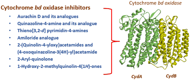
Cytochrome bd (cyt-bd) oxidase, one of the two terminal oxidases in the Mycobacterium tuberculosis (Mtb) oxidative phosphorylation pathway, plays an indispensable role in maintaining the functionality of the metabolic pathway under stressful conditions. However, the absence of this oxidase in eukaryotic cells allows researchers to select it as a potential drug target for the synthesis of anti-tubercular (anti-TB) molecules. Cyt-bd inhibitors have often been combined with cytochrome bcc/aa3 super-complex inhibitors in anti-TB drug regimens to achieve a desired bactericidal response. The functional redundancy between both the terminal oxidases is responsible for this. The cryo-EM structure of cyt-bd oxidase from Mtb (PDB ID: 7NKZ) further accelerated the research to identify its inhibitor. Herein, we have summarized the reported anti-TB cyt-bd inhibitors, insight into the rationale behind targeting cyt-bd oxidase, and an outline of the architecture of Mtb cyt-bd oxidase.


image file: d3md00587a-p1.tif

Pallavi Saha

Ms. Pallavi Saha has obtained her Bachelor in Pharmacy degree in 2019 from NSHM Knowledge Campus, Kolkata (affiliated to MAKAUT) West Bengal. She has completed her Masters from National Institute of Pharmaceutical Education and Research, Mohali, Punjab India in 2021. Her Masters dissertation was based on design and synthesis topoisomerase II α inhibitor based anti-cancer compounds. Currently, she is pursuing her Ph.D. at IIT-BHU, Varanasi, India under the supervision of Dr. Deepak K. Sharma. Her present work is focused on design and synthesis of anti-tubercular compounds targeting oxidative phosphorylation pathway.

image file: d3md00587a-p2.tif

Samarpita Das

Ms. Samarpita Das has obtained her Bachelor in Pharmacy degree in 2018 from Gupta College of Technological Sciences (affiliated to MAKAUT), Asansol, West Bengal. She has completed her Masters from National Institute of Pharmaceutical Education and Research, Mohali, Punjab India in 2020. Her Masters dissertation was based on design and synthesis of novel quinoline-based antimalarial compounds. Currently, she is a DST INSPIRE fellow at IIT-BHU, Varanasi, India under the supervision of Dr. Deepak K. Sharma. Her present work is focused on design and synthesis of small fluorescent molecule-based detectors.

image file: d3md00587a-p3.tif

Harish K. Indurthi

Mr. Harish K. Indurthi obtained his Bachelor in Pharmacy degree in 2017 from Acharya Nagarjuna University, Guntur, Andhra Pradesh. He has completed his Masters from National Institute of Pharmaceutical Education and Research, Mohali, Punjab India in 2019. He has worked on transition metal-free synthetic chemistry as a part of his Masters dissertation. Currently, he is enrolled as a Ph.D. at IIT-BHU, Varanasi, India under the supervision of Dr. Deepak K. Sharma. His doctoral studies are focused on bioluminescence-based monitoring of tumor progression and treatment by apoptotic pathway.

image file: d3md00587a-p4.tif

Rohit Kumar

Mr. Rohit Kumar obtained his Bachelor's in Pharmacy in 2020 from Apeejay Stya University, Gurugram, Haryana, and his Master's from IIT (BHU), Varanasi, Uttar Pradesh India in 2022. He is currently pursuing his PhD at IIT (BHU) under the supervision of Dr. Deepak Kumar. He is working on the synthesis of novel compounds for anti-TB activity.

image file: d3md00587a-p5.tif

Arnab Roy

Mr. Arnab Roy completed his Bachelor in Pharmacy in 2019 from NSHM Knowledge Campus (affiliated to MAKAUT), Kolkata, West Bengal. He obtained his MS (Pharm.) degree from National Institute of Pharmaceutical Education and Research (NIPER), Mohali, Punjab where he worked on evaluation of anti-hyperlipidemic activity of Sea buckthorn plant extracts in type-2 diabetes induced dyslipidemia model as a part of his dissertation. He is currently pursuing his Ph.D. at National Institute of Pharmaceutical Education and Research (NIPER), Hyderabad, India under the supervision of Dr. Nitin Pal Kalia. His doctoral studies focus on exploring the terminal oxidases for developing a therapeutic regimen against Mtb.

image file: d3md00587a-p6.tif

Nitin Pal Kalia

Dr. Nitin Pal Kalia is an Assistant Professor in the Department of Biological Sciences at National Institute of Pharmaceutical Education and Research (NIPER), Hyderabad, India. Dr. Kalia obtained his M.Sc. Biotechnology degree from Guru Nanak Dev University, Amritsar and further completed his Ph.D. from Clinical Microbiology Division, Indian Institute of Integrative Medicine, Jammu under the supervision of Dr. Inshad Ali Khan. During his PhD, his research work focused on developing efflux pump inhibitors against ESKAPE pathogens. Dr. Kalia then joined NIAID-NIH Bethesda, USA as a visiting fellow where he was involved in evaluation of new chemical entities by additional profiling using cross-screening against Mtb mutants containing resistance mutations in promiscuous drug targets (MmpL3, DprE1). He further continued his postdoctoral research at Lee Kong Chian School of Medicine, NTU Singapore in the research group of Dr. Kevin Pethe where he explored the terminal oxidases of the Electron transport chain of Mtb. Dr. Kalia has been awarded the prestigious DBT – Ramalingaswami fellowship from Govt. of India. Dr. Kalia's current research work focuses on developing novel inhibitors targeting the electron transport chain and cell wall synthesis of Mtb.

image file: d3md00587a-p7.tif

Deepak K Sharma

Dr. Deepak K Sharma is an assistant professor in the Department of Pharmaceutical Engineering and technology, IIT-BHU, Varanasi, India. Dr. Sharma has a B. Pharmacy from the Guru Gobind Singh College of Pharmacy, Kurukshetra University and M. Pharmacy from ISF college of Pharmacy, Punjab Technical University. Dr. Sharma received his Ph.D. in Medicinal Chemistry at Academy of Scientific and Innovative Research (AcSIR), CSIR-IIIM, Jammu, working in the lab of Dr. Debaraj Mukherjee. The focus of his PhD research work was target based synthesis of medicinally important compounds inspired from microbial natural product scaffold. Dr. Sharma was postdoctoral fellow in the research group of Emeritus Prof. Alan P. Kozikowski in University of Illinois, Chicago, USA and Prof. Stephen C. Miller in Massachusetts Medical School, USA. Dr. Sharma has achieved SERB-National postdoctoral fellowship award and most prestigious DBT Ramalinga Swami fellowship from Govt. of India. Dr. Sharma current research work is design and synthesis of diindolylmethane analogues for anticancer activity, bioluminescence-based monitoring of tumor progression and design and synthesis of novel anti-tubercular compounds.


Introduction

Discovered in 1882 by Robert Koch, tuberculosis (TB) is an airborne complex, communicable disease arising from infection caused by a Gram-negative aerobic pathogen, Mycobacterium tuberculosis (Mtb).1 Although a pulmonary bacterium, Mtb can cause infection throughout the body.2 According to reports on infectious diseases by the World Health Organization (WHO), TB is the 2nd most leading cause of death globally after COVID-19. Following COVID-19, there is a rising trend of TB infection, which has delayed the timeline of the “End TB Strategy” by the WHO. The rate of TB infection increased by 3.6% worldwide in 2020–2021. In 2020, 10.1 million people were affected by TB, which increased to 10.6 million in 2021. Out of this, 450[thin space (1/6-em)]000 cases were caused by drug-resistant Mtb strains.1

Available treatment protocol for drug-sensitive TB infection involves the administration of a regimen of four drugs, isoniazid (INH), rifampicin (RIF), pyrazinamide (PZA), and ethambutol (EMB) over the course of six months.3 Ardently following this four-drug regimen over such a long period compromises patient compliance as well as elevates the risk of the advent of multi-drug-resistant (MDR-Mtb) and extensively drug-resistant (XDR-Mtb) strains. MDR-TB comes with the development of resistance to INH and RIF in patients, whereas, in the case of XDR-TB-affected patients, resistance to fluoroquinolones and aminoglycosides (capreomycin, kanamycin, and amikacin) is accompanied by persistent INH and RIF resistance.4–8

Although curable, TB treatment is posed with the challenge of emergence of resistant strains9 as Mtb showcases a unique property of survival under stressful conditions, such as oxygen sparsity and nutrient unavailability. Extraordinary metabolic flexibility allows the mycobacterium to exist in a quiescent and antibiotic-tolerant state under stress, which can easily return to its normal growth phase upon restoration of favourable conditions.10–12 This metabolic plasticity enables the emergence of drug-resistant Mtb strains, making the treatment of TB notably difficult in the current environmental scenario. To tackle the increasing burden of TB, a refined treatment strategy with a shortening of treatment period along with prevention from the development of resistant strains is of dire necessity.4–8

An extended treatment period can be dealt with if the growth of resistance to existing dosage regimen be inhibited. To prevent occurrence of resistance, new drug leads with fresh drug targets residing in the distinct biological pathways of the micro-organism have to be explored and identified. This approach will help to escape the shortcomings of existing drug regimen, as the new molecules will be immune to the metabolic activity of the mycobacterial enzymes. Consequently, the treatment protocol is expected to be simplified and disease management will be easier by decreasing the treatment period, curtailing the treatment cost, and improving patient compliance.13–16

Research suggests that the energy requirement of Mtb is sufficed by oxidative phosphorylation as, unlike some microorganisms, it cannot survive simply based on substrate-level phosphorylation.17a,b In Mtb, oxidative phosphorylation occurs via the electron transport chain (ETC). So, the discovery of novel TB-inhibitory agents specifically targeting oxidative phosphorylation components is much more rational. After 40 years of constant setbacks, the FDA approved bedaquiline (BDQ) as a new anti-tubercular drug for the treatment of MDR-TB on 31st of December, 2012. BDQ acts by targeting mycobacterial ETC. Approval of this novel anti-TB agent gave a new direction to the tiring years of ongoing drug hunt18,19 Nevertheless, the successful advance of BDQ was overshadowed by the emergence of clinical resistance less than three years after its introduction to medical use. The rapid emergence of resistance is most likely linked to the absence of potent companion drugs. Hence, the need for drug combinations targeting various components of ETC was highly required.20–23Mtb possesses two terminal oxidase enzymes in its respiratory chain: cytochrome bcc1/aa3 (cyt-bcc1/aa3) super complex and cytochrome bd oxidase (cyt-bd).17,24,25 Telacebec (Q203), an imidazopyridine derivative, presently under clinical trial, is among the very few drugs acting via ETC bcc1/aa3 super complex.26 Although Q203 showed promising responses till phase II of the clinical trial, it only managed to generate a bacteriostatic reaction. From this point onwards, the discovery of cyt-bd inhibitors to ensure bactericidal response was thoroughly carried out as it was found that the action of cyt-bd compensated the inhibitory response of cyt-bcc1/aa3. A drug combination of both the terminal oxidase inhibitors might produce a synergistic response, ensuring a complete lethal response to mycobacterial growth.27,28 Cyt-bd provides resistance to Mtb from multiple antibiotics, including BDQ. Therefore, introducing cyt-bd inhibitors in drug regiments should be a wise approach to securing inhibitory response against drug-resistant TB strains.17b,36 In this review, we have provided a detailed outline of the constitutional features of cyt-bd oxidase, versed in research advancements, and literature reported cytochrome bd inhibitors.

Oxidative phosphorylation and the rationale behind targeting Cyt-bd oxidase

In Mtb, the ETC, responsible for the oxidative phosphorylation, is located across the plasma membrane. This biological pathway of Mtb is composed of five complexes, which together are employed in the synthesis of ATP by utilization of the proton motive force (PMF).9,29,30 Electrons enter the ETC by the action of complex I (NADH dehydrogenase) at the cost of oxidation of NADH to NAD+. The complex II (succinate dehydrogenase) also performs the same function. From complex I and II, electrons move to the complex III (cyt-bcc1) and IV (cyt-aa3). Complex III and IV form the terminal oxidase super complex cyt-bcc1/aa3. Cyt-bcc1/aa3 reduces oxygen to water following the transfer of the received electrons to oxygen.9,31 Another terminal oxidase is complex V (cyt-bd oxidase), belonging to the menaquinol oxidase family, which performs the same function as cyt-bcc1/aa3. During electron transfer across the respiratory chain enzymes, protons are pumped across the plasma membrane of Mtb, generating PMF across the cytoplasmic membrane. Next, the F1–F0 ATP synthase produces ATP from ADP and inorganic phosphate (Pi) by utilizing this PMF (Fig. 1).29 Due to its ATP-generating ability, oxidative phosphorylation is an indispensable metabolic process in them. From high-density mutagenesis and deletion studies, scientists have inferred that Mtb is unable to satisfy its energy requirement via substrate-level phosphorylation alone and direly requires oxidative phosphorylation for its viability.31,32
image file: d3md00587a-f1.tif
Fig. 1 Schematic representation of oxidative phosphorylation pathway in Mtb.

In the absence of stress, cyt-bcc1/aa3 acts as the major terminal oxidase in Mtb, generating an ample amount of ATP. Under stressful conditions like a hypoxic state, nutrient scarcity, and unfavourable environmental states, cyt-bcc1/aa3 is usually inhibited, and Mtb enters into a non-replicating dormant state.

In the dormant state of Mtb, the metabolism rate reduces for the long-term survival of bacteria. Even in the dormant stage of Mtb, an uninterrupted ATP supply is still needed via oxidative phosphorylation.17b To counteract and bypass this stress, the cyt-bd, the alternate terminal oxidase, comes into play and continues the ATP synthesis on behalf of cyt-bcc1/aa3.17a,33,34 Thus, cyt-bd has the potential to efficiently compensate the function of cyt-bcc1/aa3.

On the other hand, an increased tendency of induction of cyt-bd in Mtb has been observed following treatment of TB with current front-line drugs (cell wall biosynthesis inhibitors) and drugs acting via oxidative phosphorylation pathway. We assume that this may be the reason for the failure of newer anti-TB drug BDQ.35a Furthermore, in vitro treatment of Mtb with chlorpromazine and clofazimine dramatically increased the expression of cyt-bd subunits cydAB.35b Combination of amoxicillin and clavulanic acid, known as augmentin, upregulates cydAB expression, suggesting targeting of an alternate, less energy-efficient pathway.35c Due to the engagement of cyt-bd in the emergence of drug-resistant TB strains and its pioneer role in maintaining the seamless operation of the oxidative phosphorylation pathway under any adverse situation, the ultimate urge for reporting novel cyt-bd inhibitors was felt.

Cyt-bd is a non-proton pumping terminal oxidase with a higher affinity for oxygen than cyt-bcc1/aa3. Q203 is a potent cyt-bcc1/aa3 inhibitor in a nM concentration range. Despite having such appreciable inhibitory potential to its target enzyme, due to the functional redundancy between cyt-bcc1/aa3 and cyt-bd, Q203 only managed to produce a bacteriostatic response. This obligatory role of cyt-bd oxidase in the survival of Mtb under rigorous conditions with a sufficient supply of energy makes it a suitable target for the design and synthesis of novel anti-tubercular molecules.35d Besides, cyt-bd is present as a terminal oxidase only in the prokaryotic ETC,36 which makes it a safer target for the design of novel molecules barring any consequences on the eukaryotic receptors.

Architecture of cyt-bd oxidase

Previously, researchers were dependent on the crystal structure of cyt-bd from Geobacillus thermodenitrificans (PDB ID: 5DOQ) and Escherichia coli (PDB ID: 64X4) for anti-TB drug designing due to the unavailability of the cryo-EM structure of the cyt-bd oxidase from Mtb. As there exists some structural and mechanistic variation in each class of cyt-bd, these sources were insufficient to bridge the need for structural details required for considering cyt-bd as a drug target in anti-TB drug discovery. In 2021, Safarian et al. first reported the cryo-EM structure of the cyt-bd oxidase from Mtb at a resolution of 2.5 Å (PDB ID: NKZ). This work of Safarian et al. had generated a clear vision about the structure of Mtb cyt-bd.37–40

Cyt-bd, a pseudo-symmetrical heterodimer, is an assembly of two subunits, namely, CydA and CydB. It is encoded by the cydAB gene cluster. The absence of any other orphan genes encoding for accessory subunits of Mtb cyt-bd, may confirm the non-existence of any additional subunit in the cyt-bd structure. Each subunit of cyt-bd is made of nine transmembrane helices, where eight are arranged in two groups of four helices each, and the ninth one is placed as a peripheral helix. CydA subunit, possessing the three prosthetic heme groups, quinol binding site, and oxidation domain (Q-loop), constitutes the catalytic domain of this enzyme (Fig. 2).37


image file: d3md00587a-f2.tif
Fig. 2 Cryo-EM structure of the cyt-bd oxidase from Mtb. (a) Surface representation of the cyt-bd oxidase. CydA subunit is marked in yellow color and CydB subunit marked in green color. (b) The ribbon diagram of Cyt-bd Mtb. Dashed red circles in the diagram refers to the locations of accessory transmembrane helices, present in other bd oxidase structures. Reprinted with permission from ref. 37. Copyright 2022, Nature Communications.

The three heme groups in CydA subunit, heme d, heme b558, and heme b595 are placed in a triangular fashion.37 Heme b558 is situated along the plane of the heme binding site, heme b595 is placed orthogonally to it. To the proximity of heme b558, a strictly conserved glutamate residue is present as an axial ligand. In the near vicinity of heme b558, heme d is placed as an axial ligand. Heme d is a dioxygen reduction site and is connected to heme b558via well conserved histidine residue. Entry of oxygen to this dioxygen reduction site takes place via the oxygen channel which is spaced from CydB to heme d (Fig. 3).37,38,41–43


image file: d3md00587a-f3.tif
Fig. 3 (a) Orientation of the three heme cofactors in triangular fashion at CydA domain. (b) The oxygen conducting channel indicated in brown color, initiating from the membrane plane and speeded towards the active site. Reprinted with permission from ref. 37. Copyright 2022, Nature Communications.

CydB subunit of Mtb contributes to the major deviation in architecture from other classes of cyt-bd in different species. The place occupied by ubiquinone-8 (UQ-8) moiety in the B subunit of E. coli is occupied by a series of Trp and Phe residues in Mtb. These aromatic amino acid residues introduce van-der-Waals contacts between transmembrane helices 3, 4, 5, and 8; thereby stabilizing the entire structure. CydB subunit of Geobacillus thermodenitrificans is devoid of these aromatic interactions as well as observed in Mtb, due to the absence of the corresponding aromatic amino acid residues.37,39

Cyt-bd of various species are categorized into either L or S subfamilies depending on the length of the Q-loop.39 Though cyt-bd of Mtb shares some structural similarities to the L subfamily, it is not part of either class due to its variation in Q loop components length.40 The N-terminal (QN) domain of Q-loop is highly disordered and comprised of amino acid residues from Pro256.A to Val309.A (Fig. 4). This region is marked into two short helical components Qh1 and Qh2 followed by a large disordered loop region. Qh1 includes the conserved residues, Lys258.A and Glu263.A and is employed in linking the quinols to the propionate A residue of hemeb558. Qh2 extends from Tyr296.A to Ala303.A.37,45 The QN domain also contains a disulfide bond between Cys266.A and Cys285.A residues which is a signature of the mycobacterium species. This disulfide bond is responsible for rigidification the disordered Q-loop along with diminishing the flexibility between QN and Qh1 (Fig. 4).37 The C-terminal domain (QC) is a rigid and ordered segment, composed of Thr310.A to Asn333.A amino acid residues (Fig. 4). This region contains a small helix, Qh3, which expands along the periplasmic surface of transmembrane helices 6 and 7 and is extended up to the large periplasmic loop (PL8). PL8 is the connecting link between transmembrane helices 8 and 9. At this point, close proximity of the following aromatic amino acid residues: Tyr321.A, Phe325.A, Tyr330.A of Qh3, and Pro401.A, Trp402.A, Pro406.A of PL8, a hydrophobic cluster is generated which further helps in stabilizing the Qh3-PL8 interaction. The C-terminal part of the PL-8 forms multiple hydrogen bonds with the N-terminal half of PL-8 (Fig. 4).37,42–44


image file: d3md00587a-f4.tif
Fig. 4 (a) Left: the mycobacterial Q-loop is organized into the disordered QN (blue) and a rigid, well-ordered QC segment (cyan). Right: van-der-Waals interaction cluster between Qh3 and the periplasmic loop 8 (PL8) of CydA. (b) Top view of a superposition of CydA snapshots obtained from MD simulations performed in presence and absence of the Cys266.A–Cys285.A disulfide bond. Positions of cysteine residues are indicated. The red dashed circle indicates the location of the intact disulfide bond. CydA (oxidized), yellow; CydA (reduced), violet. Reprinted with permission from ref. 37. Copyright 2022, Nature Communications.

A menaquinone-9 (MK-9) binding pocket is present in the vicinity of the disulfide bond of the QN domain, which is comprised of porphyrin scaffold of heme b595, Arg8.A and Trp9.A from transmembrane helix 1, and Met397.A of transmembrane helix 9. MK-9, in its oxidized state, remains bound to this pocket. Trp9.A residue contributes majorly to the fabrication of this pocket and protection of MK-9 from surrounding milieu. Naphthoquinone group of MK-9 shows stable van-der-Waals interactions with Met397.A. In oxidized state, MK-9 shows π–π interactions with either Trp9.A or heme b595 and intermittent H-bonding with Arg8.A and Trp9.A amino acids (Fig. 5).37


image file: d3md00587a-f5.tif
Fig. 5 Allosteric MK-9 binding site of cyt-bd Mtb. Left: side view and Right: front view of the MK-9 binding pocket containing residues of transmembrane helix 1, transmembrane helix 9, and the heme b595. Reprinted with permission from ref. 37. Copyright 2022, Nature Communications.

Presence of additional solvent accessible areas has been reported from the MD simulation assay results.37 There exists a water filled channel with a diameter of 20 Å on the cytoplasmic interphase of the cyt-bd, which is shallowed to a channel of 4 Å and runs between the transmembrane helices 2 and 3 of both CydA and CydB. The following amino acid residues of Asp59.B, Glu106.A, Ser107.A, Ser139.A, and Glu62.B of CydA and CydB have established a hydrogen bond network in this channel, connecting it to the dioxygen reduction site of heme d (propionate group of heme d). This narrow channel is the proton delivery pathway.37

Literature reported cyt-bd inhibitors

With the approval of BDQ by the FDA for the treatment of drug-resistant TB patients and later identification of Q203 as an emerging anti-TB agent in combination therapy with cyt-bd inhibitors, a rising trend in identifying more potent cyt-bd inhibitors has been noted. Lu et al.46 reported the first potent cyt-bd inhibitor against Mtb in 2018. In the last six years, numerous cyt-bd inhibitors with diverse core structures and side chain modifications have been reported. All reported literature has individually enlightened the novelty of selecting cyt-bd as a drug target. In this section, we have summarized the reported cyt-bd inhibitors by different research groups.

In 2018, Lu et al. reported Aurachin D (1), a menaquinone analogue, (Fig. 6), as a potential cyt-bd oxidase inhibitor of Mtb when administered in combination with Q203.461, which is a vitamin-K analogue was first isolated from Stigmatella aurantiaca strain Sga15.47


image file: d3md00587a-f6.tif
Fig. 6 Structure of aurachin-D (1).

While determining the minimal inhibitory concentration (MIC) value of 1, a MIC value greater than 100 μg ml−1 was noted, which confirmed the necessity of blocking cyt-bcc/aa3 along with cyt-bd for achieving the bactericidal response. In combination therapy with Q203, 1 has decreased the MIC of Q203 from 10 nM to 1.5 nM. Further, they conducted a killing-kinetics experiment with Mtb H37Rv strain to validate their hypothesis. The reduction in the colony-forming unit (cfu) using combination therapy confirmed that the enhancement of potency of Q203 is directly correlated to the concentration of 1 (Fig. 7).46 Oxygen consumption rate assay (OCR) on inverted membrane vesicles (IMVs) from Mtb strain mc26020 showed that both the drugs, Q203 and 1, individually can produce up to 60% inhibitory response at 10 μM and 25 μM concentrations, respectively. But, when combined, the maximum inhibitory effect was achieved with 400 nM of each drug inhibition (Fig. 8).46 Despite such promising inhibitory potential against Mtb, less penetration of 1 through the bacterial cell wall due to poor solubility and toxic off-target effects limit its use and encourage the introduction of modifications in the core structure.48,49


image file: d3md00587a-f7.tif
Fig. 7 Kill-kinetics for combination therapy of cyt-bcc/aa3 and cyt-bd inhibitors; A) plot of log CFU ml−1vs. incubation time for Q203 and 1 at different concentrations as marked within graph, B) plot of enhancement of killing post 1 addition (25 μg ml−1) to Q203 treated sample, compared to killing by Q203 in monotherapy vs. incubation time. Reprinted with permission from ref. 46. Copyright 2018, Nature.

image file: d3md00587a-f8.tif
Fig. 8 Plot of relative inhibition (%) of OCR assay on IMV vs. drug concentration in μM; (A) the plot is for monotherapy with Q203, (B) the plot is for monotherapy with 1, and (C) the plot is for monotherapy and combination therapy with Q203 + 1. KCN in 10 mM concentration has been used as control. Reprinted with permission from ref. 46. Copyright 2018, Nature.

Lee et al. reported a quinazolin-4-amine derivative, ND-011992 (2), as an efficient inhibitor of mycobacterial ETC via inhibition of cyt-bd oxidase in their research work in 2018 (Fig. 9). Objective of their work was focused around establishing 2 + Q203 as a bactericidal drug regimen, active against both replicating as well as non-replicating, antibiotic-tolerant strains.50


image file: d3md00587a-f9.tif
Fig. 9 Structure of ND-011992 (2).

Out of 53 initially screened molecules, only two showed effective results in facial whole cell screening assay against M. bovis BCG. Out of these two, compound 2 was chosen as the representative molecule for future studies. In combination with Q203, 2 gave IC50 values of 0.5–1.6 μM in M. bovis BCG, 2.8–4.2 μM in Mtb H37Rv strains, respectively.50 OCR assay showed that both Q203 and 2, individually do not arrest the growth of Mtb, whereas, when administered in combination, they successfully arrested the oxygen consumption in M. bovis BCG with an IC50 value of 0.8 μM (Fig. 10). The potential of combination therapy was further validated by MitoXpress® Xtra oxygen consumption assay against M. bovis BCG and Mtb strains. They performed OCR on IMVs of Mycobacterium smegmatis mc2155 and M. smegmatis ΔcydAB mutant expressing the Mtb cydABDC operon. The result from IMV assay was consistent with the previous OCR assay results (Fig. 11).50 To establish the mode of action of 2, transcriptional responses of H37Rv to Q203, 2, and (Q203 + 2) were evaluated by RNA-sequence study where down regulation of gene expression in presence of the drug combination confirmed the action of 2 against cyt-bd oxidase.50


image file: d3md00587a-f10.tif
Fig. 10 Oxygen consumption assay of Q203 and ND-011992 (2) in M. bovis BCG using methylene blue as an indicator. Reprinted with permission from ref. 50. Copyright 2018, EMBO press.

image file: d3md00587a-f11.tif
Fig. 11 Plot of % OCR at varied concentration of 2 on energized (with NADH) IMVs of M. smegmatis. The parental strain is indicated by green triangles, ΔcydAB knockout strains by blue circles, and ΔcydAB complemented with Mtb CydABDC+ by red squares. Concentration of Q203 was kept at 1 μM. 100% OCR refers to the OCR of the untreated samples for each strain. Reprinted with permission from ref. 50. Copyright 2018, EMBO press.

Compound 2 was evaluated in combination therapy with Q203 against clinical isolates of drug resistant strains 123-20-0015, 123-20-0091 (XDR) and 123-20-0041, 123-20-0047 (MDR) where they reported a MIC of ≤1 μM for 2 when used in presence of Q203 (at 100 nM). Next, frequency of resistance (FOR), which implies the incidence of spontaneous mutations, was evaluated for 2. The FOR value to Q203 was not increased on the addition of 2. The cydABDC operon and all other genes except QcrB are free of mutations, which results in an extremely low FOR (<3.7 × 10¹⁰) to 2. The precise necessity of the 2 interacting residues for cyt-bd function or an unidentified off-target effect could explain this. Later on, it was found that the development of resistance to the drug combination was strictly due to the emergence of mutation to Q203.50

Although significantly inhibitory, poor pharmacological profile limits the application of 2 in combination with Q203. The compound is fairly stable in mouse plasma, murine, human microsomes, and simulated gastric fluid, but it exhibits low solubility and permeability in Caco-2 model. It has a 58% bioavailability in mice, a moderate volume of distribution, and very low systemic clearance. Due to such poor ADME profile, it has resulted in a prolonged t1/2 of 64 h. Less than optimum pharmacokinetic profile promotes the introduction of further modification to structural aspects of 2. Additionally, the comparable bactericidal response of the drug combination to that of BDQ encourages the exploration of a three-drug combination of BDQ, Q203, and 2 as a potential drug regimen for the treatment of TB.50

Hopfner et al. reported the development of thieno[3,2-d] pyrimidin-4-amines as novel cyt-bd oxidase inhibitors (Fig. 12) in 2021. They performed an ATP depletion assay and screened all their synthesized derivatives against three TB strains, namely, M. bovis, Mtb H37Rv, Mtb N0145. Studies showed that all the compounds were more active only when administered in combination with Q203 as opposed to when administered singly, and therefore, all the subsequent evaluations were carried out based on the IC50 values of the drug combination of synthesized molecules with Q203. This synergistic response also confirmed the target specificity of the synthesized molecules towards cyt-bd.51


image file: d3md00587a-f12.tif
Fig. 12 Structure of thieno[3,2-d] pyrimidin-4-amine derivatives.

An array of 50 compounds were screened out as “hits” from their established compound library, out of which, two series of compounds based on thieno[2,3-d]pyrimidine-4-amines and thieno[3,2-d]pyrimidine-4-amines, were found to have considerable inhibitory potential. For lead identification, they further proceeded with the thieno[3,2-d]pyrimidine-4-amine class and selected compound 3 as the representative compound.51

Total 13 compounds they had synthesized and assayed further to explore the effect of introducing modifications in order to improve ATP IC50 value.51 All their compounds had shown ATP IC50 values in permissible range only when used in combination therapy with Q203. Out of all the synthesized compounds, 4 was identified as the most potent one with ATP IC50 values 5.8 ± 1.06 μM, 18.9 ± 9.03 μM and 8.5 ± 2.38 μM respectively against BCG, H37Rv and N0145 strains.51Fig. 12. Structure of thieno[3,2-d] pyrimidin-4-amine derivatives.

In 2021, Hopfner et al. reported N-phenethyl-quinazolin-4-yl-amines as potential cyt-bd oxidase inhibitors following multiple steps of screening and substrate explorations (Fig. 13). They had screened all their synthesized derivatives via an ATP depletion assay against three TB strains, M. bovis, Mtb H37Rv, Mtb N0145 and studied the response of their library of compounds both individually and in combination with Q203. None of the compounds was active in the absence of Q203; hence, all the further evaluations were carried out based on the IC50 values of the combination of drugs.52 They started the inhibitory potential studies with three classes of compounds, namely, thieno[3,2-d]pyrimidin-4-amines, 2, and N-phenethylquinazolin-4-amines. Compound 5, the “hit compound” of N-phenethylquinazolin-4-amine class, showed IC50 values of 11 μM and 27 μM, respectively, against M. bovis BCG and Mtb H37Rv strains.51,52


image file: d3md00587a-f13.tif
Fig. 13 Structure of N-phenethyl-quinazolin-4-yl-amine derivatives (5–8).

They also explored the effects of chemical modifications on N-phenethylquinazolin-4-amine on anti-cyt-bd activity. Initially they synthesized 10 compounds by making alterations in the phenethylaniline moiety, keeping quinazoline core unchanged and studied the cyt-bd inhibitory ability following ATP depletion assay against the following strains: M. bovis BCG and Mtb strains (H37Rv and N0145). From the 10 derivatives, they chose compound 6 as the representative compound and synthesized another set of 11 compounds having modifications on the quinazoline core. Screening of these 11 compounds were done in the same way as the first 10 compounds.52

From the response studies of all the compounds both, individually and in combination with Q203, it was observed that compounds 7 and 8 showed the most potent cyt-bd inhibitory activity, with sub-micromolar concentrations of 0.1 μM for compound 7 and 0.2 μM for compound 8 against Mtb N0145 strain, respectively.52

Hards et al. reported the compound 3,5-diamino-6-(benzofuran-2-yl)-N-carbamoylpyrazine-2-carboxamide 9, a 6-substituted derivative of the FDA-approved diuretic amiloride, as a potential anti-TB agent in 2022 (Fig. 14).53,54 Compound 9 may produce its anti-TB response via two targets of ETC pathway, cyt-bd and to some extent, F1Fo-ATP synthase enzyme.53


image file: d3md00587a-f14.tif
Fig. 14 Structure of amiloride and its potent anti-TB derivative 9.

From MIC assay result, the initial potency of 9 was found to be 4 μM against Mtb H37Rv strain. Later on, cyt-bd specificity property, 9 was evaluated by OCR assay on IMVs and results clearly showed the cyt-bd inhibitory potential of 9 with an IC50 value of 21.2 μM.53 A strong inhibition was reported on using 9 with Q203, marking the synergistic response of both the molecules. Further experiments confirmed that the mild ATP synthase inhibitory property of 9. Such inhibitory response shows promise in utilization of this compound to treat BDQ resistant TB strains and furthermore, its ability to target multiple steps of ETC has initiated a vision for multi-component targeting drug development (Fig. 15).53


image file: d3md00587a-f15.tif
Fig. 15 Compound 9 inhibits cyt-bd and, to a lesser or indirect extent, the F1Fo-ATP synthase. Reprinted with permission from ref. 53. Copyright 2022, Nature Portfolio.

Kumar et al. reported the design and synthesis of two series of compounds, (quinazoline 4-yloxy)acetamide derivatives and (4-oxoquinazoline-3(4H)-yl)acetamide derivatives as cyt-bd oxidase inhibitors and further conducted multiple virtual screening and biological studies.55 They adopted the scaffold hopping technique on three previously reported anti-TB compounds: 2, 1, and 2-(quinolin-4-yloxy)acetamide based derivative40,50,56 and designed the first series of compounds by linking the quinazoline moiety of 2 with the acetamide chain of 2-(quinolin-4-yloxy)acetamide class of compounds, while second series of compounds were generated by amalgamation of 4-oxoquinazoline moiety of 1 with the acetamide side chain of 2-(quinolin-4-yloxy)acetamide derivatives (Fig. 16.)55


image file: d3md00587a-f16.tif
Fig. 16 Designing of (quinazoline 4-yloxy)acetamide and (4-oxoquinazoline-3(4H) yl)acetamide derivatives by scaffold hopping approach.

Twenty-nine synthesized compounds of both series were initially screened against two strains of Mtb, wild-type Mtb H37Rv and the deletion-mutated strain of Mtb, lacking the cytochrome b (QcrB) subunit of bc1 complex. Of these, only two compounds had inhibitory potential at a concentration below 32 μM. These two compounds, S-021-0601 (10) with MIC 8 μM and S-021-0607 (11) (Fig. 17) with MIC 16 μM were further subjected to growth inhibition assay for both the Mtb strains.55 Though MIC values for the mutated strains were reported to be 8 and 16 μM for 10 and 11, respectively, in wild type strain the MIC values were as high as 128 μM and 256 μM, respectively. Due to the compensatory function of the cyt bcc1/aa3 super complex, such high values were reported.55 When the two lead compounds were tested individually for synergistic response with Q203, only 11 showed synergism (Fig. 18).55 To evaluate inhibitory potential against cyt-bd, the authors explored the bactericidal response of 10 and 11 under hypoxic conditions to non-replicating TB strains, and both the compounds produced strong bactericidal response resulting in ∼1.9[thin space (1/6-em)]log10 reduction at 32 μM concentration. In conjunction with Q203, 10 and 11 showed an approximate four times enhancement in potency (Fig. 19).55 Total cellular ATP depletion assay of 10 and 11 in monotherapy and combination therapy with Q203 indicated that an appreciable reduction of ATP level is produced only in combination with Q203 (Fig. 20).55


image file: d3md00587a-f17.tif
Fig. 17 Structure of 2-(quinolin-4-yloxy)acetamide derivatives 10 and 11.

image file: d3md00587a-f18.tif
Fig. 18 Examination of the synergistic response of cyt-bd and known cyt-bc1 complex inhibitor following combination therapy. Reprinted with permission from ref. 55. Copyright 2022, Elsevier.

image file: d3md00587a-f19.tif
Fig. 19 Plot of log10 reduction in CFU in 3 days (mean ± SD of two independent experiments) on non-replicating Mtb at hypoxic condition for 3 days with incubation at 37 °C. Reprinted with permission from ref. 55. Copyright 2022, Elsevier.

image file: d3md00587a-f20.tif
Fig. 20 Plot of relative luminescence units (RLU) following treatment of various inhibitors both in monotherapy and combination therapy on Mtb for 15 h. Percentage values over the bars indicates percentage depletion in ATP compared to untreated (none) culture. Reprinted with permission from ref. 55. Copyright 2022, Elsevier.

A methylene blue assay was conducted to measure the oxygen consumption inhibitory potential of each compound. When administered as monotherapy, none of the compounds managed to retain the blue color, confirming the lack of bactericidal response generation. But when used in combination therapy with Q203 + 10 or 11, desired inhibitory response was achieved.55

Lastly, on establishing SAR from all the assay results and co-relating that with molecular docking analysis results, they concluded that their assay results agree with the docking results against two PDB IDs: 7NKZ and 7OSE. 10 and 11 are the most potent compounds of the series, exhibiting stable H-bond and pi–pi interaction at their binding site. Both the compounds individually produce bactericidal response under hypoxic conditions; response is further improved by using combination therapy with Q203.55

Lawer et al. reported a new series of 1 analogues as potential cyt-bd inhibitors in the year 2022 (Fig. 21). They had evaluated the cyt-bd inhibitory potential of all their synthesized derivatives following MIC and OCR assay on IMVs. For determining the MIC values, they used either Mtb mc26230 or M. smegmatis mc2155 strains. For preparing IMV for their study, M. smegmatis (Msmeg) ΔcydAB overexpressing the Mtb cyt-bd expression construct (CydABCD+) was utilized and TB47 was used as cyt-bcc:aa3 super complex inactivator. Seeking guidance from the observations of both the assay results, they further constructed a SAR profile of all the synthesized compounds to establish a clear vision of effects of varying substitution at different positions of 1.57


image file: d3md00587a-f21.tif
Fig. 21 Compound 1 analogues, 12 and 13 bearing citronellyl and farnesyl side chain respectively.

In their research, they had explored the effect of altering the side chain length and functionality of 4-(1H)-quinolone and effect of introducing substitutions at the C5, C6, C7 and C8 position of 4-(1H)-quinolone moiety and observed that making any major alteration in side chain is not preferable and farnesyl side chain is the most preferred choice. Substituting farnesyl to citronellyl side chain was the only alternative variation tolerated. In case of introducing substitution on the aromatic ring, fluorine substitution at C6, C7 position; hydroxy substitution at C5, C6 and methoxy substitution at C5, C6, C7 were tolerated.57

Two compounds (12 and 13) of the series managed to show appreciable inhibitory response along with the parent compound 1. Compound 12 bearing citronellyl side chain is the most potent one of the series with MIC value of 4 μM, and compound 13 bearing C6-fluoro-substitution and farnesyl side chain had a MIC value of 8 μM against the mutant strains. The appreciable MIC values of these two compounds, which are in the same range as of 1, thereby initiated a research scope of introducing further modifications to them for harbouring superior inhibitory responses.57

Jeffreys et al. in the year 2023 reported the inhibitory potential of 2 arylquinolone derivatives against cyt-bd oxidase following a series of biological assays, in vitro and in vivo studies, in mono and combination therapy with other ETC components inhibitors separately.58 For their studies they choose a compound library consisting of five classes of 2-arylquinolone derivatives, having a previous record of Plasmodium falciparum ETC inhibitory action; where template 1 are pyridyl oxygen-linked quinolone derivatives,62 template 2 are arylamine quinolone derivatives,62 template 3 are quinolones bearing bisaryl side chain substitution,61 template 4 are compounds with no linker in between aryl groups in the side chain and have varying pyridyl functionality59 and lastly template 5 with either pyrazole or triazole substitution on the side chain.60

Out of the five templates, template 4 and 5 were not refereed further for drug discovery due to their poor Mtb inhibitory potential.58 Among the first three templates, cyt-bd inhibitory potential of the series are in the following order: template 3 > template 2 > template 1. Template 3 compounds have reduced inhibitory potential of Mtb growth than template 2, maybe due to increased lipophilicity and bulkiness of template 1 and 3.58

From their preliminary growth suppression assay results of all the compound on H37Rv strain, they selected the compound CK-2-63 (14) of template 3 for their further experimental analysis. In OCR measurement experiment (Fig. 22), 14 alone produced a partial reduction of oxygen consumption at a high concentration. When given in combination therapy separately with BDQ and Q203, 14 (in 3.5 μM concentration) managed to produce an improved inhibitory response than monotherapy (Fig. 23).58


image file: d3md00587a-f22.tif
Fig. 22 Structure of CK-2-63 (14), which is the representative molecule of template 3.

image file: d3md00587a-f23.tif
Fig. 23 Mtb ETC inhibitors decreases the OCR in M. smegmatis. (a) Arrows indicate the time where additions were made: GLC (glucose, 10 mM), OLG (Oligomycin, 15 μM), FCCP (15 μM), ANT (antimycin A, 15 μM), and ROT (rotenone, 15 μM). (b) Lead compound CK-2-63 (14) has stopped oxygen consumption even in aerobic conditions when administered in increments over a 60 min period (20–200 μM). (c and d) Sequential addition of Q203 (10–110 nM) or BDQ (1–56 nM) inhibited oxygen consumption in the absence (black) and presence (red) of 3.5 μM CK-2-63. Reprinted with permission from ref. 58. Copyright 2023, American Chemical Society.

From the in silico docking results against the PDB ID: 7NKZ, Jeffreys et al. predicted that binding site of 14 is in a cleft region in close vicinity to the Q-loop and this compound can be considered as a non-competitive, mixed inhibitor of menaquinone. The docking results of 14 is similar to the docking outcome of 1 at the same binding site. Both this cyt-bd inhibitors had produced π-interactions with Arg8.A and Trp9.A amino acids, the amino acids which also produced H-bond interaction with MK-9 in intermittent state (Fig. 24).37,58


image file: d3md00587a-f24.tif
Fig. 24 (a) Compound 14 indicated by purple sticks was superimposed with the menaquinone, which is indicated by orange sticks. The yellow colored group is one of the heme b prosthetic groups (b) 1 indicated by pink sticks produces interaction in the same cleft as menaquinone and 14. (c and d) Amino acid residues of the CydA domain (green sticks), and the heme prosthetic group (yellow sticks) interacting with 14 and 1. Reprinted with permission from ref. 58. Copyright 2023, American Chemical Society.

To explore the pharmacokinetic response of 14, standard 5 day alamar blue assay and mycobacterial growth indicator tubes (MGIT) assay was conducted. When 14 was added in monotherapy, a moderate inhibitory response was produced in both assays. A superior inhibitory response was noted in the MGIT assay on the use of combination therapy with BDQ and Q203 separately. Using a triple combination of 14 + BDQ + Q203, an improved response was observed, matching the inhibitory potential of INH (Fig. 25).58


image file: d3md00587a-f25.tif
Fig. 25 Combination therapy approaches lead to increases in time to positivity (TTP). Growth suppression of M. tuberculosis increases the TtP (>44 days indicates maximum measurable suppression of growth). Inhibitors included Q203 (Q, cyt-bcc/aa3 inhibitor), AWE402 (A, cytochrome bcc/aa3 inhibitor), 14 (C, cytochrome bd inhibitor), and BDQ (B, ATP synthase inhibitor). Controls are indicated in black, drugs in isolation in red, dual combinations in blue, triple combinations in purple, and the quadruple combinations in green. Reprinted with permission from ref. 58. Copyright 2023, American Chemical Society.

Time-kill kinetic experiments were further conducted to explore the pharmacodynamic profile of their lead molecule. Matching the pharmacokinetic response, here also they observed bacteriostatic response in monotherapy (both for 14 and Q203 individually) and bactericidal response in combination therapy (14 + Q203). 14 has improved the bactericidal potency of BDQ (Fig. 26).58


image file: d3md00587a-f26.tif
Fig. 26 CK-2-63 (14) combination therapies produce sterilization in time-kill experiments. Reprinted with permission from ref. 58. Copyright 2023, American Chemical Society.

In the in vivo experiments on TB-infected mouse models of both acute and chronic inbred, an unexpected diminished ability of 14 + Q203 combination therapy was due to high plasma protein binding of 14 (Fig. 27). Thus, even after having appreciable inhibitor response in in vitro experiments; poor solubility, high lipophilicity and plasma–protein binding are the obstacles that need further attention for improving the drugability of 14.58


image file: d3md00587a-f27.tif
Fig. 27 Assessment of combination therapy of ETC inhibitors using acute and chronic in vivo mouse models of TB infection. Mtb H37Rv bacterial burdens were determined from BALB/c mice using (a) acute or (b) chronic infection models. Treatment arms consists of RIF (red bars), INH (dark blue bars), Q203 (green bars), CK-2-63 (14) (light blue bars), Q203 + 14 (blue-green bars), and untreated (gray bars) for 1 month. The mean bacterial burden in the untreated mice at the initiation of treatment (day 0) is indicated by a dashed line. Reprinted with permission from ref. 58. Copyright 2023, American Chemical Society.

Zhou et al. have reported the synthesis and biological evaluation of 1-hydroxy-2-methylquinolin-4(1H)-one derivatives (15 and 16, Fig. 28) as novel cyt-bd oxidase inhibitors. From biological assay results, they reported a detailed SAR of the synthesized compounds. Lastly, they performed in silico study and established a probable mode of interactions and nature of binding of their potent compounds to the binding site of the cyt-bd i.e., in the Q-loop.63


image file: d3md00587a-f28.tif
Fig. 28 Structure of 1-hydroxy-2-methylquinolin-4(1H)-one derivatives (15 and 16) bearing –OCF3 substitution.

Following virtual screening of the available compound libraries via homology modelling and molecular docking they had selected 37 compounds from 397[thin space (1/6-em)]465 compounds on the basis of docking score. These 37 compounds were next tested against two strains: M. smegmatis (wild type) and ΔqcrCAB mutant strain (cyt-bd+) which is the deletion mutated strain of the cyt-bcc. From the MIC values, two FDA approved drugs ivacaftor (drug for treatment of cystic fibrosis) and roquinimex (immunostimulant; increases NK cells activity and macrophage cytotoxicity) were selected for further derivatization. Ivacaftor produces a MIC value of 51 μM and roquinimex 260 μM. As, both the compounds had produced no inhibition of wild type strain, the cyt-bd target specificity of both the compounds were confirmed. Their observation was further validated by microscale thermophoresis (MST) assay, where ivacaftor (Kd values = 40 μM) and roquinimex (Kd values = 13 μM) resulted moderate binding affinity.63 While designing their series of molecules they kept quinoline moiety intact due to its abundance in ivacaftor, roquinimex, 1, and some other reported cyt-bd inhibitors and introduced modifications at R1 and R2 positions (Fig. 29).


image file: d3md00587a-f29.tif
Fig. 29 Structure of A) (1), B) ivacaftor, C) roquinimex and D) the general structure of the designed molecules; all of structures share the common quinoline moiety as core the nucleus.

From initial SAR studies, they identified 15 bearing hydroxyl group at R1 and –OCF3 benzyl substitution at R2 position is the most potent one. They had observed that long R2 chain is not a favoured substitution. Next, they varied the position of the –OCF3 substitution on the aryl ring and introduced other substitutions on the aryl ring. From the MIC values, they stated that the introduction of any other functionality other than –OCF3 is detrimental to the inhibitory response. 15 and 16 bearing –OCF3 at the para and ortho positions of the benzyl group are the most potent derivatives. In the MST assay, 16 had produced a very poor binding affinity of 165 μM of Kd value. 15 has a very appreciable binding affinity, with Kd = 4.17 ± 0.73 μM. Therefore, 16 was not further considered for the remaining studies.63

In OCR assay of 1, Q203 and 15 in monotherapy and in combination therapy using methylene blue indicator (Fig. 30), the disappearance of blue color in every monotherapy indicated the necessity of using drug combination. In combination therapy, the potency of Q203 + 15 is superior to Q203 + 1. OCR assay was also performed on IMVs from M. smegmatis. In this case, a similar inhibitory profile of the studied compounds as in the former assay was noted down (Fig. 31).63


image file: d3md00587a-f30.tif
Fig. 30 Oxygen consumption assay result of compounds Q203, 1 and 15. Reprinted with permission from ref. 63. Copyright 2023, Elsevier.

image file: d3md00587a-f31.tif
Fig. 31 Inhibition of cyt-bd oxidase oxygen consumption in IMVs of M. smegmatis mc2155 of Wt, ΔqcrB, ΔcydAB and ΔcydAB pLHcyd. Reprinted with permission from ref. 63. Copyright 2023, Elsevier.

To explore the probable binding interactions at the site of action they had performed in silico docking of 15 and 16 in a similar environment as they did in the beginning of their study (Fig. 32). From the docking results, the necessity of R1 –OH was confirmed, as it is involved in favourable interaction with Asp245. The loss of binding affinity on positioning –OCF3 at ortho in 16 introduces steric hindrance at the binding site thereby validating the inconsistency of response in MIC and MST assay.63 From all the assay results of 15, in mono and combination therapy with Q203, scientist had finally concluded that potency of 15 is improved many folds on combination therapy. In monotherapy, 15 has promising response against ΔqcrCAB strain only. Simultaneous inhibition of both the terminal oxidases is responsible for such improved potency following combination therapy.63


image file: d3md00587a-f32.tif
Fig. 32 The predicted binding mode of the representative compounds with cyt-bd. A) The predicted binding mode of 15. B) The predicted binding mode of 16. Reprinted with permission from ref. 63. Copyright 2023, Elsevier.

Assays for evaluating cytochrome bd inhibitors

Quantification of intracellular ATP levels

The primary assay to evaluate the potency of ETC inhibitors is by checking the intracellular depletion of ATP levels. Mycobacteria is exposed to ten two-fold serial dilutions of compounds in combination with fixed dose of any cytochrome bcc:aa3 inhibitor and incubated for 15–24 h at 37 °C. The intracellular ATP level is determined using luminescence based BacTiter-Glo Microbial Cell Viability assay reagent. The presence of cytochrome bcc:aa3 inhibitor triggers the essentiality of cyt-bd. The assay is rapid and can be utilized for screening large library of molecules for identifying putative cyt-bd inhibitors.27 This assay is limited to analysing ATP levels and does not confirm the actual inhibition of bacterial growth or killing of the pathogen.

Oxygen consumption assays

ETC inhibitors are known to inhibit OCR due to the capacity of the drugs to interfere with respiration. Qualitative evaluation of OCR inhibition can be done using methylene blue at a final concentration of 0.001% in sealed tubes inside anaerobic jars to avoid oxygen leakage. Further, oxygen consumption can also be tested quantitatively with the use of MitoXpress oxygen probe and recording the fluorescence.27 Chemical inhibition of both terminal oxidases should arrest the oxygen consumption in Mtb. Strict anaerobic environment should be maintained to perform this assay which often become difficult.

Steady state kinetic assay

Recombinant Mtb cyt-bd decylubiquinol oxidase activity can be monitored in a UV spectrophotometer at 283 nm. The assay reaction is initiated by addition of quinol to the crude recombinant membranes. Oxidation rate of quinol is fitted in Michaelis–Menten function to obtain the specific catalytic activity. This assay is used to validate the cytochrome bd oxidase inhibition by the small molecules, and can only be done with recombinant membranes.58

Microscale thermophoresis (MST) assay

MST assay is used to evaluate the binding affinity between cytochrome-bd and the compounds. This assay is performed by labelling cytochrome-bd with NT-647 (a red fluorescent dye) and mixed with serial dilutions of the compound, further loaded into capillaries, and measured on MST instrument (Monolith NT.115).63 Overall MST signal can be plotted against compound concentrations to obtain a dose–response curve.

Conclusions

Development of novel anti-TB agents acting via distinct mechanism of action is necessary. The recently approved BDQ exerts its antimycobacterial activity by the shutdown of ATP production by inhibiting the F-type ATP synthase. It clinically validates oxidative phosphorylation as a pharmacologic target in Mtb. Nevertheless, the successful advance of BDQ was overshadowed by the emergence of clinical resistance less than 3 years after its introduction to medical use. The rapid emergence of resistance is most likely linked to the absence of potent companion drugs. Hence targeting the additional components of oxidative phosphorylation of Mtb is highly required. Q203 is a selective inhibitor of cytochrome bc1:aa3, but the drug is only bacteriostatic. This issue raises the possibility that the alternate terminal bd-type oxidase (cytochrome bd oxidase) is capable of maintaining energized membranes. In a recent study, it was observed that upon genetic deletion of the cytochrome bd oxidase (cyt-bd)-encoding genes cydAB, Q203 inhibited mycobacterial respiration completely, became bactericidal, killed drug tolerant mycobacterial, and rapidly cleared Mtb infection in vivo. Hence, identifying a small molecule that will block cyt-bd activity is highly desirable to eradicate tuberculosis. Adoption of in silico drug design approach, availability of the cryo-EM structure of the cyt-bd oxidase from Mtb (7NKZ) facilitated the work of the researchers in terms of rational designing of cyt-bd inhibitors. Quinazoline and quinoline drug scaffolds bearing lipophilic side chains have been the building block of majority of the so far reported cyt-bd inhibitors. We assume, the structural resemblance with quinol moiety may have benefited such drug scaffolds. Though most of the so far literature reported cyt-bd inhibitors are associated with many limitations yet all the reported works have constructed the primary base for progress of future research in this direction (Table 1).
Table 1 A summary of the inhibitory potential of the most potent compounds of each literature reported class of cyt-bd inhibitors
Compound Mtb strain MIC IC50 (comp. + cyt-bcc1/aa3 inhibitor) (μM) Combination therapy with bc1 inhibitor complex
a ATP IC50 value. b % OCR IC50 value.
image file: d3md00587a-u1.tif (1) H37Rv >100 μg ml−1 In combination of 1 (25 μg ml−1) + Q203, MIC value of Q203 was reduced to 1.5 nM from 10 nM (ref. 46)
H37Rv bd-KO >100 μg ml−1
image file: d3md00587a-u2.tif (2) M. bovis BCG 0.5–1.6a 2 + Q203 (100 nM) against XDR and MDR showed MIC of ≤1 μM for 2 (ref. 50)
H37Rv 2.8–4.2a
image file: d3md00587a-u3.tif (4) M. bovis BCG 5.8 ± 1.06a In monotherapy, 4 produces ATP IC50 value > 50 μM as the inhibitory response was compensated by the action of cyt-bc1/aa3.51 In combination therapy of 4 + Q203, ATP IC50 value was reduced to 6.2–7.3 μM range due to synergistic inhibitory response51
H37Rv 5.8 ± 1.06a
N0145 8.5 ± 2.38a
image file: d3md00587a-u4.tif (7) M. bovis BCG 0.8 ± 0.144a ATP IC50 value > 25 μM was noted down when 7 and 8 was administered in monotherapy. On using drug combination with Q203 (100 nM concentration), ATP IC50 was reduced to the range of 0.1–8 μM (ref. 52)
H37Rv 5.8 ± 0.616a
N0145 0.1 ± 0.017a
image file: d3md00587a-u5.tif (8) M. bovis BCG 0.8 ± 0.077a
H37Rv 7.6 ± 1.079a
N0145 0.2 ± 0.057a
image file: d3md00587a-u6.tif (9) mc26206 9 μM 10.88–39.09a A strong inhibition was reported on using 9 + Q203 combination therapy, marking the synergistic response of both the terminal oxides inhibitors53
AtpE(A63P) 5 μM
Rv0678 (G65fs) 12 μM
H37Rv 4 μM
image file: d3md00587a-u7.tif (10) H37Rv-wild type 128 μM
ΔqcrB mutant 8 μM
image file: d3md00587a-u8.tif (11) H37Rv-wild type 256 μM
ΔqcrB mutant 16 μM
image file: d3md00587a-u9.tif (12) M. smegmatis mc2 155 (WT) >512 μM 0.35b The low IC50 values, utilizing the combination treatment of TB47 and 12, or 13 highlighted the necessity of inhibiting both the terminal oxidases57
M. smegmatis mc2 155 (Δqcr mutant)
mc2 6230 4 μM
image file: d3md00587a-u10.tif (13) mc2 155 (WT) >512 μM 0.25b
mc2 155 (Δqcr mutant)
mc2 6230 8 μM
image file: d3md00587a-u11.tif (14) H37Rv 5 μM In combination therapy with BDQ and Q203 separately, 14 (in 3.5 μM concentration) produces an improved inhibitory response than monotherapy due to inhibition of multiple components of ETC at a time.58 In combination therapy of 14 + Q203, desired bactericidal response was achieved58
image file: d3md00587a-u12.tif (15) M. smegmatis ΔqcrCAB 6.25 Potency of 15 was improved many folds in combination therapy with Q203 (ref. 63)
M. smegmatis WT >400


Conflicts of interest

There are no conflicts to declare.

Acknowledgements

The authors thank the Department of Biotechnology (DBT), India for the Ramalingaswami Research Support Grant (BT/RLF/Re-entry/54/2018). Pallavi Saha and Rohit Kumar are grateful to IIT (BHU) and the Ministry of Education, India for providing a teaching assistant fellowship.

Notes and references

  1. Global Tuberculosis report 2022, 2022, http://apps.who.int/bookorders Search PubMed.
  2. D. G. Russell, Immunol. Rev., 2011, 240, 252–268 CrossRef CAS PubMed .
  3. World Health Organization & World Health Organization, Treatment of tuberculosis: guidelines, World Health Organization, 4th edn, 2010, https://iris.who.int/handle/10665/44165 Search PubMed.
  4. C. R. Horsburgh, C. E. Barry and C. Lange, N. Engl. J. Med., 2015, 373, 2149–2160 CrossRef CAS PubMed .
  5. A. J. J. Wood, N. Engl. J. Med., 1993, 322, 784–791 CrossRef PubMed .
  6. G. B. Migliori, R. Loddenkemper, F. Blasi and M. C. Raviglione, Eur. Respir. J., 2007, 29, 423–427 CrossRef CAS PubMed .
  7. A. A. Velayati, M. R. Masjedi, P. Farnia, P. Tabarsi, J. Ghanavi, A. H. Zia Zarifi and S. E. Hoffner, Chest, 2009, 136, 420–425 CrossRef PubMed .
  8. J. C. Palomino and A. Martin, Antibiotics, 2014, 3, 317–340 CrossRef PubMed .
  9. J. Chakaya, M. Khan, F. Ntoumi, E. Aklillu, R. Fatima, P. Mwaba, N. Kapata, S. Mfinanga, S. E. Hasnain, P. D. M. C. Katoto, A. N. H. Bulabula, N. A. Sam-Agudu, J. B. Nachega, S. Tiberi, T. D. McHugh, I. Abubakar and A. Zumla, Int. J. Infect. Dis., 2021, 113, S7–S12 CrossRef CAS PubMed .
  10. L. G. Wayne and C. D. Sohaskey, Annu. Rev. Microbiol., 2001, 55, 139–163 CrossRef CAS PubMed .
  11. H. I. M. Boshoff and C. E. Barry, Nat. Rev. Microbiol., 2005, 3, 70–80 CrossRef CAS PubMed .
  12. M. B. Harbut, B. Yang, R. Liu, T. Yano, C. Vilchèze, B. Cheng, J. Lockner, H. Guo, C. Yu, S. G. Franzblau, H. M. Petrassi, W. R. Jacobs, H. Rubin, A. K. Chatterjee and F. Wang, Angew. Chem., Int. Ed., 2018, 57, 3478–3482 CrossRef CAS PubMed .
  13. A. I. Zumla, S. H. Gillespie, M. Hoelscher, P. P. J. Philips, S. T. Cole, I. Abubakar, T. D. McHugh, M. Schito, M. Maeurer and A. J. Nunn, Lancet Infect. Dis., 2014, 4, 327–340 CrossRef PubMed .
  14. A. W. Selassie, C. Pozsik, D. Wilson and P. L. Ferguson, Ann. Epidemiol., 2005, 15, 519–525 CrossRef PubMed .
  15. J. P. Owens, M. O. Fofana and D. W. Dowdy, Int. J. Tuberc. Lung Dis., 2013, 17, 590–596 CrossRef CAS PubMed .
  16. C. Lienhardt, A. Vernon and M. C. Raviglione, Curr. Opin. Pulm. Med., 2010, 16, 186–193 CAS .
  17. (a) G. M. Cook, C. Greening, K. Hards and M. Berney, Energetics of Pathogenic Bacteria and Opportunities for Drug Development, in: Adv Microb Physiol, Academic Press, 2014, pp. 1–62 Search PubMed ; (b) D. P. S. Chang and X. L. Guan, Metabolites, 2021, 2, 88 CrossRef PubMed .
  18. M. Mirsaeidi, Int. J. Mycobact., 2013, 2, 1–2 CrossRef PubMed .
  19. E. Segala, W. Sougakoff, A. Nevejans-Chauffour, V. Jarlier and S. Petrella, Chemotherapy, 2012, 56, 2326–2334 CAS .
  20. N. A. Ismail, S. V. Omar, L. Joseph, N. Govender, L. Blows, F. Ismail, H. Koornhof, A. W. Dreyer, K. Kaniga and N. Ndjeka, EBioMedicine, 2018, 28, 136–142 CrossRef PubMed .
  21. G. Degiacomi, J. C. Sammartino, V. Sinigiani, P. Marra, A. Urbani and M. R. Pasca, Front. Microbiol., 2020, 11, 1–8 CrossRef PubMed .
  22. R. C. Hartkoorn, S. Uplekar and S. T. Cole, Antimicrob. Agents Chemother., 2014, 58, 2979–2981 CrossRef PubMed .
  23. D. Almeida, T. Ioerger, S. Tyagi, S. Y. Li, K. Mdluli, K. Andries, J. Grosset, J. Sacchettini and E. Nuermberger, Chemotherapy, 2016, 60, 4590–4599 CAS .
  24. P. A. Black, R. M. Warren, G. E. Louw, P. D. Van Helden, T. C. Victor and B. D. Kana, Chemotherapy, 2014, 58, 2491–2503 Search PubMed .
  25. M. Urban, V. Šlachtová and L. Brulíková, Eur. J. Med. Chem., 2021, 12, 113139 CrossRef PubMed .
  26. K. Pethe, P. Bifani, J. Jang, S. Kang, S. Park, S. Ahn, J. Jiricek, J. Jung, H. K. Jeon, J. Cechetto, T. Christophe, H. Lee, M. Kempf, M. Jackson, A. J. Lenaerts, H. Pham, V. Jones, M. J. Seo, Y. M. Kim, M. Seo, J. J. Seo, D. Park, Y. Ko, I. Choi, R. Kim, S. Y. Kim, S. Lim, S. A. Yim, J. Nam, H. Kang, H. Kwon, C. T. Oh, Y. Cho, Y. Jang, J. Kim, A. Chua, B. H. Tan, M. B. Nanjundappa, S. P. S. Rao, W. S. Barnes, R. Wintjens, J. R. Walker, S. Alonso, S. Lee, J. Kim, S. Oh, T. Oh, U. Nehrbass, S. J. Han, Z. No, J. Lee, P. Brodin, S. N. Cho, K. Nam and J. Kim, Nat. Med., 2013, 19, 1157–1160 CrossRef CAS PubMed .
  27. N. P. Kalia, E. J. Hasenoehrl, N. B. A. Rahman, V. H. Koh, M. L. T. Ang, D. R. Sajorda, K. Hards, G. Grüber, S. Alonso, G. M. Cook, M. Berney and K. Pethe, Proc. Natl. Acad. Sci. U. S. A., 2017, 114, 7426–7431 CrossRef CAS PubMed .
  28. A. Moosa, D. A. Lamprecht, K. Arora, C. E. Barry, H. I. M. Boshoff, T. R. Ioerger, A. J. C. Steyn, V. Mizrahi and D. F. Warner, Chemotherapy, 2017, 61, 1–5 Search PubMed .
  29. D. Bald, C. Villellas, P. Lu and A. Koul, MBio, 2017, 8, 1–11 CrossRef PubMed .
  30. D. A. Lamprecht, P. M. Finin, M. A. Rahman, B. M. Cumming, S. L. Russell, S. R. Jonnala, J. H. Adamson and A. J. C. Steyn, Nat. Commun., 2016, 7, 12393 CrossRef CAS PubMed .
  31. G. M. Cook, K. Hards, E. Dunn, A. Heikal, Y. Nakatani, C. Greening, D. C. Crick, F. L. Fontes, K. Pethe, E. Hasenoehrl and M. Berney, Microbiol. Spectrum, 2017, 5, 295–315 Search PubMed .
  32. S. P. S. Rao, S. Alonso, L. Rand, T. Dick and K. Pethe, Proc. Natl. Acad. Sci. U. S. A., 2008, 105, 11945–11950 CrossRef CAS PubMed .
  33. V. B. Borisov, R. B. Gennis, J. Hemp and M. I. Verkhovsky, Biochim. Biophys. Acta, Bioenerg., 2011, 1807, 1398–1413 CrossRef CAS PubMed .
  34. T. Mogi and H. Miyoshi, J. Biochem., 2009, 145, 395–401 CrossRef CAS PubMed .
  35. (a) L. Mascolo and D. Bald, Prog. Biophys. Mol. Biol., 2020, 152, 55–63 CrossRef CAS PubMed ; (b) K. Arora, B. Ochoa-Montaño, P. S. Tsang, T. L. Blundell, S. S. Dawes, V. Mizrahi, T. Bayliss, C. J. Mackenzie, L. A. T. Cleghorn, P. C. Ray, P. G. Wyatt, E. Uh, J. Lee, C. E. Barry 3rd and H. I. Boshoff, Antimicrob. Agents Chemother., 2014, 58, 6962–6965 CrossRef PubMed ; (c) S. Mishra, P. Shukla, A. Bhaskar, K. Anand, P. Baloni, R. K. Jha, A. Mohan, R. S. Rajmani, V. Nagaraja, N. Chandra and A. Singh, Efficacy of β-lactam/β-lactamase inhibitor combination is linked to WhiB4-mediated changes in redox physiology of Mycobacterium tuberculosis, eLife, 2017, 6, e25624 CrossRef PubMed ; (d) T. Beites, K. O'Brien, D. Tiwari, C. A. Engelhart, S. Walters, J. Andrews, H. J. Yang, M. L. Sutphen, D. M. Weiner, E. K. Dayao, M. Zimmerman, B. Prideaux, P. V. Desai, T. Masquelin, L. E. Via, V. Dartois, H. I. Boshoff, C. E. Barry, S. Ehrt and D. Schnappinger, Nat. Commun., 2019, 10, 4970 CrossRef PubMed .
  36. M. Berney, T. E. Hartman and W. R. Jacobs, MBio, 2014, 5, 1–3 CrossRef PubMed .
  37. S. Safarian, H. K. Opel-Reading, D. Wu, A. R. Mehdipour, K. Hards, L. K. Harold, M. Radloff, I. Stewart, S. Welsch, G. Hummer, G. M. Cook, K. L. Krause and H. Michel, Nat. Commun., 2021, 12, 5236 CrossRef CAS PubMed .
  38. S. Safarian, A. Hahn, D. J. Mills, M. Radloff, M. L. Eisinger, A. Nikolaev, J. Meier-Credo, F. Melin, H. Miyoshi, R. B. Gennis, J. Sakamoto, J. D. Langer, P. Hellwig, W. Kühlbrandt and H. Michel, Science, 2019, 366, 100–104 CrossRef CAS PubMed .
  39. A. Theßeling, T. Rasmussen, S. Burschel, D. Wohlwend, J. Kägi, R. Müller, B. Böttcher and T. Friedrich, Nat. Commun., 2019, 10, 5138 CrossRef PubMed .
  40. S. Safarian, C. Rajendran, H. Müller, J. Preu, J. D. Langer, S. Ovchinnikov, T. Hirose, T. Kusumoto, J. Sakamoto and H. Michel, Science, 2016, 352, 583–586 CrossRef CAS PubMed .
  41. W. Wang, Y. Gao, Y. Tang, X. Zhou, Y. Lai, S. Zhou, Y. Zhang, X. Yang, F. Liu, L. W. Guddat, Q. Wang, Z. Rao and H. Gong, Nat. Commun., 2021, 12, 4621 CrossRef CAS PubMed .
  42. T. Friedrich, D. Wohlwend and V. B. Borisov, Int. J. Mol. Sci., 2022, 23, 3166–3187 CrossRef CAS PubMed .
  43. V. B. Borisov, R. B. Gennis, J. Hemp and M. I. Verkhovsky, Biochim. Biophys. Acta, Bioenerg., 2011, 1807, 1398–1413 CrossRef CAS PubMed .
  44. E. Sviriaeva, M. S. Subramanian Manimekalai, G. Grüber and K. Pethe, ACS Infect. Dis., 2020, 6, 1697–1707 CrossRef CAS PubMed .
  45. T. Mogi, S. Akimoto, S. Endou, T. Watanabe-Nakayama, E. Mizuochi-Asai and H. Miyoshi, Biochemistry, 2006, 45, 7924–7930 CrossRef CAS PubMed .
  46. P. Lu, A. H. Asseri, M. Kremer, J. Maaskant, R. Ummels, H. Lill and D. Bald, Sci. Rep., 2018, 8, 2625–2631 CrossRef PubMed .
  47. B. Kunze, G. Hofleá and H. Reichenbach, J. Antibiot., 1987, 3, 258–265 CrossRef PubMed .
  48. L. Dejon and A. Speicher, Tetrahedron Lett., 2013, 54, 6700–6702 CrossRef CAS .
  49. X. W. Li, J. Herrmann, Y. Zang, P. Grellier, S. Prado, R. Müller and B. Nay, Beilstein J. Org. Chem., 2013, 9, 1551–1558 CrossRef PubMed .
  50. B. S. Lee, K. Hards, C. A. Engelhart, E. J. Hasenoehrl, N. P. Kalia, J. S. Mackenzie, E. Sviriaeva, S. M. S. Chong, M. S. S. Manimekalai, V. H. Koh, J. Chan, J. Xu, S. Alonso, M. J. Miller, A. J. C. Steyn, G. Grüber, D. Schnappinger, M. Berney, G. M. Cook, G. C. Moraski and K. Pethe, EMBO Mol. Med., 2021, 13, 1–16 Search PubMed .
  51. S. M. Hopfner, B. S. Lee, N. P. Kalia, M. J. Miller, K. Pethe and G. C. Moraski, RSC Med. Chem., 2021, 12, 73–77 RSC .
  52. S. M. Hopfner, B. S. Lee, N. P. Kalia, M. J. Miller, K. Pethe and G. C. Moraski, Appl. Sci., 2021, 11, 9092–9105 CrossRef CAS PubMed .
  53. K. Hards, C. Y. Cheung, N. Waller, C. Adolph, L. Keighley, Z. S. Tee, L. K. Harold, A. Menorca, R. S. Bujaroski, B. J. Buckley, J. D. A. Tyndall, M. B. McNeil, K. Y. Rhee, H. K. Opel-Reading, K. Krause, L. Preiss, J. D. Langer, T. Meier, E. J. Hasenoehrl, M. Berney, M. J. Kelso and G. M. Cook, Commun. Biol., 2022, 5, 166–181 CrossRef CAS PubMed .
  54. J. E. Baer, C. B. Jones, S. A. Spitzer and H. F. Russo, J. Pharmacol. Exp. Ther., 1967, 157, 472–485 CAS .
  55. A. Kumar, N. Kumari, S. Bhattacherjee, U. Venugopal, S. Parwez, M. I. Siddiqi, M. Y. Krishnan and G. Panda, Design, Eur. J. Med. Chem., 2022, 242, 114639–114649 CrossRef CAS PubMed .
  56. K. Pissinate, A. D. Villela, V. Rodrigues, B. C. Giacobbo, E. S. Grams, B. L. Abbadi, R. V. Trindade, L. Roesler Nery, C. D. Bonan, D. F. Back, M. M. Campos, L. A. Basso, D. S. Santos and P. Machado, ACS Med. Chem. Lett., 2016, 7, 235–239 CrossRef CAS PubMed .
  57. A. Lawer, C. Tyler, K. Hards, L. M. Keighley, C. Y. Cheung, F. Kierek, S. Su, S. S. Matikonda, T. McInnes, J. D. A. Tyndall, K. L. Krause, G. M. Cook and A. B. Gamble, ACS Med. Chem. Lett., 2022, 13, 1663–1669 CrossRef CAS PubMed .
  58. L. N. Jeffreys, A. Ardrey, T. A. Hafiz, L. A. Dyer, A. J. Warman, N. Mosallam, G. L. Nixon, N. E. Fisher, W. D. Hong, S. C. Leung, G. Aljayyoussi, J. Bibby, D. V. Almeida, P. J. Converse, N. Fotouhi, N. G. Berry, E. L. Nuermberger, A. M. Upton, P. M. O'Neill, S. A. Ward and G. A. Biagini, ACS Infect. Dis., 2023, 9, 221–238 CrossRef CAS PubMed .
  59. W. D. Hong, P. D. Gibbons, S. C. Leung, R. Amewu, P. A. Stocks, A. Stachulski, P. Horta, M. L. S. Cristiano, A. E. Shone, D. Moss, A. Ardrey, R. Sharma, A. J. Warman, P. T. P. Bedingfield, N. E. Fisher, G. Aljayyoussi, S. Mead, M. Caws, N. G. Berry, S. A. Ward, G. A. Biagini, P. M. O'Neill and G. L. Nixon, J. Med. Chem., 2017, 60, 3703–3726 CrossRef CAS PubMed .
  60. W. David Hong, S. C. Leung, K. Amporndanai, J. Davies, R. S. Priestley, G. L. Nixon, N. G. Berry, S. Samar Hasnain, S. Antonyuk, S. A. Ward, G. A. Biagini and P. M. O'Neill, ACS Med. Chem. Lett., 2018, 9, 1205–1210 CrossRef CAS PubMed .
  61. C. Pidathala, R. Amewu, B. Pacorel, G. L. Nixon, P. Gibbons, W. D. Hong, S. C. Leung, N. G. Berry, R. Sharma, P. A. Stocks, A. Srivastava, A. E. Shone, S. Charoensutthivarakul, L. Taylor, O. Berger, A. Mbekeani, A. Hill, N. E. Fisher, A. J. Warman, G. A. Biagini, S. A. Ward and P. M. O'Neill, J. Med. Chem., 2012, 55, 1831–1843 CrossRef CAS PubMed .
  62. L. Voggu, S. Schlag, R. Biswas, R. Rosenstein, C. Rausch and F. Götz, J. Bacteriol., 2006, 188, 8079–8086 CrossRef CAS PubMed .
  63. Y. Zhou, M. Shao, W. Wang, C. Y. Cheung, Y. Wu, H. Yu, X. Hu, G. M. Cook, H. Gong and X. Lu, Eur. J. Med. Chem., 2023, 245, 114896–114908 CrossRef CAS PubMed .

This journal is © The Royal Society of Chemistry 2024
Click here to see how this site uses Cookies. View our privacy policy here.