
Characteristics of laser-induced plasma generated in water and in air with different nanosecond laser pulse durations
Canxu
Zhai
,
Ye
Tian
 *,
Longshang
Wang
,
Ziwen
Jia
,
Ying
Li
,
Yuan
Lu
,
Jinjia
Guo
,
Wangquan
Ye
and
Ronger
Zheng
*,
Longshang
Wang
,
Ziwen
Jia
,
Ying
Li
,
Yuan
Lu
,
Jinjia
Guo
,
Wangquan
Ye
and
Ronger
Zheng
College of Physics and Optoelectronic Engineering, Ocean University of China, Qingdao 266100, China. E-mail: ytian@ouc.edu.cn
First published on 9th November 2023
Abstract
The formation of laser-induced plasma on a solid target depends strongly on the laser pulse parameters and the ambient conditions. In this work, the characteristics of laser-induced plasma generated in water and in air were investigated with two laser pulse durations of 6 ns and 17 ns. It was shown that the responses of laser-induced breakdown spectroscopy (LIBS) signals on the laser pulse duration in water and in air are different. In water, the 17 ns laser induces a brighter plasma and a stronger LIBS signal, whereas in air, the 6 ns laser is better. Such differences were attributed to the different plasma growth mechanisms in water and in air. The plasma growth in air is driven by strong laser-supported absorption waves, where the 6 ns laser with a higher peak irradiance corresponds to a stronger shock wave and more efficient heating on the plasma. Whereas for plasma growth in water, a great amount of laser energy is consumed in the material phase transitions, and in the mechanical effects of bubble expansion and shock wave propagation. The underwater plasma is far less heated by the laser pulse, and more easily suffers from the multiple breakdowns of water producing multiple shock waves and multi-plasmas. By using a longer laser pulse, which has a lower peak irradiance, the underwater plasma can absorb more laser energy over a longer laser pulse duration, and the multiple breakdown phenomenon can be suppressed. This results in a higher quality of underwater LIBS signals. The present work gives insights into the mechanism of plasma growth in water and in air, and provides suggestions for the choice of laser pulses in the relevant LIBS analyses.
1. Introduction
Laser-induced plasma is formed from the ablation between a high-energy laser pulse and a target sample. Due to the high density and temperature, laser-induced plasma is characterized by strong optical emissions involving atomic, ionic, or molecular spectra.1 The optical emission could be used to analyze the chemical composition of the target sample, which is referred to as laser-induced breakdown spectroscopy (LIBS).2 It has developed into an important technique in analytical chemistry with the advantages of multi-elemental analysis, no or little sample preparation, and real-time and rapid analysis. LIBS has been used in various fields, especially under extreme conditions, including deep space,3,4 high temperature,5 low pressure or a vacuum6,7etc., where in situ and non-contact detection capabilities are in high demand. However, the analytical performance of LIBS is often limited due to the transient nature of the plasma. The characteristics of the plasma change dramatically during plasma evolution, which depend strongly on the laser pulse parameters and the environmental conditions.8,9 Therefore, knowledge of the characteristics of plasma under different laser ablation conditions is essential for the better use of laser-induced plasma as a spectroscopic emission source for LIBS applications.Most LIBS studies are conducted in a gaseous environment, and the characteristics of plasma produced in air or in other gases have been extensively studied.10–12 Recently, LIBS has also been shown to be a promising technique for oceanic applications, and several underwater LIBS instruments have been successfully developed for the in situ analysis of submerged mineral deposits.13–15 The characteristics of the plasma generated in water with regard to fundamental aspects of underwater LIBS are gaining interest. Dell'Aglio et al. investigated the peculiarities of laser-induced plasma in water by comparing the evolution of plasma in water and in air.16 They showed that plasma in water is strongly confined to a high-density state due to the formation of a cavitation bubble compressing the plasma at the initial stage of the expansion. The underwater plasma emission is characterized by a high continuum of radiation and a short lifetime. In contrast, plasma in air shows rapid expansion behavior, followed by a transition from non-ideal plasma to an ideal plasma where clear and discrete emission lines can be observed. This means significantly lower detection sensitivity for underwater LIBS compared with LIBS analysis in a gaseous environment.
To enhance the sensitivity of underwater LIBS, the pulse duration of a nanosecond laser has been shown to be a key factor that influences the intensity of underwater LIBS signals. Sakka et al. first examined pulse duration effects on the spectral profile of Cu emission lines with different pulse durations from 19 to 150 ns.17 They demonstrated that a long pulse laser is favorable for underwater LIBS analysis. Less-broadened spectral lines and a weak continuum can be obtained by using a long-pulse laser, due to relatively slow heating by the laser pulse that causes a larger and less-dense plasma.18 The reason for the significant advantage of a long-pulse laser was also investigated with shadowgraph images of early-stage cavitation bubbles.19 Meanwhile, several studies also reported the influence of nanosecond laser pulse duration on the characteristics of the plasma produced in air. Matsumoto et al. compared the emission spectra of zirconium metal in air obtained with fiber-optic LIBS for a normal-pulse laser of 6 ns and a long-pulse laser of 100 ns.20 The plasma evolution behavior and spectral features were considerably different depending on the pulse duration. Bai et al. studied the influences of laser fluence and pulse duration on the characteristics of the plasma induced on an aluminum target in argon gas.21 Two laser pulse durations of 4 ns and 25 ns were compared and the morphology and internal structure of the plasma were investigated using different laser fluences and pulse durations. Although the pulse duration effects have been separately studied both in water and in air, there is a large dispersion in the laser pulse parameters and other experimental conditions used. A comprehensive comparison between the plasma characteristics in water and in air is still necessary. Such a comparison is helpful for a better understanding not only for underwater LIBS but also for other fields, such as pulsed laser ablation in liquid (PLAL) for the production of nanostructures22–24 or for laser material micro-processing in a liquid.25,26
In our previous work, we have investigated the spatial–temporal characteristics of the laser-induced plasma produced in bulk water,27,28 and also demonstrated the influences of several key factors, including the laser wavelength,29 the laser focusing geometry,30 and the ambient water pressure.31 The purpose of the present work is to make a systematic comparison of the characteristics of the plasma generated on a metal target in water and in air, with two different laser pulse durations of 6 ns and 17 ns. The LIBS signals, plasma images, shadowgraph images, and ablation craters were measured and compared, and the plasma growth mechanisms under different laser ablation conditions are discussed.
2. Experimental setup
A schematic diagram of the experimental setup is shown in Fig. 1. A Q-switched Nd:YAG laser (Beamtech Optronics, Dawa 200) with a wavelength of 1064 nm was used as the ablation source. The laser was operated at two pulse durations (FWHM) of 6 ns and 17 ns by varying the laser flash-pump voltage. The same laser energy of 20 mJ was used for the two laser pulses. The repetition rate of the laser pulse was 0.2 Hz to ensure simultaneous acquisition of multiple signals from the plasma and avoid the cross-effects caused by interactions of the successive laser pulses.32 The laser passed through a half-wave plate and a Glan prism which allowed adjustment of the laser pulse energy. The laser pulse energy and pulse width were monitored with a photodiode (Thorlabs, DET10A2) connected to an oscilloscope (Tektronix MDO3024). Fig. 1 shows the temporal profiles of the 6 ns and 17 ns laser pulses measured by the photodiode with a laser energy of 20 mJ. The pulse-to-pulse stabilities of the 6 ns and 17 ns lasers are 1.8% and 7.8% by calculating the relative standard deviations of the single-shot laser energies. A long-pass dichroic mirror (Thorlabs, DMLP 900) was used to transmit the laser beam and reflect the plasma emission light. The laser beam was then focused into a quartz cuvette filled with deionized water by a pair of quartz plano-convex lenses L1 (f = 38.1 mm and 38.1 mm). The size of the quartz cuvette was 80 × 80 × 80 mm and the thickness of the cuvette wall was 2 mm. The target was mounted on a X–Y–Z stage and was submerged in water. A relatively small distance between the water surface and target surface of 15 mm (laser transmission distance in water) was used, considering the fact that part of the laser energy can be absorbed by water, especially with the 1064 nm laser. The laser focal position was set as 0.8 mm below the target surface to avoid water breakdown. The diameter of the laser spot on the target surface was about 250 μm based on the measured crater size. For laser ablation in air, the water solution in the quartz cuvette was drained off. The laser focal position was kept as 0.8 mm below the target surface, which is the same as the case in water for the purpose of comparison. Pure metals of aluminum (99.9%) and copper (99.9%) were used. The metals were polished and cleaned with an alcohol solution before the experiments. A water pump was employed to refresh the water in the cuvette, which reduced interference from metal nanoparticles sputtered into the water during the continuous laser ablation. | ||
| Fig. 1 Schematic diagram of the experimental setup for the diagnostics of laser-induced plasma in water with two laser pulse durations of 6 ns and 17 ns. HWP: half-wave plate; GP: Glan prism; DM: dichroic mirror; PD: photodiode; OSC: oscilloscope; L: lens. | ||
For the spectroscopic measurements, the plasma emission was reflected by the dichroic mirror and collected backward by a plano-convex lens L2 (f = 50.8 mm). The emission spectra were then recorded by a fiber spectrometer (Avantes ULS2048-USB2) with a wavelength range from 300 nm to 800 nm and a spectral resolution of 0.3 nm. For the acquisition of shadowgraph images, a probe laser (Purple Jade, Penny 100) operated at 532 nm and 10 ns pulse duration passed through the laser ablation region above the target, and was used for the back illumination. The shadowgraph images were recorded on a CCD camera (Daheng, ME2P) equipped with a camera lens L3 (Nikon AF, 24 mm, f/2.8D). A narrow band filter with a central wavelength of 532 nm and a bandwidth of 7 nm was placed in front of the CCD, to block spontaneous emissions from the plasma and allow only the 532 nm laser to be detected. By blocking the 532 nm laser, the time-integrated plasma images can also be obtained by the CCD camera. With this system, LIBS spectra, plasma emission images, and shadowgraph images of cavitation bubbles and shock waves can be simultaneously acquired under different experimental conditions. The timings between the two lasers, spectrometer, and CCD camera were controlled by a digital delay generator (CIQTEK, ASG 8100). Channels A and B of the delay generator were used to synchronize the 1064 nm laser (ablation laser for plasma generation) and the 532 nm laser (probe laser for shadowgraph imaging), while channels C and D were used to trigger the spectrometer and the CCD camera, respectively. In addition, the 3D topography of the ablated craters on the target surface was measured by a white light interferometer (Atometrics, ER230) with a high lateral and depth resolution of 50 nm.
3. Results and discussion
3.1. LIBS spectra obtained from the plasma induced in water and in air
Firstly, LIBS spectra were compared between the two laser pulses of 6 ns and 17 ns. Fig. 2a and b show the LIBS spectra of Al and Cu obtained in water, and Fig. 2c and d show the LIBS spectra of Al and Cu obtained in air. To acquire the LIBS signals underwater, the detection delay was optimized as 500 ns with an integration time of 1 ms and an accumulation of 20 laser pulses. Whereas in air, the plasma emission is significantly stronger with a much longer persistence time compared with the underwater plasma,16 so a longer delay of 15 μs was used to avoid saturation of the detector. In Fig. 2, the Al I 394.4 nm and Al I 396.2 nm lines as well as the AlO molecular bands centered at 455 nm and 470 nm (B2Σ+ → X2Σ+) can be identified for the aluminum plasma, and the Cu I 510.6 nm, Cu I 515.3 nm, Cu I 570.0 nm, and Cu I 578.2 nm lines can be identified for the copper plasma. We can see first that for the underwater plasma, the intensities of the spectral lines are clearly higher with the 17 ns laser than those with the 6 ns laser. The Al I 396.2 nm and Cu I 510.6 nm lines were enhanced by factors of 1.8 and 2.3, respectively. This is consistent with the work of Sakka et al. that showed the underwater plasma can be more efficiently heated with a long-pulse nanosecond laser and the quality of LIBS signals can be greatly improved by increasing the laser pulse duration.17,18 However, for the plasma in air, the 6 ns laser has clearly stronger LIBS signals than the 17 ns laser. This is also consistent with the work of Bai et al. where the LIBS signals of Al I lines obtained in argon gas were compared using the 4 ns and 25 ns laser pulses.33 In particular, we can see that the improvement in the Cu I 515.3 nm line (upper energy level of 6.2 eV) is more remarkable than the Cu I 510.6 nm line (upper energy level of 3.8 eV), indicating higher temperature of the plasma induced by the 6 ns laser. This can be attributed to the higher peak irradiance of the 6 ns laser that leads to more violent laser ablation in air, which will be further discussed in the following with the plasma imaging results. Whereas for the 17 ns laser, it appears to have a self-absorption effect by checking the line profiles of the Cu emission lines, which correspond to an inhomogeneous and low-temperature plasma with the 17 ns laser.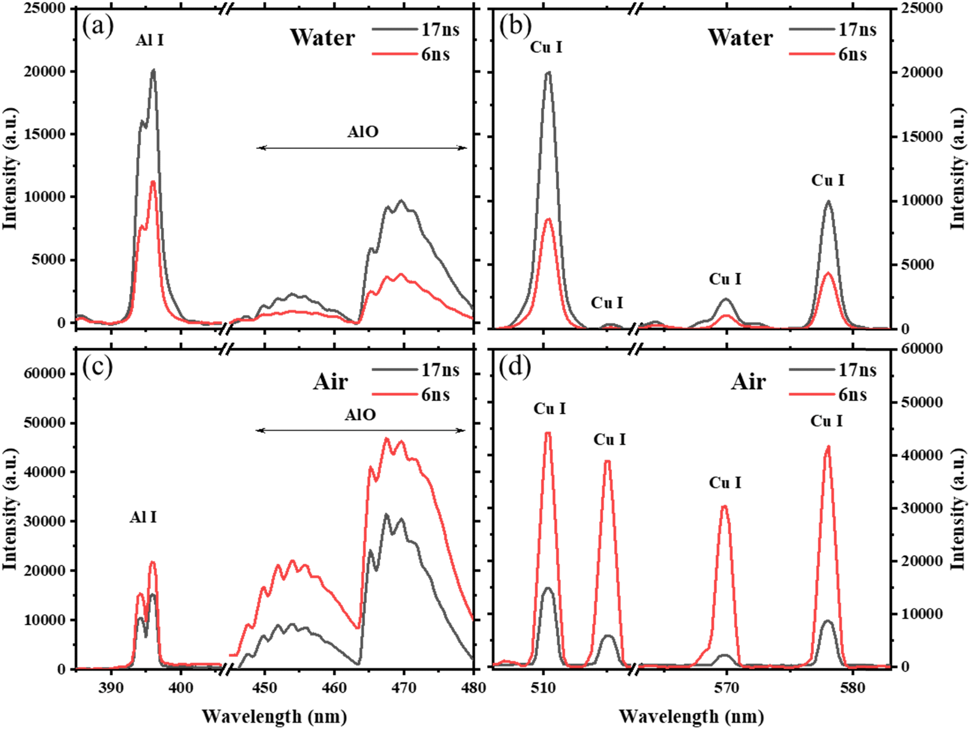 | ||
| Fig. 2 LIBS spectra obtained with the 6 ns and 17 ns lasers for the Al emission lines (a) and Cu emission lines (b) in water, and the Al emission lines (c) and Cu emission lines (d) in air. | ||
From the results in Fig. 2, we can conclude that the LIBS signal responses on the laser pulse duration in water and in air are different, and such differences are also reflected in the signal stability, which represents one of the key factors with regard to the repeatability of LIBS. We calculated the relative standard deviations (RSDs) of the 6 ns and 17 ns lasers for the Al and Cu emission lines in water and in air, respectively. Considering the fact that the underwater LIBS signal ablated at a fixed position changes dramatically as the number of laser shots increases,34 the RSD was calculated from 20 single-shot spectra with the irradiation position changed shot-by-shot. The results are shown in Fig. 3. We can see that for the underwater plasma (Fig. 3a), the RSDs of the Al atomic lines, AlO molecular bands, and Cu atomic lines are clearly lower with the 17 ns laser. The average RSD values of these emission lines are 61.5% and 34.4%, for the 6 ns and 17 ns lasers, respectively. Note that the intrinsic energy fluctuation of the 17 ns laser (RSD of 7.8%) is higher than that of the 6 ns laser (1.8%), which further emphasized the improvement in the LIBS signal stability with the 17 ns laser. Conversely, for the plasma in air, the 6 ns laser has clearly lower RSDs than the 17 ns laser, with an average RSD value of 29.5% vs. 46.5%. Therefore, from the spectral results, it showed that the 17 ns laser is beneficial to underwater LIBS in that both stronger signal intensity and higher stability can be obtained. Whereas for the traditional LIBS measurement in air, the 6 ns laser is more efficient. Considering the fact that LIBS spectral features are directly related to the characteristics of the plasma, we will show this in the following section with plasma imaging and shadowgraph imaging results obtained under the different laser ablation conditions.
 | ||
| Fig. 3 RSDs of the Al and Cu emission lines obtained with the 6 ns and 17 ns lasers for plasma induced in water (a) and in air (b). | ||
3.2. Plasma images and shadowgraph images in water and in air
The plasma images obtained in water and in air with the 6 ns and 17 ns lasers are shown in Fig. 4. We can see first that the plasma in air has a much larger size and stronger emission intensity compared with the plasma in water. This is due to the significantly different properties between water and air that lead to significantly different plasma. The incompressible nature of water with a high thermal conductivity will cause strong confinement and quenching effects for the plasma induced in water, which corresponds to a dense plasma with a small size confined near the target surface.16 This is consistent with the spectral results in Fig. 2 that the underwater LIBS signals are much weaker with larger line broadening compared with the signals in air. | ||
| Fig. 4 Time-integrated plasma emission images obtained with the (a) 17 ns laser in water, (b) 6 ns laser in water, (c) 17 ns laser in air, and (d) 6 ns laser in air. | ||
The laser pulses of 6 ns and 17 ns also show obvious differences in the plasma images. For the underwater plasma, the 17 ns laser induces a brighter plasma that is more concentrated on the target surface, while the 6 ns laser induces a much longer plasma along the laser direction with an irregular plasma morphology. The quite dispersed distribution of the plasma emission indicates multiple breakdowns of water above the target with the 6 ns laser, that degrade the quality of underwater LIBS signals. Note that the breakdown threshold of water is closer to that of a solid when the threshold difference between air and solid is compared.35 Therefore, the 6 ns laser, which has a higher peak irradiance, is more likely to suffer from the phenomenon of water breakdown. For the 17 ns laser, although the plasma is confined in a small area near the target surface, the longer laser pulse duration could provide continuous heating of the plasma at the trailing edge of the laser pulse,19 which helps to improve the quality of underwater LIBS signals, as observed in Fig. 2 and 3. However, for the plasma in air, the 6 ns laser induces a brighter plasma, while the 17 ns laser induces a smaller plasma with a non-uniform emission distribution. This means insufficient excitation of the plasma with the longer laser pulse of 17 ns whose peak irradiance is lower, and this is also consistent with the self-absorption phenomenon of the Cu emission lines, as observed in Fig. 2d.
To show the mechanical effects of cavitation bubbles and shock waves formed during laser ablation in water, shadowgraph images were measured at two delay times of 1 μs and 100 μs, and the results are shown in Fig. 5a and b. We can see that at 1 μs, the bubble of the 17 ns laser had a hemispherical shape and adhered closely to the target surface. In contrast, the bubble of 6 ns shows a clearly irregular shape which is similar to irregular plasma morphology of 6 ns, as shown in Fig. 4b. Moreover, accompanying the irregular bubble, multiple shock waves were observed. This proves the occurrence of multiple independent breakdowns that form multiple plasmas in water above the target surface. The formation of multiple plasmas in water leads to a shielding effect for the initial plasma from laser heating, which reduces the plasma temperature as well as the emission intensity. Also, the multiple breakdowns in water is a random event that occurs when the laser irradiance at a certain position of the laser focal region exceeds the breakdown threshold of water,36 leading to a large pulse-to-pulse plasma fluctuation and therefore low stability of underwater LIBS signals. By increasing the laser pulse duration from 6 ns to 17 ns, the multiple breakdown phenomenon can be greatly suppressed.
 | ||
| Fig. 5 Shadowgraph images with the 6 ns and 17 ns lasers obtained in water at a delay of 1 μs (a), in water at a delay of 100 μs (b), in air at a delay of 200 ns (c), and in air at a delay of 1 μs (d). The laser pulse duration and delay time are indicated in each image. | ||
At a longer delay of 100 μs, the bubble expands significantly and reaches a radius of 1.88 mm for the 17 ns laser and 2.08 mm for the 6 ns laser. We mention that the light observed inside the bubble at this time is not plasma emission. It is actually the excitation light (from the probe laser) used for the shadowgraph illumination passing through the bubble. According to cavitation bubble dynamics theory, the bubble energy is proportional to the maximum bubble radius,37 which implies that more laser energy was converted to the bubble mechanical energy for the shorter laser pulse of 6 ns. Furthermore, the propagation distance of the shock wave at 1 μs was 1.59 mm for the 17 ns laser and 1.77 mm for the 6 ns laser, indicating stronger shock wave energy from the 6 ns laser. From an energy balance point of view, the more laser energy is converted to the mechanical effects of bubble expansion and shock wave propagation, the less energy is converted to the plasma radiation. This also explains the underwater plasma emission results observed above.
For the shadowgraph images obtained in air, as shown in Fig. 5c and d, it is clear that the 6 ns laser produces both a larger plasma and a stronger shock wave. During the plasma generation process, the expansion velocity of the initial plasma is faster than the sound velocity of the medium, and the high-pressure and high-temperature plasma heated by the laser pulse compresses the surrounding gas that produces the shock wave.38 Therefore, it shows that the 6 ns laser with a higher peak irradiance could provide more efficient heating on the plasma, driving the propagation of a faster shock wave. Here, what is more important is that the responses of plasma emissions to the laser pulse duration in water and in air are different. This reveals the different laser–target–plasma interactions and the plasma growth mechanisms in water and in air, which will be discussed in the following section.
3.3. Discussion of the plasma growth mechanisms in water and in air
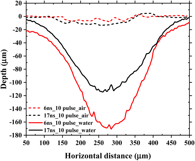
The laser ablation craters were measured by white light interferometry after a sequence of laser ablations from one-pulse ablation to ten-pulse ablation in water. The depth profiles of the ablation craters obtained with the 6 ns and 17 ns lasers are shown in Fig. 6. We can see that with the 17 ns laser, the craters are clearly shallower. This is consistent with the work of Sakka et al., which showed that longer nanosecond laser pulses correspond to smaller ablation craters.39 More material mass can be ablated by the 6 ns laser than by the 17 ns laser, which means that a larger proportion of laser energy is consumed in the phase transition of melting and vaporization of the material in the ablation crater. The greater mass ablated from the target also leads to stronger mechanical effects of bubble expansion and shock wave propagation, as described above. Here, we mention that the deeper crater of the 6 ns laser does not correspond to higher underwater LIBS signals. This is different from laser ablation in air where greater mass removal generally corresponds to a larger number density of the emitting particles and then stronger LIBS signals. | ||
| Fig. 6 Depth profiles of the ablation craters obtained with the 6 ns and 17 ns lasers after a sequence of laser ablations from one-pulse ablation to ten-pulse ablation in water. | ||
Fig. 7 gives a comparison of the ablation craters in water and in air. We can see that the crater depth in air is significantly smaller than that in water. This indicates a much higher laser ablation rate underwater, which agrees well with the work of Sakka et al.18 and relevant work in laser material micro-processing.40,41 This phenomenon can be attributed to the much stronger confinement effect of water that leads to much higher pressure and density for the underwater plasma. The higher plasma pressure on the target surface significantly enhances the laser ablation efficiency in water.25 Another reason is that, for laser ablation in air, the initial plasma interacts strongly with the laser pulse through inverse bremsstrahlung absorption, which corresponds to a strong plasma shielding effect that reduces the laser energy deposition on the target surface. Such shielding is more efficient for laser ablation in air than in water. If we compare the difference between the 6 ns and 17 ns laser ablations in air, we find a slightly deeper crater for the 17 ns laser. This is consistent with the weaker plasma emission observed in air with the 17 ns laser. Less plasma shielding occurs with the longer laser pulse, which improves the laser energy deposition on the target surface.
 | ||
| Fig. 7 Comparisons between the depth profiles of ablation craters obtained in water and in air with the 6 ns and 17 ns lasers after a sequence of ten-pulse ablation. | ||
The mechanisms of plasma growth in air and in water with different laser pulses are further discussed and illustrations of the characteristics of plasma are given in Fig. 8. For a nanosecond LIBS-based condition, plasma growth in air is characterized by laser-supported absorption waves, including the two main regimes of laser-supported combustion (LSC) waves and laser-supported detonation (LSD) waves.42–44 LSC applies when the laser irradiance is low and the induced shock wave cannot completely ionize the surrounding gas. The laser interacts directly with the ablation vapor near the target surface, forming a layered plasma structure with lower temperature at its periphery which is prone to self-absorption.45 LSD applies when the laser irradiance is high and the high-pressure shock wave ionizes the neutral gas surrounding the plasma. The neutral gas then absorbs more laser energy through inverse bremsstrahlung, and “pulls” forward the plasma expansion, resulting in a longer and more homogeneous plasma. Strong plasma shielding also occurs within this regime, thus blocking the laser from reaching the target surface. According to the observed results above, the 6 ns laser ablation plasma in air is more likely to be described by the LSD regime. It produces a larger plasma with a stronger emission intensity and a stronger shock wave, together with higher quality LIBS signals. Whereas the 17 ns laser, which has a lower peak irradiance, produces a non-uniform plasma with weak emission and self-absorption of the spectral lines, which probably corresponds to the LSC regime.
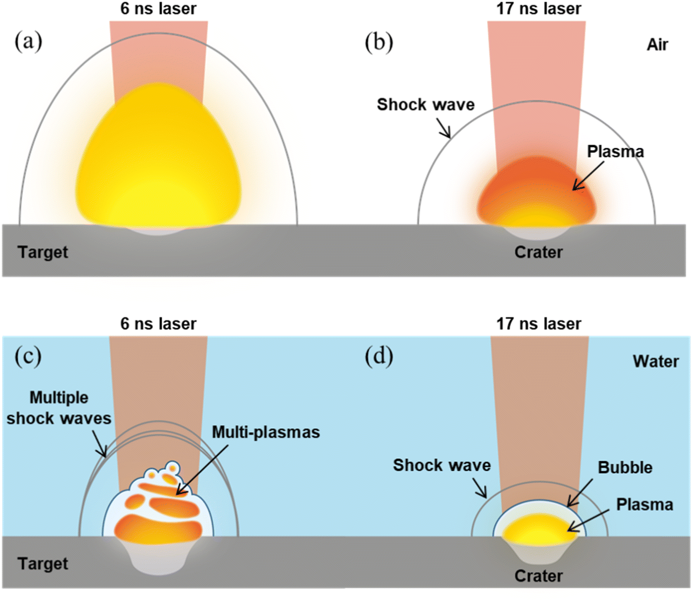 | ||
| Fig. 8 Illustrations of the characteristics of plasma generated in air with the 6 ns laser (a), in air with the 17 ns laser (b), in water with the 6 ns laser (c), and in water with the 17 ns laser (d). | ||
The plasma growth in water is quite different. Although the underwater plasma has a much larger ablation crater, the radiation of the underwater plasma is much weaker than that of the plasma in air. The incompressible nature of water will cause a significantly stronger mechanical effect, but significantly lower plasma radiation. The underwater plasma was confined in a very small area near the target surface with a quite high inner pressure, leading to a much higher laser ablation rate and producing a much larger crater. A great amount of laser energy is consumed in the material phase transitions of melting and vaporization, and in the mechanical effects of bubble expansion and shock wave propagation. Unlike laser ablation in air, where a strong laser-supported absorption wave exists, the underwater plasma is far less heated by the laser pulse due to the great energy consumption in the phase transitions and the mechanical effects. Therefore, compared with the plasma in air, the underwater plasma is characterized by a large ablation crater and high particle number density, but low temperature. That is why the underwater LIBS signals are quite weak with much broadened emission lines compared with the LIBS signals in air.
Another significant difference is that the breakdown threshold of water is closer to that of a solid target, compared with air. This means that with the same laser conditions, laser ablation in water more easily suffers from the multiple breakdown of water above the target, and produces multiple shock waves and multiple plasmas. The plasma and bubble morphologies can be greatly disordered with severe pulse-to-pulse fluctuation due to the multiple breakdowns. The quality of underwater LIBS signals is therefore greatly degraded in this case. However, by increasing the laser pulse duration from 6 ns to 17 ns, the multiple breakdown phenomenon can be greatly suppressed because of the lower peak irradiance of the longer laser pulse. Instead, the mechanical effects are mild with less laser damage to the target surface, while the plasma can absorb more laser energy over a longer laser pulse duration. This results in a higher temperature of the plasma with a longer emission lifetime, which clearly improves the quality of underwater LIBS signals. Again for traditional LIBS in air, the 6 ns laser is indeed a good choice.
4. Conclusions
In this work, the characteristics of laser-induced plasma generated in water and in air were investigated and compared, with two laser pulse durations of 6 ns and 17 ns. It was shown that the responses of LIBS signals to the laser pulse duration in water and in air are different. In water, the 17 ns laser induces a brighter plasma and a stronger LIBS signal, while in air, the 6 ns laser is better. Such differences were attributed to the different plasma growth mechanisms in water and in air. The plasma growth in air is driven by strong laser-supported absorption waves, where the 6 ns laser with higher peak irradiance corresponds to a stronger shock wave and more efficient heating on the plasma. Whereas for plasma growth in water, a great amount of laser energy is consumed in the material phase transitions, and in the mechanical effects of bubble expansion and shock wave propagation. The underwater plasma is therefore far less heated by the laser pulse with much lower plasma temperature and emission intensity compared with the plasma in air.Moreover, the breakdown threshold of water is closer to that of a solid target, compared with air. Thus, the underwater plasma more easily suffers from the multiple breakdowns of water, producing multiple shock waves and multiple plasmas. The underwater LIBS signals can be greatly degraded due to the multiple breakdowns. By using a longer laser pulse that has a lower peak irradiance, the multiple breakdown phenomenon can be suppressed, and the underwater plasma can absorb more laser energy over a longer laser pulse duration. This results in stronger underwater LIBS signals with a higher pulse-to-pulse stability. The present work gives insights into the mechanism of plasma growth in water and in air, and provides suggestions for the choice of laser pulses in relevant LIBS analyses.
Conflicts of interest
The authors declare no conflict of interest.Acknowledgements
This work was supported by the National Natural Science Foundation of China (No. 12174359 and 61975190), the Shandong Provincial Natural Science Foundation, China (No. ZR2023YQ059), and the Fundamental Research Funds for the Central Universities (No. 202261009).References
- C. Aragón and J. A. Aguilera, Characterization of laser induced plasmas by optical emission spectroscopy: a review of experiments and methods, Spectrochim. Acta, Part B, 2008, 63, 893–916, DOI:10.1016/j.sab.2008.05.010.
- D. A. Cremers and L. J. Radziemski, Handbook of Laser-Induced Breakdown Spectroscopy, John Wiley & Sons, 2013 Search PubMed.
- P.-Y. Meslin, O. Gasnault, O. Forni, S. Schröder, A. Cousin, G. Berger, S. M. Clegg, J. Lasue, S. Maurice, V. Sautter, S. Le Mouélic, R. C. Wiens, C. Fabre, W. Goetz, D. Bish, N. Mangold, B. Ehlmann, N. Lanza, A.-M. Harri, R. Anderson, E. Rampe, T. H. McConnochie, P. Pinet, D. Blaney, R. Léveillé, D. Archer, B. Barraclough, S. Bender, D. Blake, J. G. Blank, N. Bridges, B. C. Clark, L. DeFlores, D. Delapp, G. Dromart, M. D. Dyar, M. Fisk, B. Gondet, J. Grotzinger, K. Herkenhoff, J. Johnson, J.-L. Lacour, Y. Langevin, L. Leshin, E. Lewin, M. B. Madsen, N. Melikechi, A. Mezzacappa, M. A. Mischna, J. E. Moores, H. Newsom, A. Ollila, R. Perez, N. Renno, J.-B. Sirven, R. Tokar, M. de la Torre, L. d'Uston, D. Vaniman and A. Yingst, Soil Diversity and Hydration as Observed by ChemCam at Gale Crater, Mars, Science, 2013, 341, 1238670, DOI:10.1126/science.1238670.
- W. Xu, X. Liu, Z. Yan, L. Li, Z. Zhang, Y. Kuang, H. Jiang, H. Yu, F. Yang, C. Liu, T. Wang, C. Li, Y. Jin, J. Shen, B. Wang, W. Wan, J. Chen, S. Ni, Y. Ruan, R. Xu, C. Zhang, Z. Yuan, X. Wan, Y. Yang, Z. Li, Y. Shen, D. Liu, B. Wang, R. Yuan, T. Bao and R. Shu, The MarSCoDe Instrument Suite on the Mars Rover of China's Tianwen-1 Mission, Space Sci. Rev., 2021, 64, 1–58, DOI:10.1007/s11214-021-00836-5.
- L. Sun, H. Yu, Z. Cong, Y. Xin, Y. Li and L. Qi, In situ analysis of steel melt by double-pulse laser-induced breakdown spectroscopy with a Cassegrain telescope, Spectrochim. Acta, Part B, 2015, 112, 40–48, DOI:10.1016/j.sab.2015.08.008.
- C. Li, Q. Li, L. Li, B. Men, H. Wu, D. Wu, R. Hai, X. Wu and H. Ding, Characteristic of spatiotemporal evolution of hydrogen isotope in laser-induced plasma under low-pressure environment, Spectrochim. Acta, Part B, 2023, 206, 106735, DOI:10.1016/j.sab.2023.106735.
- X. Bai, R. Hai, Z. He, X. Wang, J. Mu, H. Wu, C. Li, D. Wu, G. Xu, Z. Hu, F. Ding and H. Ding, Quantitative analysis of tungsten in steel by one-point calibration laser-induced breakdown spectroscopy in vacuum, Spectrochim. Acta, Part B, 2023, 206, 106724, DOI:10.1016/j.sab.2023.106724.
- A. De Giacomo, M. Dell'Aglio, O. De Pascale and M. Capitelli, From single pulse to double pulse ns-laser induced breakdown spectroscopy under water: elemental analysis of aqueous solutions and submerged solid samples, Spectrochim. Acta, Part B, 2007, 62, 721–738, DOI:10.1016/j.sab.2007.06.008.
- A. De Giacomo, M. Dell'Aglio, R. Gaudiuso, S. Amoruso and O. De Pascale, Effects of the background environment on formation, evolution and emission spectra of laser-induced plasmas, Spectrochim. Acta, Part B, 2012, 78, 1–19, DOI:10.1016/j.sab.2012.10.003.
- C. G. Parigger, Review of spatiotemporal analysis of laser-induced plasma in gases, Spectrochim. Acta, Part B, 2021, 179, 106122, DOI:10.1016/j.sab.2021.106122.
- J. Buday, P. Pořízka and J. Kaiser, Imaging laser-induced plasma under different laser irradiances, Spectrochim. Acta, Part B, 2020, 168, 105874, DOI:10.1016/j.sab.2020.105874.
- Y. Tian, E. B. Sokolova, R. Zheng, Q. Ma, Y. Chen and J. Yu, Characteristics of the ablation plume induced on glasses for analysis purposes with laser-induced breakdown spectroscopy, Spectrochim. Acta, Part B, 2015, 114, 7–14, DOI:10.1016/j.sab.2015.09.024.
- B. Thornton, T. Takahashi, T. Sato, T. Sakka, A. Tamura, A. Matsumoto, T. Nozaki, T. Ohki and K. Ohki, Development of a deep-sea laser-induced breakdown spectrometer for in situ multi-element chemical analysis, Deep Sea Res., Part I, 2015, 95, 20–36, DOI:10.1016/j.dsr.2014.10.006.
- T. Takahashi, S. Yoshino, Y. Takaya, T. Nozaki, K. Ohki, T. Ohki, T. Sakka and B. Thornton, Quantitative in situ mapping of elements in deep-sea hydrothermal vents using laser-induced breakdown spectroscopy and multivariate analysis, Deep Sea Res., Part I, 2020, 158, 103232, DOI:10.1016/j.dsr.2020.103232.
- C. Liu, J. Guo, Y. Tian, C. Zhang, K. Cheng, W. Ye and R. Zheng, Development and field tests of a deep-sea laser-induced breakdown spectroscopy (LIBS) system for solid sample analysis in seawater, Sensors, 2020, 20, 7341, DOI:10.3390/s20247341.
- M. Dell'Aglio, V. Gardette, S. Jantzi and A. De Giacomo, Comparison between laser induced plasmas in gas and in liquid, J. Appl. Phys., 2021, 129, 233303, DOI:10.1063/5.0039625.
- T. Sakka, H. Oguchi, S. Masai, K. Hirata, Y. H. Ogata, M. Saeki and H. Ohba, Use of a long-duration ns pulse for efficient emission of spectral lines from the laser ablation plume in water, Appl. Phys. Lett., 2006, 88, 061120, DOI:10.1063/1.2172235.
- T. Sakka, S. Masai, K. Fukami and Y. H. Ogata, Spectral profile of atomic emission lines and effects of pulse duration on laser ablation in liquid, Spectrochim. Acta, Part B, 2009, 64, 981–985, DOI:10.1016/j.sab.2009.07.018.
- T. Sakka, A. Tamura, A. Matsumoto, K. Fukami, N. Nishi and B. Thornton, Effects of pulse width on nascent laser-induced bubbles for underwater laser-induced breakdown spectroscopy, Spectrochim. Acta, Part B, 2014, 97, 94–98, DOI:10.1016/j.sab.2014.05.009.
- A. Matsumoto, H. Ohba, M. Toshimitsu, K. Akaoka, A. Ruas, T. Sakka and I. Wakaida, Fiber-optic laser-induced breakdown spectroscopy of zirconium metal in air: special features of the plasma produced by a long-pulse laser, Spectrochim. Acta, Part B, 2018, 142, 37–49, DOI:10.1016/j.sab.2018.01.012.
- X. Bai, Q. Ma, M. Perrier, V. Motto-Ros, D. Sabourdy, L. Nguyen, A. Jalocha and J. Yu, Experimental study of laser-induced plasma: influence of laser fluence and pulse duration, Spectrochim. Acta, Part B, 2013, 87, 27–35, DOI:10.1016/j.sab.2013.05.019.
- D. Zhang, B. Gokce and S. Barcikowski, Laser synthesis and processing of colloids: fundamentals and applications, Chem. Rev., 2017, 117, 3990–4103, DOI:10.1021/acs.chemrev.6b00468.
- M. Dell'Aglio, A. De Giacomo, S. Kohsakowski, S. Barcikowski, P. Wagener and A. Santagata, Pulsed laser ablation of wire-shaped target in a thin water jet: effects of plasma features and bubble dynamics on the PLAL process, J. Phys. D: Appl. Phys., 2017, 50, 185204, DOI:10.1088/1361-6463/aa652a.
- M. Dell'Aglio, V. Motto-Ros, F. Pelascini, I. B. Gornushkin and A. De Giacomo, Investigation on the material in the plasma phase by high temporally and spectrally resolved emission imaging during pulsed laser ablation in liquid (PLAL) for NPs production and consequent considerations on NPs formation, Plasma Sources Sci. Technol., 2019, 28, 085017, DOI:10.1088/1361-6595/ab369b.
- S. Zhu, Y. Lu, M. Hong and X. Chen, Laser ablation of solid substrates in water and ambient air, J. Appl. Phys., 2001, 89, 2400–2403, DOI:10.1063/1.1342200.
- N. Smirnov, S. Kudryashov, A. Rudenko, D. Zayarny and A. Ionin, Pulse width and ambient medium effects during ultrashort-pulse laser ablation of silicon in air and water, Appl. Surf. Sci., 2021, 562, 150243, DOI:10.1016/j.apsusc.2021.150243.
- Y. Tian, S. Hou, L. Wang, X. Duan, B. Xue, Y. Lu, J. Guo and Y. Li, CaOH molecular emissions in underwater laser-induced breakdown spectroscopy: spatial–temporal characteristics and analytical performances, Anal. Chem., 2019, 91, 13970–13977, DOI:10.1021/acs.analchem.9b03513.
- Z. Jia, D. Li, Y. Tian, H. Pan, Q. Zhong, Z. Yao, Y. Lu, J. Guo and R. Zheng, Early dynamics of laser-induced plasma and cavitation bubble in water, Spectrochim. Acta, Part B, 2023, 206, 106713, DOI:10.1016/j.sab.2023.106713.
- Y. Tian, B. Xue, J. Song, Y. Lu, Y. Li and R. Zheng, Comparative investigation of laser-induced breakdown spectroscopy in bulk water using 532-and 1064-nm lasers, Appl. Phys. Express, 2017, 10, 072401, DOI:10.7567/APEX.10.072401.
- Y. Tian, L. Wang, B. Xue, Q. Chen and Y. Li, Laser focusing geometry effects on laser-induced plasma and laser-induced breakdown spectroscopy in bulk water, J. Anal. At. Spectrom., 2019, 34, 118–126, 10.1039/C8JA00282G.
- Z. Jia, Y. Tian, H. Pan, T. Li, Y. Li, Q. Zhong, Z. Yao, Y. Lu, J. Guo, W. Ye and R. Zheng, Dynamics of laser-induced plasma and cavitation bubble at high pressures and the impacts on underwater LIBS signals, Spectrochim. Acta, Part B, 2023, 209, 106793, DOI:10.1016/j.sab.2023.106793.
- M. R. Kalus, S. Barcikowski and B. Goekce, How the Physicochemical Properties of the Bulk Material Affect the Ablation Crater Profile, Mass Balance, and Bubble Dynamics During Single-Pulse, Nanosecond Laser Ablation in Water, Chem.–Eur. J., 2021, 27, 5978–5991, DOI:10.1002/chem.202005087.
- X. Bai, Q. Ma, V. Motto-Ros, J. Yu, D. Sabourdy, L. Nguyen and A. Jalocha, Convoluted effect of laser fluence and pulse duration on the property of a nanosecond laser-induced plasma into an argon ambient gas at the atmospheric pressure, J. Appl. Phys., 2013, 113, 013304, DOI:10.1063/1.4772787.
- A. Matsumoto, Y. Shimazu, S. Yae and T. Sakka, Effect of repeated irradiation on laser-induced breakdown spectroscopy of copper immersed in a sodium chloride aqueous solution and normalization with bubble collapse time, J. Anal. At. Spectrom., 2023 10.1039/D3JA00268C.
- J. R. Bettis, Correlation among the laser-induced breakdown thresholds in solids, liquids, and gases, Appl. Opt., 1992, 31, 3448–3452, DOI:10.1364/AO.31.003448.
- P. K. Kennedy, D. X. Hammer and B. A. Rockwell, Laser-induced breakdown in aqueous media, Prog. Quantum Electron., 1997, 21, 155–248, DOI:10.1016/S0079-6727(97)00002-5.
- A. Vogel, S. Busch and U. Parlitz, Shock wave emission and cavitation bubble generation by picosecond and nanosecond optical breakdown in water, J. Acoust. Soc. Am., 1996, 100, 148–165, DOI:10.1121/1.415878.
- J. L. Gottfried, Influence of exothermic chemical reactions on laser-induced shock waves, Phys. Chem. Chem. Phys., 2014, 16, 21452–21466, 10.1039/C4CP02903H.
- T. Sakka, H. Oguchi, S. Masai and Y. H. Ogata, Quasi Nondestructive Elemental Analysis of Solid Surface in Liquid by Long-pulse Laser Ablation Plume Spectroscopy, Chem. Lett., 2007, 236, 508–509, DOI:10.1246/cl.2007.508.
- H. W. Kang, H. Lee and A. J. Welch, Laser ablation in a liquid-confined environment using a nanosecond laser pulse, J. Appl. Phys., 2008, 103, 083101, DOI:10.1063/1.2905314.
- X. Wang, K. Ando, N. Feng and T. Nakajima, Single-shot nanosecond laser ablation of Ni in low and high viscosity liquids at different temperatures, Appl. Surf. Sci., 2022, 592, 153361, DOI:10.1016/j.apsusc.2022.153361.
- J. Yu, Q. Ma, V. Motto-Ros, W. Lei, X. Wang and X. Bai, Generation and expansion of laser-induced plasma as a spectroscopic emission source, Front. Phys., 2012, 7, 649–669, DOI:10.1007/s11467-012-0251-2.
- J. Buday, P. Pořízka and J. Kaiser, Imaging laser-induced plasma under different laser irradiances, Spectrochim. Acta, Part B, 2020, 168, 105874, DOI:10.1016/j.sab.2020.105874.
- Y. Zhao, L. Zhang, S. Wang, J. Han, G. Xia, W. Ma, L. Dong, W. Yin, L. Xiao, S. Jia and Y. Jin, Species distribution in laser-induced plasma on the surface of binary miscible alloy, Spectrochim. Acta, Part B, 2020, 173, 105987, DOI:10.1016/j.sab.2020.105987.
- F. Rezaei, G. Cristoforetti, E. Tognoni, S. Legnaioli, V. Palleschi and A. Safi, A review of the current analytical approaches for evaluating, compensating and exploiting self-absorption in Laser Induced Breakdown Spectroscopy, Spectrochim. Acta, Part B, 2020, 169, 105878, DOI:10.1016/j.sab.2020.105878.
| This journal is © The Royal Society of Chemistry 2024 |