
 Open Access Article
 This Open Access Article is licensed under a
 Creative Commons Attribution 3.0 Unported Licence
Investigating serum concentration profiles of orally ingested short-chain fatty acid supplements
Christopher G.
Green
 a,
Marilyn L. Y.
Ong
ab,
Samantha N.
Rowland
a,
Tindaro
Bongiovanni
cd,
Lewis J.
James
a,
Tom
Clifford
a,
Stephen J.
Bailey
a and
Liam M.
Heaney
*a
a,
Marilyn L. Y.
Ong
ab,
Samantha N.
Rowland
a,
Tindaro
Bongiovanni
cd,
Lewis J.
James
a,
Tom
Clifford
a,
Stephen J.
Bailey
a and
Liam M.
Heaney
*a
aSchool of Sport, Exercise and Health Sciences, Loughborough University, Loughborough, UK. E-mail: L.M.Heaney2@Lboro.ac.uk
bExercise and Sports Science Programme, School of Health Sciences, Health Campus, Universiti Sains Malaysia, 16150 Kubang Kerian, Kelantan, Malaysia
cPlayer Health & Performance Department, Palermo Football Club, Palermo, Italy
dDepartment of Biomedical and Neuromotor Sciences (DIBINEM), University of Bologna, Bologna, Italy
First published on 30th October 2024
Abstract
Acetate, propionate, and butyrate are naturally-occurring short-chain fatty acids (SCFAs) derived from bacterial metabolism of dietary fibre and have been associated with numerous positive health outcomes. All three acids have been shown to offer unique physiological and metabolic effects and, therefore, could be targeted for co-ingestion as part of a nutritional/medicinal plan. However, a better understanding of the outcomes of supplementing in combination on circulating concentration profiles is necessary to confirm uptake efficacy. This study sought to investigate the acute circulating concentration profiles of acetate, propionate, and butyrate following oral supplementation. Three experimental trials were conducted including investigations to understand the impact of capsule coating on circulating concentration profiles, the effect of supplementation dose on uptake kinetics, and the outcome of a short, repeated, supplementation routine on circulating levels. Serum samples were analysed for SCFA content using a quantitative GC-MS assay. It was observed that an acid-resistant coated capsule caused a delayed and blunted blood concentration response, with the non-acid resistant trial displaying earlier and more intense peak serum concentrations. For dose comparison investigations, all SCFAs peaked within 60 min and returned to baseline concentrations by 120 min post-supplementation. A graded dose relationship was present for propionate and butyrate when considering the total circulating exposure across a 240 min monitoring period. In addition, a one-week, twice-daily, repeated supplementation protocol resulted in no changes in basal serum SCFA concentrations. Overall, these data indicate that acetate, propionate, and butyrate display relatively similar circulating concentration profiles following oral co-ingestion, adding knowledge to help inform supplementation strategies for future outcomes where acute elevation of circulating SCFAs is desired.
Introduction
Short-chain fatty acids (SCFAs) are metabolites derived from the degradation of dietary fibre, other non-digestible carbohydrates, and proteins, by commensal bacteria in the gastrointestinal (GI) tract.1,2 The majority of microbiota-derived produced SCFA content (i.e. acetic [C2], propionic [C3], and butyric [C4] acids) are used as an energy substrate by colonic enterocytes, whilst approximately 15% of SCFA molecules reach peripheral circulation.3 These SCFAs are also found naturally in food products. For example, acetic acid is the principal acid in vinegar,4 propionic acid is found in fermented foods and shellfish,5 and butyric acid is present in dairy products.6 The interaction between bacterially-derived metabolites and intestinal cellular processes has identified the beneficial links between SCFA production and gastrointestinal (GI) health (e.g., lower risk of colorectal cancer).1,7 However, it is also of interest to better understand the potential wider health benefits, such as reduced chronic inflammation, through increasing the availability of SCFAs within the systemic circulation.1 The rationale for improved health status with elevated systemic SCFAs relates to the presence of G-coupled protein receptors (GPCRs), GPR41, GPR43 and GPR109a (also known as free fatty acid receptors, or FFARs) on cells and tissues throughout the body.1 SCFAs are ligands for GPCRs which, when activated, drive outcomes considered beneficial to health, predominantly related to reduced pro-inflammatory cytokine production and improved immune cell function.1,8,9 This suggests that SCFAs can positively impact human physiology beyond their known benefits within the gut. These benefits may be related to the severity/presence of disease,10 immune health,11 inflammatory conditions,12 the respiratory system,8,9 as well as the potential to benefit exercise performance and/or recovery.13Owing to the potential health benefits conferred by circulating SCFAs, coupled with the inherently low contribution of SCFAs absorbed from the gut, research has sought to understand the physiological and metabolic impact of exogenously supplemented SCFAs. This includes the chronic supplementation of acetic acid containing supplements (e.g., apple cider vinegar) in obese and overweight adults to assess multiple metabolic responses (such as plasma glucose and lipid profiles; see Valdes et al. for meta-analysis),14 and butyrate supplements for GI tract-associated conditions (e.g., Crohn's disease, irritable bowel syndrome, etc.),15–17 and for patients with metabolic syndrome.18,19 Importantly, evidence suggests that the major SCFAs (i.e. acetate, propionate, and butyrate) can induce positive physiological outcomes,1 including acute effects on appetite and energy expenditure.20–23 Furthermore, all three of these acids have been shown to offer unique physiological and metabolic effects. For example, acetate is incorporated into cholesterol to a greater extent than propionate and butyrate whilst propionate is the primary SCFA used by the liver for gluconeogenesis.3 Therefore, these SCFAs could rationally be targeted for co-ingestion as part of a nutritional/medicinal plan. However, a better understanding on the outcomes of supplementing in combination on circulating concentration profiles is necessary to confirm uptake efficacy. To date, no data have been reported to demonstrate the circulating concentration profiles of SCFA co-ingestion. These outcomes would be informative to compare bioavailability and elimination at dosages relevant to those amenable for production of a commercial nutritional product (i.e. within orally ingestible capsules).
Currently, there are no guidelines nor formal recommendations for oral supplementation of SCFAs. Most commercially available SCFA supplements (predominantly in differing forms of butyrate salts) are marketed for gut health. However, the wide-ranging potential physiological effects require the understanding of systemic bioavailability following supplementation. Furthermore, as SCFAs can be absorbed in the proximal regions of the GI tract,24,25 understanding the effect of early and delayed availability of SCFA supplements is important to provide a practical approach to systemic uptake that bypasses their use at distal GI regions. Previous research has investigated the acute (within 3 h) bioavailability of propionate, via oral sodium propionate ingestion,21 and acetate, via vinegar ingestion.26 However, these supplementation protocols included repeated ingestion over a short period of time and/or high intake masses/volumes, and thus did not assess responses to a single dose which would be more comparable to the use of a commercial health supplement. Conversely, data relating specifically to the acute serum concentrations of orally ingested butyrate salts are not available. Whilst these studies offer insight to absorption locations and concentration changes, the investigation of SCFA co-ingestion at commercially relevant dosing strategies is necessary to understand the potential physiological exposure of exogenous SCFAs. This will aid in the assessment for the potential use of SCFAs as health supplements with the intention to interact with whole body energy production, inflammation, and immune function.
Overall, this study aimed to investigate three elements of oral SCFA supplementation. Firstly, the influence of capsule coating (i.e. acid/non-acid resistant) on acute serum SCFA uptake. Secondly, the assessment of a dose comparison experiment on acute serum concentrations of SCFAs following co-ingestion (acetate, propionate, and butyrate) and, thirdly, the impact of a one-week repeated supplementation period on basal SCFA concentrations. It was hypothesised that capsule coating, dosage, and repeated supplementation would all influence the circulating levels of each supplemented SCFA.
Methods
This study included single blinded experiments to investigate serum concentrations of orally ingested SCFA supplements. This included an investigation into the impact of capsule composition on bioavailability (referred to herein as the capsule experiment), an assessment of the acute (240 min) serum concentrations profiles of co-ingested SCFA supplements (acute experiment), and the impact of short-term supplementation (7 days) of SCFAs on basal serum levels (chronic experiment). All procedures were approved by the Loughborough University Ethics Review Sub-Committee (Human Participants, approval ref: 6688). All participants provided written informed consent to participate in the study and were free to withdraw at any point. To assess the tolerance of the SCFA supplements in all experiments, participants were qualitatively asked to report any incidence of gastrointestinal discomfort or any other information relating to the consumption of the supplement.Capsule experiment
Participants and standardisation
Five healthy adults (3 male, 2 female) [mean (SD), 30 (8) years, 81.6 (16.2) kg] were recruited to this initial pilot experiment and completed two trials which investigated the encapsulation of sodium acetate and calcium propionate (both food chemical codex grade, Merck, Gillingham, UK) in different capsule compositions. A comparison for butyrate was not possible due to a bulk form food-grade butyrate salt not being commercially available. The two trials included the use of a hydroxypropyl methylcellulose (HPMC) capsule designed to break down immediately following ingestion (referred to herein as HPMC, VCaps® Plus, Lonza, Basel, Switzerland), alongside capsules coated with an acid-resistant formula designed to survive the low pH of the stomach and to dissolve once entering the higher pH environment of the small intestine (referred to herein as delayed release, DRCaps®, Lonza, Basel, Switzerland). For both trials, size 000 opaque white capsules were filled with 2 g of sodium acetate (1439 mg acetate content split across two capsules) and 2 g of calcium propionate (1570 mg propionate content split across two capsules). Participants refrained from strenuous exercise and alcohol consumption and made note of food and drink consumption for the 24 h prior to the first laboratory visit, replicating this for the subsequent visit. All sessions started between 0800–0930 h following an overnight fast (no food or drink consumption other than plain water after 2200 h the previous night). Participants performed the trials in a randomised, counterbalanced manner to avoid trial order bias and were blinded to which capsule was being ingested at each trial.Procedure
A schematic of trial procedures is provided in Fig. 1A. Upon arrival to the laboratory, body mass was recorded, and participants were provided with a standardised breakfast meal (40 g of sugared cornflakes cereal with 130 mL of semi-skimmed UHT milk). Following breakfast, a cannula was inserted into an antecubital vein and a baseline blood sample was collected 45 min following cessation of eating. Details on the procedure for blood sampling are provided later. To control for potential artefacts of postural shift, participants rested in a semi-recumbent position for 10 min prior to the collection of all blood samples. Participants then orally ingested the supplements with plain water as quickly as was comfortable (<10 min). Serum samples were collected at 30-, 60-, 120-, 180-, and 240 min post-capsule ingestion. To allow for a wash out period of the ingested SCFAs, each trial was separated by at least 7 days.21 | ||
| Fig. 1 Timeline of experimental procedures. (A) Capsule and acute experiment trial day procedures. Numbers represent time as min post-supplementation. (B) Chronic experiment procedure, 24-hour sample (Day 1), supplementation period (Day 1 to Day 6) and Day 7 sample. | ||
Acute experiment
Participants and standardisation
Fifteen healthy adults (11 male, 4 female) [28 (6) years, 78.6 (15.1) kg] completed three trials which investigated the serum concentration profiles of acetate, propionate, and butyrate over a 240 min period following supplementation at three different dosages. The minimum sample size required to identify a large difference (Cohen's d = 0.8) between the three trials was calculated as 12 (β − 1 = 0.8, α = 0.05). The trials were defined by the SCFA dose consumed and are referred to as a double dose, single dose, or control dose (i.e. no SCFAs). The single dose trial was defined as the maximum mass of SCFA salt that could be packed into one size 000 capsule for acetate and propionate (1 g), and the manufacturer recommended dose for butyrate (1.5 g). The double dose was defined as two times the single dose.In the double dose trial, participants consumed 2 g of sodium acetate and 2 g of calcium propionate, each split across two opaque size 000 VCaps® Plus capsules (four size 000 capsules in total). Additionally, they consumed 3 g (2374 mg butyrate content, split across four transparent size 00 HPMC capsules) of a commercially available sodium butyrate supplement (BodyBio, Cambridge, UK). In the single dose trial, participants ingested half of the SCFA content to that of the double dose experiment (one opaque size 000 capsule for each of acetate and propionate, and two transparent size 00 capsules for butyrate), along with 1 g of low sodium salt (to mimic the acetate/propionate capsules, LoSalt, East Kilbride, UK) and 2 g of medium chain triglyceride (MCT) powder (to mimic the butyrate capsules, both products from Bulk, Colchester, UK). The placebo mixture was pre-mixed in a ratio of 1![[thin space (1/6-em)]](https://www.rsc.org/images/entities/char_2009.gif) :2 for LoSalt:MCT powder and distributed across two size 000 opaque capsules and two size 00 transparent capsules. As acetate and butyrate supplements contained sodium, a low sodium salt was used within the placebo capsules to partially match the increased sodium intake without causing repeated high intake of daily sodium chloride. In addition, MCT powder was used due to its presence in the commercially available butyrate supplement. In the control trial, participants consumed 2 g of low sodium salt and 4 g of MCT powder (across four size 000 opaque capsules and four transparent size 00 capsules). Participants refrained from strenuous exercise and alcohol consumption and were asked to note food and drink consumption for the 24 h prior to laboratory visits. Food and drink consumption before the first trial was repeated for the subsequent visits. All sessions started between 0800–0930 h following an overnight fast (no food or drink consumption other than plain water after 2200 h the previous night). Participants performed the trials in a randomised, counterbalanced manner to avoid trial order bias and were blinded to which dose was being ingested at each trial. The trial day procedures mimicked those completed for the capsule experiment and were separated by at least 7 days since last SCFA intake to ensure adequate washout (Fig. 1A).
:2 for LoSalt:MCT powder and distributed across two size 000 opaque capsules and two size 00 transparent capsules. As acetate and butyrate supplements contained sodium, a low sodium salt was used within the placebo capsules to partially match the increased sodium intake without causing repeated high intake of daily sodium chloride. In addition, MCT powder was used due to its presence in the commercially available butyrate supplement. In the control trial, participants consumed 2 g of low sodium salt and 4 g of MCT powder (across four size 000 opaque capsules and four transparent size 00 capsules). Participants refrained from strenuous exercise and alcohol consumption and were asked to note food and drink consumption for the 24 h prior to laboratory visits. Food and drink consumption before the first trial was repeated for the subsequent visits. All sessions started between 0800–0930 h following an overnight fast (no food or drink consumption other than plain water after 2200 h the previous night). Participants performed the trials in a randomised, counterbalanced manner to avoid trial order bias and were blinded to which dose was being ingested at each trial. The trial day procedures mimicked those completed for the capsule experiment and were separated by at least 7 days since last SCFA intake to ensure adequate washout (Fig. 1A).
Chronic experiment
Analyses
In brief, 100 μL of serum was mixed with 100 μL 1 M hydrochloric acid and 100 μL of an internal standard mixture. The internal standard mixture contained 6 μg mL−1 of each deuterium labelled SCFA in MTBE. Samples were vortexed for 30 s before centrifugation at 5400g for 15 min at 15 °C. The organic layer (∼50 μL) was transferred to a low volume autosampler vial for analysis. All samples, including three quality control (QC) samples at low, medium, and high concentrations were placed in a randomised sequence for analysis. The QCs contained 100, 200 and 375 ng mL−1 of propionate and butyrate for low, medium, and high QCs, respectively. Due to the higher concentrations of acetate in circulating blood, the acetate concentrations for QC samples were 10× that of propionate and butyrate.
An injection volume of 3 μL was used and each sample was analysed in duplicate. The inlet and transfer line temperatures were set to 250 °C. The electron ionisation source temperature was set to 230 °C and had a fixed ionisation energy of 70 eV applied. The quadrupole mass analyser temperature was set to 150 °C. Purified helium at a constant flow rate of 2 mL min−1 was used as a carrier gas. The GC oven was programmed with a double ramp temperature increase protocol. The initial temperature was set to 80 °C for 1 min before linearly increasing to 127 °C at a rate of 10 °C min−1. From this point, the oven temperature increased linearly at a rate of 30 °C min−1 until a temperature of 181 °C was reached. The run time was 7.5 min followed by a post-run temperature hold of 2 min at 230 °C. To ensure no carryover of samples, a blank run (100% MTBE) was implemented after every duplicate injection. The quadrupole mass analyser was operated in scheduled selected ion monitoring mode. Mass Hunter software (Version B.07.00; Agilent Technologies, Stockport, UK) was used for GC-MS data acquisition and monitoring.
Statistical analyses
Statistical analyses were performed using STATA MP (v17, StataCorp, Texas, USA) and IBM SPSS (v28, IBM, Illinois, USA). All data were analysed for normality using the Shapiro–Wilk test with a p value threshold of 0.05 set for violations of normality. Where normality assumptions were violated, data were log-transformed and reassessed for normality. Log-transformation and consequent analysis was performed on all total area under the curve (tAUC) and maximum concentration (Cmax) data in the acute experiment and at baseline, 24 h and Day 7 comparisons in the chronic experiment. Due to the robustness of linear mixed-effects models (LMM) to manage data that severely violate parametric assumptions,28 all data analyses using LMM were performed on untransformed data.LMM were used to analyse serum SCFA concentrations over time in the capsule and acute experiments with participants modelled as random effects. Restricted maximum-likelihood estimation and small sample inference using the Kenward–Rogers degrees of freedom method were implemented in the model. Where a difference and/or interaction was reported, post-hoc contrasts were performed and adjusted for multiple comparisons using a false discovery rate (FDR) of 5% following the Benjamini–Hochberg method.29
Total area under the curve (tAUC), maximum serum concentration (Cmax) and time of maximum concentration (Tmax) were calculated by cubic splines using the pkcollapse command in STATA MP.
Paired sample t-tests were used to compare trials for tAUC and Cmax in the capsule experiment and to compare concentrations between baseline and 24 h, and baseline and Day 7 in the chronic experiment. A 3 × 1 repeated-measures ANOVA with a main effect for trial was used to compare trial differences for tAUC and Cmax in the acute experiment, and the baseline SCFA concentrations in the chronic experiment. For all ANOVAs, where assumptions of sphericity were violated, a Greenhouse–Geisser correction was applied. Where significant trial effects were observed, post-hoc paired-sample t-tests were subject to Benjamini–Hochberg FDR correction.
Data are presented as mean (standard error of the mean, SEM) unless otherwise stated. All p values included within the text satisfied the Benjamini–Hochberg FDR correction and are reported as the raw p value alongside the critical value (VCRIT) each was compared to in the FDR correction. An alpha value was set at p < 0.05.
Results
Capsule experiment
 | ||
| Fig. 2 Serum concentration and tAUC of acetate (A and B) and propionate (C and D) following ingestion of two forms of capsule formulation. 0 min = Baseline. Diamonds with a solid line indicate the early release trial, triangles with a dashed line indicated the delayed trial. For A and C the markers refer to the mean values, with error bars visualising the standard error of the mean. For B and D the grey lines with numbers 1–5 represent individual participant values (n = 5), the horizontal lines refer to the mean values, with error bars visualising the standard error of the mean. *Different between trials; adifferent to baseline in early release trial only. bDifferent to baseline in delayed trial only (all, p < 0.05). | ||
The tAUC (Fig. 2B) for acetate was not different between capsule types. The Cmax for acetate was not different between trials [7920 (2010) vs. 7171 (1041) ng mL−1, for HPMC and delayed release respectively]. The mode for Tmax of acetate in the HPMC trial (4 participants) was 60 min and 120 min in the delayed release trial (3 participants).
The tAUC (Fig. 2D) for propionate was not different between capsule types. The Cmax of propionate was not different between trials [460 (113) vs. 306 (72) ng mL−1, for HPMC and delayed release respectively]. The mode for Tmax of propionate in both the HPMC trial (5 participants) and delayed release trial (3 participants) was 60 min.
Acute experiment
 | ||
| Fig. 3 Serum concentration and tAUC of acetate (A and B), propionate (C and D), and butyrate (E and F) following ingestion of three different doses of SCFA supplements. 0 min = Baseline. Circles with a solid line indicate the double dose trial, squares with a dashed line indicate the single dose trial, and triangles with a dotted line indicate the control trial. For A, C and E the markers refer to the mean values, with error bars visualising the standard error of the mean. For B, D and F the grey lines represent individual participant values (n = 15), the horizontal lines refer to the mean values with error bars visualising the standard error of the mean. ‡Difference between the double dose trial and the control trial; †difference between the single dose trial and the control trial; #difference between the double dose trial and the single dose trial; adifferent to baseline in the double dose trial. bDifferent to baseline in the single dose trial (all p < 0.05). | ||
There was a trial effect on tAUC for acetate (p = 0.004). Post-hoc analysis confirmed that the tAUC was greater in the double dose trial compared to the control trial (p = 0.004, VCRIT = 0.033), and greater in the single dose trial compared to the control trial (p < 0.001, VCRIT = 0.017). No differences in acetate tAUC were observed between the single dose trial and the double dose trial (Fig. 3B).
There was a trial effect on Cmax for acetate (p < 0.001). Post-hoc analysis showed that Cmax was greater in the double dose trial and the single dose trial compared to the control trial (both, p < 0.001, VCRIT = 0.017). No difference between the single dose trial and the double dose trial was observed for acetate (Table 1). The mode for Tmax for acetate was at 60 min in both the double dose trial (7 participants) and the single dose trial (7 participants).
| C max (ng mL−1) | |||
|---|---|---|---|
| Control | Single | Double | |
| Control = control dose trial; single = single dose trial; double = double dose trial. Values represent mean (standard error of the mean).a Difference between double dose trial and the control trial.b Difference between the single dose and the control trial.c Difference between double dose trial and the single dose trial (all p < 0.05). | |||
| Acetate | 5902 (973) | 8743 (1454)b | 10193 (1575)a |
| Propionate | 255 (41) | 668 (119)b | 1489 (359)a,c |
| Butyrate | 77 (13) | 711(298)b | 1275 (389)a,c |
There was a trial effect on tAUC for propionate (p < 0.001). Post-hoc analysis confirmed that the tAUC was greater in the double dose trial compared to both the single dose trial (p = 0.002, VCRIT = 0.050) and the control trial (p < 0.001, VCRIT = 0.017), and greater in the single dose trial compared to the control trial (p < 0.001, VCRIT = 0.017) (Fig. 3D).
There was a trial effect on Cmax for propionate (p < 0.001). Post-hoc analysis showed that the Cmax was greater in the single dose trial and the double dose trial (both p < 0.001, VCRIT = 0.017) compared to the control trial. The Cmax was also greater in the double dose trial compared to the single dose trial (p = 0.016, VCRIT = 0.050) (Table 1). The mode for Tmax was 60 min in the double dose trial (14 participants) and in the single dose trial (9 participants).
There was a trial effect on tAUC for butyrate (p < 0.001). Post-hoc analysis confirmed that the tAUC was greater in the double dose trial compared to both the single dose trial (p = 0.005, VCRIT = 0.050) and the control trial (p < 0.001, VCRIT = 0.017), and greater in the single dose trial compared to the control trial (p < 0.001, VCRIT = 0.017) (Fig. 3F).
There was a trial effect on Cmax for butyrate (p < 0.001). Post-hoc analysis confirmed that the Cmax was greater in the single dose trial and the double dose trial (both p < 0.001, VCRIT = 0.017) compared to the control trial. The Cmax was greater in the double dose trial compared to the single dose trial (p = 0.033, VCRIT = 0.050) (Table 1). The mode for Tmax in the double dose trial was 60 min (13 participants), and at 30 min in the single dose trial (8 participants).
Chronic experiment
The baseline concentrations of SCFAs in serum were not different between trials. Paired sample t-tests identified that no difference was observed between baseline concentration and at 24 h nor at Day 7 for all SCFAs. This demonstrated that the short-term supplementation did not alter basal levels of acetate, propionate, or butyrate (Fig. 4).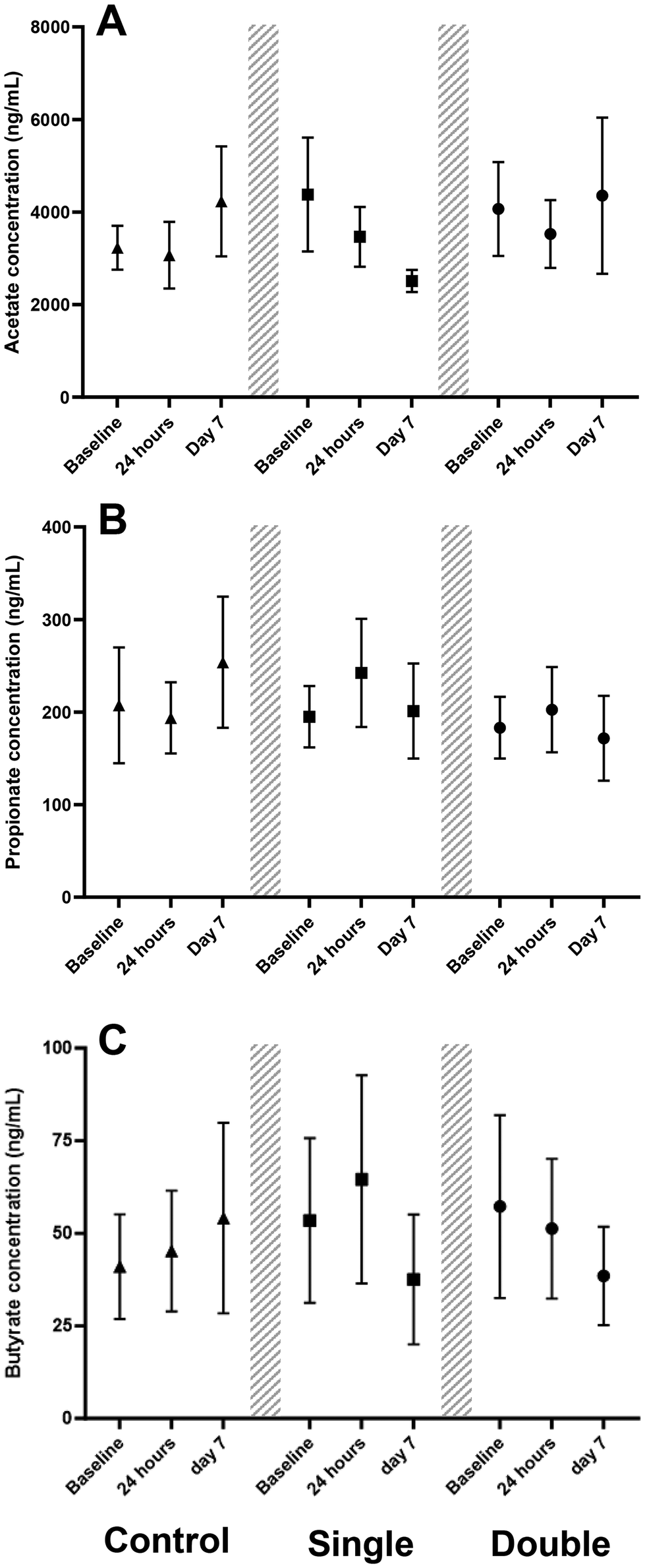 | ||
| Fig. 4 Serum concentration of acetate (A), propionate (B), and butyrate (C) at baseline, 24 h post-acute ingestion and after 7 days (Day 7) of twice daily consumption of the short-chain fatty acid supplements. Triangles indicate the control dose trial, squares indicate the single dose trial, and circles indicate the double dose trial. The markers refer to the mean values, with error bars visualising the standard error of the mean (n = 8). | ||
Discussion
This set of experiments investigated the influence of different supplementation parameters on circulating levels of acetate, propionate, and butyrate following oral co-ingestion. An initial experiment demonstrated that encapsulation of acetate and propionate in HPMC capsules induced an earlier (60 min vs. 120 min) peak serum concentration compared to capsules designed to resist the acidic stomach environment. Following this, a dose comparison investigation was performed to understand the acute serum concentrations of orally ingested acetate, propionate, and butyrate. As this investigation was performed in HPMC capsules, levels of all SCFAs reached peak values within 60 min and returned to baseline by 120 min. Peak serum concentrations of propionate and butyrate were shown to be increased following the double dose trial when compared to the single dose trial, with acetate levels not different. This was also the case for total load (assessed by tAUC) where propionate and butyrate, but not acetate, showed increasing total load in a stepwise manner as dose increased, demonstrating an overall increased systemic delivery of propionate and butyrate. Finally, basal circulating concentrations of SCFAs were not altered following a short-term (7-day) supplementation period.The results observed from the capsule experiment suggest that no clear advantage is gained using acid resistant capsule formulations when systemic uptake of SCFAs following exogenous supplementation is the desired outcome. Whilst the tAUC for both acids were similar, the lower Cmax values observed in the delayed trial could be due to a broader disintegration time profile leading to similar circulatory load of the SCFAs spread over a longer time period. This finding, combined with the ease of availability and lower production costs of HPMC capsules, mean that the use of HPMC capsules is recommended for future investigations to assess the impact of SCFA supplementation on metabolic/physiological processes that occur distally to the GI tract.
With respect to serum concentration characteristics observed following co-ingestion of acetate, propionate, and butyrate, the patterns were consistent with the serum acetate profile observed previously following both the intake of a vinegar-based drink and encapsulated vinegar ingestion.26 The rapid increase in circulating SCFA concentrations is likely explained by passive absorption from the stomach.25 Based on the expected gastric transit time for healthy adults and capsule formulation,30 the contents of the capsule would have likely been in the stomach fluid within 30 min of supplementation.31 The inclusion of a standardised breakfast prior to supplementation will have provided improved reproducibility with respect to gastric emptying/transit behaviour, as well as to minimise the impact of acetate production in the fasted state. Due to the pKa of these SCFAs being ∼4–5, the majority of ingested SCFA molecules would be in the associated (non-ionic) forms which are lipid soluble and able to cross the stomach epithelium.25 The absence of a difference in peak and total acetate levels between the single dose trial and double dose trial are likely attributable to the fact that basal acetate levels are much higher and more variable than those observed for propionate and butyrate, leading to more inter-individual variability and thus reducing the group-wide impact of acetate supplementation dose on subsequent systemic concentrations.
Previous studies assessing the effects of exogenous SCFA supplementation have targeted delivery to the distal regions of the GI tract.20–23,32 This is partly to simulate the production of SCFAs by the gut microbiota but also due to the positive physiological and health impacts of SCFAs on cells in this region.1 However, exogenously increasing peripheral SCFA levels via oral supplementation may offer further physiological and health benefits.1,9–13 The peak concentrations reached in the single dose trial and the double dose trial have previously been shown to acutely affect fuel utilisation (e.g., increased fat oxidation) and appetite hormone secretion (e.g., increased peptide YY and glucagon like peptide-1 secretion)20–23 as well as be at sufficient levels for GPCR activation.1 Whilst increasing SCFA production via the gut microbiota is possible, substantial changes to the diet (e.g., increase in dietary fibre) and/or regular supplementation with appropriate pre/probiotics would be required. However, due to intestinal use and metabolism in the liver,3 this may not elevate peripheral concentrations of SCFAs to the levels observed in this study. Previous work has shown that it is possible to acutely increase circulating concentrations following colonic delivery of SCFAs at physiologically relevant concentrations,22,23 and via oral supplementation of propionate supplements designed to reach the intestines.20,21 However, the peak concentrations reached within the periphery following colonic delivery of SCFAs were ∼3-fold, ∼7-fold, and ∼9-fold lower than in the present double dose trial for acetate, propionate, and butyrate, respectively, despite comparable total load of exogenous SCFAs.22,23 Differences in supplementation strategy (5 doses over a 2-hour window)21 and supplement type (inulin propionate ester)20 make it difficult to directly compare previous values of peak propionate concentrations to those in the present study. However, it must be noted that peripheral propionate concentrations reached in both the single dose trial and the double dose trial exceeded those seen previously following ∼7 g of oral sodium propionate ingestion, although no data beyond three hours where serum propionate concentrations may have peaked were provided by the authors.21 Although the acute physiological effects were not measured in the present study, the consistent serum concentration profiles and greater peripheral concentrations (in comparison to colonic delivery strategies) suggest that ingestion of SCFAs will result in a peripheral peak within 60 min following ingestion. This ingestion timeline can be applied in future research assessing the acute rise of peripheral SCFA concentrations on physiological processes.3 An interesting observation in this work was that the Tmax point for butyrate differed between the single dose (30 min) and double (60 min) dose trials. This response was not seen in the acetate and propionate trials, and thus the reason for this in the butyrate trials is not known and requires further clarification. To this point, it is not possible to attribute the metabolic fate of these molecules due to information not being available on whether the SCFAs are metabolised, incorporated into other molecules, taken into tissues, or excreted, which may have played a role in the different dynamics noted for butyrate. Nonetheless, this work provides important data for the acute changes in serum concentrations of SCFAs where the desired outcome is to maximise systemic availability following an oral dose.
Although clear rises in circulating SCFAs were observed following ingestion, the repeated intake of SCFAs across a 7-day period did not alter basal serum levels. This supports the observations from the acute experiment in that SCFAs are eliminated, distributed, or metabolised within 120 min. This mirrors previous observations where a four-week supplementation of butyrate did not alter basal plasma concentrations in healthy participants.18,19 The rapid utilisation and hepatic metabolism of butyrate were processes suggested to explain the lack of rise in circulating levels following sustained supplementation.18,19 In addition, previous studies have shown that serum SCFA levels return to pre-ingestion levels within an hour of peaking,3,22,23,26 which is similar to the present study where the estimated elimination time following peak levels was 60–120 min. This profile may be influenced by uptake of SCFAs into tissues which express the relevant transporter proteins (such as skeletal muscle), interactions with relevant GPCRs, assimilation into other molecules (e.g., long chain triglycerides), rapid oxidation, and/or rapid excretion.1,3,33 This suggests that the maintenance of elevated peripheral SCFA availability with exogenous supplementation may not be possible without ingestion at regular and unrealistic intervals and/or doses. To increase basal circulating levels of SCFAs, it may be necessary to stimulate gut bacteria-derived production of SCFAs (i.e., targeting the gut microbiome composition and function through prebiotic or probiotic mechanisms); however, limitations with this approach have been described previously. Although the presence of ingested SCFAs in their original form may be transient, the timing and/or dosing of repeated supplementation protocols may be non-critical if chronic physiological benefits can still be identified. Further work into both acute and chronic supplementation would enhance our knowledge within this area.
Importantly, the SCFA supplements were well tolerated with no major side effects reported by participants, including no reports of GI distress/disturbance. However, a mildly unpleasant taste and smell were reported relating to the butyrate supplements. Additionally, the size 000 capsules used for aspects of the supplementation protocols were reported as mildly-to-moderately uncomfortable to swallow by most participants. Whilst this capsule size was chosen to maximise the possible dose using fewer capsules, it is recommended that future studies use capsules smaller than 000 size to maintain user comfort, albeit this will require a trade-off between capsule quantity and desired dose of SCFA delivery.
It is important to note the limitations to the current study. For example, it was not possible to compare the absorption profile of butyrate in delayed release capsules due to lack of commercial availability; however, it is presumed that this profile would follow that of acetate and propionate with delayed and blunted serum concentrations. Importantly, although the number of participants exceeded the target size based on power calculations, the overall small sample size of the investigation means that these data may not be generalisable to the wider population. In addition, the unequal sex ratio (i.e. male-to-female) alongside the work being completed in younger, healthy individuals, limits the overall ability to understand whether demographic differences may exist. Furthermore, additional sampling timepoints (e.g., at 15 min intervals) would have improved the definition of Cmax and more confidently mapped the rise and fall in serum concentrations. Whilst dietary intake and fibre were not actively standardised, all participants were asked to maintain habitual dietary intake and followed a repeated food consumption diary in the 24 h prior to each trial. This gives the confidence that dietary fibre intake was stable within each individual participants’ habitual behaviour. Finally, despite participants confirming full compliance with supplement ingestion, alongside not returning any spare supplements to the laboratory, it cannot be guaranteed that all supplements were consumed across the investigatory periods.
Conclusions
In conclusion, oral co-ingestion of acetate, propionate, and butyrate in capsules designed to disintegrate in the proximal region of the GI tract increase circulating concentrations, with a dose-dependent manner observed for propionate and butyrate ingestion. The serum concentration profiles of the SCFAs were similar across molecules, with peak circulating concentrations reached at 30–60 min post-ingestion, returning to baseline within 120 min. Whilst definitive dosage recommendations cannot be drawn from the present data, the physiologically relevant peak concentrations observed, as well as the consistent timing of bioavailability following oral supplementation, provide valuable information to aid researchers in developing and designing supplementation protocols for future research in this ever-growing area.Author contributions
CGG – data curation, formal analysis, investigation, methodology, preparation, writing – original draft; MLYO – investigation, supervision, writing – review & editing; SNR – supervision, writing – review & editing; TB – supervision, writing – review & editing; LJJ – resources, writing – review & editing; TC – supervision, writing – review & editing; SJB – supervision, writing – review & editing; LMH – conceptualization, funding acquisition, methodology, project administration, resources, supervision, preparation, writing – original draft, writing – review & editing.Data availability
The data reported within this manuscript are available on the Loughborough University Research Repository at https://doi.org/10.17028/rd.lboro.27264402.Conflicts of interest
LJJ, TC, SJB, and LMH have received research funding, consultancy payments, and/or speaker fees from various industry partners, but none related to the research described within this report.Acknowledgements
The authors would like to thank Lonza and BodyBio for providing in-kind support through the provision of empty capsules and sodium butyrate supplements, respectively. We thank the participants for their time and commitment to volunteer in this study. This is a summary of independent research funded by the School of Sport, Exercise and Health Sciences (Loughborough University) and carried out at the National Institute for Health and Care Research (NIHR) Leicester Biomedical Research Centre (BRC). The views expressed are those of the author(s) and not necessarily those of the NIHR or the Department of Health and Social Care. CGG is supported by a Doctoral Research Scholarship administered by the School of Sport, Exercise and Health Sciences and the Doctoral College at Loughborough University. MLYO is supported by the Ministry of Higher Education Malaysia and Universiti Sains Malaysia Post-Doctoral Fellowship. SNR is supported by external research funding awarded to LMH from Biopolis S.L. The funders played no role in the study design or execution, nor in the production of this manuscript.References
- A. Koh, F. De Vadder, P. Kovatcheva-Datchary and F. Bäckhed, Cell, 2016, 165, 1332–1345 CrossRef CAS PubMed.
- T. Bongiovanni, M. O. L. Yin and L. M. Heaney, Int. J. Sports Med., 2021, 42, 1143–1158 CrossRef CAS.
- E. Boets, S. V. Gomand, L. Deroover, T. Preston, K. Vermeulen, V. De Preter, H. M. Hamer, G. Van den Mooter, L. De Vuyst, C. M. Courtin, P. Annaert, J. A. Delcour and K. A. Verbeke, J. Physiol., 2017, 595, 541–555 CrossRef CAS PubMed.
- H. O. Santos, W. M. A. M. de Moraes, G. A. R. da Silva, J. Prestes and B. J. Schoenfeld, Clin. Nutr. ESPEN, 2019, 32, 1–7 CrossRef PubMed.
- H.-J. Lee, H.-J. Ahn, C.-S. Kang, J.-C. Choi, H.-J. Choi, K.-G. Lee, J.-I. Kim and H.-Y. Kim, Food Control, 2010, 21, 217–220 CrossRef CAS.
- A. Pituch, J. Walkowiak and A. Banaszkiewicz, Prz. Gastroenterol., 2013, 8, 295–298 Search PubMed.
- E. E. Blaak, E. E. Canfora, S. Theis, G. Frost, A. K. Groen, G. Mithieux, A. Nauta, K. Scott, B. Stahl, J. van Harsselaar, R. van Tol, E. E. Vaughan and K. Verbeke, Benefic. Microbes, 2020, 11, 411–455 CAS.
- S. Ashique, G. De Rubis, E. Sirohi, N. Mishra, M. Rihan, A. Garg, R.-J. Reyes, B. Manandhar, S. Bhatt, N. K. Jha, T. G. Singh, G. Gupta, S. K. Singh, D. K. Chellappan, K. R. Paudel, P. M. Hansbro, B. G. Oliver and K. Dua, Chem.-Biol. Interact., 2022, 368, 110231 CrossRef CAS PubMed.
- M. G. Machado, V. Sencio and F. Trottein, Infect. Immun., 2021, 89, e0018821 CrossRef.
- J. Tan, C. McKenzie, M. Potamitis, A. N. Thorburn, C. R. Mackay and L. Macia, in Advances in Immunology, Academic Press Inc., 2014, vol. 121, pp. 91–119 Search PubMed.
- E. Ciarlo, T. Heinonen, J. Herderschee, C. Fenwick, M. Mombelli, D. Le Roy and T. Roger, Sci. Rep., 2016, 6, 37944 CrossRef CAS.
- R. F. Mcloughlin, B. S. Berthon, M. E. Jensen, K. J. Baines and L. G. Wood, Am. J. Clin. Nutr., 2017, 106, 930–945 CrossRef CAS.
- M. L. Y. Ong, C. G. Green, T. Bongiovanni and L. M. Heaney, Benefic. Microbes, 2023, 14, 565–590 CAS.
- D. S. Valdes, D. So, P. A. Gill and N. J. Kellow, J. Acad. Nutr. Diet., 2021, 121, 895–914 CrossRef PubMed.
- T. Banasiewicz, Ł. Krokowicz, Z. Stojcev, B. F. Kaczmarek, E. Kaczmarek, J. Maik, R. Marciniak, P. Krokowicz, J. Walkowiak and M. Drews, Colorectal Dis., 2013, 15, 204–209 CrossRef CAS.
- A. Di Sabatino, R. Morera, R. Ciccocioppo, P. Cazzola, S. Gotti, F. P. Tinozzi, S. Tinozzi and G. R. Corazza, Aliment. Pharmacol. Ther., 2005, 22, 789–794 CrossRef CAS PubMed.
- L. Krokowicz, B. F. Kaczmarek, P. Krokowicz, Z. Stojcev, J. Mackiewicz, J. Walkowiak, M. Drews and T. Banasiewicz, Travel Med. Infect. Dis., 2014, 12, 183–188 CrossRef PubMed.
- K. E. C. Bouter, G. J. Bakker, E. Levin, A. V. Hartstra, R. S. Kootte, S. D. Udayappan, S. Katiraei, L. Bahler, P. W. Gilijamse, V. Tremaroli, M. Stahlman, F. Holleman, N. A. W. Van Riel, H. J. Verberne, J. A. Romijn, G. M. Dallinga-Thie, M. J. Serlie, M. T. Ackermans, E. M. Kemper, K. Willems Van Dijk, F. Backhed, A. K. Groen and M. Nieuwdorp, Clin. Transl. Gastroenterol., 2018, 9, 155 CrossRef.
- M. C. P. Cleophas, J. M. Ratter, S. Bekkering, J. Quintin, K. Schraa, E. S. Stroes, M. G. Netea and L. A. B. Joosten, Sci. Rep., 2019, 9, 775 CrossRef PubMed.
- E. S. Chambers, A. Viardot, A. Psichas, D. J. Morrison, K. G. Murphy, S. E. K. Zac-Varghese, K. MacDougall, T. Preston, C. Tedford, G. S. Finlayson, J. E. Blundell, J. D. Bell, E. L. Thomas, S. Mt-Isa, D. Ashby, G. R. Gibson, S. Kolida, W. S. Dhillo, S. R. Bloom, W. Morley, S. Clegg and G. Frost, Gut, 2015, 64, 1744–1754 CrossRef CAS.
- E. S. Chambers, C. S. Byrne, K. Aspey, Y. Chen, S. Khan, D. J. Morrison and G. Frost, Diabetes, Obes. Metab., 2018, 20, 1034–1039 CrossRef CAS.
- C. M. van der Beek, E. E. Canfora, K. Lenaerts, F. J. Troost, S. W. M. O. Damink, J. J. Holst, A. A. M. Masclee, C. H. C. Dejong and E. E. Blaak, Clin. Sci., 2016, 130, 2073–2082 CrossRef CAS PubMed.
- E. E. Canfora, C. M. Van Der Beek, J. W. E. Jocken, G. H. Goossens, J. J. Holst, S. W. M. Olde Damink, K. Lenaerts, C. H. C. Dejong and E. E. Blaak, Sci. Rep., 2017, 7, 2360 CrossRef PubMed.
- M. G. Schmitt, K. H. Soergel and C. M. Wood, Gastroenterology, 1976, 70, 211–215 CrossRef CAS.
- D. R. Saunders, Nutr. Res., 1991, 11, 841–847 CrossRef CAS.
- S. S. Sugiyama, T. F. Fushimi, M. K. Kishi, S. I. Irie, S. T. Tsuji, N. H. Hosokawa and T. K. Kaga, J. Nutr. Sci. Vitaminol., 2010, 56, 226–269 Search PubMed.
- J. T. Bain, M. W. Taal, N. M. Selby, J. C. Reynolds and L. M. Heaney, J. Mass Spectrom. Adv. Clin. Lab, 2022, 25, 36–43 CrossRef CAS PubMed.
- H. Schielzeth, N. J. Dingemanse, S. Nakagawa, D. F. Westneat, H. Allegue, C. Teplitsky, D. Réale, N. A. Dochtermann, L. Z. Garamszegi and Y. G. Araya-Ajoy, Methods Ecol. Evol., 2020, 11, 1141–1152 CrossRef.
- Y. Benjamini and Y. Hochberg, J. R. Stat. Soc. Ser. B Stat. Method, 1995, 57, 289–300 CrossRef.
- J. Graff, K. Brinch and J. L. Madsen, Clin. Physiol., 2001, 21, 253–259 CrossRef CAS.
- S. Stegemann, S. Vishwanath, R. Kumar, D. Cade, M. Lowery, K. Hutchinson, M. Morgen, C. Lee and A. Goodwin, Comparative Human In vivo Study of an Immediate Release Tablet Over-Encapsulated by Gelatin and Hydroxypropyl Methyl Cellulose Capsules - Impact Of Dissolution Rate on Bioequivalence, Am. Pharm. Rev., 2015, 11, 38–45 Search PubMed.
- A. Roda, P. Simoni, M. Magliulo, P. Nanni, M. Baraldini, G. Roda and E. Roda, World J. Gastroenterol., 2007, 13, 1079–1084 CrossRef CAS PubMed.
- J. Frampton, K. G. Murphy, G. Frost and E. S. Chambers, Nat. Metab., 2020, 2, 840–848 CrossRef CAS.
| This journal is © The Royal Society of Chemistry 2024 |