
 Open Access Article
 This Open Access Article is licensed under a Creative Commons Attribution-Non Commercial 3.0 Unported Licence
A pH-neutral bioactive glass coated 3D-printed porous Ti6Al4V scaffold with enhanced osseointegration†
Xinguang
Wang‡
ab,
Qirui
Guo‡
cd,
Yizhen
He
ab,
Xiao
Geng
ab,
Cheng
Wang
ab,
Yang
Li
ab,
Zijian
Li
ab,
Caimei
Wang
e,
Dong
Qiu
 *cd and
Hua
Tian
*ab
*cd and
Hua
Tian
*ab
aDepartment of Orthopedics, Peking University Third Hospital, Beijing, 100191, China. E-mail: tianhua@bjmu.edu.cn
bEngineering Research Center of Bone and Joint Precision Medicine, Ministry of Education, Beijing, 100191, China
cBeijing National Laboratory for Molecular Sciences, CAS Research/Education Center for Excellence in Molecular Sciences, Institute of Chemistry, Chinese Academy of Sciences, Beijing, 100190, China. E-mail: dqiu@iccas.ac.cn
dUniversity of Chinese Academy of Sciences, Beijing, 100049, China
eBeijing 3D Printing Orthopedic Application Engineering Technology Research Center, Beijing, 102200, China
First published on 29th November 2022
Abstract
Osseointegration is vital for the success of non-degradable implants like those made of titanium alloys. In order to promote osseointegration, implants are made porous, providing space for bone ingrowth. Despite extensive optimization of the pore geometry and porosity, bone ingrowth into implants is still marginal; further modification to promote bone ingrowth as well as osseointegration becomes paramount. In this study, a pH neutral bioactive glass with the composition of 10.8% P2O5–54.2% SiO2–35% CaO (mol%; hereinafter referred to as PSC) was successfully coated on 3D-printed porous Ti6Al4V scaffolds using an in situ sol–gel method. This PSC coating is strongly bonded to the substrate and quickly induces the formation of hydroxyapatite on the scaffold surface upon contact with body fluid. In vitro, the PSC-coated Ti6Al4V scaffolds showed superior biocompatibility, cell proliferation promotion, cell adhesion, osteogenic differentiation and mineralization compared to their bare counterparts, implying better osseointegration. In vivo experiments confirmed this expectation; after being implanted, the coated scaffolds had more bone ingrowth and osseointegration, and consequently, higher push-out strength was achieved, proving the validity of the proposed concept in this study. In conclusion, PSC coating on 3D-printed porous Ti6Al4V scaffolds can improve osteogenesis, bone ingrowth, and osseointegration. Together with the versatility of this in situ sol–gel coating method, titanium alloy implants with better biological performances may be developed for immediate clinical applications.
1. Introduction
With the development of 3D printing technology, 3D-printed titanium alloy (Ti6Al4V) porous implants have been widely used in orthopedics and stomatology, with good clinical outcomes.1,2 Compared with traditional non-porous implants, 3D-printed Ti6Al4V porous implants can reduce the elastic modulus and stress shielding by adjusting the pore size, geometry and porosity, and can promote the growth of trabecular bone into the pores, realizing better osseointegration between the bone and the implant.3–5 However, the inherent bio-inertness of titanium alloys restricts the degree of bone ingrowth, thus osseointegration is still rather limited and the risk of prosthesis loosening remains fairly high.6,7 Undoubtedly, this is adverse to implant survival time and the clinical effect of surgery.8,9 Therefore, the surface modification of 3D-printed Ti6Al4V implants has recently become an emerging focus.10 Biogenic growth factors, such as bone morphogenetic protein-2,11 type-I collagen,12 and functional hydrogel coating,13 have been introduced to promote bone ingrowth and osseointegration; however, they have certain disadvantages such as high cost, complicated manufacturing process, unstable quality, and difficult to preserve nature.8 Mineral coating, for example Ca/P by microarc oxidation,14 was also developed on 3D-printed Ti6Al4V porous implants with lower cost and stable quality, while its promotion of bone ingrowth was far from satisfactory, significantly hampering their clinical applications.15In recent years, bioactive glasses (BGs) have received increasing attention16–18 because they show the best performance on osteogenesis and angiogenesis in the absence of biogenic growth factors,19 the two essential elements for ideal bone regeneration materials.20,21 Since Larry Hench created a kind of BG named 45S5, it has been developed and applied in clinical practice.22 45S5 is as effective as an iliac crest graft to maintain correction and achieve fusion in adolescent idiopathic scoliosis.23 Besides, studies have also shown that 45S5 coating can be realized on the surface of 3D-printed microporous titanium alloy scaffolds, which has good biocompatibility and can promote the attachment, proliferation and differentiation of human bone marrow stromal cells (BMSCs).24 However, 45S5 was prepared using the melt-quenching method, and the high pH of the dissolved product, low degradation rate, crystallization during sintering, and small specific surface area of the product lead to its relatively low biological activity.25
The newly developed pH-neutral BG with the composition of 10.8% P2O5–54.2% SiO2–35% CaO (mol%; hereinafter referred to as PSC)26,27 is especially promising because it has circumvented two most striking drawbacks of classical BGs, namely, the high pH and low degradation rate.25,28 PSC can provide a relatively neutral pH (about 7.8) and release large amounts of phosphate ions and silicate ions when immersed in body fluids, which were beneficial to new bone growth.25 Compared to 45S5 and beta-tricalcium phosphate (β-TCP), PSC showed better promotion of the osteogenic and angiogenic differentiation of BMSCs.28 However, direct coating of PSC on scaffolds is not straightforward because PSC powders are hard to be sprayed and firmly attached to the surface. Instead, as PSC is made from a sol–gel process, a coating may be prepared by spaying the sol onto the surface. Unfortunately, the standard sol–gel process for PSC is very time-consuming (the gelling usually takes more than two weeks), thus the timely formation of coating is unrealistic.25
In this study, a fast sol–gel process for PSC preparation is developed, where the gelling time is reduced to a few minutes, thus enabling in situ formation of PSC coating. In detail, PSC was successfully coated onto the surface of 3D-printed porous Ti6Al4V scaffolds, and its effects on proliferation, adhesion, and mineralization of rat BMSCs and in vivo performances of osteogenesis, bone ingrowth, and osseointegration in a rabbit femoral defect model were evaluated.
2. Materials and methods
2.1 Design and manufacture of 3D-printed Ti6Al4V porous scaffolds
We designed 2 types of 3D-printed Ti6Al4V scaffolds: discs with a diameter of 7 mm and a thickness of 2 mm for material characterization experiments and cell experiments and cylinders with a diameter of 5 mm and a height of 6 mm for animal experiments (ESI† Fig. S1). The 3D-printed Ti6Al4V scaffolds in material characterization and animal experiments were porous with a pore size of 640 mm and a diameter of 400 mm, and the micropores are distributed in a diamond cubic lattice structure. This parameter has proved beneficial to bone ingrowth in previous studies.4The manufacturing process of the 3D-printed Ti6Al4V porous scaffolds is as follows. The model of the 3D-printed scaffolds was imported into an electron beam melting rapid prototyping machine (Arcam AB, Molndal, Sweden) in a standard triangulation language format for layer-by-layer 3D printing using Ti6Al4V powders as the printing material. Subsequently, the scaffolds were ultrasonically cleaned in acetone, ethanol, and deionized water for 30 min.
2.2 Preparation of PSC-coated 3D-printed porous Ti6Al4V scaffolds
Preparation of PSC: 50% phytic acid (PA) (Sigma-Aldrich, St. Louis, MO, USA) aqueous solution was dried and dissolved in ethanol for further use. Based on the composition of PSC (10.8% P2O5: 54.2% SiO2: 35% CaO, mol%), tetraethylorthosilicate and PA were added into the as-formed phytic acid/ethanol solution. After 2 h of mixing under magnetic stirring, the as-prepared calcium methoxyethoxide solution was added to the mixture and further stirred for another 0.5 h. A clear and brownish-yellow PSC sol was obtained.
2.3 Characterization of PSC bulk and PSC coating
Fourier transform infrared spectroscopy (FTIR) (Bruker Equinox 55, Nexus, USA) was performed on PSC before and after the reaction with SBF. The scanning step size is 4 cm−1, and the range is 4000–400 cm−1.
2.4 In vitro cell experiments
2.5 Animal experiments
This animal experimental protocol was approved by the Laboratory Animal Welfare Ethics Branch of Peking University Institutional Review Board of Peking University Biomedical Ethics Committee (Approval Number: LA2021036).![[thin space (1/6-em)]](https://www.rsc.org/images/entities/char_2009.gif) 00000 units of penicillin were injected intramuscularly per day for the first 3 d after surgery to prevent infection.
00000 units of penicillin were injected intramuscularly per day for the first 3 d after surgery to prevent infection.
At 4 weeks postoperatively, 6 rabbits were randomly selected in the Ti and Ti-PSC groups. After being euthanized with excessive CO2, the distal femur specimens on the bilateral sides of each rabbit were taken out, with a total of 12 specimens in each group. Micro-computed tomography (micro-CT) analysis was performed on all 12 specimens, followed by 6 specimens randomly selected for bone histological analysis and the other 6 specimens for push-out tests. At 8 weeks postoperatively, the remaining 6 rabbits in each group were euthanized, and a similar experimental protocol was conducted.
2.6 Statistical analysis
Continuous variables consistent with normal distribution were described by mean ± standard deviation (SD), and comparisons between two groups were performed using an independent sample t-test. SPSS software version 25.0 (SPSS, Inc., Chicago, IL, USA) was used for statistical analysis, and P values <0.05 were considered statistically significant.3. Results and discussion
3.1 Fast sol–gel process for PSC preparation
A magnified version of video was taken to illustrate this rapid sol–gel process (ESI† Video S1 and Video S2). As shown in the video, the flowable sol-precursor quickly gelled once combined with water (ESI† Video S1) or steam (ESI† Video S2). After calcination of the above-sol, PSC was obtained. Since the concentration and usage of the precursor for PSC coating was thinner and less, the moisture in the air was enough to make it gelled in a short time, so a fast sol–gel process for PSC coating preparation was developed. This was really faster than the previous method reported by C. Y. Cui et al.,25 in which the gelling takes more than two weeks.3.2 Characterization of the PSC and PSC coating
| Parameters | Ti group | Ti-PSC group | T value | P value |
|---|---|---|---|---|
| Elastic modulus (MPa) | 1160 ± 122 | 1303 ± 108 | −1.751 | 0.131 |
| Stiffness (N mm−1) | 3799 ± 401 | 4265 ± 355 | −1.745 | 0.132 |
| Yield strength (N) | 1090 ± 100 | 1132 ± 154 | −0.465 | 0.658 |
| Compression strength (N) | 1364 ± 134 | 1327 ± 102 | 0.427 | 0.684 |
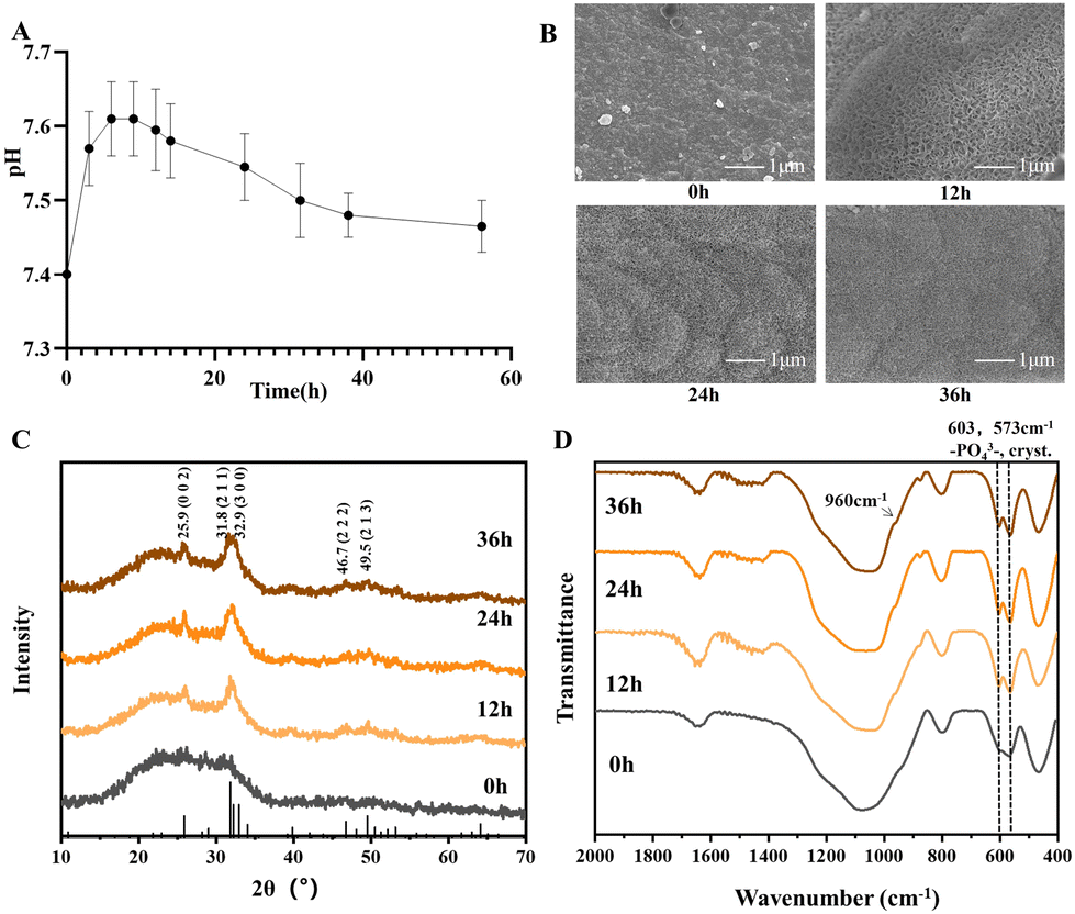 | ||
| Fig. 1 pH and bioactivity test of PSC. (A) pH of PSC dissolutions. The pH value of PSC dissolutions only slightly increased from 7.4 to 7.6. (B) SEM of PSC in SBF for 0, 12, 24 and 36 h, respectively. Characteristic morphology of HA was found in 12 h and grew denser with prolonged immersion in 24 h and 36 h. (C) XRD of PSC in SBF for 0, 12, 24 and 36 h. Characteristic peaks of HA (25.9°, 31.7°, 32.8°, 39.7°, 46.6°, 49.6°, 53.2°, and 64.1°; JCPDS: 09-0432) were found. (D) FTIR of PSC in SBF for 0, 12, 24 and 36 h. Double absorption peaks at 607 cm−1 and 567 cm−1 as well as an enhanced band at 960 cm−1 were observed. | ||
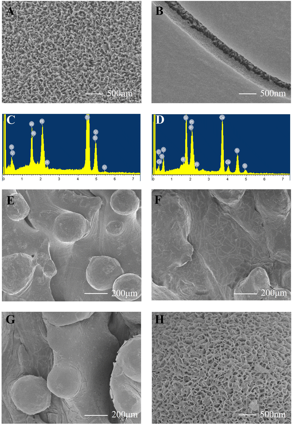 | ||
| Fig. 2 Surface characterizations of scaffolds and adhesion strength of the PSC coating. (A) SEM of alkali-treated scaffolds. Many pin-shaped pores appeared on the scaffolds after the alkali treatment. (B) SEM of PSC-coated scaffolds. PSC was successfully coated on 3D-printed porous Ti6Al4V scaffolds and scaffolds could be observed in the cracks of the coating. (C) EDS of Ti6Al4V scaffolds. The main elements in Ti6Al4V scaffolds were Ti, Al, and V. (D) EDS of PSC-coated scaffolds. The main elements in the surface of PSC-coated scaffolds were Ti, Si, Ca, P, and O. (E) SEM of scaffolds of Ti6Al4V. Partially melted titanium alloy powder was seen. (F) SEM of PSC-coated scaffolds before ultrasonication tests. PSC coating was seen on the surface of the Ti6Al4V scaffold. (G) SEM of PSC-coated scaffolds after ultrasonication tests. No PSC-coating fragments were found to detach from the scaffold. (H) SEM of PSC-coated scaffolds in SBF for 12 h. Many needle-like aggregates appeared on the surface of PSC-coated scaffolds, a feature implying the likely formation of HA. | ||
3.2 In vitro cell experiments
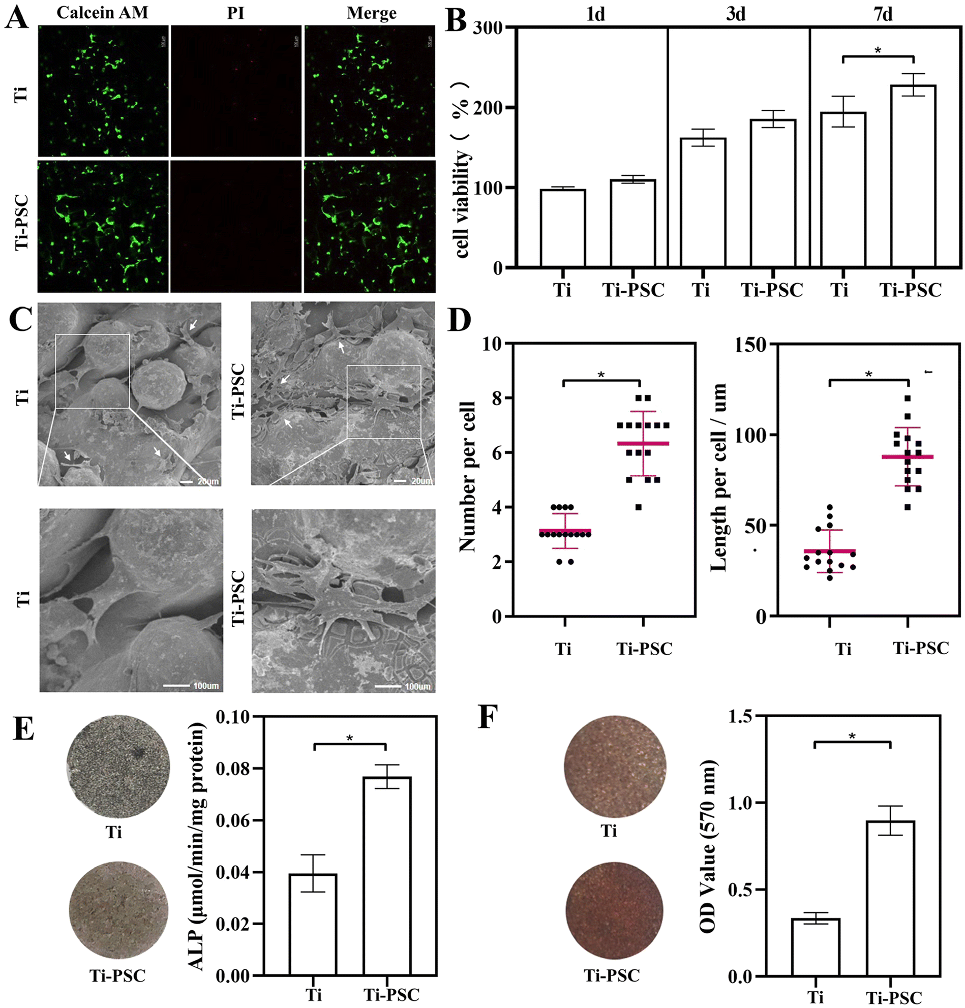 | ||
| Fig. 3 Biocompatibility, cell proliferation, cell adhesion, cell differentiation and cell mineralization on two groups of scaffolds. (A) Live/dead staining of the scaffolds after 24 h of cell culture. Calcein-AM stained live cells and propidium iodide (PI)-stained dead cells. The intensity and coverage of green fluorescence were higher, and red fluorescence was less in the Ti-PSC group. (B) Cell proliferation on two groups of scaffolds. The cells in the Ti-PSC group were more viable than those in the Ti group at any interval, especially a significant difference (P < 0.05) occurred on Day 7. (C) SEM of BMSCs on two groups of scaffolds for 48 h. BMSCs in the Ti-PSC group covered a larger area, and the pseudopodia showed a filament-like structure. (D) The number of pseudopodia and the length of BMSCs on two groups of scaffolds for 2 days. The number of pseudopodia and the length of BMSCs in the Ti-PSC group were higher than those of the Ti group (P < 0.05). (E) ALP staining and quantitative assay on two groups of scaffolds for 14 days. There were more grey-black precipitates on the surface of the Ti-PSC group scaffolds with a higher amount of ALP (P < 0.05). (F) Alizarin red staining and semi-quantitative assay on two groups of scaffolds for 28 days. There were more dark red calcium nodules on the surface of the Ti-PSC group scaffolds with a higher amount of alizarin red (P < 0.05). | ||
Quantitative tests were performed on the adhesion results. 15 cells within the SEM scope were randomly selected, and the number of pseudopodia and the length of the cells were recorded. The results are shown in Fig. 3D. Compared with the Ti group, BMSCs possessed twice as many pseudopodia (the number of pseudopodia in the Ti group was 3.1 ± 0.6, while the number of pseudopodia in the Ti-PSC group was 6.3 ± 1.2. P < 0.05). Moreover, the cell length in the Ti group was 35.8 ± 11.8, while the cell length in the Ti-PSC group was 87.9 ± 16.1. BMSCs cells had a wider coverage in the Ti-PSC group (2.4 times that of the Ti group with P < 0.05), which means that PSC coating is beneficial for cell adhesion.
To summarize, the PSC-coated Ti6Al4V scaffolds showed superior biocompatibility, cell proliferation promotion, cell adhesion, osteogenic differentiation and mineralization compared to their bare counterparts in vitro cell experiments. Considering the fact that acidity or strong alkalinity was harmful to osteogenesis and high Si and P concentrations in the appropriate range were beneficial to cell proliferation and differentiation, PSC, with a relatively neutral pH and higher Si and P concentrations, could promote the biocompatibility, proliferation, adhesion, osteogenic differentiation and mineralization of BMSCs.25,26
3.3 Animal surgery
All surgical procedures on 24 rabbits were performed successfully, and no complications such as death and wound infection occurred within 8 weeks after surgery. All the scaffolds were in the original position, and there was no obvious infection or heterotopic ossification at the operative site when the distal femur specimens were taken out.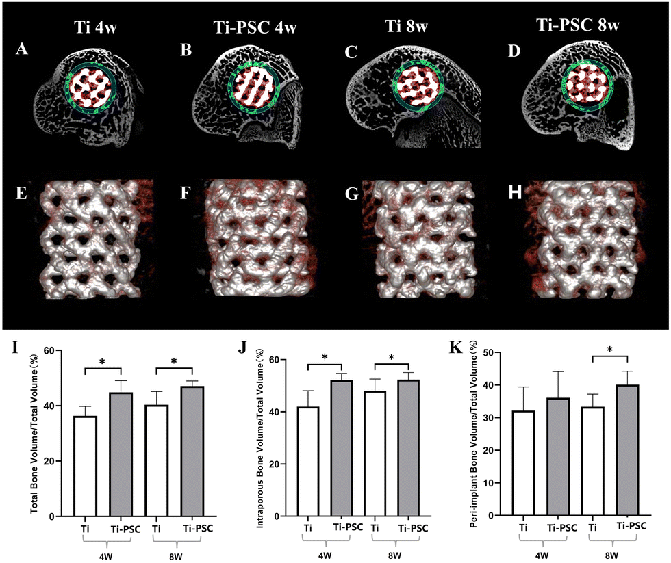 | ||
| Fig. 4 Osteogenesis, bone in-growth, and osseointegration of Ti and Ti-PSC group scaffolds at 4 and 8 weeks in micro-CT. (A–D) Representative cross-sectional image of micro-CT (A: Ti group at 4 weeks; B: Ti-PSC group at 4 weeks; C: Ti group at 8 weeks; D: Ti-PSC group at 8 weeks). The scaffold was labelled in white. New bone in the intra-porous region was labelled in red and new bone in the peri-implant region was labelled in green. (E–H) Representative 3D-reconstructed images of micro-CT (E: Ti group at 4 weeks; F: Ti-PSC group at 4 weeks; G: Ti group at 8 weeks; H: Ti-PSC group at 8 weeks). The scaffold was labelled in silver and new bone was labelled in red. (I–K) Total (I), intra-porous (J), and peri-implant (K) Bone Volume/Total Volume of the Ti group scaffold and Ti-PSC group scaffold at 4 and 8 weeks in Micro CT. *: P < 0.05. | ||
| Parameters | 4 w | 8 w | ||
|---|---|---|---|---|
| Ti group (%) | Ti-PSC group (%) | Ti group (%) | Ti-PSC group (%) | |
| a At the same time, P < 0.05 when compared with Ti and Ti-PSC groups. | ||||
| Total BV/TV | 36.29 ± 3.33 | 44.69 ± 4.02a | 40.34 ± 4.71 | 47.20 ± 1.80a |
| Intra-porous BV/TV | 42.16 ± 6.20 | 52.10 ± 2.44a | 47.89 ± 4.58 | 52.31 ± 2.71a |
| Peri-implant BV/TV | 32.09 ± 7.06 | 36.20 ± 8.04 | 33.35 ± 4.10 | 40.18 ± 4.08a |
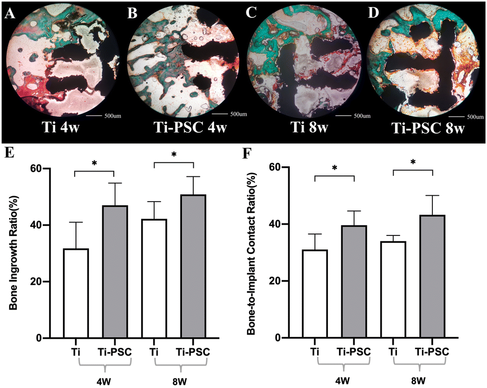 | ||
| Fig. 5 Osteogenesis, bone in-growth, and osseointegration of Ti group scaffolds and Ti-PSC group scaffolds at 4 and 8 weeks in bone histology. (A–D) Representative images of bone histology: (A: Ti group at 4 weeks; B: Ti-PSC group at 4 weeks; C: Ti group at 8 weeks; D: Ti-PSC group at 8 weeks): The scaffold was stained black, new bone was stained green, and osteoid tissues were stained red or orange. (E) Bone ingrowth proportion of Ti group scaffolds and Ti-PSC group scaffolds at 4 and 8 weeks in bone histology. (F) Bone-to-implant contact ratio (BICR) of Ti group scaffolds and Ti-PSC group scaffolds at 4 and 8 weeks in bone histology. *: P < 0.05. | ||
Meanwhile, we also found that BICR of the Ti-PSC group was higher than that of the Ti group at 4 and 8 weeks (4 weeks: 39.55% ± 4.96% vs.31.15% ± 5.40%, P < 0.05; 8 weeks: 43.32% ± 6.93% vs. 33.98% ± 2.13%, P < 0.05), indicating that the Ti-PSC group had better osteointegration. Our previous study also found that in the repair of calvarial bone defects in rats, the new bone tissues with lamellar bone morphology in the PSC group were more organized and continuous, suggesting that the PSC does not simply promote osteogenesis, more importantly, this osteogenesis is not disordered, i.e., the PSC can indeed promote bone remodeling and osseointegration.28
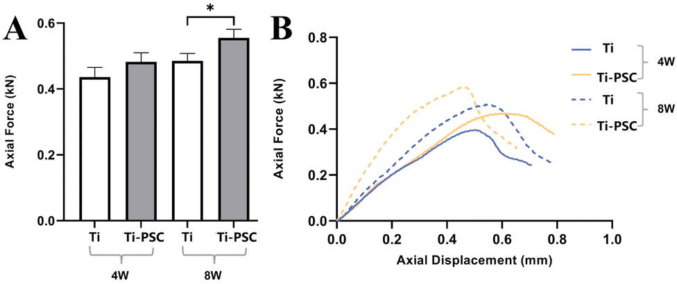 | ||
| Fig. 6 Fixation strength of Ti and Ti-PSC group scaffolds at 4 and 8 weeks in the push-out test. (A) Maximum push-out force of the Ti group scaffold and Ti-PSC group scaffold at 4 and 8 weeks in the push-out test. *: P < 0.05.(B) Representative axial force-displacement curves of the Ti group scaffold and Ti-PSC group scaffold at 4 and 8 weeks in the push-out test. | ||
In Fig. 6B, the representative axial force-displacement curves of the two groups at 4 and 8 weeks are shown. The axial force-displacement curve of the Ti-PSC group was steeper than that of the Ti group at 4 weeks and 8 weeks, that is, to produce the same axial displacement of implant, the needed axial force of the Ti-PSC group was greater. This also indicates that the scaffolds of the Ti-PSC group had greater fixation strength of scaffold and bone, which was a compositive index of osteogenesis, bone in-growth, and osseointegration, and was also an objective expression of the implant stability.39,40 Therefore, the PSC-coated 3D-printed Ti6Al4V porous implant is expected to improve the stability of the implant in clinical practice and has a good prospect for clinical application.
4. Conclusions
In this study, a pH neutral bioactive glass (PSC) was successfully coated on 3D-printed porous Ti6Al4V scaffolds with good adhesion strength. This in situ sol–gel method for fabricating PSC coating was simple, quick, and convenient. PSC-coated Ti6Al4V scaffolds showed superior biocompatibility, cell proliferation promotion, cell adhesion, osteogenic differentiation and mineralization compared to their bare counterparts in vitro and had more bone ingrowth and osseointegration as well as higher push-out strength of the implant in vivo. PSC coating is expected to improve the stability of the 3D-printed porous Ti6Al4V implants, which may be developed for immediate clinical applications.Author contributions
Xinguang Wang: conceptualization, methodology, software, validation, formal analysis, investigation, writing – original draft, and visualization; Qirui Guo: methodology, software, validation, formal analysis, investigation, and writing – original draft; Yizhen He: methodology, software, validation, formal analysis, investigation, and writing – original draft; Xiao Geng: methodology, validation, formal analysis, investigation, and data curation; Cheng Wang: methodology, validation, formal analysis, and investigation; Yang Li: methodology, formal analysis, and investigation; Zijian Li: formal analysis and investigation; Caimei Wang: formal analysis, investigation, and writing – review & editing; Dong Qiu: conceptualization, formal analysis, investigation, data curation, writing – review & editing, supervision, project administration, and funding acquisition. Hua Tian: conceptualization, formal analysis, investigation, data curation, writing – review & editing, supervision, project administration, and funding acquisition.Conflicts of interest
All authors have read the declaration of interests and declare that they have no competing interests.Acknowledgements
This work was supported by the Natural Science Foundation of Beijing Municipality (grant number: 19L2160). The authors acknowledge the research support from the Department of Laboratory Animal Science of Peking University Health Science Center in the animal experiment and the Beijing AKEC Medical Co., Ltd in the manufacture of 3D-printed Ti6Al4V porous scaffolds.Notes and references
- H. H. Malik, A. R. Darwood, S. Shaunak, P. Kulatilake, A. A. El-Hilly, O. Mulki and A. Baskaradas, J. Surg. Res., 2015, 199, 512–522 CrossRef PubMed.
- C. Serrano, H. van den Brink, J. Pineau, P. Prognon and N. Martelli, J. Cranio-Maxillofac. Surg., 2019, 47, 1387–1397 CrossRef PubMed.
- V. Karageorgiou and D. Kaplan, Biomaterials, 2005, 26, 5474–5491 CrossRef CAS PubMed.
- J. Lv, Z. Jia, J. Li, Y. Wang, J. Yang, P. Xiu, K. Zhang, H. Cai and Z. Liu, Adv. Eng. Mater., 2015, 17, 1391–1398 CrossRef CAS.
- J. Parthasarathy, B. Starly, S. Raman and A. Christensen, J. Mech. Behav. Biomed. Mater., 2010, 3, 249–259 CrossRef PubMed.
- J. Liu, N. B. Mohd Rafiq, L. M. Wong and S. Wang, Front. Chem., 2021, 9, 768007 CrossRef CAS PubMed.
- B. G. X. Zhang, D. E. Myers, G. G. Wallace, M. Brandt and P. F. M. Choong, Int. J. Mol. Sci., 2014, 15, 11878–11921 CrossRef PubMed.
- Z. Jing, T. Zhang, P. Xiu, H. Cai, Q. Wei, D. Fan, X. Lin, C. Song and Z. Liu, Biomed. Mater., 2020, 15, 052003 CAS.
- S. B. Goodman, Z. Yao, M. Keeney and F. Yang, Biomaterials, 2013, 34, 3174–3183 CrossRef CAS PubMed.
- X. Sheng, A. Wang, Z. Wang, H. Liu, J. Wang and C. Li, Front. Bioeng. Biotechnol., 2022, 10, 850110 CrossRef PubMed.
- J. Lv, P. Xiu, J. Tan, Z. Jia, H. Cai and Z. Liu, Biomed. Mater., 2015, 10, 035013 CrossRef PubMed.
- Y. Zhao, L. Bai, Y. Zhang, R. Yao, Y. Sun, R. Hang, X. Chen, H. Wang, X. Yao, Y. Xiao and R. Hang, Biomaterials, 2022, 288, 121684 CrossRef CAS PubMed.
- X. Li, K. Xu, Y. He, B. Tao, K. Li, C. Lin, J. Hu, J. Wu, Y. Wu, S. Liu, L. Peng, H. Wang and K. Cai, Biomaterials, 2022, 287, 121683 CrossRef CAS PubMed.
- P. Xiu, Z. Jia, J. Lv, C. Yin, Y. Cheng, K. Zhang, C. Song, H. Leng, Y. Zheng, H. Cai and Z. Liu, ACS Appl. Mater. Interfaces, 2016, 8, 17964–17975 CrossRef CAS PubMed.
- R. Ni, Z. Jing, C. Xiong, D. Meng, C. Wei and H. Cai, Ann. Transl. Med., 2022, 10, 710 CrossRef CAS PubMed.
- J. R. Jones, Acta Biomater., 2013, 9, 4457–4486 CrossRef CAS PubMed.
- B. Begines, C. Arevalo, C. Romero, Z. Hadzhieva, A. R. Boccaccini and Y. Torres, ACS Appl. Mater. Interfaces, 2022, 14, 15008–15020 CrossRef CAS PubMed.
- J. C. Moses and B. B. Mandal, ACS Appl. Mater. Interfaces, 2022, 14, 14961–14980 CrossRef CAS PubMed.
- A. A. El-Rashidy, J. A. Roether, L. Harhaus, U. Kneser and A. R. Boccaccini, Acta Biomater., 2017, 62, 1–28 CrossRef CAS PubMed.
- X. V. Bui, V. B. Nguyen, T. T. H. Le and Q. M. Do, Glass Phys. Chem., 2013, 39, 64–66 CrossRef CAS.
- S. Ali, I. Farooq and K. Iqbal, Saudi Dent. J., 2014, 26, 1–5 CrossRef PubMed.
- F. Baino, S. Hamzehlou and S. Kargozar, J. Funct. Biomater., 2018, 9, 25 CrossRef PubMed.
- B. Ilharreborde, E. Morel, F. Fitoussi, A. Presedo, P. Souchet, G.-F. Penneçot and K. Mazda, J. Pediatr. Orthop., 2008, 28, 347–351 CrossRef PubMed.
- X. Ye, S. Leeflang, C. Wu, J. Chang, J. Zhou and Z. Huan, Materials, 2017, 10, 1244 CrossRef PubMed.
- C. Y. Cui, S. N. Wang, H. H. Ren, A. L. Li, D. Qiu, Y. H. Gan and Y. M. Dong, RSC Adv., 2017, 7, 22063–22070 RSC.
- A. Li and D. Qiu, J. Mater. Sci.: Mater. Med., 2011, 22, 2685–2691 CrossRef CAS PubMed.
- A. L. Li, H. H. Ren, Y. Cui, C. Wang, X. J. Zhou, H. Lin and D. Qiu, J. Non-Cryst. Solids, 2017, 475, 10–14 CrossRef CAS.
- H. Zhao, G. Liang, W. Liang, Q. Li, B. Huang, A. Li, D. Qiu and D. Jin, Mater. Sci. Eng., C, 2020, 116, 111249 CrossRef CAS PubMed.
- P. Yu, F. Lu, W. Zhu, D. Wang, X. Zhu, G. Tan, X. Wang, Y. Zhang, L. Li and C. Ning, Appl. Surf. Sci., 2014, 313, 947–953 CrossRef CAS.
- J. Ryu, S. H. Ku, H. Lee and C. B. Park, Adv. Funct. Mater., 2010, 20, 2132–2139 CrossRef CAS.
- Z. Jing, R. Ni, J. Wang, X. Lin, D. Fan, Q. Wei, T. Zhang, Y. Zheng, H. Cai and Z. Liu, Bioact. Mater., 2021, 6, 4542–4557 CrossRef CAS PubMed.
- K. Maekawa, K. Shimono, M. Oshima, Y. Yoshida, B. Van Meerbeek, K. Suzuki and T. Kuboki, J. Oral Rehabil., 2009, 36, 362–367 CrossRef CAS PubMed.
- S.-H. Jun, B. M. W. Chang, H.-P. Weber and J.-J. Kwon, Int. J. Oral Maxillofac. Implants, 2010, 25, 985–990 Search PubMed.
- P. Xiu, Z. Jia, J. Lv, C. Yin, Y. Cheng, K. Zhang, C. Song, H. Leng, Y. Zheng, H. Cai and Z. Liu, ACS Appl. Mater. Interfaces, 2016, 8, 17964–17975 CrossRef CAS PubMed.
- H. Yang, Q. Zhu, H. Qi, X. Liu, M. Ma and Q. Chen, Materials, 2018, 11, 1540 Search PubMed.
- T. R. Arnett, J. Nutr., 2008, 138, 415S–418S CrossRef CAS PubMed.
- T. Kokubo and H. Takadama, Biomaterials, 2006, 27, 2907–2915 CrossRef CAS PubMed.
- K. Pluta, A. Sobczak-Kupiec, O. Półtorak, D. Malina and B. Tyliszczak, J. Biomed. Mater. Res., Part A, 2018, 106, 1941–1950 CrossRef CAS PubMed.
- H. Liu, W. Li, C. Liu, J. Tan, H. Wang, B. Hai, H. Cai, H. J. Leng, Z. J. Liu and C. L. Song, Biofabrication, 2016, 8, 045012 CrossRef PubMed.
- H. Spece, C. Basgul, C. E. Andrews, D. W. MacDonald, M. L. Taheri and S. M. Kurtz, J. Biomed. Mater. Res., Part B, 2021, 109, 1436–1454 CrossRef CAS PubMed.
Footnotes |
| † Electronic supplementary information (ESI) available. See DOI: https://doi.org/10.1039/d2tb02129c |
| ‡ Xinguang Wang and Qirui Guo contributed equally to this work. |
| This journal is © The Royal Society of Chemistry 2023 |