
 Open Access Article
 This Open Access Article is licensed under a Creative Commons Attribution-Non Commercial 3.0 Unported Licence
Ion transport in two-dimensional flexible nanoporous membranes
Yechan
Noh
 a and
Narayana R.
Aluru
*b
a and
Narayana R.
Aluru
*b
aDepartment of Mechanical Science and Engineering, University of Illinois at Urbana-Champaign, Urbana, Illinois 61801, USA
bWalker Department of Mechanical Engineering, Oden Institute for Computational Engineering & Sciences, University of Texas at Austin, Austin 78712, USA. E-mail: aluru@utexas.edu
First published on 14th June 2023
Abstract
Ion transport is a fundamental mechanism in living systems that plays a role in cell proliferation, energy conversion, and maintaining homeostasis. This has inspired various nanofluidic applications such as electricity harvesting, molecular sensors, and molecular separation. Two dimensional (2D) nanoporous membranes are particularly promising for these applications due to their ultralow transport barriers. We investigated ion conduction across flexible 2D membranes via extensive molecular dynamics simulations. We found that the microscopic fluctuations of these membranes can significantly increase ion conductance, for example, by 320% in Cu-HAB with 0.5 M KCl. Our analysis of ion dynamics near the flexible membranes revealed that ion hydration is destabilized when the membrane fluctuated within a specific frequency range leading to improved ion conduction. Our results show that the dynamic coupling between the fluctuating membrane and ions can play a crucial role in ion conduction across 2D nanoporous membranes.
Introduction
Living systems, including humans, rely on ion transport as a fundamental mechanism for various physiological processes, such as maintaining homeostasis,1 regulating cell proliferation,1 generating electricity,2 and producing energy-storage molecules.3,4 Ion transport across nanometer-sized pores exhibits unique characteristics such as selective ion transport,5–8 activation by stimuli,9,10 and mechanosensitive ion transport.11,12 These features of ion transport have inspired a wide range of applications such as osmotic power generation,13,14 ion separation,15,16 DNA sequencing,17,18 and water purification.19–22 Recently, several 2D nanoporous membranes have been successfully fabricated using bottom-up approaches23–25 and these advances are expected to further advance nanopore applications owing to their ultralow transport barrier, uniform pore size, high pore density, and excellent flexibility. Several studies have focused on ion-transport phenomena across the 2D membranes providing a microscopic understanding on the effect of diameter,26–28 surface charge density,26,29 functional groups,30 mechanical strain,31,32 ion concentration,26 and pore–pore interactions.33–35 However, there is a lack of understanding of how the exceptional flexibility of 2D nanoporous membranes affects microscopic ion transport.Over the past few decades, researchers have studied various aspects of ion transport across 2D nanoporous membranes. In 2008, Sint et al.36 studied ion transport through functionalized graphene nanopores using molecular dynamics (MD) simulations. They demonstrated that a graphene nanopore could be an anion or cation-selective membrane depending on the functional groups. In 2013, He et al.30 presented a computational study demonstrating that a graphene nanopore with 0.43 nm diameter and three carboxylate groups exhibited voltage-dependent selectivity (i.e., cation and anion-selective at low and high voltages, respectively). In 2014, Suk and Aluru27 studied ion transport in sub-5 nm graphene nanopores using MD simulations. They demonstrated that the density and mobility of ions in a nanopore deviate from their bulk value, and continuum theory with corrected density and mobility can account for MD simulation results. In 2019, Fang et al.31 studied the effect of mechanical strain on ion conduction through a graphene-embedded crown. They observed strain-sensitive ion transport, where ion current increases by an order of magnitude when 2% strain is applied. In 2020, Noh and Aluru26 demonstrated that 2D nanopores are electrically imperfect, and ion conductance is not entirely governed by surface conductance. In 2022, Jiang et al.37 demonstrated a pressure-induced enhancement of ion conductance in single-layer graphene nanopores, which cannot be explained by the linear electrokinetic theory. Jiang et al. further demonstrated that ion conductance increases due to the pressure-driven transport of capacitively accumulated ions near the graphene surface, creating nonlinear electrohydrodynamic coupling. However, many prior MD simulations overlooked nanopore flexibility, assuming nanopores to be rigid. This assumption may not align with experimental studies, potentially leading to inconsistencies between simulated predictions and experimental results. In our current investigation, we aim to bridge this gap by accounting for nanopore flexibility.
Prior research in the fields of molecular biology and nanofluidics have demonstrated the importance of surface fluctuations in various microscopic phenomena. In 1999, Kohen et al.38 studied biological catalysts and observed that the vibrational modes of enzymes are a major factor in their catalytic activity. In 2001, Bernèche and Roux8 studied the KcsA potassium channel and observed that the fluctuations of the channel structure can considerably affect the free energy profiles and ion selectivity. In 2004, Noskov et al.5 studied the KcsA channel and observed that carbonyl groups exhibit liquid-like fluctuations that are important for ion selectivity. In 2015, Ma et al.39 studied water transport in a double-walled carbon nanotube and observed that the longitudinal vibration mode of the CNT creates oscillatory friction and enhances diffusion of confined water. In 2018, Marbach et al.40 theoretically showed that the effective diffusion of confined fluids within wiggling surfaces can be increased or decreased depending on the spectrum of surface fluctuations. In 2022, Noh and Aluru21 demonstrated that water permeation in a 2D flexible membrane is higher (up to 102%) than that in its rigid counterpart and that water permeation is maximized when the membrane frequency matches the vibrational frequency of water molecules. In subsequent work,41 they demonstrated vibrational coupling-induced enhancement of water transport is more significant in small diameter pores (water flow increases up to 500% in a 0.75 nm diameter pore). In 2022, Lyu et al.42 studied chloride conduction in multilayered COF membranes and concluded that membrane flexibility is an important factor for predicting experimentally measured chloride conductivity.
In the present study, we analyze how the flexibility of 2D nanoporous membranes affects ion transport. We study ion transport considering two different types (flexible and rigid) of nanoporous membranes and observe a considerable difference between ion current in the flexible membrane and its rigid counterpart. To understand the mechanisms arising from the membrane flexibility, we analyze the pore expansion, ion dynamics near the membranes, and microscopic fluctuations of the membrane and nearby ions.
Results and discussion
The primary focus of this study is to understand how the flexibility of a membrane affects ion conduction. Toward this, we designed MD simulations as shown in Fig. 1a. The nanoporous membrane was immersed in an electrolyte solution, and the edges of the membrane were constrained while the rest of the membrane fluctuates freely. For comparison, we also considered a rigid membrane, where all membrane atoms were fixed. A constant external electric field was applied along the z-direction of the system to generate an electrophoretic ion current. We considered two single-layer nanoporous membranes (Fig. 1b and c): Cu-HAB25,43 and GNM-3.21 GNM-3 is a graphene nanomesh.44 The graphene-based nanopore membrane was actively studied for various applications20,22,30,36,45,46 and was recently synthesized on a large scale with a consistent pore size.23 Cu-HAB25,43 is a metal–organic framework (MOF)-based 2D membrane. MOF-based membranes have shown great potential in numerous applications.15,19,21,25,47–49 In this study, GNM-3 and Cu-HAB are hydrogenated and assumed to be defect free. Fig. 1d and e demonstrate fluctuations in nanoporous membranes where the amplitudes of the fluctuations are a few angstroms, which is comparable to the size of nanopore we consider. Fig. 1f and g show the current–voltage response for 2D nanoporous membranes. For both membranes, we observe a linear relation between ion current and applied voltage within the tested voltage range. Interestingly, ion conductance (i.e., the slope of the current–voltage curve) in flexible Cu-HAB is approximately twice that of its rigid counterpart, where mechanical deformations and fluctuations are suppressed. | ||
| Fig. 1 Ion transport across 2D flexible nanoporous membranes. a. MD simulation setup for ion transport. The 2D membrane is immersed in an electrolyte solution, and its edges are constrained. A cross-membrane electric potential difference is applied. Atomic structure of b. Cu-HAB and c. GNM-3. Fluctuations of d. Cu-HAB and e. GNM-3 (bottom) and its distribution (top). The opaque colors indicate the mean position of the membrane. The transparent colors indicate the membrane fluctuations. Current–voltage characteristics in f. Cu-HAB and g. GNM-3 membranes. The plotted ion currents are divided by the number of nanopores (i.e., 12) in the membrane. The solid lines represent a linear fit to the MD data. | ||
On the other hand, ion conductance in the flexible GNM-3 membrane is marginally higher (9%) than that of its rigid counterpart. As a result, ion conductance in flexible Cu-HAB is slightly higher than in flexible GNM-3, although the pore size of GNM-3 is 25% higher than that of Cu-HAB. Fig. 2a and b show the number of ion species transported across the membranes over time. The transport of potassium and chloride ions increases linearly and is higher in the flexible membranes compared to their respective rigid counterparts. Also, it is shown that both cation and anion currents increase and this results in reduced ion selectivity. Specifically, we measured the anion selectivity using the formula:  , where I− represents anion current and I+ represents cation current. In rigid Cu-HAB, the anion selectivity is S− = 0.68, but the anion selectivity is reduced to S− = 0.49 in flexible Cu-HAB. This is attributed to the slightly enlarged pore diameter in flexible membrane and the vibrational coupling effect. Fig. 2a and b show a slight electroosmotic flow that is created by anion selectivity due to the positive charge of the nanopores. This hydrodynamic flow can slightly increase the anion transport, while slightly decreasing the cation transport. We note that the fluctuations of the membrane do not create directional water flow, as the system considered in this work is ideally symmetrical.
 | ||
| Fig. 2 Ion transport for various parameters. The number of molecules transported across the membranes per pore, npass, over time in a. Cu-HAB and b. GNM-3 at 0.5 V. In the plot, npass for water molecules are reduced by the factor of the ratio between the densities of water and ion (i.e., 55 for 1.0 M solution). Current–voltage curves for different concentrations of KCl solution at 298 K in c. Cu-HAB and d. GNM-3. Current–voltage curves for different types of ions for 1.0 M concentration at 298 K in e. Cu-HAB and f. GNM-3. Current–voltage curves for different temperatures for 1.0 M KCl solution in g. Cu-HAB and h. GNM-3. The symbols with solid and transparent colors represent flexible and rigid membranes, respectively. The solid and dashed lines represent a linear fit to the MD data. i. The increase in ion conductance in flexible membrane over its rigid counterpart is given by ε = (Gflexible − Grigid)/Grigid. The effect of different parameters (concentration, type of ion and temperature) is shown. | ||
To better understand the phenomenon, we conducted parametric studies considering different concentrations of ions, types of ions, and temperatures. Fig. 2c shows that ion conductance is much higher in flexible Cu-HAB than in its rigid counterpart for all the tested concentrations. Similarly, ion conductance in flexible GNM-3 (Fig. 2d) is higher than in its rigid counterpart for all the tested concentrations. However, the difference in ion conductance between the flexible and rigid membranes is lower for GNM-3 membranes than for Cu-HAB membranes at the same concentration (Fig. 2i). Notably, ion conductance in 0.5 M KCl is approximately 320% higher in the flexible Cu-HAB membrane and 130% higher in the flexible GNM-3 membrane than in their respective rigid counterparts. We attribute this to the enhanced ion transport through the membrane surface in low concentration, which will be discussed further later in this paper. For the types of ions considered (i.e., KCl, NaCl and NaBr), we observed that the conductance was considerably higher in the flexible Cu-HAB membrane (Fig. 2e) and slightly higher in the flexible GNM-3 membrane (see Fig. 2f) compared to their rigid counterparts. Fig. 2g and h show that ion conductance increases with the increase in temperature for both membranes, but the ratio of conductance increase due to the membrane flexibility does not change much for different temperatures (Fig. 2i).
To understand the physical mechanism governing increased ion conductance due to the membrane flexibility, we analyzed pore expansion in flexible 2D membranes. Fig. 3a shows that the pore radius is marginally larger in the flexible membranes than in the rigid membranes for Cu-HAB and GNM-3. The difference in pore radii of the rigid and flexible membranes for Cu-HAB and GNM-3 is 3.8% and 2.9%, respectively. The larger pore radii difference in Cu-HAB compared to that in GNM-3 is due to the lower mechanical stiffness in Cu-HAB (34.0 N m−1) compared to that in GNM-3 (61.2 N m−1).21 Ionic conductance theory can be used to analyze the contribution of pore expansion to ion conductance. Electrophoretic ion current is given by  ,26,50–52 where κ is the ionic conductivity, R is the nanopore radius, and Δϕ is the electric potential difference. Using this theory, the nanopore enlargement in flexible membranes can only cause a small increase (up to 4%) in ionic conductance.
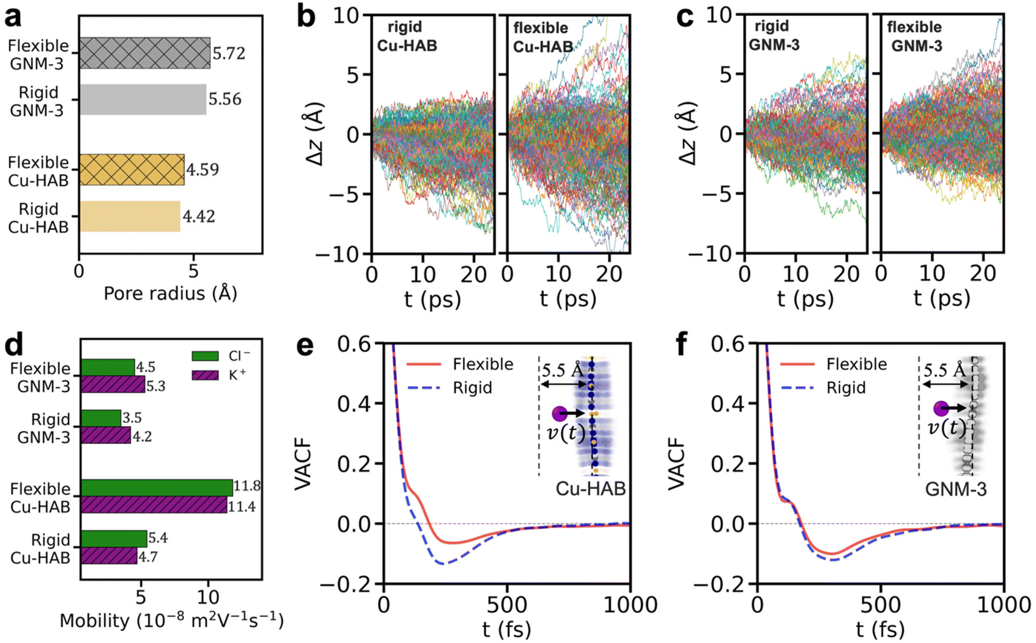 | ||
| Fig. 3 Pore expansion and increase in ion mobility in flexible membranes. a. Pore radius. Trajectories of potassium ions near b. rigid Cu-HAB (left), flexible Cu-HAB (right), c. rigid GNM-3 (left), and flexible GNM-3 (right). The displacement is given by Δz(t) = z(t) − z(t0) for −5.5 Å ≤ z(t0) ≤ 0, where t0 is the reference time. d. Mobility of ions near the membranes. Velocity autocorrelation function (VACF) of potassium ions near e. Cu-HAB and f. GNM-3. VACF is given by 〈ν(t + t0)ν(t0)〉/〈ν(t0)ν(t0)〉 for −5.5 Å ≤ z(t0) ≤ 0. The insets show the “near the membranes” region used in this study. | ||
Membrane fluctuation, arising from membrane flexibility, is another important mechanism that has been shown to increase water transport at a certain frequency.21 To understand the effect of membrane fluctuation on the ion conduction, we analyzed the trajectories of ions near the membranes. Fig. 3b and c show the trajectories of potassium ions near the membranes with respect to time. The potassium ions diffuse more actively in the vicinity of the flexible Cu-HAB membrane compared to the rigid Cu-HAB membrane. For quantitative comparison, ion mobility is calculated using the Einstein–Smoluchowski equation,  , where D is the diffusion coefficient, q is the charge of a particle, kB is the Boltzmann constant, and T is the temperature. Ion mobilities near the 2D flexible membranes (within 5.5 Å from the membrane) are shown in Fig. 3d. Ion mobility near the flexible Cu-HAB is about 117 to 142% greater than near the rigid Cu-HAB. On the other hand, ion mobility increases by about 25 to 29% near flexible GNM-3 compared to its rigid counterpart. On the other hand, the average ion concentration near the flexible membranes decreases slightly compared to the rigid membrane (1% decrease in GNM3 and 4% decrease in Cu-HAB). These indicate that the high ion conductance near flexible membranes is due to the high ion mobility near flexible membranes. The high ion mobility near the flexible membrane conforms with the study by Lyu et al.,42 which states that ion conductivity is higher in a flexible multilayered covalent organic framework compared to that in the rigid counterpart. It is noted that the ion mobility near the rigid membranes was lower compared to bulk mobility (7.6 × 10⁻⁸ m2 V−1 s−1 for potassium ion). However, ion mobility near the flexible Cu-HAB is higher compared to bulk mobility. Several studies have reported ion mobility higher than its bulk value in nanoscale channels/pores. Duan and Majumdar reported that the ion mobility in a 2 nm deep hydrophilic nanochannel is about 250% higher than bulk mobility.53 Ma et al.54 reported that ion mobility in a 3 nm nanopore can be higher or lower compared to bulk mobility, depending on the ion concentration. In addition, studies showing the enhanced diffusion dynamics of fluid molecules confined to wiggling surfaces40 and the longitudinal phonon mode of carbon nanotubes39 supports our observations of improved ion mobility in flexible nanoporous membranes.
To further understand the impact of membrane fluctuations on ion mobility, we analyzed the velocity autocorrelation function (VACF) of ions near the membrane region. Fig. 3e and f show the VACF of ions near the membrane region. For Cu-HAB and GNM-3 membranes, ions experience forward scattering (i.e., positive value of VACF) for the first few hundred femtoseconds, followed by backscattering (i.e., negative value of VACF) for the remainder of time. Fig. 3e shows that the fluctuations of Cu-HAB enhances the forward scattering and reduces backscattering. As a result, the ion mobility is increased. The ion mobility is calculated by the time integration of VACF:  , where ν is the velocity of a particle, t0 is the reference time, and the angle brackets denotes the time and ensemble average. On the other hand, the VACFs for ions near the GNM-3 membranes (Fig. 3f) show that forward scattering is almost the same, and backscattering is slightly reduced in the flexible GNM-3 membrane compared to that in the rigid GNM-3 membrane. This analysis of VACFs indicates that the high ion conduction in fluctuating membranes is due to the dynamic coupling between the fluctuating membranes and nearby ions. This membrane–ion coupling is a surface phenomenon, and this mechanism leads to higher enhancements at low ion concentration where the surface conductance is dominant.26,55 The impact of ion concentration on the enhanced ion conduction in flexible 2D membranes will be further investigated in a future study.
To understand the membrane-dependent increase in conductance, we analyzed how the frequency spectrum associated with membrane fluctuations affects nearby ion mobility. For this, additional MD simulations were run using oscillating membranes at various frequencies ωs (Fig. 4a). In this conceptual study, the membrane oscillates in the out-of-plane direction following a simple sine function, and its average vibrational energy is assumed to be equal to the thermal energy. The VACFs of ions near the oscillating membranes are calculated (Fig. 4b). The oscillation of membrane enhances the forward scattering and reduces the backscattering, similar to what is observed in the flexible Cu-HAB. The degree of that effect (forward scattering enchancement and backscattering reduction) depends on the membrane frequency and is maximized at the membrane frequency of around 200 cm−1. In addition, the ion conductance is considerably improved when the membrane oscillates with frequencies of 200 cm−1 (∼135% increase compared to rigid counterpart). The conductance improvement is almost diminished in frequencies higher than 800 cm−1. We compared the range of frequency where the ion conduction is improved by computing the vibrational density of states (VDOS) for Cu-HAB and GNM-3. The VDOS is given by  . The VDOS for Cu-HAB and GNM-3 (Fig. 4d) show that the Cu-HAB membrane exhibits high vibrational density in frequencies lower than 800 cm−1, where the ion conductance is improved. The GNM-3 membrane exhibits lower vibrational density compared to that exhibited by Cu-HAB in that frequency regime where ion conductance is improved. This explains the membrane-dependent conductance increase between the Cu-HAB and GNM-3 membranes.
 | ||
| Fig. 4 Ion transport through oscillating membranes. a. System for MD simulation. The entire membrane oscillates harmonically with a frequency ωs in the out-of-plane direction. b. The VACF for potassium ions near the membrane for various ωs. c. Ion conductance for various ωs. The gray dashed line represents the conductance in rigid Cu-HAB. d. Vibrational density of states for the flexible Cu-HAB, the flexible GNM-3, and ion–water stretching mode (membrane fluctuation data are obtained from the system described in Fig. 1a). e. Schematic of dehydration near the oscillating membrane. f. Average hydration time near membranes for different vibrational characteristics. | ||
We have identified two distinct frequency regimes in the resonance of a clamped membrane. The wiggling motions of the membrane, with frequencies significantly lower (approximately 0.05 cm−1 for a membrane with Lm = 4.62 nm) than those of hydrated ions, are influenced by membrane size, shape, and tension. However, due to their low frequencies, these motions do not create significant vibrational coupling with nearby hydrated ions. Conversely, the atomic oscillations of the membrane, with frequencies ranging between 5–1000 cm−1, exhibit strong vibrational coupling and are not influenced by the membrane's size.
To further understand the physical origin of the frequency range where conductance is high, we studied the vibrational frequency of hydrated ions. Ion dehydration28,30 is one of the important mechanisms governing ion transport in angstrom-scale. The ion–water stretching mode (Fig. 4e) is the vibrational mode that is related to the ion dehydration. The VDOS of the ion–water stretching modes (see the bottom plot of Fig. 4d) exhibit a maximum at the frequency of around 200–300 cm−1, where the ion conductance is maximum. Moreover, we found that the vibrational match between a membrane and the ion–water stretching mode destabilizes the hydration shell of ions, enhancing ion conduction. The average hydration time τhyd is calculated by integrating the residence time autocorrelation function of water in the first hydration shell. Fig. 4f displays the average hydration time for ions near membranes with different vibrational characteristics. The longest average hydration time is observed near the rigid Cu-HAB, where the atomic fluctuations are suppressed. This implies that ions near the rigid membrane form a relatively stable hydration shell that can hinder ion transport through angstrom-scale pores. The hydration time is low near fluctuating membranes, and the average hydration time decreases as the membrane frequency approaches 200–300 cm−1. This suggests that the ion conduction and ion mobility near the membrane surface are improved due to the destabilized ion hydration when the frequency of the membrane fluctuation matches the ion–water stretching mode. This demonstrates that the type of electrolyte may potentially influence the degree of vibrational coupling and the enhancement of conductance. In terms of the effect of temperature, an increase in the temperature of system can increase the frequency of both the membrane and the hydrated ions. However, such frequency changes can only marginally alter the degree of vibrational coupling. This study expands the knowledge of ion transport by showing that the microscopic fluctuation of a membrane is an important factor in ion conduction across nanoporous membranes.
Conclusion
We observed that ion conductance in a flexible Cu-HAB membrane is approximately 102% higher compared to its rigid counterpart, whereas it is 9% higher in the flexible GNM-3 membrane compared to its rigid counterpart. Both potassium and chloride ions experience faster transport across flexible membranes. The increase in conductance exists irrespective of variations in ion concentration, ion type, and temperature. We observed a marginal pore expansion in flexible membranes, which slight increase in ionic conductance. Ion mobility is about 117% to 142% higher near flexible Cu-HAB and about 25% to 29% higher near flexible GNM-3 than their respective rigid counterparts. We found that the vibrational coupling between the fluctuating membrane and the ion–water stretching mode (200–300 cm−1) destabilizes the ion hydration, which explains the membrane-dependent conductance enhancement between Cu-HAB and GNM-3. Our findings highlight the importance of microscopic fluctuations of 2D membranes in ion conduction. In addition, this phenomenon holds potential for manipulation of ion conduction by engineering the fluctuations of the membrane, which can be applied to various applications such as water purification, molecular separation, and electricity generation.Methods
We ran comprehensive MD simulations for ion transport across nanoporous membranes. As depicted in Fig. 1a, the membrane is immersed in a 1.0 M KCl aqueous solution (we also tested different types of ions and different concentrations for some cases). The edges of a membrane are fixed, and the rest of the membrane is allowed to fluctuate during simulations. For comparison, we also considered cases with rigid membranes, where all the atoms of the membranes are fixed. To generate the electric potential gradient, we applied a uniform external electric field perpendicular to the membrane.56 For all systems, the dimension perpendicular to the membrane is 5.80 nm. Periodic boundary conditions were applied in all directions. We considered two types of 2D membranes: Cu-HAB and GNM-3 (Fig. 1b and c).21 The size of the Cu-HAB membrane is 4.62 nm by 4.00 nm, and the size of the GNM-3 membrane is 5.01 nm by 4.43 nm. Both membranes contain 12 pores in a periodic cell. The electrolyte was modeled using the Coulombic and LJ potentials, and the membranes were modeled using the ReaxFF force field.57 In the case of an oscillating membrane, the entire Cu-HAB membrane oscillates in the direction perpendicular to the membrane following A![[thin space (1/6-em)]](https://www.rsc.org/images/entities/char_2009.gif) sin2πωst, where A is the amplitude, ωs is the frequency, and t is the time. The vibrational energy was assumed to be equal to the thermal energy
sin2πωst, where A is the amplitude, ωs is the frequency, and t is the time. The vibrational energy was assumed to be equal to the thermal energy  , where N is the number of atoms, kB is the Boltzmann constant, and T is the temperature. The flexible simple point charge water model58 was used for water. The potential parameters described by Joung et al.59 were used for ions. ReaxFF potential parameters were adopted from Wood et al.60 for GNM-3 and from Monti et al.61 for Cu-HAB. The membrane–liquid interactions were modeled based on LJ and Coulombic potentials, and the LJ parameters are obtained from the Lorentz–Berthelot mixing rule (LJ parameters from Siu et al.62 for hydrogen, Jorgensen et al.63 for nitrogen, and Heinz et al.64 for copper). The cut-off distance of the LJ potential was 1.2 nm. At the initial stage of the MD simulation, the energy of the system was minimized. The partial charges of the membrane were calculated using the charge equilibration (Qeq) method65 and fixed during the simulation. The long-range Coulombic interactions were calculated using the particle–particle particle–mesh method66 with an accuracy of 10−5. The temperature of the system was maintained using the Nosé–Hoover thermostat at around the target temperature (298 K for most cases and 358 K for a few cases). Furthermore, atomic trajectories were computed in an NVT ensemble with 0.5 fs time interval.67 The system was equilibrated for 1 ns and the data were obtained for 4 ns to calculate ion current. Ion current was calculated by counting the number of ions passing through the pores per unit time, and the mean squared displacement of ions was used to calculate the diffusion coefficient. We utilized the large-scale atomic/molecular massively parallel simulator (LAMMPS)68 for MD simulations and the open visualization tool (OVITO)69 for atomic visualizations.
Author contributions
Y. N. performed simulations, analysis, and wrote the manuscript under the guidance of N. A.Conflicts of interest
The authors declare no competing interests.Acknowledgements
The work on graphene nanomembranes was supported by the Center for Enhanced Nanofluidic Transport (CENT), an Energy Frontier Research Center funded by the U.S. Department of Energy, Office of Science, Basic Energy Sciences (award # DE-SC0019112). All other aspects of this work were supported by the National Science Foundation (NSF) under grants 2140225 and 2137157. The computing power is provided by the Extreme Science and Engineering Discovery Environment (XSEDE) granted by NSF grant no. OCI1053575 and Blue Waters supercomputing center, awarded by the State of Illinois and NSF, OCI-0725070, ACI-1238993.References
- K. Kunzelmann, J. Membr. Biol., 2005, 205, 159–173 CrossRef CAS PubMed.
- A. L. Gotter, M. A. Kaetzel and J. R. Dedman, Comp. Biochem. Physiol., Part A: Mol. Integr. Physiol., 1998, 119, 225–241 CrossRef CAS PubMed.
- K. Xiao, L. Jiang and M. Antonietti, Joule, 2019, 3, 2364–2380 CrossRef CAS.
- M. A. El-Sayed, Acc. Chem. Res., 1992, 25, 279–286 CrossRef CAS.
- S. Y. Noskov, S. Bernèche and B. Roux, Nature, 2004, 431, 830–834 CrossRef CAS PubMed.
- C. Duran, C. H. Thompson, Q. Xiao and H. C. Hartzell, Annu. Rev. Physiol., 2010, 72, 95–121 CrossRef CAS PubMed.
- R. B. Stockbridge, L. Kolmakova-Partensky, T. Shane, A. Koide, S. Koide, C. Miller and S. Newstead, Nature, 2015, 525, 548–551 CrossRef CAS PubMed.
- S. Bernèche and B. Roux, Nature, 2001, 414, 73–77 CrossRef PubMed.
- M. Zhou, J. H. Morais-Cabral, S. Mann and R. MacKinnon, Nature, 2001, 411, 657–661 CrossRef CAS PubMed.
- S.-L. Cai, Y.-B. Zheng, S.-H. Cao, X.-H. Cai and Y.-Q. Li, Chem. Commun., 2016, 52, 12450–12453 RSC.
- D. Basu and E. S. Haswell, Curr. Opin. Plant Biol., 2017, 40, 43–48 CrossRef CAS PubMed.
- A. Anishkin, S. H. Loukin, J. Teng and C. Kung, Proc. Natl. Acad. Sci. U. S. A., 2014, 111, 7898–7905 CrossRef CAS PubMed.
- Y. Feng, W. Zhu, W. Guo and L. Jiang, Adv. Mater., 2017, 29, 1702773 CrossRef PubMed.
- J. Feng, M. Graf, K. Liu, D. Ovchinnikov, D. Dumcenco, M. Heiranian, V. Nandigana, N. R. Aluru, A. Kis and A. Radenovic, Nature, 2016, 536, 197 CrossRef CAS PubMed.
- X. Li, H. Zhang, P. Wang, J. Hou, J. Lu, C. D. Easton, X. Zhang, M. R. Hill, A. W. Thornton, J. Z. Liu, B. D. Freeman, A. J. Hill, L. Jiang and H. Wang, Nat. Commun., 2019, 10, 2490 CrossRef PubMed.
- H. Qi, Z. Li, Y. Tao, W. Zhao, K. Lin, Z. Ni, C. Jin, Y. Zhang, K. Bi and Y. Chen, Nanoscale, 2018, 10, 5350–5357 RSC.
- S. J. Heerema and C. Dekker, Nat. Nanotechnol., 2016, 11, 127–136 CrossRef CAS PubMed.
- K. Liu, J. Feng, A. Kis and A. Radenovic, ACS Nano, 2014, 8, 2504 CrossRef CAS PubMed.
- Z. Cao, V. Liu and A. Barati Farimani, Nano Lett., 2019, 19, 8638–8643 CrossRef CAS PubMed.
- D. Cohen-Tanugi and J. C. Grossman, Nano Lett., 2012, 12, 3602–3608 CrossRef CAS PubMed.
- Y. Noh and N. R. Aluru, Nano Lett., 2022, 22, 419–425 CrossRef CAS PubMed.
- Y. Yang, X. Yang, L. Liang, Y. Gao, H. Cheng, X. Li, M. Zou, R. Ma, Q. Yuan and X. Duan, Science, 2019, 364, 1057–1062 CrossRef CAS PubMed.
- C. Moreno, M. Vilas-Varela, B. Kretz, A. Garcia-Lekue, M. V. Costache, M. Paradinas, M. Panighel, G. Ceballos, S. O. Valenzuela, D. Peña and A. Mugarza, Science, 2018, 360, 199–203 CrossRef CAS PubMed.
- Y. Zhong, B. Cheng, C. Park, A. Ray, S. Brown, F. Mujid, J.-U. Lee, H. Zhou, J. Suh, K.-H. Lee, A. J. Mannix, K. Kang, S. J. Sibener, D. A. Muller and J. Park, Science, 2019, 366, 1379–1384 CrossRef CAS PubMed.
- D. Feng, T. Lei, M. R. Lukatskaya, J. Park, Z. Huang, M. Lee, L. Shaw, S. Chen, A. A. Yakovenko, A. Kulkarni, J. Xiao, K. Fredrickson, J. B. Tok, X. Zou, Y. Cui and Z. Bao, Nat. Energy, 2018, 3, 30–36 CrossRef CAS.
- Y. Noh and N. R. Aluru, ACS Nano, 2020, 14, 10518–10526 CrossRef CAS PubMed.
- M. E. Suk and N. R. Aluru, Ion transport in sub-5 nm graphene nanopores, J. Chem. Phys., 2014, 140, 084707 CrossRef PubMed.
- S. Sahu, M. Di Ventra and M. Zwolak, Nano Lett., 2017, 17, 4719–4724 CrossRef CAS PubMed.
- Z. Li, Y. Qiu, Y. Zhang, M. Yue and Y. Chen, J. Phys. Chem. C, 2019, 123, 15314–15322 CrossRef CAS.
- Z. He, J. Zhou, X. Lu and B. Corry, ACS Nano, 2013, 7, 10148–10157 CrossRef CAS PubMed.
- A. Fang, K. Kroenlein, D. Riccardi and A. Smolyanitsky, Nat. Mater., 2019, 18, 76–81 CrossRef CAS PubMed.
- A. Smolyanitsky, A. Fang, A. F. Kazakov and E. Paulechka, Nanoscale, 2020, 12, 10328–10334 RSC.
- A. Gadaleta, C. Sempere, S. Gravelle, A. Siria, R. Fulcrand, C. Ybert and L. Bocquet, Phys. Fluids, 2014, 26, 012005 CrossRef.
- K. Yazda, K. Bleau, Y. Zhang, X. Capaldi, T. St-Denis, P. Grutter and W. W. Reisner, Nano Lett., 2021, 21, 4152–4159 CrossRef CAS PubMed.
- M. Tsutsui, K. Yokota, I. W. Leong, Y. He and T. Kawai, Cell Rep. Phys. Sci., 2022, 3, 101065 CrossRef CAS.
- K. Sint, B. Wang and P. Král, J. Am. Chem. Soc., 2008, 130, 16448–16449 CrossRef CAS PubMed.
- X. Jiang, C. Zhao, Y. Noh, Y. Xu, Y. Chen, F. Chen, L. Ma, W. Ren, N. R. Aluru and J. Feng, Sci. Adv., 2022, 8, eabj2510 CrossRef CAS PubMed.
- A. Kohen, R. Cannio, S. Bartolucci, J. P. Klinman and J. P. Klinman, Nature, 1999, 399, 496–499 CrossRef CAS PubMed.
- M. Ma, F. Grey, L. Shen, M. Urbakh, S. Wu, J. Z. Liu, Y. Liu and Q. Zheng, Nat. Nanotechnol., 2015, 10, 692–695 CrossRef CAS PubMed.
- S. Marbach, D. S. Dean and L. Bocquet, Nat. Phys., 2018, 14, 1108–1113 Search PubMed.
- Y. Noh and N. R. Aluru, Phys. Rev. E, 2022, 106, 025106 CrossRef CAS PubMed.
- B. Lyu, M. Wang, Z. Jiang and J. Jiang, J. Membr. Sci., 2022, 658, 120754 CrossRef CAS.
- N. Lahiri, N. Lotfizadeh, R. Tsuchikawa, V. V. Deshpande and J. Louie, J. Am. Chem. Soc., 2017, 139, 19–22 CrossRef CAS PubMed.
- J. Bai, X. Zhong, S. Jiang, Y. Huang and X. Duan, Nat. Nanotechnol., 2010, 5, 190–194 CrossRef CAS PubMed.
- G. Tronci, F. Raffone and G. Cicero, Appl. Sci., 2018, 8, 1547 CrossRef.
- S. Zhao, J. Xue and W. Kang, J. Chem. Phys., 2013, 139, 114702 CrossRef PubMed.
- L. E. Kreno, K. Leong, O. K. Farha, M. Allendorf, R. P. Van Duyne and J. T. Hupp, Chem. Rev., 2012, 112, 1105–1125 CrossRef CAS PubMed.
- J. Lee, O. K. Farha, J. Roberts, K. A. Scheidt, S. T. Nguyen and J. T. Hupp, Chem. Soc. Rev., 2009, 38, 1450–1459 RSC.
- H. Wang, Q.-L. Zhu, R. Zou and Q. Xu, Chem, 2017, 2, 52–80 CAS.
- R. M. M. Smeets, U. F. Keyser, D. Krapf, M.-Y. Wu, N. H. Dekker and C. Dekker, Nano Lett., 2006, 6, 89–95 CrossRef CAS PubMed.
- A. Kumar, K.-B. Park, H.-M. Kim and K.-B. Kim, Nanotechnology, 2013, 24, 495503 CrossRef PubMed.
- S. W. Kowalczyk, A. Y. Grosberg, Y. Rabin and C. Dekker, Nanotechnology, 2011, 22, 315101 CrossRef PubMed.
- C. Duan and A. Majumdar, Nat. Nanotechnol., 2010, 5, 848–852 CrossRef CAS PubMed.
- J. Ma, K. Li, Z. Li, Y. Qiu, W. Si, Y. Ge, J. Sha, L. Liu, X. Xie, H. Yi, Z. Ni, D. Li and Y. Chen, J. Am. Chem. Soc., 2019, 141, 4264–4272 CrossRef CAS PubMed.
- D. Stein, M. Kruithof and C. Dekker, Phys. Rev. Lett., 2004, 93, 035901 CrossRef PubMed.
- J. Gumbart, F. Khalili-Araghi, M. Sotomayor and B. Roux, Biochim. Biophys. Acta, Biomembr., 2012, 1818, 294–302 CrossRef CAS PubMed.
- A. C. T. van Duin, S. Dasgupta, F. Lorant and W. A. Goddard, J. Phys. Chem. A, 2001, 105, 9396–9409 CrossRef CAS.
- Y. Wu, H. L. Tepper and G. A. Voth, J. Chem. Phys., 2006, 124, 024503 CrossRef PubMed.
- I. S. Joung and T. E. Cheatham, J. Phys. Chem. B, 208, 112, 9020–9041 CrossRef PubMed.
- M. A. Wood, A. C. T. van Duin and A. Strachan, J. Phys. Chem. A, 2014, 118, 885–895 CrossRef CAS PubMed.
- S. Monti, C. Li and V. Carravetta, J. Phys. Chem. C, 2013, 117, 5221–5228 CrossRef CAS.
- S. W. I. Siu, K. Pluhackova and R. A. Böckmann, J. Chem. Theory Comput., 2012, 8, 1459–1470 CrossRef CAS PubMed.
- W. L. Jorgensen, D. S. Maxwell and J. Tirado-Rives, J. Am. Chem. Soc., 1996, 118, 11225–11236 CrossRef CAS.
- H. Heinz, R. A. Vaia, B. L. Farmer and R. R. Naik, J. Phys. Chem. C, 2008, 112, 17281–17290 CrossRef CAS.
- A. K. Rappe and W. A. Goddard, J. Phys. Chem., 1991, 95, 3358–3363 CrossRef CAS.
- R. W. Hockney and J. W. Eastwood, Computer Simulation Using Particles, McGraw-Hill, New York, 1981 Search PubMed.
- B. D. Jensen, K. E. Wise and G. M. Odegard, J. Comput. Chem., 2015, 36, 1587–1596 CrossRef CAS PubMed.
- S. Plimpton, J. Comput. Phys., 1995, 117, 1–19 CrossRef CAS.
- A. Stukowski, Modell. Simul. Mater. Sci. Eng., 2010, 18, 015012 CrossRef.
| This journal is © The Royal Society of Chemistry 2023 |