Controllable dynamics of complex DNA nanostructures

Wen Wang *ab, Yue Shen ab and Bryan Wei *c
aBGI Research, BGI, Shenzhen 518083, China
bGuangdong Provincial Key Laboratory of Genome Read and Write, Shenzhen 518120, China. E-mail: wangwen4@genomics.cn
cSchool of Life Sciences, Tsinghua University-Peking University Center for Life Sciences, Center for Synthetic and Systems Biology, Tsinghua University, Beijing 100084, China. E-mail: bw@tsinghua.edu.cn

Received 22nd October 2022 , Accepted 26th January 2023

First published on 20th February 2023


Abstract

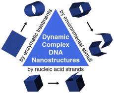
In the past four decades, a variety of self-assembly design frameworks have led to the construction of versatile DNA nanostructures with increasing complexity and controllability. The controllable dynamics of DNA nanostructures has garnered much interest and emerged as a powerful tool for conducting sophisticated tasks at the molecular level. In this minireview, we summarized the controllable reconfigurations of complex DNA nanostructures induced by nucleic acid strands, environmental stimuli and enzymatic treatments. We also envisioned that with the optimization of response time, sensitivity and specificity, dynamic DNA nanostructures have great promise in applications ranging from nanorobotics to life sciences.


Introduction

The predictability and programmability of Watson–Crick base pairing make DNA a superb intelligent material in nanotechnology research for the construction of a large collection of nanoscale structures and devices. With four decades of rapid development, versatile DNA nanostructures with increasing complexity and controllability have been constructed with a variety of creative and disparate self-assembly strategies.1–3 Apart from the complex static nanoarchitectures from expanded design space, reconfigurable and autonomous devices, such as DNA walkers and circuits,4–6 molecular motors,7,8 nanorobots,9 and stimuli-responsive nanostructures10,11 have emerged as functional machines that enable a plethora of sophisticated applications such as cargo sorting,12 drug delivery,13,14 and artificial nanomachinery15–17 at the molecular level. Besides the development to achieve higher structural order and higher construction complexity, controllable dynamics has become one of the hot spots to further advance the capability of rational design in DNA nanotechnology. Complex DNA nanostructures (e.g., DNA origami constructs) are becoming appealing candidates for integrating intricate moving parts and multiple functional modules (e.g., therapeutic modules of high valences). It would be very challenging to realize the same advanced functionalities imbedded in the complex constructs with simpler constructs composed of few strands.

In this minireview, we aim to outline the recent progress of controllable reconfigurations of complex DNA nanostructures. A general strategy to construct dynamic DNA structures is to incorporate reconfigurable elements into static DNA structures. We first summarize the commonly used reconfiguration mechanisms in Fig. 1. The capability of responding to a wide range of environmental changes offered by these reconfigurable elements sets the foundation of controllable dynamics, such as the formation of DNA duplexes, ion-induced conformational changes of special structural motifs, and aptamer reconfigurations upon specific target recognition. Enzymatic reactions such as strand elongation, cleavage, and ligation are other efficient approaches for driving controllable reconfigurations. We next summarize the design and construction of dynamic DNA nanostructures according to the different reconfiguration mechanisms, mainly focusing on the reconfigurations of megadalton complex nanostructures.


image file: d2nr05872c-f1.tif
Fig. 1 Commonly used reconfiguration mechanisms for constructing dynamic DNA structures. (A) Formation of a DNA duplex. (B) Toehold-mediated strand displacement reaction. (C) Stacking transformation of an isomer. (D) Formation of a DNA triplex. (E) Formation of a quadruplex structure. (F) Reconfiguration of an aptamer structure upon specific targeting. (G) Enzymatic elongation. (H) Enzymatic cleavage. (I) Enzymatic ligation.

Reconfigurations induced by nucleic acid strands

Nucleic acid strands are the most frequently used molecular triggers to drive dynamic DNA structural reconfigurations, especially through toehold-mediated strand displacement reactions,18–20 in which one single-stranded DNA (ssDNA) of two partially complementary strands is displaced by another fully complementary strand to form a more stable DNA duplex. The first example of strand displacement based creation and reconfiguration of unique topological structures using a ‘fold-and-cut’ strategy was presented by Yan and colleagues. They demonstrated the reconfiguration of the Möbius strips into programmable topological objects such as supercoiled ring and catenane structures by cutting along specific edges using strand displacement reactions (Fig. 2A).21 Afterwards, Yan's group developed a ‘fold-release-fold’ strategy of releasing and recombining staple strands through strand displacement to transform a simple DNA origami structure into two kinds of more complex and quasi-fractal structures step by step (Fig. 2B).22 Yin and colleagues developed a general framework for complex reconfigurations of single-stranded tile and brick structures. The original two-dimensional (2D) rectangular molecular canvas can be “cut” into diverse prescribed shapes including two full sets of alphabets using DNA strand displacement reactions (Fig. 2C).23 Wei and colleagues demonstrated a reversible global conformational change in an origami with a blocks linked by connecting staples (BLCS) system. Upon the removal of a specific set of tie staples via strand displacement and the addition of another set of tie staples, the structure reconfigured from one conformation to another in a reversible manner (Fig. 2D).24 Ke and colleagues constructed a three-dimensional (3D) reconfigurable plasmonic tripod regulated by tuning the interarm angles of the tripod via strand displacement to control the length of the struts (Fig. 2E).25 Kwon and colleagues demonstrated a reconfigurable DNA accordion rack structure that changes conformation in response to the length of the DNA lock input (Fig. 2F).26 Ke and colleagues reported a modular design strategy for the programmable reconfiguration of the dimensions and curvatures of DNA origami structures. Selected modular units of short double-stranded DNA (dsDNA) with loops can be elongated to long modules with long dsDNA via toehold-mediated strand displacement, which resulted in controllable elongation, bending and twisting of the overall origami structures (Fig. 2G).27
image file: d2nr05872c-f2.tif
Fig. 2 Nucleic acid strand-driven dynamic DNA structures. (A) Reconfiguration of Möbius strips into programmable topological objects.21 (B) Reconfiguration of DNA origami structures into complex and quasi-fractal patterns.22 (C) Complex reconfiguration of DNA nanostructures assembled with single-stranded tiles.23 (D) Reversible global conformational change in a BLCS origami system.24 (E) A 3D reconfigurable plasmonic tripod.25 (F) A reconfigurable DNA accordion rack.26 (G) Reconfiguration of the dimensions and curvatures of DNA origami structures constructed with small modular dynamic units.27 (H) Reconfiguration of a DNA domino array consisting of DNA anti-junction units.28 (I) Reconfigurable domino origami arrays with programmable curvatures.29 (J) A 2D to 3D reconfiguration of nanostructures consisting of anti-junction units.30

In addition to strand displacement reactions, nucleic acid strands can be utilized directly to activate structural reconfigurations. Ke and colleagues showed a stepwise reconfiguration of a DNA domino array consisting of DNA anti-junction units. The addition of a molecular trigger strand initiated the isomerization of the local anti-junction unit and the structural change can transfer to neighboring units in a controllable pathway to achieve a global structural transformation (Fig. 2H).28 Besides this canonical domino array with uniformly sized DNA units, they later adjusted the sizes of the DNA units and introduced a domino array consisting of DNA units with size gradients. After the unit isomerization driven by the molecular trigger strand, the domino array can transform into a curved configuration because of the reoriented DNA helices with different lengths. The degree of curvatures can also be controlled with the design of unit size gradients (Fig. 2I).29 Song and colleagues demonstrated the realization of a controllable reconfiguration of nanostructures consisting of similar anti-junction units from 2D to 3D, in which the 2D nanoarray was modularized into three connected parts and different parts could be independently transformed by their respective trigger DNA strands (Fig. 2J).30

Reconfigurations induced by environmental stimuli

Environmental signals, such as ionic concentrations (e.g., pH value and metal ions), temperature, target molecules, light and electric/magnetic fields, can be used to control the conformational changes of nanostructures with the incorporations of special reconfigurable motifs or aptamers.

pH-responsive reconfiguration of complex DNA nanostructures can be achieved by the implementation of pH-responsive units, such as cytosine-rich i-motifs31 and pH-dependent Hoogsteen triplexes.32 Linko and colleagues presented a DNA origami nanocapsule that is capable of reversible transformation between the closing and opening states controlled by the formation and dissociation of triplex latches via Hoogsteen base pairing in response to environmental pH changes (Fig. 3A).33 Wei and colleagues demonstrated a pH-responsive reconfiguration of a four-arm junction lattice implemented with cytosine-rich angle-controlling struts. When pH dropped from neutral pH (∼7) to acidic pH (∼4.5), the conformational change of the incorporated i-motifs led to the contraction of the angle-controlling struts and resulted in a more-defined lattice shape (Fig. 3B).34


image file: d2nr05872c-f3.tif
Fig. 3 Environmental stimulus-driven dynamic DNA structures. (A) pH-controlled DNA origami nanocapsule.33 (B) pH-responsive reconfiguration of a four-arm junction lattice.34 (C) Dynamic DNA nanodevices based on the changes of the cation concentration and temperature.35 (D) Ion-responsive reconfiguration of a DNA origami nanodevice.36 (E) Potassium-induced reconfiguration of a DNA-origami nanoarm.37 (F) Thermo-responsive actuation of a DNA origami flexor.38 (G) Light-triggered transformation of a DNA origami sphere.39 (H) An aptamer-gated DNA nanorobot.40 (I) A nucleolin-triggered DNA origami nanorobot.41

Changes in the cation concentration and temperature can also be used to trigger structural reconfigurations. Dietz and colleagues developed a strategy using shape-complementarity instead of base pairing to construct dynamic DNA origami structures, such as an actuator, a switchable gear, an unfoldable nanobook, and a nanorobot. They demonstrated that the conformations of the assemblies can be finely controlled by environmental stimuli such as changes in salt concentrations or solution temperatures (Fig. 3C).35 Castro and colleagues presented an ion-responsive reconfiguration strategy based on weak base-paring interactions that respond to environmental ionic conditions. This strategy utilizes short and weakly complementary ssDNA overhangs whose hybridization is sensitive to changes in cation concentrations as hinge arms for a DNA origami structure. This carefully fine-tuned strategy allows for reversible and repeatable structural transformations between the opening and closing states and can be driven by several different kinds of cations (Fig. 3D).36 Murata and colleagues created a DNA origami nanoarm composed of a series of repeated tension adjustable modules, which can undergo a deformation from a linear shape into an arched shape. By employing G-quadruplex-forming sequences as the bridge strands, they successfully achieved a potassium-induced reversible deformation of the DNA origami nanoarm regulated by the contraction/relaxation of G-quadruplex formation/melting (Fig. 3E).37 Baumberg and colleagues demonstrated a thermo-responsive actuation of a DNA origami flexor using the phase transition of a thermo-responsive polymer poly(N-isopropylacrylamide) (PNIPAM) incorporated into the flexor system (Fig. 3F).38

Besides the chemical and physical stimuli mentioned above, light irradiation can also be used to activate the reconfiguration of complex DNA nanostructures. With the incorporation of photo-cleavable ortho-nitrobenzyl spacers into a DNA origami capsule, Han and colleagues demonstrated a structural reconfiguration induced by exposure to light with a specific wavelength (Fig. 3G).39

Target recognition is another commonly used approach for the reconfiguration of dynamic DNA nanostructures and has great potential in smart drug delivery systems. Church and colleagues constructed an aptamer-gated DNA nanorobot for the targeted delivery of molecular payloads to the cell surface. Upon recognizing specific targets, the aptamer “lock” binds to the target instead of the partially complementary strand, resulting in the opening of the DNA origami barrel and the releasing of the loaded cargoes (Fig. 3H).40 Similarly, Zhao and colleagues developed a nucleolin-triggered DNA origami nanorobot for the selective blockage of tumor blood vessels and employed nucleolin-targeting aptamers (AS1411 DNA aptamers) as “lock” strands. The reconfiguration of opening the tubular structure into a planar rectangular structure was triggered by the specific recognition of nucleolin expressed on tumor endothelial cells, followed by the release of encapsulated thrombin (Fig. 3I).41 As shown in Fig. 3H and I, and other examples introduced more recently,42,43 multiple types of modules can be integrated into complex origami devices to achieve different functions simultaneously. Such a design scheme has brought new opportunities in targeted therapeutics.

Reconfigurations induced by enzymatic treatments

Enzymes from the molecular cloning toolbox are excellent candidates for controllable reconfigurations of complex DNA nanostructures. With diverse DNA operability (e.g., gap filling, ligating and cleaving), enzymes such as polymerase, ligase and restriction enzymes can interact with reconfigurable elements in the DNA nanostructures and lead to conformational changes. Schmidt and colleagues developed a DNA polymerase-assisted gap filling method for the structural transformations of wireframe DNA origami structures. After the designed single-stranded gap regions in the DNA origami structures were filled with DNA polymerases, the collapsed structures were transformed into full-size filled structures, and curved tube-like structures were straightened (Fig. 4A).44 Mao and colleagues developed a similar fold-and-fill (F&F) strategy to define the geometry of a DNA origami Chinese knot. The routine path of the Chinese knot was first determined using a small amount of staple strands, and then the single-stranded segments on the scaffold strand were filled with DNA polymerase to achieve the designed structure (Fig. 4B).45 Pan and colleagues showed that DNA origami structures can be trimmed into designer shapes by polymerase-triggered DNA strand displacement reactions. Staples with 3′ end toeholds were removed from the origami structures after treatment with DNA polymerase, which resulted in cutting the original structure into various geometric shapes (Fig. 4C).46 Recently, Wei and colleagues demonstrated a successful structural transformation from a planar lattice into a tubular shape upon ligation treatment, in which the weak base pairings of the short sticky ends were consolidated by DNA ligase (Fig. 4D).47 Wei and colleagues also showcased that DNA nanostructures with allosteric sites can undergo global allosteric transitions from one conformation to another upon different enzymatic processes, such as templated elongation by DNA polymerase, digestion by exonuclease and nick sealing by ligase (Fig. 4E).48 Bellot and colleagues developed an enzyme-sensitive DNA origami ‘nanoactuator’ device, which can be activated by the restriction enzyme BamHI and can then drive the configuration to the open state (Fig. 4F).49 Lin and colleagues presented a CRISPR-Cas12a-catalyzed method to reconfigure DNA origami structures, in which the unpaired scaffold or staple strands of DNA origami structures are targeted and removed by Cas12a and lead to structural transformations. Because Cas12a endonuclease does not require sequence specificity, this method provides a versatile tool for post-processing of DNA origami structures (Fig. 4G).50
image file: d2nr05872c-f4.tif
Fig. 4 Enzymatic treatment-driven dynamic DNA structures. (A) DNA polymerase-assisted transformation of a wireframe DNA origami structure.44 (B) DNA polymerase-assisted formation of a DNA origami Chinese knot.45 (C) Transformation of DNA origami structures into designer shapes by polymerase-triggered DNA strand displacement.46 (D) Transformation from a planar lattice into a tubular shape upon ligation treatment.47 (E) Allosteric transitions of DNA nanostructures upon different enzymatic processes.48 (F) A restriction enzyme-sensitive DNA origami device.49 (G) CRISPR-Cas12a-catalyzed reconfiguration of a DNA origami structure.50

Conclusion and outlook

In this minireview, we summarized the recent progress of controllable reconfigurations of complex DNA nanostructures according to different reconfiguration mechanisms. Elements for controllable dynamics have already been widely demonstrated in the field of DNA nanotechnology. Toehold-mediated strand displacement and some other mechanisms have been applied in a myriad of case studies for simple devices or assembly systems composed of few strands. Micrometer-scale constructs (e.g., ligase engineered DNA crystals51 and DNA tubes52,53) have also been assembled from several species of strands with controllable reconfigurations in response to versatile stimuli such as temperature, ionic strength, redox reagents, enzymes, and nucleic acid strands. As we can see in many examples highlighted in this review, multiple reconfiguration schemes can be utilized in megadalton complex DNA nanostructures for the implementation of intricate dynamics. With their excellent responses to specific stimuli, dynamic DNA nanostructures have great potential in a wide range of applications, such as biological sensing and imaging, smart drug delivery, data storage and encryption,54,55 and molecular robotics. On the other side of the coin, when compared to their counterparts of limited complexity, it is actually more difficult to optimize the desired construction of complex DNA nanostructures, especially for constructs to be further scaled up in size and/or quantity. Extra assembly optimization and purification processes might be necessary to get the products properly functioning for downstream applications. With the rapid development of dynamic DNA nanotechnology, emerging approaches can be optimized to achieve super-fast stimulus response (e.g., real-time), high sensitivity and specificity, and precise reconfiguration movements in series or in parallel, thus providing more application opportunities in life and materials sciences. One can also imagine synthetic constructs to mimic or even rival the sophisticated and precise dynamics of natural macromolecules and complexes.

Conflicts of interest

There are no conflicts to declare.

Acknowledgements

B. W. acknowledges the support of grant no. 2021YFF1200200 from the National Key R&D Program of China. W. W. acknowledges the support of grant no. 32201175 from the National Natural Science Foundation of China.

References

  1. N. C. Seeman and H. F. Sleiman, Nat. Rev. Mater., 2017, 3, 17068 CrossRef.
  2. H. Ramezani and H. Dietz, Nat. Rev. Genet., 2020, 21, 25–26 CrossRef PubMed.
  3. S. Dey, C. Fan, K. V. Gothelf, J. Li, C. Lin, L. Liu, N. Liu, M. A. D. Nijenhuis, B. Saccà, F. C. Simmel, H. Yan and P. Zhan, Nat. Rev. Methods Primers, 2021, 1, 13 CrossRef CAS.
  4. K. Lund, A. J. Manzo, N. Dabby, N. Michelotti, A. Johnson-Buck, J. Nangreave, S. Taylor, R. Pei, M. N. Stojanovic, N. G. Walter, E. Winfree and H. Yan, Nature, 2010, 465, 206–210 CrossRef CAS PubMed.
  5. H. Gu, J. Chao, S. J. Xiao and N. C. Seeman, Nature, 2010, 465, 202–205 CrossRef CAS PubMed.
  6. J. Chao, J. Wang, F. Wang, X. Ouyang, E. Kopperger, H. Liu, Q. Li, J. Shi, L. Wang, J. Hu, L. Wang, W. Huang, F. C. Simmel and C. Fan, Nat. Mater., 2018, 18, 273–279 CrossRef PubMed.
  7. A.-K. Pumm, W. Engelen, E. Kopperger, J. Isensee, M. Vogt, V. Kozina, M. Kube, M. N. Honemann, E. Bertosin, M. Langecker, R. Golestanian, F. C. Simmel and H. Dietz, Nature, 2022, 607, 492–498 CrossRef CAS PubMed.
  8. S. Lauback, K. R. Mattioli, A. E. Marras, M. Armstrong, T. P. Rudibaugh, R. Sooryakumar and C. E. Castro, Nat. Commun., 2018, 9, 1446 CrossRef PubMed.
  9. E. Kopperger, J. List, S. Madhira, F. Rothfischer, D. C. Lamb and F. C. Simmel, Science, 2018, 359, 296–301 CrossRef CAS PubMed.
  10. A. Kuzuya, Y. Sakai, T. Yamazaki, Y. Xu and M. Komiyama, Nat. Commun., 2011, 2, 449 CrossRef PubMed.
  11. P. Petersen, G. Tikhomirov and L. Qian, Nat. Commun., 2018, 9, 5362 CrossRef CAS PubMed.
  12. A. J. Thubagere, W. Li, R. F. Johnson, Z. Chen, S. Doroudi, Y. L. Lee, G. Izatt, S. Wittman, N. Srinivas, D. Woods, E. Winfree and L. Qian, Science, 2017, 357, eaan6558 CrossRef PubMed.
  13. E. S. Andersen, M. Dong, M. M. Nielsen, K. Jahn, R. Subramani, W. Mamdouh, M. M. Golas, B. Sander, H. Stark, C. L. Oliveira, J. S. Pedersen, V. Birkedal, F. Besenbacher, K. V. Gothelf and J. Kjems, Nature, 2009, 459, 73–76 CrossRef CAS PubMed.
  14. G. Grossi, M. Dalgaard Ebbesen Jepsen, J. Kjems and E. S. Andersen, Nat. Commun., 2017, 8, 992 CrossRef PubMed.
  15. A. Kuzyk, R. Schreiber, H. Zhang, A. O. Govorov, T. Liedl and N. Liu, Nat. Mater., 2014, 13, 862–866 CrossRef CAS PubMed.
  16. A. Peil, L. Xin, S. Both, L. Shen, Y. Ke, T. Weiss, P. Zhan and N. Liu, ACS Nano, 2022, 16, 5284–5291 CrossRef CAS PubMed.
  17. L. Liu, Q. Xiong, C. Xie, F. Pincet and C. Lin, Sci. Adv., 2022, 8, eadd1830 CrossRef CAS PubMed.
  18. B. Yurke, A. J. Turberfield, A. P. Mills, F. C. Simmel and J. L. Neumann, Nature, 2000, 406, 605–608 CrossRef CAS PubMed.
  19. D. Y. Zhang and G. Seelig, Nat. Chem., 2011, 3, 103–113 CrossRef CAS PubMed.
  20. F. C. Simmel, B. Yurke and H. R. Singh, Chem. Rev., 2019, 119, 6326–6369 CrossRef CAS PubMed.
  21. D. Han, S. Pal, Y. Liu and H. Yan, Nat. Nanotechnol., 2010, 5, 712–717 CrossRef CAS PubMed.
  22. F. Zhang, J. Nangreave, Y. Liu and H. Yan, Nano Lett., 2012, 12, 3290–3295 CrossRef CAS PubMed.
  23. B. Wei, L. L. Ong, J. Chen, A. S. Jaffe and P. Yin, Angew. Chem., Int. Ed., 2014, 53, 7475–7479 CrossRef CAS PubMed.
  24. Y. Cui, R. Chen, M. Kai, Y. Wang, Y. Mi and B. Wei, ACS Nano, 2017, 11, 8199–8206 CrossRef CAS PubMed.
  25. P. Zhan, P. K. Dutta, P. Wang, G. Song, M. Dai, S. X. Zhao, Z. G. Wang, P. Yin, W. Zhang, B. Ding and Y. Ke, ACS Nano, 2017, 11, 1172–1179 CrossRef CAS PubMed.
  26. Y. Choi, H. Choi, A. C. Lee, H. Lee and S. Kwon, Angew. Chem., Int. Ed., 2018, 57, 2811–2815 CrossRef CAS PubMed.
  27. D. Wang, L. Yu, C. M. Huang, G. Arya, S. Chang and Y. Ke, J. Am. Chem. Soc., 2021, 143, 2256–2263 CrossRef CAS PubMed.
  28. J. Song, Z. Li, P. Wang, T. Meyer, C. Mao and Y. Ke, Science, 2017, 357, eaan3377 CrossRef PubMed.
  29. D. Wang, L. Yu, B. Ji, S. Chang, J. Song and Y. Ke, Nano Lett., 2020, 20, 8236–8241 CrossRef CAS PubMed.
  30. Y. Liu, J. Cheng, S. Fan, H. Ge, T. Luo, L. Tang, B. Ji, C. Zhang, D. Cui, Y. Ke and J. Song, Angew. Chem., Int. Ed., 2020, 59, 23277–23282 CrossRef CAS PubMed.
  31. L. Chen, L. Cai, X. Zhang and A. Rich, Biochemistry, 1994, 33, 13540–13546 CrossRef CAS PubMed.
  32. K. M. Vasquez and P. M. Glazer, Q. Rev. Biophys., 2002, 35, 89–107 CrossRef CAS PubMed.
  33. H. Ijas, I. Hakaste, B. Shen, M. A. Kostiainen and V. Linko, ACS Nano, 2019, 13, 5959–5967 CrossRef CAS PubMed.
  34. W. Wang, C. Chen, S. Vecchioni, T. Zhang, C. Wu, Y. P. Ohayon, R. Sha, N. C. Seeman and B. Wei, Angew. Chem., Int. Ed., 2021, 60, 25781–25786 CrossRef CAS PubMed.
  35. T. Gerling, K. F. Wagenbauer, A. M. Neuner and H. Dietz, Science, 2015, 347, 1446–1452 CrossRef CAS PubMed.
  36. A. E. Marras, Z. Shi, M. G. Lindell, R. A. Patton, C.-M. Huang, L. Zhou, H.-J. Su, G. Arya and C. E. Castro, ACS Nano, 2018, 12, 9484–9494 CrossRef CAS PubMed.
  37. Y. Suzuki, I. Kawamata, K. Mizuno and S. Murata, Angew. Chem., Int. Ed., 2020, 59, 6230–6234 CrossRef CAS PubMed.
  38. V. A. Turek, R. Chikkaraddy, S. Cormier, B. Stockham, T. Ding, U. F. Keyser and J. J. Baumberg, Adv. Funct. Mater., 2018, 28, 1706410 CrossRef.
  39. R. E. Kohman and X. Han, Chem. Commun., 2015, 51, 5747–5750 RSC.
  40. S. M. Douglas, I. Bachelet and G. M. Church, Science, 2012, 335, 831–834 CrossRef CAS PubMed.
  41. S. Li, Q. Jiang, S. Liu, Y. Zhang, Y. Tian, C. Song, J. Wang, Y. Zou, G. J. Anderson, J. Y. Han, Y. Chang, Y. Liu, C. Zhang, L. Chen, G. Zhou, G. Nie, H. Yan, B. Ding and Y. Zhao, Nat. Biotechnol., 2018, 36, 258–264 CrossRef CAS PubMed.
  42. S. Liu, Q. Jiang, X. Zhao, R. Zhao, Y. Wang, Y. Wang, J. Liu, Y. Shang, S. Zhao, T. Wu, Y. Zhang, G. Nie and B. Ding, Nat. Mater., 2021, 20, 421–430 CrossRef CAS PubMed.
  43. Z. Wang, L. Song, Q. Liu, R. Tian, Y. Shang, F. Liu, S. Liu, S. Zhao, Z. Han, J. Sun, Q. Jiang and B. Ding, Angew. Chem., Int. Ed., 2021, 60, 2594–2598 CrossRef CAS PubMed.
  44. N. P. Agarwal, M. Matthies, B. Joffroy and T. L. Schmidt, ACS Nano, 2018, 27, 2546–2553 CrossRef PubMed.
  45. K. He, Z. Li, L. Liu, M. Zheng and C. Mao, ChemBioChem, 2020, 21, 2132–2136 CrossRef CAS PubMed.
  46. K. Chen, F. Xu, Y. Hu, H. Yan and L. Pan, Small, 2022, 18, e2201478 CrossRef PubMed.
  47. T. Bai, J. Zhang, K. Huang, W. Wang, B. Chen, Y. Li, M. Zhao, S. Zhang, C. Zhu, D. Liu and B. Wei, Nucleic Acids Res., 2022, 50, 8392–8398 CrossRef CAS PubMed.
  48. Q. Yan, Y. Wang, J. Shi and B. Wei, Nucleic Acids Res., 2020, 48, 7595–7600 CAS.
  49. Y. Ke, T. Meyer, W. M. Shih and G. Bellot, Nat. Commun., 2016, 7, 10935 CrossRef CAS PubMed.
  50. Q. Xiong, C. Xie, Z. Zhang, L. Liu, J. T. Powell, Q. Shen and C. Lin, Angew. Chem., Int. Ed., 2020, 59, 3956–3960 CrossRef CAS PubMed.
  51. M. Zheng, Z. Li, C. Zhang, N. C. Seeman and C. Mao, Adv. Mater., 2022, 34, e2200441 CrossRef PubMed.
  52. L. N. Green, A. Amodio, H. K. K. Subramanian, F. Ricci and E. Franco, Nano Lett., 2017, 17, 7283–7288 CrossRef CAS PubMed.
  53. L. N. Green, H. K. K. Subramanian, V. Mardanlou, J. Kim, R. F. Hariadi and E. Franco, Nat. Chem., 2019, 11, 510–520 CrossRef CAS PubMed.
  54. Y. Zhang, F. Wang, J. Chao, M. Xie, H. Liu, M. Pan, E. Kopperger, X. Liu, Q. Li, J. Shi, L. Wang, J. Hu, L. Wang, F. C. Simmel and C. Fan, Nat. Commun., 2019, 10, 5469 CrossRef PubMed.
  55. S. Fan, D. Wang, J. Cheng, Y. Liu, T. Luo, D. Cui, Y. Ke and J. Song, Angew. Chem., Int. Ed., 2020, 59, 12991–12997 CrossRef CAS PubMed.

This journal is © The Royal Society of Chemistry 2023