 Open Access Article
Creative Commons Attribution 3.0 Unported Licence
Identification of macrocyclic peptides which activate bacterial cylindrical proteases†
Raoul
Walther
 a,
Linda M.
Westermann
b,
Sheiliza
Carmali
 c,
Sophie E.
Jackson
a,
Heike
Brötz-Oesterhelt
 *bd and
David R.
Spring
 *a
*a
aYusuf Hamied Department of Chemistry, University of Cambridge, Lensfield Road, CB2 1EW Cambridge, UK. E-mail: spring@ch.cam.ac.uk
bInterfaculty Institute of Microbiology and Infection Medicine, Dept. of Bioactive Compounds, University of Tübingen, Auf der Morgenstelle 28, 72076 Tübingen, Germany. E-mail: heike.broetz-oesterhelt@uni-tuebingen.de
cSchool of Pharmacy, Queen's University Belfast, BT9 7BL Belfast, UK
dCluster of Excellence Controlling Microbes to Fight Infections, Germany
First published on 17th May 2023
Abstract
The caseinolytic protease complex ClpXP is an important house-keeping enzyme in prokaryotes charged with the removal and degradation of misfolded and aggregated proteins and performing regulatory proteolysis. Dysregulation of its function, particularly by inhibition or allosteric activation of the proteolytic core ClpP, has proven to be a promising strategy to reduce virulence and eradicate persistent bacterial infections. Here, we report a rational drug-design approach to identify macrocyclic peptides which increase proteolysis by ClpP. This work expands the understanding of ClpP dynamics and sheds light on the conformational control exerted by its binding partner, the chaperone ClpX, by means of a chemical approach. The identified macrocyclic peptide ligands may, in the future, serve as a starting point for the development of ClpP activators for antibacterial applications.
Introduction
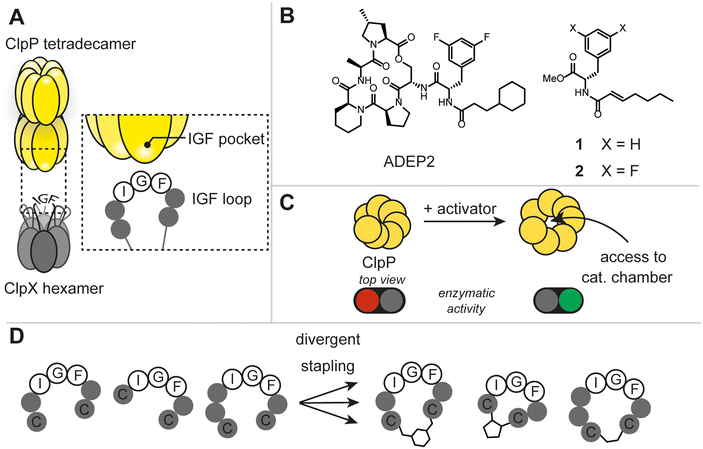
The caseinolytic protease complex (ClpXP), comprising the tetradecameric proteolytic core (ClpP) and a hexameric Clp-ATPase, ClpX, has been subject to intensive research due to its fundamental role in bacterial proteostasis, stress response and other regulatory processes (Fig. 1A).1 Despite this, rational target-selection programs in antibacterial discovery did not single out ClpXP as a promising target, presumably, because it is not essential for survival in the majority of prokaryotes.1c It was not until the discovery of the natural product-derived acyldepsipeptide antibiotics (ADEP) that ClpXP became a promising antibacterial target (Fig. 1B).2 Mode of action studies revealed that ADEP binds tightly to ClpP and inhibits its interaction with ClpX. Intriguingly, as opposed to most antibiotics used in the clinic, ADEP is not an inhibitor of enzymatic function, but instead allosterically increases enzymatic activity of ClpP and causes uncontrolled protein degradation (Fig. 1C). Mutational analyses have shown that the essential binding epitope of ClpX to ClpP is the tripeptide ‘I/LGF’.3 Prior to high resolution cryo-EM structures of ClpXP, ADEP served as a tool mimicking the binding of the ClpX tripeptide ‘IGF’ and shed light on the induced dynamics and structural consequences on ClpP.1a,b To date, ClpX-induced structural and mechanistic consequences continue to be a debated topic. For instance, in the three ClpXP cryo-EM studies published to date, substantial differences in axial pore diameter were observed.4 Thus, we envisaged that increasing the arsenal of more ‘ClpX-like’ ligands could contribute to the discussion and clarify some of the unknowns. This work aims at answering 1) whether the rational design of peptidomimetic macrocycles bearing the IGF binding epitope through a two-component stapling approach is feasible, 2) whether a focused library can deliver ligands against ClpP, and 3) whether binding results in any regulatory control of the protease.Results and discussion
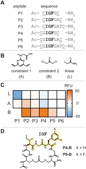
Due to the lack of prior known peptide-based low molecular weight ligands to ClpP (other than ADEP), we opted for a diversity-oriented stapling strategy5 varying the linear peptide sequence and the two-component staples for macrocyclization (Fig. 1D). For macrocyclization, we selected a two-component peptide stapling strategy.6 Specifically, we used a cysteine bis-alkylation method,7 due to its straightforward synthetic implementation and potential for further chemical functionalization, such as fluorescent tags for microscopy or cell-penetrating tags.8 Considering the structural knowledge available, we kept the evolutionary conserved tripeptide binding epitope ‘IGF’ of ClpX constant and varied the number of flanking amino acids based on the natural epitope from Escherichia coli (E. coli).As a starting point, we chose six peptide sequences (P1–P6, Fig. 2A) bearing C- and N-terminal cysteine residues for ring closure via alkylation. Linear peptide precursors P1–P6 were prepared by conventional Fmoc-based solid-phase peptide synthesis (see ESI† for detailed experimental protocols). Peptides were synthesized using rink-amide resin resulting in C-terminal amides and the N-terminus was capped with acetic anhydride. The staples were varied with respect to introduced carbon chain length (three, five, and seven) to explore the impact of macrocycle size and rigidity on binding, namely 1,3 dichloroacetone (B),7b 1,3-bis(bromomethyl)benzene,‡7c and 4,6-divinylpyrimidine (A),7a respectively (Fig. 2B, see ESI† for detailed experimental protocols).
Next, we screened for enzymatic activation of ClpP to quickly assess the chemical library for binding and understand whether IGF-bearing macrocyclic peptides could modulate ClpP enzymatic function. Modulation of ClpP was assessed using a biochemical assay monitoring the enzymatic function of the protease based on previously reported successes for the identification of allosteric activators of ClpP.9 In the absence of an allosteric ligand, such as ADEP, the apical pore of ClpP is constricted, only allowing small peptides to enter the catalytic chamber. In contrast, ADEP binding to the apical hydrophobic pocket results in apical pore opening to a diameter of ∼20 Å, allowing access of unstructured proteins to the catalytic chamber of the protease (Fig. 1C). FITC-casein, a protein labeled with fluorescein to the extent of self-quenching, is a suitable substrate for this assay, as it can only enter the proteolytic core upon apical pore opening and is indirectly a measure for the conformational state of the protease.2,10 Enzymatic degradation of FITC-casein alleviates self-quenching giving rise to an increased fluorescence intensity and an easy readout for our purpose to identify any ligand that positively modulates the enzymatic activity of recombinant E. coli ClpP (Ec.ClpP).
Ec.ClpP was incubated for 24 h at 37 °C in the presence of a peptide library, or vehicle (DMSO) and fluorescence intensity was measured thereafter (Fig. 2C and S1†). All linear peptide controls (P1–P6-L) showed no increase in fluorescence over the negative control. The macrocycles based on stapling with A showed only marginal increase in fluorescence and this indicates that a 7-atom staple is too large. Constriction of the linear peptide sequences with the smaller staple B showed an impact on the overall fluorescence intensity, with P2-B and P4-B (Fig. 2D) demonstrating a significant increase of fluorescence intensity over the DMSO-treated sample. This was reproducibly confirmed in three independent experiments (varying the batch of enzyme). Further evaluation of P2-B revealed that the presence or absence of Ec.ClpP did not significantly change the fluorescence signal and that the basal fluorescence response was significantly higher than the DMSO control, rendering it a false positive hit (Fig. S2†). Fortunately, P4-B did not exhibit this behaviour and was used in all further experiments. The initial focused library screen illustrated that peptide linear sequence, chemical constraint, and macrocycle size play an intricate role in selecting an IGF conformation which activates Ec.ClpP function.
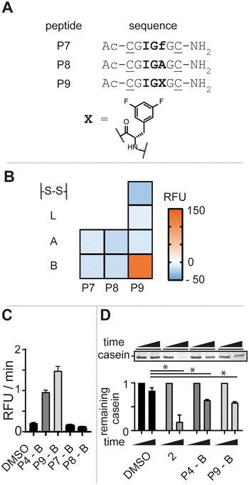
To further gauge the sequence specificity of the tripeptide epitope IGF and rule out unspecific binding, we synthesized peptides P7 and P8 substituting L-phenylalanine for its enantiomer D-phenylalanine (f) and L-alanine (A), respectively (Fig. 3A, see ESI† for detailed experimental protocols). Again, the isolated macrocycles were subjected to our enzymatic assay and the negative control peptide macrocycles showed no stimulation of enzymatic activity (Fig. 3B). The requirement of the natural configuration and sequence of the IGF loop and of the ADEP Phe linker for activation supported the notion that the increase in fluorescence was a direct consequence of P4-B binding to ClpP and inducing a conformational change reminiscent of ADEP2 and other activators.9b,11
ADEP structure–activity studies revealed that the introduction of two fluoro-substituents on the phenylalanine residue markedly increased the bioactivity against Staphylococcus aureus (S. aureus).12 We thus opted to synthesize bis-fluorinated macrocyclic analogue to improve the activity of the lead macrocycle (see ESI† for detailed experimental protocols). Enzymatic activity was not observed for any of the synthesized analogues (P9-A, P9-S-S, or P9-L) other than the macrocycle with constraint B, P9-B, in agreement with our previous studies (Fig. 3B). P9-B exhibited a 1.5-fold improved activity over P4-B at equimolar concentrations, whereas the control macrocyclic peptides P7-B and P8-B were devoid of activity (Fig. 3C). Further validation of the P4/9-B-dependent control of Ec.ClpP function was confirmed by SDS-PAGE analysis of beta-casein digestion (Fig. 3D).§
Given the high sequence conservation of the tripeptide epitope of ClpX across bacterial phyla,3 we were interested in understanding whether P4-B and P9-B could activate other homologues of ClpP. To this end, we expressed and isolated S. aureus ClpP (Sa.ClpP), one of the most relevant pathogens where ClpP activators have shown clinical potential.2,13
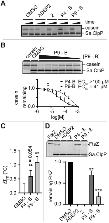
We monitored the digestion of beta-casein by Sa.ClpP in the presence of ADEP2, 2 (Fig. 1B), P4-B, and P9-B (Fig. 2D). P4-B and P9-B were able to activate the enzymatic function of Sa.ClpP, indicating binding to other ClpP homologues (Fig. 4A). This observation was dose-dependent with an observed effective dose (EC50) of 41 μM for P9-B and >100 μM for P4-B (Fig. 4B). Binding was further validated by differential scanning fluorimetry and a mild stabilization of Sa.ClpP was observed in presence of P4-B and P9-B with an apparent increase in melting temperature of 0.6 ± 0.2 and 1.1 ± 0.2 °C, respectively (Fig. 4C and S3†).
Prior to high-resolution cryo-EM structures of ClpXP, much of the ClpX induced allostery has been deduced from ADEP binding to ClpP. Key structural features induced by ADEP are the observed axial pore opening and the selection for the extended state of ClpP aligning the catalytic triad in its active geometry. Additionally, ADEP assembles oligomeric forms of ClpP to its active tetradecamer. The enzymatic digestion experiments strongly suggest that the identified macrocycles induce proteolysis in a concentration-dependent manner. Now, we set out to answer whether P4/9-B are able to stabilize the tetradecameric form of Sa.ClpP. We artificially destabilized tetradecameric Sa.ClpP by changing the buffer from a compatible HEPES (pH 7.0) to an inactivating Tris-HCl buffer (pH 7.6). We speculate that the destabilization is due to a change in pH.14 The oligomeric state was monitored by gel-filtration experiments. ADEP2 and 2 (ref. 15) were able to retain the tetradecamer in Tris-HCl, unlike P4/9-B (Fig. S4†). This experiment demonstrates that ADEP-based high affinity ligands are able to stabilize oligomeric state of ClpP, whereas our ‘ClpX’-like ligands did not show the same stabilization, presumably due to their lower intrinsic affinity¶ and require much higher concentrations (due to solubility issues of the macrocycles, concentrations exceeding 0.2 mM were not attempted). ClpX overcomes this obstacle by means of avidity and ClpX mutants lacking one or two ‘IGF’-loops show compromised association and dissociation kinetics to ClpP.16
Lastly, we set out to answer to what extent ADEP and other allosteric activators of ClpP resemble the activation process of ClpX. To this end, we set up a biochemical assay and monitored the digestion of FtsZ, an essential protein for cell division, which is the major determinant for the filamentation phenotype induced by ADEP-activated ClpP.17 Similarly to the digestion of beta-casein, ADEP2 and 2-treated samples activated Sa.ClpP, and FtsZ was digested. Incubation of P4-B and P9-B with ClpP also induced the digestion of FtsZ, yet less efficiently (Fig. 4D). This experiment is in agreement with the enzymatic digestion experiment of beta-casein and suggests that the structural and dynamic consequences of our IGF epitopes on ClpP are not substrate dependent, but are a result of the presence of the identified macrocycle, and strongly support a mechanism of axial pore opening. We performed docking studies of the identified macrocycles into the same pocket where ADEP binds (see ESI† for full details). P9-B showed an improved docking score over P4-B, in agreement with our experimental data (Table S4†).
The identified macrocyclic peptides require higher concentrations and longer incubation times than the high affinity ligands ADEP2 and 2. Nevertheless, the experiments illustrate that mimicking the IGF-loops of ClpX without its controlling elements results in proteolytically active ClpP and this holds promise for the development of macrocyclic peptide activators of ClpP. Of note, at this stage we cannot rule out that our identified macrocycles bind at another site than the suggested allosteric site of the IGF binding loop of ClpX. There is precedence where dipeptides and the protease inhibitor bortezomib are able to increase enzymatic function of ClpP homologues through another mechanism.11
Conclusions
In summary, we succeeded in identifying macrocyclic peptide ligands against ClpP through a diversity-oriented stapling approach. Our biochemical analysis strongly suggests that the identified lead macrocyclic peptides (P4-B and P9-B) containing the IGF binding epitope induce enzymatic activity. The current study is the first report on successfully targeting the ClpP hydrophobic pocket by a synthetic macrocyclic peptide and lays a foundation for the rational design of ‘ClpX-like’ macrocyclic peptide activators of ClpP.Author contributions
Project was conceptualized by RW. Experiments were designed and performed by RW. Sa.ClpP was isolated by RW and LMW. RW and SC designed docking experiment, SC performed and interpreted data. SEJ, HBO and DRS supervised the project. All authors contributed to data interpretation and manuscript writing.Conflicts of interest
There are no conflicts to declare.Acknowledgements
Drs. Tim Schober, Francisco Javier Pérez-Areales, Soňa Krajčovičová are acknowledged for valuable discussions during the preparation of this manuscript. Jack Barber and Eva Přáda Brichtová are greatly acknowledged for assistance in the isolation and purification of Ec.ClpP. RW was supported by a postdoctoral fellowship by the Independent Research Fund Denmark (grant – 0170-00001B). The Spring group research was supported by grants from the Engineering and Physical Sciences Research Council, the Biotechnology and Biological Sciences Research Council and Medical Research Council. This work was funded in part by UKRI grants. HBO gratefully acknowledges the Deutsche Forschungsgemeinschaft (German Research Foundation, DFG) for funding by the TRR 261, project-ID 398967434, and infrastructural support by the Cluster of Excellence EXC 2124: Controlling Microbes to Fight Infection (CMFI, project ID 390838134). For the purpose of Open Access, the author has applied a CC BY public copyright license to any Author Accepted Manuscript (AAM) version arising.Notes and references
- (a) M. F. Mabanglo and W. A. Houry, J. Biol. Chem., 2022, 298, 101781 CrossRef CAS PubMed; (b) I. T. Malik and H. Brötz-Oesterhelt, Nat. Prod. Rep., 2017, 34, 815–831 RSC; (c) E. Culp and G. D. Wright, J. Antibiot., 2017, 70, 366–377 CrossRef CAS PubMed.
- H. Brötz-Oesterhelt, D. Beyer, H.-P. Kroll, R. Endermann, C. Ladel, W. Schroeder, B. Hinzen, S. Raddatz, H. Paulsen, K. Henninger, J. E. Bandow, H.-G. Sahl and H. Labischinski, Nat. Med., 2005, 11, 1082–1087 CrossRef PubMed.
- Y.-I. Kim, I. Levchenko, K. Fraczkowska, R. V. Woodruff, R. T. Sauer and T. A. Baker, Nat. Struct. Biol., 2001, 8, 230–233 CrossRef CAS PubMed.
- (a) F. T. F. Tsai and C. P. Hill, eLife, 2020, 9, e56501 CrossRef PubMed; (b) C. Gatsogiannis, D. Balogh, F. Merino, S. A. Sieber and S. Raunser, Nat. Struct. Mol. Biol., 2019, 26, 946–954 CrossRef CAS PubMed; (c) X. Fei, T. A. Bell, S. Jenni, B. M. Stinson, T. A. Baker, S. C. Harrison and R. T. Sauer, eLife, 2020, 9, e52774 CrossRef CAS PubMed; (d) Z. A. Ripstein, S. Vahidi, W. A. Houry, J. L. Rubinstein and L. E. Kay, eLife, 2020, 9, e52158 CrossRef CAS PubMed.
- For recent selected examples see: (a) L. Peraro, Z. Zou, K. M. Makwana, A. E. Cummings, H. L. Ball, H. Yu, Y.-S. Lin, B. Levine and J. A. Kritzer, J. Am. Chem. Soc., 2017, 139, 7792–7802 CrossRef CAS PubMed; (b) S. M. Guéret, S. Thavam, R. J. Carbajo, M. Potowski, N. Larsson, G. Dahl, A. Dellsén, T. N. Grossmann, A. T. Plowright, E. Valeur, M. Lemurell and H. Waldmann, J. Am. Chem. Soc., 2020, 142, 4904–4915 CrossRef PubMed; (c) J. E. Montgomery, J. A. Donnelly, S. W. Fanning, T. E. Speltz, X. Shangguan, J. S. Coukos, G. L. Greene and R. E. Moellering, J. Am. Chem. Soc., 2019, 141, 16374–16381 CrossRef CAS PubMed; (d) M. G. Ricardo, A. M. Ali, J. Plewka, E. Surmiak, B. Labuzek, C. G. Neochoritis, J. Atmaj, L. Skalniak, R. Zhang, T. A. Holak, M. Groves, D. G. Rivera and A. Dömling, Angew. Chem., 2020, 59, 5235–5241 CrossRef CAS PubMed; (e) M. G. Ricardo, D. Llanes, L. A. Wessjohann and D. G. Rivera, Angew. Chem., Int. Ed., 2019, 58, 2700–2704 CrossRef CAS PubMed; (f) Y. Wu, J. Williams, E. D. D. Calder and L. J. Walport, RSC Chem. Biol., 2021, 2, 151–165 RSC.
- (a) J. Iegre, N. S. Ahmed, J. S. Gaynord, Y. Wu, K. M. Herlihy, Y. S. Tan, M. E. Lopes-Pires, R. Jha, Y. H. Lau, H. F. Sore, C. Verma, D. H. O'Donovan, N. Pugh and D. R. Spring, Chem. Sci., 2018, 9, 4638–4643 RSC; (b) J. Iegre, P. Brear, D. J. Baker, Y. S. Tan, E. L. Atkinson, H. F. Sore, D. H. O'Donovan, C. S. Verma, M. Hyvönen and D. R. Spring, Chem. Sci., 2019, 10, 5056–5063 RSC; (c) Y. Wu, F. Villa, J. Maman, Y. H. Lau, L. Dobnikar, A. C. Simon, K. Labib, D. R. Spring and L. Pellegrini, Angew. Chem., Int. Ed., 2017, 56, 12866–12872 CrossRef CAS PubMed.
- For recent selected examples see: (a) N. S. Robertson, S. J. Walsh, E. Fowler, M. Yoshida, S. M. Rowe, Y. Wu, H. F. Sore, J. S. Parker and D. R. Spring, Chem. Commun., 2019, 55, 9499–9502 RSC; (b) N. Assem, D. J. Ferreira, D. W. Wolan and P. E. Dawson, Angew. Chem., Int. Ed., 2015, 54, 8665–8668 CrossRef CAS PubMed; (c) H. Jo, N. Meinhardt, Y. Wu, S. Kulkarni, X. Hu, K. E. Low, P. L. Davies, W. F. DeGrado and D. C. Greenbaum, J. Am. Chem. Soc., 2012, 134, 17704–17713 CrossRef CAS PubMed; (d) P. T. Hart, P. Hommen, A. Noisier, A. Krzyzanowski, D. Schüler, A. T. Porfetye, M. Akbarzadeh, I. R. Vetter, H. Adihou and H. Waldmann, Angew. Chem., Int. Ed., 2021, 60, 1813–1820 CrossRef PubMed; (e) M. Wendt, R. Bellavita, A. Gerber, N.-L. Efrém, T. van Ramshorst, N. M. Pearce, P. R. J. Davey, I. Everard, M. Vazquez-Chantada, E. Chiarparin, P. Grieco, S. Hennig and T. N. Grossmann, Angew. Chem., Int. Ed., 2021, 60, 13937–13944 CrossRef CAS PubMed.
- (a) J. Iegre, J. S. Gaynord, N. S. Robertson, H. F. Sore, M. Hyvönen and D. R. Spring, Adv. Ther., 2018, 1, 1800052 CrossRef; (b) L. Reguera and D. G. Rivera, Chem. Rev., 2019, 119, 9836–9860 CrossRef CAS PubMed.
- (a) N. P. Lavey, J. A. Coker, E. A. Ruben and A. S. Duerfeldt, J. Nat. Prod., 2016, 79, 1193–1197 CrossRef CAS PubMed; (b) E. Leung, A. Datti, M. Cossette, J. Goodreid, S. E. McCaw, M. Mah, A. Nakhamchik, K. Ogata, M. El Bakkouri, Y.-Q. Cheng, S. J. Wodak, B. T. Eger, E. F. Pai, J. Liu, S. Gray-Owen, R. A. Batey and W. A. Houry, Chem. Biol., 2011, 18, 1167–1178 CrossRef CAS PubMed.
- M. W. Thompson, S. K. Singh and M. R. Maurizi, J. Biol. Chem., 1994, 269, 18209–18215 CrossRef CAS PubMed.
- (a) J. Felix, K. Weinhäupl, C. Chipot, F. Dehez, A. Hessel, D. F. Gauto, C. Morlot, O. Abian, I. Gutsche, A. Velazquez-Campoy, P. Schanda and H. Fraga, Sci. Adv., 2019, 5, eaaw3818 CrossRef CAS PubMed; (b) T. Akopian, O. Kandror, R. M. Raju, M. UnniKrishnan, E. J. Rubin and A. L. Goldberg, EMBO J., 2012, 31, 1529–1541 CrossRef CAS PubMed.
- (a) B. Hinzen, S. Raddatz, H. Paulsen, T. Lampe, A. Schumacher, D. Häbich, V. Hellwig, J. Benet-Buchholz, R. Endermann, H. Labischinski and H. Brötz-Oesterhelt, ChemMedChem, 2006, 1, 689–693 CrossRef CAS PubMed , for further optimization of the ADEP core; (b) D. W. Carney, K. R. Schmitz, J. V. Truong, R. T. Sauer and J. K. Sello, J. Am. Chem. Soc., 2014, 136, 1922–1929 CrossRef CAS PubMed.
- B. P. Conlon, E. S. Nakayasu, L. E. Fleck, M. D. LaFleur, V. M. Isabella, K. Coleman, S. N. Leonard, R. D. Smith, J. N. Adkins and K. Lewis, Nature, 2013, 503, 365–370 CrossRef CAS PubMed.
- I. T. Malik, J. D. Hegemann and H. Brötz-Oesterhelt, Int. J. Mol. Sci., 2022, 23, 465 CrossRef CAS PubMed.
- D. W. Carney, C. L. Compton, K. R. Schmitz, J. P. Stevens, R. T. Sauer and J. K. Sello, ChemBioChem, 2014, 15, 2216–2220 CrossRef CAS PubMed.
- A. J. Amor, K. R. Schmitz, T. A. Baker and R. T. Sauer, Protein Sci., 2019, 28, 756–765 CrossRef CAS PubMed.
- P. Sass, M. Josten, K. Famulla, G. Schiffer, H.-G. Sahl, L. Hamoen and H. Brötz-Oesterhelt, Proc. Natl. Acad. Sci. U. S. A., 2011, 108, 17474–17479 CrossRef CAS PubMed.
Footnotes |
| † Electronic supplementary information (ESI) available. See DOI: https://doi.org/10.1039/d3md00136a |
| ‡ Initial stapling attempts resulted in poor aqueous solubility of the resulting macrocycles and concomitant difficulties during purification. Thus, this sequence of macrocycles was abandoned. |
| § A positive control 2 (ref. 15) (Fig. 1B) was included in the assay. |
| ¶ Affinity refers in this context to the estimated EC50 for casein degradation not to the binding constant. |
| This journal is © The Royal Society of Chemistry 2023 |


