 Open Access Article
Creative Commons Attribution 3.0 Unported Licence
Correction: Long-chain polyunsaturated fatty acids and extensively hydrolyzed casein-induced browning in a Ucp-1 reporter mouse model of obesity
Liufeng
Mao
ab,
Jiwen
Lei
ab,
Marieke H.
Schoemaker
c,
Bingxiu
Ma
d,
Yan
Zhong
e,
Tim T.
Lambers
c,
Eric A. F.
Van Tol
c,
Yulai
Zhou
d,
Tao
Nie
*abf and
Donghai
Wu
*gh
aCAS Key Laboratory of Regenerative Biology, Joint School of Life Sciences, Guangzhou Medical University, Guangzhou 511436, China
bGuangzhou Institutes of Biomedicine and Health, Chinese Academy of Sciences, Guangzhou 510530, China
cMead Johnson Pediatric Nutrition Institute, Global R&D, Middenkampweg 2, 6545 CJ, Nijmegen, The Netherlands
dSchool of Pharmaceutical Sciences, Jilin University, Changchun, 130012, Jilin, China
eMead Johnson Pediatric Nutrition Institute, Global R&D, 15th floor, East building of New Hualian Mansion, No. 755 Middle Huaihai Road, Shanghai 200020, China
fCentral Laboratory of the First Affiliated Hospital of Jinan University, Guangzhou 510630, China. E-mail: nietaoly@gmail.com; Fax: +86-020-32015297; Tel: +86-020-32015297
gCAS Key Laboratory of Regenerative Biology, Joint School of Life Sciences, Guangzhou Institutes of Biomedicine and Health, Chinese Academy of Sciences, Guangzhou 510530, China
hGuangzhou Medical University, Guangzhou 511436, China. E-mail: wu_donghai@gibh.ac.cn; Fax: +86-020-32015250; Tel: +86-020-32015250
First published on 23rd October 2023
Abstract
Correction for ‘Long-chain polyunsaturated fatty acids and extensively hydrolyzed casein-induced browning in a Ucp-1 reporter mouse model of obesity’ by Liufeng Mao et al., Food Funct., 2018, 9, 2362–2373, https://doi.org/10.1039/C7FO01835E.
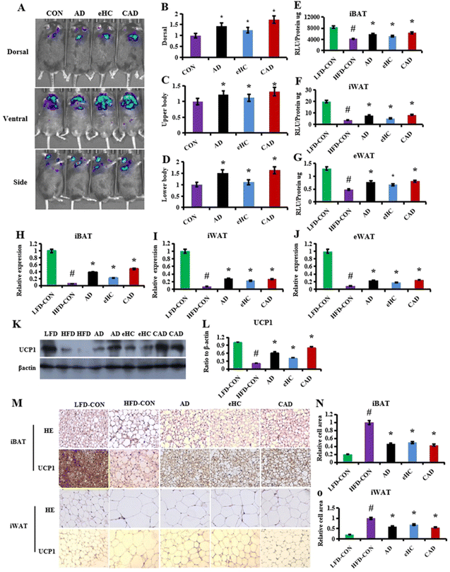
The authors regret that there was an error in Fig. 3M where some images were duplicated. The corrected Fig. 3 is shown below.
The Royal Society of Chemistry apologises for these errors and any consequent inconvenience to authors and readers.
| This journal is © The Royal Society of Chemistry 2023 |

