Current status and prospects of MOFs in controlled delivery of Pt anticancer drugs

Jinyi Chen ab, Zhixin Zhang b, Jiaxin Ma b, Alireza Nezamzadeh-Ejhieh c, Chengyu Lu b, Ying Pan *ab, Jianqiang Liu *abd and Zhi Bai *a
aThe First Dongguan Affiliated Hospital, Guangdong Medical University, Dongguan, 523808, China. E-mail: pangying@gdmu.edu.cn; weidayou100@163.com
bGuangdong Provincial Key Laboratory of Research and Development of Natural Drugs, and School of Pharmacy, Guangdong Medical University, Guangdong Medical University Key Laboratory of Research and Development of New Medical Materials, Dongguan, 523808, China. E-mail: jianqiangliu2010@126.com
cChemistry Department, Shahreza Branch, Islamic Azad University, Shahreza, Isfahan, Iran
dAffiliated Hospital of Guangdong Medical University, Zhanjiang, Guangdong, China

Received 9th February 2023 , Accepted 24th March 2023

First published on 24th March 2023


Abstract

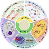
Cancer has become the second leading reason for death in the world. Still, cancer therapy development is exceptionally challenging because the tumor microenvironment is very complex, and individual tumors are very different. In recent years, researchers have found that platinum-based drugs in the form of metal complexes can effectively solve tumor resistance. In this regard, metal–organic frameworks (MOFs) as suitable carriers with high porosity are also exceptional in the biomedical field. Therefore, this article reviews the application of platinum as an anticancer drug and the composite anticancer properties of platinum and MOF materials and prospects for its future development, which provides a new direction for further research in the biomedical field.


1. Introduction

With their unique anti-cancer mechanism and broad spectrum, platinum drugs are considered to be one of the most widely used chemotherapeutic agents in clinical practice. However, there are still some pressing issues that need to be addressed. Cisplatin is a first-generation platinum drug that is used in anti-cancer therapy and has a place in anti-cancer therapy because of its ability to distribute rapidly into tissues and cause DNA damage to cancer cells, but it is a treatment that causes kidney damage as many oxygen radicals are generated during hydration and metabolism. The nephrotoxicity of cisplatin and its derivatives is related to the interaction of oligopeptides such as sulfur-containing proteins or glutathione, which cause organelle and cytoplasmic damage. The endoplasmic reticulum and mitochondrial apoptotic pathways or cascade reactions induced by complementary and cysteine proteins also cause severe nephrotoxicity to some extent, and cisplatin is also highly hematotoxic and neurotoxic.1–5 On this basis, different cisplatin derivatives have been prepared to address the deficiencies of cisplatin, so that different targeting agents have been introduced into the molecular scaffold of the gold-denominated platinum(II) complex, or the platinum(II) complex has been oxidatively reduced to a reductively activated platinum(IV) prodrug, or different ligands have been modulated so that the axial ligands of the platinum(II) prodrug are altered to enhance its biological response. Of interest is their incorporation into drug delivery devices to form nanodrug delivery systems, especially novel nanoparticle carriers formed via liposomes.6 Based on first-generation platinum drugs, second-generation platinum drugs, namely carboplatin and nedaplatin, which have lower toxicities and are equivalent to cisplatin, have been considered for development. These drugs can be combined with paclitaxel and other drugs to treat lung cancer.

Like cisplatin, carboplatin kills cancer cells by causing damage to DNA, has a non-specific cell cycle, is also drug resistant, and is a hindrance to cancer treatment. Currently, carboplatin is usually used synergistically with other drugs to enhance its therapeutic effect and avoid its drawbacks, such as: (i) in combination with myelosuppressive agents, which increases the risk of myelosuppression; and (ii) in combination with paclitaxel, which prevents the delayed excretion of paclitaxel and increases host toxicity. Although there is no cross-resistance between carboplatin and non-platinum drugs, there is cross-resistance between carboplatin and cisplatin and there is still room for improvement.7 Oxaliplatin, a third-generation platinum drug, was the first platinum drug to be resistant to tumor cells, and it prevents the binding of repair proteins to DNA by introducing the hydrophobic ligand cyclohexanediamine. The beauty of oxaliplatin is the use of an oxalate moiety as the leaving group, which releases oxalate that chelates free calcium ions, leading to severe neurotoxicity of the drug.8 Oxaliplatin is the first platinum-based drug with significant efficacy in colon cancer. It has no cross-resistance with cisplatin and carboplatin and has excellent therapeutic effects, although its drug interactions are not yet known. It is often used clinically in combination with 5-fluorouracil, capecitabine and calcium formyl tetrahydrofolate in the treatment of colon cancer, in addition to changing the ligand structure and preparing drug delivery systems for cancer treatment. Lobaplatin is a third-generation platinum drug that is less toxic than oxaliplatin and readily emulsifies with other drugs, forming “oil-in-drug” particles that can be deposited in the lesion. In addition, lobaplatin is commonly used for transcatheter arterial chemoembolization because its pH is close to the normal physiological pH of the human body.9–12

The development of these three generations of platinum drugs has shown that platinum drugs are widely used as essential drugs for the treatment of common malignancies. Most conventional platinum nanoparticles use their own antioxidant effect for anti-tumor therapy, or even more, they use external energy to generate active oxygen-like species that can inhibit the growth of tumors. However, it is difficult to maximize the effects of platinum drugs in the fight against cancer with platinum nanoparticles alone and, therefore, covalently combining them with other biomolecules to endow them with greater properties is an effective way to enhance their effects. Platinum(IV) prodrugs can be reduced to platinum(II) analogs with cytotoxic activity in both cancer and normal cells, but they still suffer from long hydrolysis times and poor stability to bioreductants, and their toxicity and resistance greatly hinder their role in anticancer therapy. The choice of photoactivated platinum(IV) precursors can solve the problem of passive activation of platinum(IV) precursors, but their stability in the dark is low and they also produce DNA damaging singlet oxygen under hypoxic conditions in cancer cells. Therefore, azide-based complexes were created to overcome the problem of chemotherapeutic drug delivery by combining different polymers, biological target molecules, permeation enhancers or nanoparticles. Meanwhile, the results of ligand-targeted delivery or protease-activation studies of platinum anticancer precursors are exciting, and the use of platinum(IV) complexes for targeted delivery opens up unlimited possibilities for anticancer therapy.13–15 The use of nanoparticle-encapsulated platinum drugs can improve the bioavailability and reduce the toxicity of platinum drugs. Commonly used nanoparticles include liposome, albumin and microsphere formulations. Liposome nanoparticles, used in specific biomolecular interactions to actively target and deliver higher doses of platinum at the site of swollen cancer and reduce non-targeted toxicity, will provide effective drug release with future precision design, offering a bright future for anti-cancer therapy.16 Nevertheless, they still suffer from a low encapsulation rate, poor drug loading capacity, low in vivo clearance and unstable safety.

Compared to transition metal materials that are used in catalyzed reactions,17,18 MOF materials are a class of three-dimensional structured porous materials with high porosity, adjustable pore size and large specific surface area using metal ions as linkages and organic ligands,19–22 making them suitable drug carriers to solve the problem of low drug loading capacity of nano-formulations.23–32 Meanwhile, researchers can create different MOF materials with special properties by changing metal ions or organic ligands.33–37 For example, the backbones of some MOF materials readily collapse when encountering acids so that they can release drugs through backbone collapse at the sites of inflammation or tumors in vivo. There are three main types of MOF materials available: in situ encapsulation, two-step encapsulation, and active molecules as ligands.38–40 MOF materials can adsorb and release large amounts of therapeutic drugs (ibuprofen, procainamide, nitric oxide, etc.), and making MOFs into nanometallic frameworks (NMOFs) as carriers has better results. NMOFs, with infinitely adjustable platforms in their middle, have imaging and therapeutic functions after loading other drugs, and can be considered for use in multifunctional-in-one targeted drug systems.41 Each step of developing MOF carriers, from the beginning with small-molecule drug loading, to the later loading of large-molecule drugs (e.g. proteins), and more recent polymeric nanoparticle encapsulation or membrane coating, has shown great advances in drug delivery by MOFs, and the targeting of catalyst-dependent nanomedicines has also been investigated for its encouraging role in anti-cancer therapy.42 However, this approach is only applicable to drugs with good thermal stability and, therefore, more post-synthetic modifications are currently used to load drugs. As a rare metal, platinum can be used in metal complexes with organic molecules to form antitumor drugs or as a dopant with other metals to prepare MOF materials and encapsulate drugs for clinical therapeutics.43–49Fig. 1a illustrates the history of research into platinum-containing metal oxides in cancer therapy and other treatments. Fig. 1b displays pie charts showing the approximate proportion of current research on platinum-containing prodrugs and platinum-containing metal oxides in the biomedical field, with a detailed breakdown of the applications of platinum-containing metal oxides.


image file: d3dt00413a-f1.tif
Fig. 1 (a) Axis of the development of platinum-containing MOFs in therapy. (b) Proportion of the different forms of platinum in clinical applications. Reproduced with permission from ref. 50–58.

2. MOF materials carrying platinum for biological research and medical applications

In the previous paragraph, we summarized the various biomedical applications of platinum itself as a therapeutic agent, but in any case, it has some unavoidable toxicity toward organs. To achieve safer and more secure therapeutic effects, researchers have decided to use some nano-particle delivery systems that encapsulate platinum drugs to improve their bioavailability and reduce their toxicity to the organism, thus facilitating their development in biomedicine.

As a new metal–organic backbone, MOF materials can play different roles by forming complexes between metal ions and organic ligands. At the same time, they can be used as good drug carriers to carry drugs for delivery because of their large specific surface areas and large pores.

2.1 Biological applications of Pt-MOFs

Platinum is a notable precious metal, capable of forming anti-tumor drugs in the form of metal complexes with organic molecules, which can then be carried on MOFs for the treatment of diseases of the body, and used for doping with other metals to build new MOF materials with different efficacies, which can then be delivered via other therapeutic drugs for clinical treatment. In both approaches, platinum plays a different role where the fundamental differences lie in the form in which it is bound to the MOF material. In the first form of binding, platinum is involved in the composition of the material through doping, hence the “/”, but the principle can be further subdivided into two categories: one in which platinum is used as a metal junction to build the MOF backbone, where the resulting MOF material is usually bimetallic; and the other in which platinum is used as a foreign substance. The other form is where Pt is doped into the existing MOF material but is neither involved in the construction of the MOF material nor encapsulated in the outer layer of the MOF. In addition to the above-mentioned binding forms, in the second form of platinum–MOF binding, platinum essentially acts as part of the drug in the treatment. The final binding to the MOF material is usually encapsulated, and the symbol “@” is often used to connect the drug and the MOF material in writing such MOFs.

2.2 Application of Pt/MOFs

Platinum nanoparticles have optical, magnetic, high temperature and oxidation resistance, as well as high corrosion resistance, and other potential properties, while having good biocompatibility and catalytic performance, high conductivity, promote electron transfer and other excellent characteristics, which make them excel in the fields of medicine, the environment, solar and fuel cells and electrocatalysts, and have been widely studied by researchers. By counting all the research literature on platinum-based MOFs, it can be found that their most extensive application is in the biomedical field, and their application can be roughly divided into three areas: as a biopharmaceutical analysis platform, as a photocatalyst, and as a drug delivery carrier.

Pt NPs can be used in combination with MOFs for antimicrobial therapy. As shown in Fig. 2, Yu et al. doped platinum atoms into a porphyrin metal–organic skeleton to form PCN-222-Pt for antibacterial and anti-inflammatory use, providing a convenient and non-invasive new method for the treatment of periodontitis without antibiotics.57 In the past, dentists used mechanical debridement and antibiotic administration to treat patients with periodontitis. Still, such therapies would destroy the oral environment and be prone to drug resistance. Therefore, it is necessary to study the clinical treatment without antibiotics.59,60 In the early stage of non-invasive and antibiotic-free therapy, scholars thought of using a metal or its oxides as an alternative to antibiotics to kill bacteria through ion release. Although such methods have a significant effect, they have not been put into clinical treatment due to the long treatment time.61–63 After scholars continued to explore in recent years, more and more nanomaterials and precious metals were found to have good enzyme-like activity,64–70 with MOF materials having more abundant catalytic sites due to their highly porous structure.70 Based on this reason, in the PCN-222-Pt composite material, researchers improved the oxidase activity of PCN-222 by doping with a noble metal platinum atom with its enzyme-like activity providing the MOF material with the same enzyme-like activity. At the same time, the platinum atom can spontaneously generate reactive oxygen species, thereby destroying the bacterial biofilm so that the material can achieve 98.69% anti-Staphylococcus aureus and 99.91% anti-Escherichia coli resistance.


image file: d3dt00413a-f2.tif
Fig. 2 Preparation and application of the PCN-222-Pt antibacterial ointment. Reproduced with permission from ref. 57.

In antibacterial therapy, there are usually two ways to kill bacteria. One is to destroy bacterial biofilms to kill bacteria, and the other is to achieve sterilization through photocatalysis. Luo et al. co-doped graphene oxide (GO) and Pt NPs with NH2-MIL-125 to form NH2-MIL-125-GO-Pt for photocatalytic sterilization. In previous photocatalytic experiments using MOF materials, their poor photocatalytic efficiencies due to the shortcomings of pure MOFs, have been greatly improved, such as their wide band gap, their fast recombination rate of photogenerated carriers and weak visible light absorption.71 In the doping of the material, there is a Schottky junction between platinum and the MOF, so there is a large amount of charge at the interface. The excellent conductivity of GO causes the effective separation and transfer of the photogenerated electron–hole (e/h) pairs, resulting in improved photocatalytic efficiency and the ability to kill bacteria.58

We can see from the pie chart in Fig. 1b that the combination of platinum and the MOF is most commonly used in cancer treatment, and that current cancer treatment is no longer limited to traditional treatment methods such as chemotherapy and radiotherapy. New types of sonodynamic therapy (SDT) and photodynamic therapy (PDT) have been well developed and have attracted the attention of many researchers. Although these two kinetic treatments are non-invasive and efficient tumor treatment methods, they are both affected by the hypoxic environment of the tumor, thereby reducing their treatment efficiency.72,73 To solve the hypoxia problem in such therapies, Gao et al. synthesized Sm-TCPP-Pt/TPP composites by doping platinum nanoparticles with catalase-like activity with the synthesized porphyrin skeleton. This material has a unique two-dimensional structure, which can promote the diffusion of reactive oxygen species, thus providing an oxygen-rich therapeutic environment during PDT and overcoming the defects in traditional PDT.51 As shown in Fig. 3, Ren et al. also used platinum nanozymes and chromium-containing porphyrin skeletons to prepare PCN-224/Pt composites and loaded them with doxorubicin for treatment. Such composites can convert endogenous hydrogen peroxide into oxygen, thereby alleviating the hypoxic environment of tumor tissue. At the same time, the oxygen produced in the process can down-regulate the expression of hypoxia-inducible factors in the body, thereby enhancing the SDT sensitivity and improving the therapeutic effect.56


image file: d3dt00413a-f3.tif
Fig. 3 (a) The procedure of DOX@PCN-224/Pt NP synthesis. (b) Schematic of the enhanced catalytic SDT-chemo combination cancer therapy driven by Pt nanozyme-decorated PCN-224 NPs. Reproduced with permission from ref. 56.

The above two studies show that in PDT and SDT treatments, scholars currently tend to combine platinum as a catalase-like nanozyme with different MOF materials50,95,96 to improve the hypoxic environment of tumor tissue. In selecting MOF materials, researchers tend to use porphyrin-based MOF materials with rich metal-active sites and good stability to bind to platinum to the maximum extent, and release more oxygen and better enhance the therapeutic effects of PDT and SDT.

2.3 Application of Pt@MOFs

In the previous paragraph, we summarized the application of platinum nanoparticles doped into MOF materials. In addition to doping, MOF-encapsulated platinum nanoparticles can also play a great role in the biomedical field. Like the Pt/MOFs mentioned above, Pt@MOFs can also improve tumor tissue hypoxia. The hypoxia of the tumor microenvironment is fundamentally caused by an increase in oxygen consumption caused by tumor tissue proliferation and a decrease in oxygen transport caused by tumor spread.97–100 This situation will affect the weakening of the efficacy of PDT and SDT mentioned above; in addition, it will also change cell metabolism, directly or indirectly affect the function of almost all immune cells, so as to cause immune escape, weaken the immunogenicity of tumor cells, which will lead to the body being unable to accurately identify tumor cells as “foreign” components, so that tumor cells proliferate and spread through the body, and eventually continue to worsen the cancer.101–104 Based on the above reasons, to better solve the hypoxia problem in the tumor microenvironment, researchers have combined the excellent properties of MOF materials and platinum nanoparticles to prepare many efficient new composite materials to improve the hypoxia dilemma.

Due to the excellent reductive ability of PDA and its coordination sites toward metal ions, it has been used as a reductant to reduce Pt4+ ions to Pt nanoparticles. As shown in Fig. 4, Wang et al. proposed a new composite material, PDA-Pt@PCN-FA, by encapsulating polydopamine (PDA) and platinum nanoparticles with the zirconium-containing porphyrin MOF material PCN to form a new composite material PDA-Pt@PCN-FA. As shown in Fig. 4, the SEM, DLS and TEM images of PDA-Pt@PCN-FA proved that the synthesis of PDA-Pt@PCN-FA was successful. In this composite material, platinum nanoparticles are still used as catalase-like nanozymes to catalyze endogenous hydrogen peroxide into oxygen. However, unlike the doping form mentioned above, polydopamine is used as the core in this material to stabilize the in situ growth of platinum nanoparticles. The outer layer of this structure is composed of zirconium porphyrins. This composite structure enables each layer to perform its function without mutual interference.


image file: d3dt00413a-f4.tif
Fig. 4 (a) Schematic of the core–shell manufacture for enhanced tumor therapy. (b) SEM and DLS (inset) images of PDA-Pt. (c) TEM image of PDA-Pt. (d) SEM and DLS (inset) images of PDA-Pt@PCN. (e) TEM image of PDA-Pt@PCN. (f) Schematic illustration of PDA-Pt@PCN. (g) STEM-HAADF image and the corresponding elemental mapping images of PDA-Pt@PCN. (h) X-ray photoelectron spectroscopy (XPS) peaks of PDA-Pt and PDA-Pt@PCN. Reproduced with permission from ref. 50.

The shortcomings of the components that may interfere with each other when platinum is doped are improved.50,74–76 As shown in Fig. 5, Liu et al., who used a similar encapsulation method, added the platinum nanoparticle mimicking catalase to the center of the porphyrin skeleton and innovatively embedded a gold nanoparticle mimicking glucose oxidase into the porphyrin skeleton shell by doping, and then added folic acid to modify it, and finally synthesized a PCN@Pt@PCN-Au-FA composite.52 In addition to catalyzing the endogenous hydrogen peroxide into oxygen conversion, the material also accelerates glucose consumption. It uses self-produced hydrogen peroxide as a substrate, enabling photodynamic therapy to synergize with starvation therapy to enhance treatment efficiency.76 This method is a cascade catalytic model that can be applied to nano-enzyme combination therapy. Cascade catalytic reactions are widespread in organisms. The former reaction product becomes the latter reaction substrate in the chain enzymatic reaction. At the same time, every reaction will produce an amplification effect. The cascade catalytic reaction can significantly improve the reaction efficiency.77,78 Based on this reaction mechanism, Liu et al. constructed a Pt@PCN222-Mn enzyme-like cascade catalytic system, which showed a high activity efficiency of superoxide dismutase and catalase and extremely high active oxygen scavenging ability. This study broadens the development of cascade nanozymes in the biomedical field and opens up new ideas for cascade catalytic reactions in cancer treatment.54


image file: d3dt00413a-f5.tif
Fig. 5 Schematic for the catalytic cascade-enhanced synergistic cancer therapy driven by dual inorganic nanozyme-engineered porphyrin MOFs (PCNs). Reproduced with permission from ref. 52.

At present, researchers are not satisfied with the effect of independent PDT treatment and are committed to exploring the combination of PDT and other therapies, such as the combination of PDT and chemotherapy that can reverse the drug resistance of tumor cells.79,80 Liu et al. used an amino-functionalized MOF to deposit porous gold nanoparticles and coated them with platinum nanoenzymes to produce Pt@UiO-66-NH2@Aushell-Ce6, which was combined with photodynamic and photothermal therapy in vitro and in vivo, and achieved sound synergistic therapeutic effects.81 Chen et al. constructed DOX-Pt-tipped Au@ZIF-8 by integrating the anticancer drug DOX and a plasma Pt/Au bimetallic heterostructure into the ZIF-8 backbone. This material has photothermal (PT) and computed tomography (CT) imaging capabilities and can also provide a nanotherapeutic platform having a plasma-enhanced chemiluminescent effect.82 Also of note is image synergy photodynamic therapy.83 However, the complex and difficult-to-target tumor microenvironment has dramatically hindered the research of this method.84,85 Since intracellular glutathione (GSH) often causes drug resistance in cancer cells, and clinically used anticancer drugs do not preferentially accumulate in tumor tissues, doctors have to use high-dose therapeutic drugs during treatment to compensate for non-ideal drug distribution and cell resistance.53,86 Based on the above reasons, as shown in Fig. 6, Wang et al. developed a composite material TBD-Pt(IV)@MOF-199, which can release the encapsulated cisplatin prodrug after cellular uptake. At the same time, the up-regulation of glutathione levels in cancer cells will reduce drug release, thus avoiding the injury of normal cells.87 After entering the body, MOF-199 will consume GSH in cancer cells and dissociate it to release the cisplatin prodrug. Under light, the cisplatin prodrug can produce oxygen and emit bright fluorescence for synergistic image-guided PDT.


image file: d3dt00413a-f6.tif
Fig. 6 (A) Synthesis of Pt(IV)@MOF-199 and TBD-Pt(IV)@MOF-199 NPs; (B) GSH depletion and hypoxia relief endowed by TBD-Pt(IV)@MOF-199 NPs in the tumor microenvironment for photo-chemotherapy. Reproduced with permission from ref. 87.

In the clinical treatment of cancer, doctors often need to administer drugs to patients. At present, the common method is to administer drugs by injection. However, although anticancer drugs can kill or inhibit tumor cells, owing to their low selectivity they will also affect normal tissues of the human body. Therefore, patients often suffer severe adverse reactions such as bone marrow suppression and liver and kidney function damage during their treatment. Thus, drug delivery carriers and targeted therapy have received extensive attention from scholars. Currently, the common drug delivery carriers in clinical treatment are liposome preparations, albumin preparations and microsphere preparations. Still, there are problems with these, such as low encapsulation efficiency and drug loading, low in vivo clearance rate, and unstable safety. Based on the above reasons, researchers have begun to use MOF materials with high drug loading, good in vivo clearance effect, and good biocompatibility for drug loading and delivery. It is worth mentioning that the modification of the MOF surface can also endow the composite material with good targeting.

For example, hyaluronic acid, as an acidic mucopolysaccharide, can bind to the CD44 receptor on the surface of the tumor and can promote the endocytosis of hyaluronic acid in tumor cells by binding to the receptor. Therefore, modifying the drug carrier can make the drug carrier attain good tumor targeting and can also relatively prolong its circulation time in vivo.88 In recent years, more and more cancer treatments have matured, but some cancer cells have always been difficult to eradicate, and such cancer cells are often prone to drug resistance.89 At present, the common treatment methods are palliative, so scholars have begun to explore a way to cure such cancers at source by killing cancer mitochondria. Targeting mitochondria is currently considered an excellent way to eradicate drug-resistant cancer cells. It destroys the power source of cancer cells by directly inhibiting mitochondria, thereby inducing cancer cell death.90 Such methods avoid the steps of administration of cancer cells, so they do not awaken the drug resistance mechanism of cancer cells.91 Although this method has good effect, there are still two problems.

First, there is no critical difference between mitochondria of normal cells and cancer cells, so the current drug selectivity for mitochondria is low. Second, the accumulation of drugs in mitochondria is low, and sometimes there is a problem that mitochondria cannot be killed entirely. For this reason, as shown in Fig. 7, Xing et al. used the MOF material ZIF-90, which has particular targeting and ATP responsiveness to mitochondria, to encapsulate drugs for killing tumor cell mitochondria.55,92,93 In terms of drug selection, they chose to encapsulate cisplatin (DDP) for administration. Fig. 7 shows that SEM, TEM, XRD and DLS characterization revealed that ZIF-90 had a high loading capacity for DDP. Therefore, eventually, they formed a ZIF-90@DDP composite for clinical trials.94


image file: d3dt00413a-f7.tif
Fig. 7 (a) ZIF-90@DDP preparation and (b) the probable mechanism of mitochondria-targeting zeolitic imidazole frameworks that bypass the resistance pathway of DDP in the resistance human ovarian cancer cell line A2780/DDP. The concentration of Pt in cancer cells and mitochondria of (c) A2780 and (d) A2780/DDP incubated with DDP or ZIF-90@DDP for 4 or 12 h, respectively. Mean ± SD (n = 3), **P < 0.01. Cell viability of A2780 and A2780/DDP incubated with (e) DDP and (f) ZIF-90@DDP after 24 h at different concentrations of Pt. Mean ± SD (n = 3), *P < 0.05. Reproduced with permission from ref. 94. The MOF materials mentioned in this paper are classified according to the role and application of platinum in Table 1.
Table 1 Platinum-containing MOFs in the treatment of disease
The role of platinum MOF type Synthetic method Applications Ref.
Generation of reactive oxygen species PCN-222-Pt Theoretical calculations Antimicrobial therapy 57 and 59–70
Modifying effect NH2-MIL-125-GO-Pt Hydrothermal method Antimicrobial therapy 58
In situ reduction method
Catalyze H2O2 into O2 Sm-TCPP-Pt/TPP Photodynamic therapy 51, 72 and 73
Catalyze H2O2 into O2 PCN-224/Pt In situ reduction method Sonodynamic therapy 56, 72 and 73
Catalyze H2O2 into O2 PDA-Pt@PCN-FA In situ reduction method Photodynamic therapy 50
Catalyze H2O2 into O2 PCN@Pt@PCN-Au-FA Cascade catalytic reaction 52 and 74–76
Catalytic H2O2 Pt@PCN222-Mn Cascade catalytic reaction 54, 77 and 78
Nano-enzyme Pt@UiO-66-NH2@Aushell-Ce6 One-step solution-phase strategy Photodynamic combined photothermal therapy 79–81
Nano-enzyme DOX-Pt-tipped Au@ZIF-8 Chemo-phototherapy 82–85
Oxygen generation TBD-Pt(IV)@MOF-199 Image synergy photodynamic therapy 53, 86 and 87
Anti-cancer drugs ZIF-90@DDP One-step coating Destroying tumor mitochondria 55 and 88–94


3. Conclusion

This paper reviews the recent applications of Pt combined with MOF materials in the biomedical field, in which Pt is combined with the MOF in two ways. One is doping with the MOF, and the other is in the form of encapsulation with the MOF. Of these two types of binding, the encapsulated form enables the composite material to be formed with each layer functioning in its way and without interfering with each other, thus allowing the material to be built into a nano-platform reaction system that can provide better therapeutic results. The vast majority of applications for combining platinum and MOF materials are centered on clinical oncology therapies, such as alleviating the hypoxic environment of tumor tissue to improve the efficacy of PDT and SDT, or maximizing the therapeutic effect of the materials as a circulating system in vivo through a cascade of catalytic reactions. One of the most unique and novel approaches is the targeted killing of tumor mitochondria to eradicate drug-resistant cancers, which addresses the current challenge of tumor cell eradication and has a promising future. A significant trend in recent research is to use the peroxidase-like activity of platinum to mimic biological enzymes and then combine them with a stable porphyrin-based backbone to build a good cascade catalytic system for biomedical applications. However, the shortcoming is that such materials are currently only used to alleviate the hypoxic environment of tumor tissues, and the scope of research is small. Based on the analysis of the characteristics and advantages of this method, we believe that further applications of the combined nanoenzyme and two-dimensional MOF structure can be explored in the future, such as in biosensing and catalysis. Due to its excellent therapeutic effect, it can also be used in antibacterial therapy for deep bacterial killing through cascade catalysis. In addition, although there are various forms of MOF materials that can be combined with platinum, they lack some organ or tissue targeting, so we believe that it is necessary to explore more targeted materials for MOF modification in future research, which can improve drug delivery and reduce the damage to the organism.

Author contributions

Y. Pan, Z. Bo and J. Q. Liu planed and conducted the review; Jinyi Chen, Jiaxin Ma, Zhixin Zhang, C. Y. Lu and Alireza Nezamzadeh-Ejhieh reviewed the draft. All the authors collectively finalized the paper.

Conflicts of interest

The authors declare that they have no known competing financial interests or personal relationships that could have appeared to influence the work reported in this paper.

Acknowledgements

This research was partially funded by Special Funds for Scientific Technological Innovation of Undergraduates in Guangdong Province (pdjh2023a0028, pdjh2023b0232, pdjh2023b0234, pdjh2023b0237, pdjh2022a0216, pdjh2022b0225 and pdjh2022b0224), the National Innovation and Entrepreneurship training program for college students (202210571001, 202210571004, 202210571012, S202210571074, S202210571092, S202210571093, S202210571102 and S202210571109), the Guangdong Basic and Applied Basic Research Foundation (2021A1515011616), and the Featured Innovation Project of Guangdong Province (2022KTSCX045). Dr Ying Pan thanks the China Scholarship Council.

References

  1. P. G. Rose, B. N. Bundy, E. B. Watkins, J. T. Thigpen, G. Deppe, M. A. Maiman, D. L. Clarke-Pearson and S. Insalaco, N. Engl. J. Med., 1999, 340, 1144–1153 CrossRef CAS PubMed .
  2. R. Arriagada, B. Bergman, A. Dunant, T. L. Chevalier, J. P. Pignon and J. Vansteenkiste, N. Engl. J. Med., 2004, 350, 351–360 CrossRef PubMed .
  3. M. Raudenska, J. Balvan, M. Fojtu, J. Gumulec and M. Masarik, Metallomics, 2019, 11(7), 1182–1199 CrossRef CAS PubMed .
  4. M. Liu, L. Cui, X. Li, C. Xia, Y. Li, R. Wang, F. Ren, H. Liu and J. Chen, Thorac. Cancer, 2021, 12(6), 924–931 CrossRef CAS PubMed .
  5. G. Huang, Q. Zhang, C. Xu, L. Chen and H. Zhang, Toxicol. Res., 2022, 11(3), 385–390 CrossRef PubMed .
  6. T. C. Johnstone, K. Suntharalingam and S. J. Lippard, Chem. Rev., 2016, 116(5), 3436–3486 CrossRef CAS PubMed .
  7. R. Q. Pan, J. M. Wang, F. Qi and R. Z. Liu, Oncol. Lett., 2017, 14, 4741–4745 CrossRef PubMed .
  8. T. S. Maughan, R. A. Adams, C. G. Smith, A. M. Meade, M. T. Seymour and R. H. Wilson, Lancet, 2011, 11, 2103–2114 CrossRef PubMed .
  9. M. J. McKeage, Expert Opin. Invest. Drugs, 2001, 10(1), 119–1128 CrossRef CAS PubMed .
  10. Y. Wu, X. Y. Xu, F. Yan, W. L. Sun, Y. Zhang, D. L. Liu and B. Shen, OncoTargets Ther., 2019, 12, 4849–4857 CrossRef CAS PubMed .
  11. Y. Li, B. Liu, F. Yang, Y. Yu, A. Zeng, T. Ye, W. Yin, Y. Xie, Z. Fu and C. Zhao, Biomed. Pharmacother., 2016, 83, 1239–1246 CrossRef CAS PubMed .
  12. H. Y. Dai, L. Liu, S. K. Qin, X. M. He and S. Y. Li, Biomed. Pharmacother., 2011, 65(3), 137–141 CrossRef CAS PubMed .
  13. A. Abed, M. Derakhshan, M. Karimi, M. Shirazinia, M. Mahjoubin-Tehran, M. Homayonfal, M. R. Hamblin, S. A. Mirzaei, H. Soleimanpour, S. Dehghani, F. F. Dehkordi and H. Mirzaei, Front. Pharmacol., 2022, 13, 797804 CrossRef CAS PubMed .
  14. M. Imran, W. Ayub, I. S. Butler and M. Zia-ur-Rehman, Coord. Chem. Rev., 2018, 37, 405–429 CrossRef .
  15. T. W. Hambley, J. Biol. Inorg. Chem., 2019, 24(4), 457–466 CrossRef CAS PubMed .
  16. H. S. Oberoi, N. V. Nukolova, A. V. Kabanov and T. K. Bronich, Adv. Drug Delivery Rev., 2013, 65(13–14), 1667–1685 CrossRef CAS PubMed .
  17. (a) Y. F. Wang, C. J. Wang, Q. Z. Feng, J. J. Zhai, S. S. Qi, A. G. Zhong, M. M. Chu and D. Q. Xu, Chem. Commun., 2022, 58, 6653–6656 RSC ; (b) W. B. Yao, J. J. Wang, A. G. Zhong, S. L. Wang and Y. L. Shao, Org. Chem. Front., 2020, 7, 3515–3520 RSC ; (c) W. B. Yao, L. L. He, D. M. Han and A. G. Zhong, J. Org. Chem., 2019, 84, 14627–14635 CrossRef CAS PubMed .
  18. (a) W. B. Yao, J. L. Wang, A. G. Zhong, J. S. Li and J. G. Yang, Org. Lett., 2020, 22, 8086–8090 CrossRef CAS PubMed ; (b) W. B. Yao, J. L. Wang, Y. P. Lou, H. J. Wu, X. X. Qi, J. G. Yang and A. G. Zhong, Org. Chem. Front., 2021, 8, 4554–4559 RSC .
  19. (a) S. Chen, R. Huang, J. Zou, D. Liao, J. Yu and X. Jiang, Ecotoxicol. Environ. Saf., 2020, 191, 110194 CrossRef CAS PubMed ; (b) Y. Hu, J. Liao, D. Wang and G. Li, Anal. Chem., 2014, 86, 3955–3963 CrossRef CAS PubMed ; (c) M. H. Pham, G. T. Vuong, A. T. Vu and T. O. Do, Langmuir, 2011, 27, 15261–15267 CrossRef CAS PubMed .
  20. C. D. Wu and W. B. Lin, Angew. Chem., Int. Ed., 2005, 44, 1958–1961 CrossRef CAS PubMed .
  21. Y. Y. Zhong, C. Chen, S. Liu, C. Y. Lu, D. Liu, Y. Pan, H. Sakiyama, M. Muddassir and J. Q. Liu, Dalton Trans., 2021, 50, 18016–18026 RSC .
  22. (a) C. Y. Rao, L. Y. Zhou, Y. Pan, C. Y. Lu, X. Y. Qin, H. Sakiyama, M. Muddassir and J. Q. Liu, J. Alloys Compd., 2022, 897, 163178 CrossRef CAS ; (b) Y. Y. Li, D. Q. C. Li, T. R. Qin, Z. Shi, P. K. Fu, D. Q. Xiong and X. Y. Dong, Appl. Organomet. Chem., 2023, 37, e6920 CAS ; (c) X. Y. Dong, Z. Shi, D. Q. C. Li, Y. Y. Li, N. An, Y. J. Shang, H. Sakiyama, M. Muddassir and C. D. Si, J. Solid State Chem., 2023, 318, 123713 CrossRef CAS ; (d) T. R. Qin, Z. Shi, W. J. Zhang, X. Y. Dong, N. An, H. Sakiyama, M. Muddassir, D. Srivastava and A. Kumar, J. Mol. Struct., 2023, 1282, 135220 CrossRef CAS .
  23. F. Mollarasouli, S. Kurbanoglu, K. Asadpour-Zeynali and S. A. Ozkan, J. Electroanal. Chem., 2020, 856, 113672 CrossRef CAS .
  24. L. Qin, Y. Li, F. L. Liang, L. J. Li, Y. W. Lan, Z. Y. Li, X. T. Lu, M. Q. Yang and D. Y. Ma, Microporous Mesoporous Mater., 2022, 341, 112098 CrossRef CAS .
  25. L. Qin, F. L. Liang, Y. Li, J. N. Wu, S. Y. Guan, M. Y. Wu, S. L. Xie, M. S. Luo and D. Y. Ma, Inorganics, 2022, 10, 202 CrossRef CAS .
  26. L. T. Li, J. F. Zou, Y. T. Han, Z. H. Liao, P. F. Lu, A. Nezamzadeh-Ejhieh, J. Q. Liu and Y. Q. Peng, New J. Chem., 2022, 46, 19577–19592 RSC .
  27. C. Y. Rao, D. H. Liao, Y. Pan, Y. Y. Zhong, W. F. Zhang, Q. Ouyang, A. Nezamzadeh-Ejhieh and J. Q. Liu, Expert Opin. Drug Delivery, 2022, 19(10), 1183–1202 CrossRef CAS PubMed .
  28. M. M. Li, S. H. Yin, M. Z. Lin, X. L. Chen, Y. Pan, Y. Q. Peng, J. B. Sun, A. Kumar and J. Q. Liu, J. Mater. Chem. B, 2022, 10, 5105–5128 RSC .
  29. Q. J. Ding, Z. J. Xu, L. Y. Zhou, C. Y. Rao, W. M. Li, M. Muddassir, H. Sakiyama, B. L. Q. Ouyang and J. Q. Liu, J. Colloid Interface Sci., 2022, 621, 180–194 CrossRef CAS PubMed .
  30. A. Dutta, Y. Pan, J. Q. Liu and A. Kumar, Coord. Chem. Rev., 2021, 445, 214074 CrossRef CAS .
  31. W. C. Liu, Q. W. Yan, C. Xia, X. X. Wang, A. Kumar, Y. Wang, Y. W. Liu, Y. Pan and J. Q. Liu, J. Mater. Chem. B, 2021, 9, 4459–4474 RSC .
  32. Y. Z. Qiu, G. J. Tan, Y. Q. Fang, S. Liu, Y. B. Zhou, A. Kumar, M. Trivedi, D. Liu and J. Q. Liu, New J. Chem., 2021, 45, 20987–21000 RSC .
  33. Y. L. Liu, W. L. Fu, C. M. Li, C. Z. Huang and Y. F. Li, Anal. Chim. Acta, 2015, 861, 55–61 CrossRef CAS PubMed .
  34. R. Roder, T. Preiß, P. Hirschle, B. Steinborn, A. Zimpel and M. Hohn, J. Am. Chem. Soc., 2017, 139, 2359–2368 CrossRef PubMed .
  35. N. Q. Qin, A. Pan, J. Yuan, F. Ke, X. Y. Wu, J. Zhu, J. Q. Liu and J. F. Zhu, ACS Appl. Mater. Interfaces, 2021, 13, 12463–12471 CrossRef CAS PubMed .
  36. X. L. Tan, D. H. Liao, C. Y. Rao, L. Y. Zhou, Z. C. Tan, Y. Pan, A. Singh, A. Kumar, J. Q. Liu and B. H. Li, J. Solid State Chem., 2022, 314, 123352 CrossRef CAS .
  37. (a) X. Y. Dong, Y. Y. Li, D. Q. C. Li, D. H. Liao, T. R. Qin, O. Prakash, A. Kumar and J. Q. Liu, CrystEngComm, 2022, 24, 6933–6943 RSC ; (b) Z. J. Xu, Z. Y. Wu, S. Huang, K. H. Ye, Y. H. Jiang, J. Q. Liu, J. C. Liu, X. W. Lu and B. Li, J. Controlled Release, 2023, 354, 615–625 CrossRef CAS PubMed .
  38. L. U. Zhang, Z. Wang, Y. Zhang, F. Cao, K. Dong, J. Ren and X. Qu, ACS Nano, 2018, 12, 10201–10211 CrossRef CAS PubMed .
  39. H. Furukawa, K. E. Cordova, M. O'Keeffe and O. M. Yaghi, Science, 2013, 341, 1230444 CrossRef PubMed .
  40. L. Wang, Y. Han, X. Feng, J. Zhou, P. Qi and B. Wang, Coord. Chem. Rev., 2016, 307, 361–381 CrossRef CAS .
  41. R. C. Huxford, J. Della Rocca and W. Lin, Curr. Opin. Chem. Biol., 2010, 14(2), 262–268 CrossRef CAS PubMed .
  42. H. D. Lawson, S. P. Walton and C. Chan, ACS Appl. Mater. Interfaces, 2021, 13(6), 7004–7020 CrossRef CAS PubMed .
  43. J. Zhang, Y. Yuan, S. B. Xie, Y. Chai and R. Yuan, Biosens. Bioelectron., 2014, 60, 224–230 CrossRef CAS PubMed .
  44. Y. Wang, M. Zhao, J. Ping, B. Chen, X. Cao, Y. Huang, C. Tan, Q. Ma, S. Wu and Y. Yu, Adv. Mater., 2016, 28, 4149–4155 CrossRef CAS PubMed .
  45. H. Eskandari, M. Amirzehni, H. Asadollahzadeh, J. Hassanzadeh and P. A. Eslami, Sens. Actuators, B, 2018, 275, 145–154 CrossRef CAS .
  46. J. P. Patterson, P. Abellan, M. S. Denny Jr., C. Park, N. D. Browning, S. M. Cohen, J. E. Evans and N. C. Gianneschi, J. Am. Chem. Soc., 2015, 137, 7322–7328 CrossRef CAS PubMed .
  47. Y. P. Li, X. H. Zhu, S. N. Li, Y. C. Jiang, M. C. Hu and Q. G. Zhai, ACS Appl. Mater. Interfaces, 2019, 11, 11338–11348 CrossRef CAS PubMed .
  48. S. L. Yao, S. J. Liu, X. M. Tian, T. F. Zheng, C. Cao, C. Y. Niu, Y. Q. Chen, J. L. Chen, H. Huang and H. R. Wen, Inorg. Chem., 2019, 58, 3578–3581 CrossRef CAS PubMed .
  49. H. Cheng, Y. Liu, Y. Hu, Y. Ding, S. Lin, W. Cao, Q. Wang, J. Wu, F. Muhammad, X. Zhao, D. Zhao, Z. Li, H. Xing and H. Wei, Anal. Chem., 2017, 89, 11552–11559 CrossRef CAS PubMed .
  50. X. S. Wang, J. Y. Zeng, M. K. Zhang, X. Zeng and X. Z. Zhang, Adv. Funct. Mater., 2018, 28, 1801783 CrossRef .
  51. Z. G. Gao, Y. J. Li, Y. Zhang, K. W. Cheng, P. J. An, F. H. Chen, J. Chen, C. Q. You, Q. Zhu and B. W. Sun, ACS Appl. Mater. Interfaces, 2020, 12, 1963–1972 CrossRef CAS PubMed .
  52. C. Liu, J. Xing, O. U. Akakuru, L. J. Luo, S. Sun, R. F. Zou, Z. S. Yu, Q. L. Fang and A. G. Wu, Nano Lett., 2019, 19, 5674–5682 CrossRef CAS PubMed .
  53. (a) V. Torchilin, Adv. Drug Delivery Rev., 2011, 63, 131 CrossRef CAS PubMed ; (b) R. K. Jain and T. Stylianopoulos, Nat. Rev. Clin. Oncol., 2010, 7, 653 CrossRef CAS PubMed .
  54. Y. F. Liu, Y. Cheng, H. Zhang, M. Zhou, Y. J. Yu, S. C. Lin, B. Jiang, X. Z. Zhao, L. Y. Miao, C. W. Wei, Q. Y. Liu, Y. W. Lin, Y. Du, C. J. Butch and H. Wei, Sci. Adv., 2020, 6, 2695 CrossRef PubMed .
  55. C. Adhikari, A. Das and A. Chakraborty, Mol. Pharmaceutics, 2015, 12, 3158–3166 CrossRef CAS PubMed .
  56. Q. Ren, N. Yu, L. Y. Wang, M. Wen, P. Geng, Q. Jiang, M. Q. Li and Z. G. Chen, J. Colloid Interface Sci., 2022, 614, 147–159 CrossRef CAS PubMed .
  57. Y. Yu, Y. Cheng, L. Tan, X. M. Liu, Z. Y. Li, Y. F. Zheng, T. Wu, Y. Q. Liang, Z. D. Cui, S. L. Zhu and S. L. Wu, Chem. Eng. J., 2022, 431, 133279 CrossRef CAS .
  58. Y. Luo, B. Li, X. M. Liu, Y. F. Zheng, E. J. Wang, Z. Y. Li, Z. D. Cui, Y. Q. Liang, S. L. Zhu and S. L. Wu, Bioact. Mater., 2022, 18, 421–432 CrossRef CAS PubMed .
  59. K. Jepsen and S. Jepsen, Periodontol. 2000, 2016, 71, 82–112 CrossRef PubMed .
  60. B. Huang, X. Liu, Z. Li, Y. Zheng, K. W. K. Yeung, Z. Cui, Y. Liang, S. Zhu and S. Wu, Chem. Eng. J., 2021, 414, 128805 CrossRef CAS .
  61. A. Nasajpour, S. Ansari, C. Rinoldi, A. S. Rad, T. Aghaloo, S. R. Shin, Y. K. Mishra, R. Adelung, W. Swieszkowski, N. Annabi, A. Khademhosseini, A. Moshaverinia and A. Tamayol, Adv. Funct. Mater., 2017, 28, 1703437 CrossRef .
  62. T. Wu, L. Huang, J. Sun, J. Sun, Q. Yan, B. Duan, L. Zhang and B. Shi, Carbohydr. Polym., 2021, 269, 118276 CrossRef CAS PubMed .
  63. D. Li, Y. Qiu, S. Zhang, M. Zhang, Z. Chen and J. Chen, Biomed. Res. Int., 2020, 2020, 4567049 Search PubMed .
  64. L. Gao, J. Zhuang, L. Nie, J. Zhang, Y. u. Zhang, N. Gu, T. Wang, J. Feng, D. Yang, S. Perrett and X. Yan, Nat. Nanotechnol., 2007, 2, 577–583 CrossRef CAS PubMed .
  65. F. Natalio, R. André, A. F. Hartog, B. Stoll, K. P. Jochum, R. Wever and W. Tremel, Nat. Nanotechnol., 2012, 7, 530–535 CrossRef CAS PubMed .
  66. S. Dong, Y. Dong, T. Jia, S. Liu, J. Liu, D. Yang, F. He, S. Gai, P. Yang and J. Lin, Adv. Mater., 2020, 32, 2002439 CrossRef CAS PubMed .
  67. L. Wang, F. Gao, A. Wang, X. Chen, H. Li, X. Zhang, H. Zheng, R. Ji, B. Li, X. Yu, J. Liu, Z. Gu, F. Chen and C. Chen, Adv. Mater., 2020, 32, 2005423 CrossRef CAS PubMed .
  68. J. Shan, X. Li, K. Yang, W. Xiu, Q. Wen, Y. Zhang, L. Yuwen, L. Weng, Z. Teng and L. Wang, ACS Nano, 2019, 13, 13797–13808 CrossRef CAS PubMed .
  69. R. Long, K. Mao, X. Ye, W. Yan, Y. Huang, J. Wang, Y. Fu, X. Wang, X. Wu, Y. Xie and Y. Xiong, J. Am. Chem. Soc., 2013, 135, 3200–3207 CrossRef CAS PubMed .
  70. C. Liu, J. Yao, J. Hu, O. U. Akakuru, S. Sun, T. Chen and A. Wu, Mater. Horiz., 2020, 7, 3176–3186 RSC .
  71. Y. Luo, J. Li, X. Liu, L. Tan, Z. Cui, X. Feng, X. Yang, Y. Liang, Z. Li, S. Zhu, Y. Zheng, K. W. K. Yeung, C. Yang, X. Wang and S. Wu, ACS Cent. Sci., 2019, 5, 1591–1601 CrossRef CAS PubMed .
  72. J. J. Dang, H. He, D. L. Chen and L. C. Yin, Biomater. Sci., 2017, 5, 1500–1511 RSC .
  73. Z. Liu, F. Song, W. Shi, G. Gurzadyan, H. Yin, B. Song, R. Liang and X. Peng, ACS Appl. Mater. Interfaces, 2019, 11, 15426–15435 CrossRef CAS PubMed .
  74. Y. Zhang, F. M. Wang, C. Q. Liu, Z. Z. Wang, L. H. Kang, Y. Y. Huang, K. Dong, J. S. Ren and X. G. Qu, ACS Nano, 2018, 12, 651–661 CrossRef CAS PubMed .
  75. M. Comotti, C. Della Pina, R. Matarrese and M. Rossi, Angew. Chem., Int. Ed., 2004, 43, 5812–5815 CrossRef CAS PubMed .
  76. J. Park, Q. Jiang, D. Feng, L. Mao and H. C. Zhou, J. Am. Chem. Soc., 2016, 138, 3518–3525 CrossRef CAS PubMed .
  77. G. Sachdeva, A. Garg, D. Godding, J. C. Way and P. A. Silver, Nucleic Acids Res., 2014, 42, 9493–9503 CrossRef CAS PubMed .
  78. X. Lian, A. Erazo-Oliveras, J. P. Pellois and H. C. Zhou, Nat. Commun., 2017, 8, 2075 CrossRef PubMed .
  79. (a) Y. Wang, G. Wei, X. Zhang, F. Xu, X. Xiong and S. Zhou, Adv. Mater., 2017, 29, 1605357 CrossRef PubMed ; (b) A. P. Castano, P. Mroz, M. X. Wu and M. R. Hamblin, Proc. Natl. Acad. Sci. U. S. A., 2008, 105, 5495 CrossRef CAS PubMed ; (c) A. Khdair, C. Di, Y. Patil, L. Ma, Q. P. Dou, M. P. V. Shekhar and J. Panyam, J. Controlled Release, 2010, 141, 137 CrossRef CAS PubMed .
  80. (a) S. Xu, X. Zhu, C. Zhang, W. Huang, Y. Zhou and D. Yan, Nat. Commun., 2018, 9, 2053 CrossRef PubMed ; (b) X. Zhang, Z. Xi, J. O. A. Machuki, J. Luo, D. Yang, J. Li, W. Cai, Y. Yang, L. Zhang, J. Tian, K. Guo, Y. Yu and F. Gao, ACS Nano, 2019, 13, 5306 CrossRef CAS PubMed .
  81. C. Liu, L. J. Luo, L. Y. Zeng, J. Xing, Y. Z. Xia, S. Sun, L. Y. Zhang, Z. Yu, J. L. Yao, Z. S. Yu, O. U. Akakuru, M. Saeed and A. G. Wu, Small, 2018, 14, 1801851 CrossRef PubMed .
  82. M. M. Chen, H. L. Hao, W. Zhao, X. L. Zhao, H. Y. Chen and J. J. Xu, Chem. Sci., 2021, 12, 10848–10854 RSC .
  83. (a) Y. Yuan, J. Liu and B. Liu, Angew. Chem., Int. Ed., 2014, 53, 7163 CrossRef CAS PubMed ; (b) C. J. Zhang, Q. Hu, G. Feng, R. Zhang, Y. Yuan, X. Lu and B. Liu, Chem. Sci., 2015, 6, 4580 RSC .
  84. Z. Zhou, J. Song, L. Nie and X. Chen, Chem. Soc. Rev., 2016, 45, 6597 RSC .
  85. (a) H. Chen, W. Zhang, G. Zhu, J. Xie and X. Chen, Nat. Rev. Mater., 2017, 2, 17024 CrossRef CAS PubMed ; (b) J. Shi, P. W. Kantoff, R. Wooster and O. C. Farokhzad, Nat. Rev. Cancer, 2017, 17, 20 CrossRef CAS PubMed .
  86. (a) J. M. Brown and W. R. Wilson, Nat. Rev. Cancer, 2004, 4, 437 CrossRef CAS PubMed ; (b) Y. Lou, P. C. McDonald, A. Oloumi, S. mChia, C. Ostlund, A. Ahmadi, A. Kyle, U. Auf dem Keller, S. Leung, D. Huntsman, B. Clarke, B. W. Sutherland, D. Waterhouse, M. Bally, C. Roskelley, C. M. Overall, A. Minchinton, F. Pacchiano, F. Carta, A. Scozzafava, N. Touisni, J. Y. Winum, C. T. Supuran and S. Dedhar, Cancer Res., 2011, 71, 3364 CrossRef CAS PubMed .
  87. Y. B. Wang, W. B. Wu, D. Mao, C. Teh, B. Wang and B. Liu, Adv. Funct. Mater., 2020, 30, 2002431 CrossRef CAS .
  88. M. H. Yu, S. Jambhrunkar, P. Thorn, J. Z. Chen, W. Y. Gu and C. Z. Yu, Nanoscale, 2013, 1, 178–183 RSC .
  89. G. Coukos and S. C. Rubin, Obstet. Gynecol., 1998, 91, 783–792 CAS .
  90. S. Fulda, L. Galluzzi and G. Kroemer, Nat. Rev. Drug Discovery, 2010, 9, 447 CrossRef CAS PubMed .
  91. D. R. Green and J. C. Reed, Science, 1998, 281, 1309–1312 CrossRef CAS PubMed .
  92. J. Deng, K. Wang, M. Wang, P. Yu and L. Mao, J. Am. Chem. Soc., 2017, 139, 5877–5882 CrossRef CAS PubMed .
  93. J. T. Edsall, G. Felsenfeld, D. S. Goodman and F. R. N. Gurd, J. Am. Chem. Soc., 1954, 76, 3054–3061 CrossRef CAS .
  94. Y. Xing, Z. Q. Jiang, O. U. Akakurub, Y. Hed, A. G. Lid, J. Lib and A. G. Wu, Colloids Surf., B, 2020, 189, 110837 CrossRef CAS PubMed .
  95. S. Liang, X. R. Deng, Y. Chang, C. Q. Sun, S. Shao, Z. X. Xie, X. Xiao, P. A. Ma, H. Y. Zhang, Z. Y. Cheng and J. Lin, Nano Lett., 2019, 19, 4134–4145 CrossRef CAS PubMed .
  96. M. Moglianetti, E. De Luca, P. A. Deborah, R. Marotta, T. Catelani, B. Sartori, H. Amenitsch, S. F. Retta and P. P. Pompa, Nanoscale, 2016, 8, 3739–3752 RSC .
  97. V. Petrova, M. Annicchiarico-Petruzzelli, G. Melino and I. Amelio, Oncogenesis, 2018, 7, 10 CrossRef PubMed .
  98. H. Liu, W. Jiang, Q. Wang, L. Hang, Y. Wang and Y. Wang, Biomater. Sci., 2019, 7, 3706–3716 RSC .
  99. P. Vaupel and A. Mayer, Cancer Metastasis Rev., 2007, 26, 225–239 CrossRef CAS PubMed .
  100. E. Ortiz-Prado, J. F. Dunn, J. Vasconez, D. Castillo and G. Viscor, Am. J. Blood Res., 2019, 9, 1–14 CAS .
  101. A. Vito, N. El-Sayes and K. Mossman, Cells, 2020, 9, 992 CrossRef CAS PubMed .
  102. M. Sitkovsky and D. Lukashev, Nat. Rev. Immunol., 2005, 5, 712–721 CrossRef CAS PubMed .
  103. H. Van Belle, F. Goossens and J. Wynants, Am. J. Pathol., 1987, 252, H886–H893 CAS .
  104. H. R. Winn, R. Rubio, R. R. Curnish and R. M. Berne, J. Cereb. Blood Flow Metab., 1981, 1, S401–S402 CrossRef PubMed .

Footnote

These authors contributed equally to this work.

This journal is © The Royal Society of Chemistry 2023