DOI:
10.1039/D3DT00028A
(Paper)
Dalton Trans., 2023,
52, 6360-6374
A new family of luminescent iridium complexes: synthesis, optical, and cytotoxic studies†
Received
4th January 2023
, Accepted 4th April 2023
First published on 5th April 2023
Abstract
By using N,N-dibutyl-2,2′-bipyridine-4,4′-dicarboxamide as a diimine (dbbpy) and distinctive cyclometalated groups, this work reports a new family of cationic phosphorescent Ir(III) cyclometalated [Ir(C^N)2(N^N)]X compounds [C^N = difluorophenylpyridine (dfppy) a, 2,6-difluoro-3-(pyridin-2-yl)benzaldehyde (CHO-dfppy) b, and 2,6-difluoro-3-pyridin-2-yl-benzoic acid (COOH-dfppy) c; X = Cl−2a,b,c-Cl; X = PF6−2b,c-PF6]. For comparative purposes, the related complex [Ir(dfppy)2(H2dcbpy)]+ (3a-PF6) incorporating 3,3′-dicarboxy-2,2′-bipyridine as an auxiliary ligand (N^N = H2dcbpy) is also presented. All complexes have been fully characterized and their photophysical properties were investigated in detail. The theoretically calculated results obtained by density functional theory (DFT) and time-dependent density functional theory (TD-DFT) studies indicate that luminescence is derived from mixed 3ML′CT (Ir → N^N)/3LL′CT (C^N → N^N) excited states with the predominant metal-to-diimine charge transfer character. Their antineoplastic activity against tumour cell lines A549 (lung carcinoma) and HeLa (cervix carcinoma), as well as the nontumor BEAS-2B (bronchial epithelium) cell line was assessed and fluorescence microscopy studies were performed for their cellular localization. Among them, 2a-Cl exhibited the most potent anticancer activity, being higher than cisplatin. However, 2b-Cl and 2c-Cl,-PF6 were the least toxic, while 2b-PF6 and 3a-PF6 exhibited only moderate activity. Confocal microscopy studies for 2a-Cl suggest that complexes localize preferentially in the lysosomes and to a lesser extent in the cytoplasm, but ultimately causing damage to the mitochondria. Finally, the potential photodynamic behaviour of scarcely toxic complexes 2b-Cl, 2b-PF6, 2c-Cl and 3a-PF6 was also studied.
Introduction
Given their efficacy, platinum-based agents (cisplatin, carboplatin, and oxaliplatin) are amongst the most prescribed chemotherapeutics in oncologic treatments.1–10 The cytotoxicity of these drugs mainly relies on the formation of intrastrand cross-links with guanine residues, triggering the inhibition of DNA transcription and apoptosis of cancer cells.11,12 However, despite their effectiveness in cancer chemotherapy, their poor selectivity and the acquired resistance of some tumours together with the lack of effective approaches in the treatment of aggressive metastatic cancers force the development of new drugs. To this end, the design of new cisplatin-like or Pt(IV) derivatives and prodrugs to target specific receptors or tumour cells,13 including new delivery systems to reduce side effects,14,15 is a very active area that has been recently reviewed. Moreover, in recent decades, the search for nonplatinum metal-based drugs capable of combining high anticancer activity, low cytotoxicity and a mechanism of action that differs from those of the cisplatin family has increased exponentially.16–22 In this context, many transition metals have been tested as antineoplastic agents.19,23 In addition, the incorporation of new strategies, such as thermo-, chemo- and photodynamic24 therapies, and also synergistic treatments as a combination of several modalities to treat cancer are providing excellent antitumor effects. In particular, photodynamic therapy (PDT) is an emerging cancer treatment strategy that shows fewer side effects and higher selectivity than conventional therapies. It involves the excitation of a non-toxic photosensitizer (PS) with light to produce a long-lasting triplet excited state that can interact with oxygen (3O2) to produce reactive oxygen species (ROS) through type I (electron transfer) or type II (energy transfer) mechanisms. Singlet oxygen that is generated via the type II mechanism has been implicated as the most important mediator of the anticancer effects of PDT. ROS are produced in a wide range of physiological processes, particularly by mitochondria. Nevertheless, uncontrolled and excessive production of ROS, or a decreased ability of cells to scavenge ROS, gives rise to oxidative stress and subsequent damage to various cellular components, causing apoptosis and cell necrosis.25,26
In this field, organometallic iridium complexes are an attractive class of compounds that have demonstrated great potential as an alternative to platinum-based metallodrugs.27 Iridium complexes have demonstrated promising antiproliferative activity in vitro and/or in vivo through various mechanisms of action, such as disturbance of cellular redox homeostasis, interaction with proteins, or regulation of non-apoptotic pathways.28–32 Moreover, due to their exceptional photophysical properties and good cell permeability, phosphorescent cyclometalated iridium(III) complexes have been tested in cellular imaging as biomolecular probes,33–35 anticancer drugs, and photosensitizers (PSs) to produce singlet oxygen (1O2).26,36 As cyclometalated iridium complexes integrate the anticancer efficacy and excellent phosphorescence properties, they have shown great potential as theranostic agents.26,34,35,37 In particular, cationic cyclometalated iridium(III) complexes with ancillary diimine ligands [Ir(C^N)2(N^N)]+ stand out in this field because they display rich emissive-state characteristics, which include high quantum yields, large Stokes shifts, long-lasting luminescence, and good photostability, which can be tuned depending on the C^N backbone and the diimine ligand. Commonly, the 3MLCT, ligand-to-ligand charge-transfer (3LLCT), and intraligand (3IL) excited states compete for the emission depending on the energy levels of frontier orbitals. Moreover, together with their cationic nature, proper incorporation of substituents on the cyclometalated group or the diimine ligand could result in the modification of their cell permeability and their physio-chemical and biological activities.38–40
Here, we report a new family of cationic Ir(III) cyclometalated [Ir(C^N)2(N^N)]+ compounds with N,N-dibutyl-2,2′-bipyridine-4,4′-dicarboxamide as the diimine (dbbpy) and different cyclometalated backbones (2a–c) [C^N = difluorophenylpyridine (dfppy) a, 2,6-difluoro-3-(pyridin-2-yl) benzaldehyde (CHO-dfppy) b, and 2,6-difluoro-3-pyridin-2-yl-benzoic acid (COOH-dfppy) c]. Due to the low solubility of 2b-Cl and 2c-Cl, the related 2b-PF6 and 2c-PF6 complexes were prepared using PF6− as the counter anion. For comparison, the complex [Ir(dfppy)2(H2dcbpy)]+ (3a-PF6; H2dcbpy = 3,3′-dicarboxy-2,2′-bipyridine) is also included. It is remarkable that complexes bearing an acid in the cyclometalated group show increased solubility, even in water.41 Their optical properties supported by theoretical calculations are presented. Their antineoplastic activity against tumour cell lines A549 (lung carcinoma) and HeLa (cervix carcinoma) as well as the nontumor BEAS-2B (bronchial epithelium) cell lines was assessed and fluorescence microscopy studies for their cellular localization were performed. Finally, the potential photodynamic properties of the scarcely toxic complexes 2b-Cl, 2b-PF6, 2c-Cl and 3a-PF6 were also evaluated.
Results and discussion
Synthesis and characterization
The HC^N compounds 2,6-difluoro-3-(pyridin-2-yl) benzaldehyde (CHO-dfppyH)42 and 2,6-difluoro-3-pyridin-2-yl-benzoic acid (COOH-dfppyH),43 and the diimine N,N′-dibutyl-2,2′-bipyridine-4,4′-dicarboxamide (dbbpy)44 have been previously reported. The new organometallic chloride-bridged diiridium precursors [Ir(CHO-dfppy)2(μ-Cl)]2 (1b) and [Ir(COOH-dfppy)2(μ-Cl)]2 (1c) were synthesized following similar procedures to those previously established for [Ir(dfppy)2(μ-Cl)]2 (1a).45 The mononuclear complexes 2-Cl were prepared by refluxing a mixture of the chelating ligand dbbpy and the corresponding iridium precursor (1) in MeOH/CH2Cl2 for 12 h (Scheme 1). Analogous complexes with PF6 as the counter anion (2b-PF6 and 2c-PF6) could be prepared as pure complexes following a procedure described by Zhou et al.,46 instead of anion exchange reactions starting from 2-Cl derivatives. This procedure involves the in situ formation of the solvated acetonitrile complexes [Ir(CHO-dfppy)2(NCMe)2]PF6 and [Ir(COOH-dfppy)2(MeCN)2], respectively, and the subsequent treatment with the dbbpy ligand (Scheme 1), avoiding the presence of mixed counterions in the reaction media. Finally, the synthesis of [Ir(dfppy)2(H2dcbpy)]PF6 (3a-PF6) was carried out following a similar procedure to that reported by Amouri, Barbieri et al.47 for related complexes with the 4,4′-dicarboxy-2,2′-bipyridine ligand (see the ESI† for details).
 |
| | Scheme 1 Synthesis of compounds 1, 2(a–c) and 3a-PF6. | |
All of the products have been characterized using high-resolution mass spectrometry and different spectroscopic means (IR and multinuclear 1H, 13C{1H}, and 19F NMR spectra and 1H–1H COSY, 1H–13C HMBC, HSQC correlation experiments; see the ESI,† Experimental section). The ESI mass spectra of complexes 2 and 3a-PF6 confirmed the presence of the molecular peak [M]+ with the expected isotopic distribution, while the precursor complexes 1b and 1c showed peaks at m/z 665.02 (100%) and 702.06 (100%) corresponding to the bridge splitting [Ir(CHO-dfppy)2Cl + H]+ and [Ir(COOH-dfppy)2 + Na + H2O]+ species, respectively. The FTIR spectra of complexes 2 and 3a-PF6 exhibit characteristic vibration bands of C![[double bond, length as m-dash]](https://www.rsc.org/images/entities/char_e001.gif) O (1660 cm−1) and N–H of amide groups (only for 2, 3350–3250 cm−1), and the complexes with PF6 show an intense band at 800 cm−1. The NMR images of the binuclear iridium complexes in CDCl3 for 1b and in D2O/KOH mixture for 1c show the presence of only one type of chemically equivalent cyclometalated ligands, as expected for the formation of a single isomer (two isomers are possible, a meso form and a racemic pair). However, in DMSO solution two sets of cyclometalated ligands are generated, indicating the cleavage of the chloride bridging system in the presence of the highly coordinating DMSO, to form [Ir(CHO-dfppy)2Cl(DMSO)] (1b-DMSO) (Fig. S.1.1†) and [Ir(COOH-dfppy)2Cl(DMSO)] (1c-DMSO), respectively. This behaviour is not unusual and has been reported before.48–50 The 1H and 13C{1H} NMR spectra (CDCl3 for 2a,b, MeOD for 2c-Cl, and acetone-d62c-PF6, 3a-PF6; see the ESI†) showed the presence of the expected signals for a C2 symmetry with the two cyclometalated group equivalents and a symmetrical bipyridine ligand. In complexes 2, featuring the dbbpy ligand, the singlet corresponding to the H3 protons, adjacent to the amide function, appeared as the most deshielded signal (∼10.6 2a,b-Cl, 9.2 2c-Cl, 10.30 2b-PF6, 9.21 2c-PF6); however, the amide CONH occurred as a broad resonance, which is slightly downfield shifted in complexes 2-Cl relative to 2-PF6 (9.63 2a-Cl, 9.53 2b-Clvs. 9.36 2b-PF6, 8.34 2c-PF6), likely due to the interaction with Cl− (CONH⋯Cl⋯HNCO), as observed by X-ray diffraction in the solid state. In 2c-Cl, this resonance is lost, likely due to a fast exchange with MeOD. The 13C{1H} and 19F NMR spectra also confirmed the formation of the complexes. Thus, two doublet signals for the non-equivalent F6 and F8 fluorine resonances are seen in all complexes, with the additional expected doublet due to PF6− in complexes with this counter anion. Furthermore, the structures of complex 2a-Cl and [2c]+ were determined by X-ray diffraction.
O (1660 cm−1) and N–H of amide groups (only for 2, 3350–3250 cm−1), and the complexes with PF6 show an intense band at 800 cm−1. The NMR images of the binuclear iridium complexes in CDCl3 for 1b and in D2O/KOH mixture for 1c show the presence of only one type of chemically equivalent cyclometalated ligands, as expected for the formation of a single isomer (two isomers are possible, a meso form and a racemic pair). However, in DMSO solution two sets of cyclometalated ligands are generated, indicating the cleavage of the chloride bridging system in the presence of the highly coordinating DMSO, to form [Ir(CHO-dfppy)2Cl(DMSO)] (1b-DMSO) (Fig. S.1.1†) and [Ir(COOH-dfppy)2Cl(DMSO)] (1c-DMSO), respectively. This behaviour is not unusual and has been reported before.48–50 The 1H and 13C{1H} NMR spectra (CDCl3 for 2a,b, MeOD for 2c-Cl, and acetone-d62c-PF6, 3a-PF6; see the ESI†) showed the presence of the expected signals for a C2 symmetry with the two cyclometalated group equivalents and a symmetrical bipyridine ligand. In complexes 2, featuring the dbbpy ligand, the singlet corresponding to the H3 protons, adjacent to the amide function, appeared as the most deshielded signal (∼10.6 2a,b-Cl, 9.2 2c-Cl, 10.30 2b-PF6, 9.21 2c-PF6); however, the amide CONH occurred as a broad resonance, which is slightly downfield shifted in complexes 2-Cl relative to 2-PF6 (9.63 2a-Cl, 9.53 2b-Clvs. 9.36 2b-PF6, 8.34 2c-PF6), likely due to the interaction with Cl− (CONH⋯Cl⋯HNCO), as observed by X-ray diffraction in the solid state. In 2c-Cl, this resonance is lost, likely due to a fast exchange with MeOD. The 13C{1H} and 19F NMR spectra also confirmed the formation of the complexes. Thus, two doublet signals for the non-equivalent F6 and F8 fluorine resonances are seen in all complexes, with the additional expected doublet due to PF6− in complexes with this counter anion. Furthermore, the structures of complex 2a-Cl and [2c]+ were determined by X-ray diffraction.
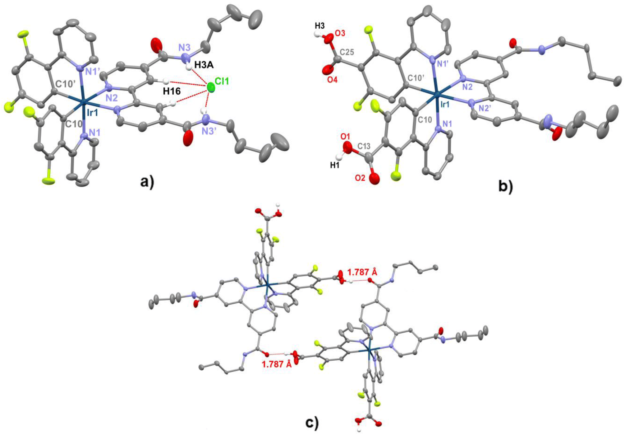
Single yellow crystals of 2a-Cl were obtained by diffusion of n-hexane into a saturated solution of the corresponding complex in CH2Cl2 at room temperature. However, slow diffusion of n-heptane into a solution of complex 2c-PF6 in acetone afforded yellow crystals that were identified as 2c-PO2F2–acetone due to partial hydrolysis of the counter anion. Both complexes crystallized in the P21/n space group and, as expected for centrosymmetric space groups, both enantiomers (Δ and Λ) are present in the lattice. A view of the cationic part with selected bond lengths and angles are presented in Fig. 1 and Table S1.† Both cations exhibited the characteristic octahedral environment around the IrIII center, with a mutual cis-disposition of C-metalated and a trans arrangement of the corresponding nitrogen atoms of the 2-(2,4-difluorophenyl)pyridine cyclometalated ligands. The bond distances and angles were comparable to those found in analogous compounds.25,51,52 In both anions, the Ir–C distances were within the expected values (∼2.01 Å). The Ir–N2 distances to the dbbpy ligand (∼2.13 Å) were longer than the corresponding Ir–N1 (∼2.05 Å, C^N) distance, in accordance with the strong trans influence of the metalated C atom. The chelating C–Ir–N(C^N) angles are around 80° while the N–Ir–N(C^N) were roughly 174.5°, similar to those observed in related complexes. A close look into the structures revealed the presence of hydrogen bonding interactions. Thus, in complex 2a-Cl, short hydrogen bonding between the Cl− anion and the H atoms of the two carboxyamide groups (CONH) is observed (Cl⋯H3–N 2.458(1) Å). Furthermore, Cl is also close to the adjacent H atom of the pyridine rings (Cl⋯H–C(16) 2.601(1) Å). These distances, which were shorter than the sum of van der Waals radii (2.95 Å), are comparable to those reported in other complexes,53–58 thus supporting the formation of an ionic pair in which the chloride acts as an acceptor of four hydrogen donors of the chelating dbbpy ligand. In its turn, in the crystal of 2c-PO2F2 the cations dimerize through short hydrogen bonding between the ketonic group of one of the carboxyamide arms and the carboxylic group of the cyclometalated ligand. The O⋯H distance (1.79 Å) and the angle O⋯H–O (164.9) are comparable to those seen in other systems.59
 |
| | Fig. 1 (a) Molecular structure of complex [Ir(dfppy)2(dbbpy)]Cl (2a-Cl), (b) view of the cation [Ir(COOH-dfppy)2(dbbpy)]+ (from structure of 2c-PO2F2), and (c) view of the dimer formed by two cations [2c]+ through hydrogen bonds between the ketonic group of one amide and the carboxylic group of one cyclometalated ligand. Selected bond distances (Å) and angles (°); 2a-Cl: Ir(1)–C(10) 2.008(2), Ir(1)–N(1) 2.049(2), Ir(1)–N(2) 2.125(2), Cl(1)⋯H(16) 2.601(1), Cl(1)⋯H(3A) 2.458(1). N(1)–Ir(1)–C(10) 80.46(8), N(1)–Ir(1)–C(10′) 95.73(8), N(2)–Ir(1)–N(2′) 77.3(1), N(1)–Ir(1)–N(1′) 174.63(9). [2c]+: Ir(1)–C(10) 2.009(4), Ir(1)–N(1) 2.042(3), Ir(1)–N(2) 2.131(3). N(1)–Ir(1)–C(10) 80.5(1), N(1′)–Ir(1)–C(10) 97.3(1), N(2)–Ir(1)–N(2′) 76.7(1), N(1)–Ir(1)–N(1′) 174.6(1). | |
Noncovalent interactions (NCI analysis) have been carried out on the ionic pair 2a-Cl. The interactions were colour coded with blue and green colours, indicating strong and moderate attractive forces, respectively, while the red and yellow ones correspond to strong and weak repulsive forces (Fig. 2). In the NCI plot, clear green surfaces between the NH and CH protons of dbbpy ligand and chloride atom developed, thus supporting the involvement of donor–acceptor interactions between the anion Cl and the H–X (X = N, C). In addition, green surfaces were also observed for intramolecular C–X⋯π (dfppy) (X = F, H).
 |
| | Fig. 2 NCI plot isosurfaces of the noncovalent interactions of 2a-Cl generated for s = 0.3. | |
Photophysical properties and theoretical calculations
Absorption spectra.
The UV–vis absorption spectra of all compounds were recorded in dimethyl sulfoxide, and the corresponding data are given in Table 1. Selected spectra for 2-Cl and 3a-PF6 are shown in Fig. 3. The counter anion for complexes 2 has negligible influence on their maxima. For complexes 2-Cl, the absorption spectra were also recorded in different solvents showing minor variations in their maxima (Table S7 and Fig. S10†). Moreover, concentration dependence studies in DMSO (1 × 10⁻⁶ to 1 × 10⁻² M) have been carried out for complexes 2-Cl, showing that the three complexes follow the Beer–Lambert Law and, thus, no remarkable ground-state aggregation phenomena occur in solution (Fig. S11†).
 |
| | Fig. 3 Absorption spectra of complexes 2-Cl and 3a-PF6 in DMSO solution (5 × 10⁻⁵ M) at 298 K. Inset: expansion of the low energy region (400–500 nm). | |
Table 1 Absorption data in DMSO solution (5 × 10⁻⁵ M) of complexes 1–3
| Complex |
λ
abs/nm (ε/×10−3/M−1 cm−1) DMSO |
|
1b-DMSO
|
293 (29.96), 333 (9.72), 366 (5.58) |
|
1c-DMSO
|
277 (60.2), 292 (52.4), 365 (9.6) |
|
2a-Cl
|
273 (62.6), 310 (33.8), 368 (9.5), 447 (1.3), 469 (0.7) |
|
2b-Cl
|
306 (23.8), 328 (13.7), 358 (4.4), 421 (1.1) |
|
2b-PF6
|
276 (43.2), 305 (32.5), 332 (19.4), 358 (7.8), 421 (1.0) |
|
2c-Cl
|
269 (54.2), 284 (60.4), 300 (52.4), 365 (9.6), 409 (1.4) |
|
2c-PF6
|
281 (51.4), 300 (34.2), 363 (8.6), 431 (2.4) |
|
3a-PF6
|
265 (41.8), 305h (20.94), 361 (6.48), 450 (0.93) |
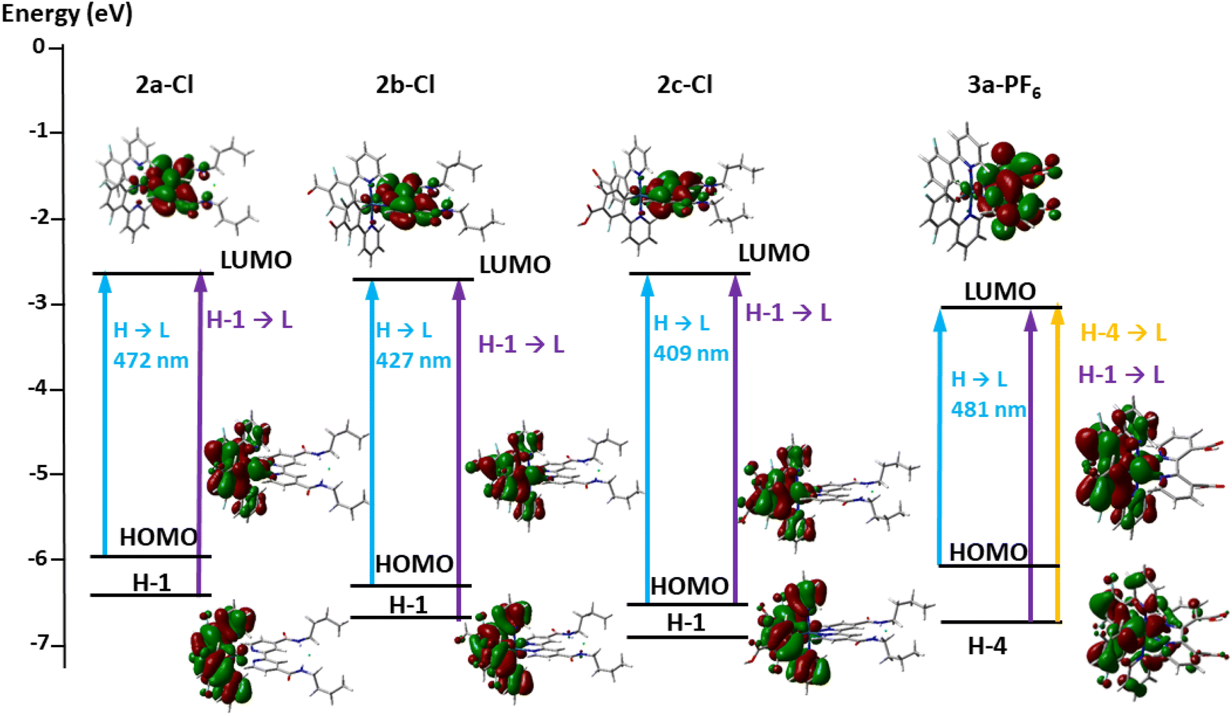
According to TD-DFT calculations on complexes 2-Cl (see the ESI†), the intense high energy absorption features in the UV region (<350 nm) are ascribed to spin allowed π–π* transitions of the ligands (1IL C^N, L′ dbbpy) with metal to ligand contribution (MLCT and ML′CT). In mononuclear complexes 2, the moderately intense band around 365 nm can be mainly related to the intense S3 transition, which is associated with H-1 to the LUMO. H-1 resides on the cyclometalated ligands (91–92%) and Ir (7%) while the LUMO is located on the dbbpy ligand. Therefore, this band mainly arises from spin allowed 1LL′CT (C^N → dbbpy) transitions with some 1ML′CT contribution. The low energy band extending in the region of λ > 400 nm (447 2a-Cl, 421 2b-Cl, 409 nm 2c-Cl) is associated with the HOMO → LUMO transitions, having an ML′CT/LL′CT character. The slightly hypsochromic shift on going from 2a-Cl to 2c-Cl is reflected in the calculations (S1, cal. 472 2a-Cl, 427 2b-Cl; 410 nm 2c), and can be attributed to the stabilization of the HOMO due to the presence of the electron withdrawing CHO and COOH substituents (Fig. 4). Due to strong spin–orbit coupling (SOC) associated with iridium, an overlapping with the spin-forbidden singlet–triplet metal-to-ligand and ligand-to-ligand transitions (3ML′CT/3LL′CT) is expected in this region. In complex 3a-PF6, having the dicarboxy-pyridine (3,3′H2dcbpy), the low energy feature appears at ca. 450 nm with a tail extending to 500 nm, also with an ML′CT/LL′CT character according to calculations (see the ESI†).
 |
| | Fig. 4 Schematic representation of selected excitation for complexes 2-Cl and 3a-PF6 in DMSO. | |
Emission spectra.
The room temperature emission spectra of the precursor complex 1c and complexes 2 and 3 were recorded in DMSO solution (Fig. 5) and in the solid state (Fig. S12†) and the data are listed in Table 2 and Table S8.† For complexes 2-Cl, the spectra were also examined in different solvents aiming to check the solvatochromic behaviour, which is common in this type of complexes (Fig. S13†). As an example, the emission spectra in DMSO solution are shown in Fig. 5a. The precursor complex 1c was highly soluble in DMSO and, as noted before, generated the solvate 1c-DMSO upon dissolution, which exhibits a slightly structured band located at 511 nm which is ascribed to a 3LC with a 3MLCT character (Fig. 5b). In the solid state the emission of 1c was red shifted to 580 nm, suggesting that the emission might have originated from molecular aggregation of the dinuclear derivative in the rigid media through ππ stacking of the cyclometalated ligands. A highest energy shoulder is observed at 540 nm that is likely due to the emission from the 3MLCT/3LC contribution on individual molecules (Fig. S13†). In DMSO solution, the complexes 2-Cl and 2-PF6 were brightly emissive and exhibited similar photophysical properties. All complexes displayed broad unstructured bands in the range of 540–565 nm, with a negligible influence of the counter anion likely due to the easy breakdown of the hydrogen bonding interactions in complexes 2-Cl. In agreement with theoretical calculations and previous works,58,60,61 this emission is attributed to a mixed 3ML′CT (Ir → N^N)/3LL′CT (C^N → N^N) excited state with a predominant ML′CT character. The photoluminescence quantum yields (PLQYs) in deaerated solutions (ϕ) were relatively high (from 44.6% for 2b-PF6 to 54.1 for 2c-PF6), with lifetimes in the range 0.58 to 0.81 μs. As expected, compared with the deoxygenated atmosphere, the emission intensity under an air atmosphere was notably reduced. In the case of complex 3a-PF6, the emission is red shifted (λmax = 605 nm) and the photoluminescence quantum yield drops to 4.2%, as does the decay to 0.31 μs. This result is not unexpected and is in accordance with the energy gap law. The measured lower lifetime of complexes 2 (0.58–081 μs) and 3a-PF6 (0.31 μs) measured in deoxygenated solutions in relation to 1c can be attributed to a notable higher metal contribution in the excited state. Comparing the complexes with the dbbpy ligand, the emission maxima follow the order (540 2b < 560 2c < 565 2a), which is consistent with the stabilization of the HOMO in complexes featuring the CHO and COOH substituents. In complex 3a-PF6, the observed red shift can be attributed to a remarkable stabilization of the target 3,3′-H2dcbpy LUMO (Fig. 4 and 5a). The reduced quantum yield of this complex (Table 2), which is reflected in the higher knr and lower kr in relation to complexes 2, could be related to the relatively strong vibrational quenching effect caused by the presence of the two carboxylic units on the bipyridine ligand.
 |
| | Fig. 5 Normalized emission spectra of complexes (a) complexes 2–3 in DMSO solution (5 × 10⁻⁴ M), (b) 1c in different media, and (c) of complex 2c-Cl in different solvents. | |
Table 2 Photophysical data in DMSO solution (5 × 10⁻⁴ M)a. Radiative (Kr) and non-radiative (Knr) constants calculated at room temperature
| Complex |
λ
em (nm) |
Energy/λem![[thin space (1/6-em)]](https://www.rsc.org/images/entities/char_2009.gif) a a |
τ (μs) aereated/deoxygenated |
φ aereated/deoxygenated |
K
rb/s−1 |
K
nrb/s−1 |
|
Predicted from DFT (B3LYP/LANL2DZ (Ir) 31G(d,p)) calculations in DMSO at 298 K, by estimating the energy difference between the optimized T1 and singlet state S0.
Values from deoxygenated solution.
|
|
1c
|
510 |
— |
10.14b |
—/<0.01 |
1.5 × ·103 |
9.7 × 10⁴ |
|
2a-Cl
|
568 |
2.2 eV/556 |
0.18/0.81 |
0.15/0.53 |
6.6 × 10⁵ |
5.7 × 10⁵ |
|
2b-Cl
|
540 |
2.5 eV/494 |
0.42/0.72 |
0.35/0.51 |
7.1 × 10⁵ |
6.8 × 10⁵ |
|
2b-PF6
|
542 |
— |
0.28/0.58 |
0.21/0.45 |
7.7 × 10⁵ |
9.6 × 10⁵ |
|
2c-Cl
|
558 |
2.4 eV/510 |
0.43/0.76 |
—/0.54 |
7.1 × 10⁵ |
6.1 × 10⁵ |
|
2c-PF6
|
558 |
— |
0.41/0.73 |
0.25/0.54 |
7.4 × 10⁵ |
6.3 × 10⁵ |
|
3a-PF6
|
605 |
1.7 eV/715 |
0.14/0.31 |
0.02/0.04 |
1.4 × 10⁵ |
3.1 × 10⁶ |
The influence of the solvent has been examined for complexes 2-Cl (see Fig. 5c, and Table S8 and Fig. S13 in the ESI† for 2c-Cl). For these complexes, the emission band gradually shifts hypsochromically as the solvent polarity decreases (2a-Cl 565 DMSO, 520 THF and 495 nm toluene; 2b-Cl 540 DMSO, 507 THF and 503 nm toluene; 2c-Cl 615 H2O, 540 DMSO, and 517 nm THF). The observed positive solvatochromism indicates that the excited state is more polar than the ground state, being stabilized by the solvent polarity and confirms the strong charge transfer nature of the excited state. For 2a-Cl, the decay and the PLQY decrease with the polarity of the solvent. This feature is very remarkable for complex 2c-Cl, featuring the COOHdfppy cyclometalated ligand, in H2O wherein the PLQY falls to 3.1% with a decay of 0.06 μs. In this complex, the exchange of carboxylic and H2O protons opens a new channel for deactivation, as reflected in the high value for knr (1.7 × 10⁻⁷ s−1).
To ascertain the detailed properties of the excited state, the optimized geometries of T1 have been calculated using the S0 geometries and the B3LYP approach for complexes 2a–c-Cl and 3a-PF6. Fig. 6 shows the optimized excited-state structures of the highest singly occupied molecular orbitals (HSOMOs), lowest singly occupied molecular orbitals (LSOMOs), and spin density distributions. In all complexes, the SOMO-1 involves Ir metal and the C^N cyclometalating group, while the SOMO is primarily localized on the bipyridine. The predicted spin densities have been localized over the bipyridine ligand and, thus, the emissions are mainly characterized by both ML′CT from iridium to bipyridine charge transfer and LL′CT from the cyclometalated to the bipyridine ligand. There is good agreement between the experimental and calculated emission wavelengths shown in Table 2 (calculated by estimating the energy difference between the optimized T1 and singlet state S0 in DMSO). The introduction of CHO (2b) and COOH (2c) substituents on the cyclometalated group caused a slight hypsochromic shift by the stabilization of the SOMO-1 in relation to 2a-Cl, while the incorporation of carboxylic units on the bipyridine provoked a remarkable bathochromic shift, which is in good agreement with the experimental data.
 |
| | Fig. 6 Calculated SOMO, SOMO-1 and spin density representations of 2 (a, b and c)-Cl and 3a-PF6 in DMSO solution. | |
Quantum yield 1O2 quantification.
In photodynamic therapy, the presence of singlet oxygen is one of the most important factors for the enhancement of the cytotoxic activity of a complex. Therefore, complexes 2b-Cl, 2b-PF6, 2c-Cl and 3a-PF6 were selected to assess their ability to generate 1O2. These complexes generate singlet oxygen (1O2) at room temperature in CH2Cl2 solution (Fig. 7). The 1O2 oxygen emission at 1270 nm was directly monitored using a near-infrared detector upon excitation at 425 nm. To determine the singlet oxygen generation, we use a reference method by UV-Visible spectroscopy in acetonitrile. ROS generation was visualized using the common ROS-capturing agent 1,3-disphenylisobenzofuran (DPBF). DPBF reacts with ROS to generate 1,2-dibenzoylbenzene, resulting in an absorbance decline at 410 nm.62,63 The activity of a mixture of each complex with DPBF in acetonitrile solution, employing [Ru(bpy)3]2 as a reference (φSΔ = 0.56),64 was measured under blue light irradiation (λ 460 nm). A clear decrease in the absorbance of the DPBF band at 410 nm (Fig. S14†) demonstrated the generation of 1O2 by the Ir(III) complexes. The values of φ obtained for all four complexes (0.32 for 2b-PF6, 0.12 for 2c-Cl, 0.10 for 3a-PF6, and 0.09 for 2b-Cl) suggest that they might have future potential as an intracellular 1O2 generator for photo-chemotherapeutic development.
 |
| | Fig. 7 Emission band of the singlet oxygen from fresh solution of 2b-PF6 (λex 425 nm). | |
Biological studies
To start, the stability of complexes 2 and 3a was supported by 1H NMR or UV-Vis spectra in DMSO solution or cellular medium, respectively, which revealed that they remained unaltered within 74 h (Fig. S15–S18 and S20†). Their photostability in both media was also assessed. The complexes were found to be stable upon blue light irradiation with a blue lamp (396 nm) for at least 10 min (Fig. S19 for 3a-PF6 and S21† for 2b,c-Cl and 3a-PF6). The cytotoxicity of complexes 2 and 3a was determined in vitro against human cell lines by an MTS-based method. We also evaluated the effect of irradiation with UV light on the antiproliferative activity of complexes 2b (Cl and PF6), 2c-Cl and 3a-PF6, their interaction with DNA and lipophilicity, as well as their cellular localization.
Cytotoxic activity and selectivity index.
The IC50 values were determined against two different human tumours (A549, lung carcinoma and HeLa, cervix carcinoma) and nontumoral BEAS-2B (bronchial epithelium) cell lines after cellular exposure to the compounds for 72 h and compared to cisplatin as reference (Table 3 and Fig. S22†).
Table 3 Cytotoxic IC50 values (μM)a and selectivity indexb of the complexes 2a-Cl, 2b-PF6 and 3a-PF6 in A549, HeLa and BEAS-2B human cell lines compared with cisplatin
| Complex |
IC50a |
SIb |
| A549 |
HeLa |
BEAS-2B |
A549 |
HeLa |
|
IC50 values presented as mean ± standard error of the mean of three different experiments.
Selectivity index (SI) = IC50 nontumor cell (BEAS-2B)/IC50 cancer cell (A549 or HeLa), as described in ref. 65, 66.
As determined in ref. 67.
As determined in ref. 68.
|
|
2a-Cl
|
3.73 ± 0.76 |
5.53 ± 0.41 |
12.32 ± 0.21 |
3.30 |
2.23 |
|
2b-PF6
|
27.17 ± 1.44 |
74.6 ± 0.72 |
51.6 ± 4.71 |
1.90 |
0.69 |
|
3a-PF6
|
32.87 ± 3.01 |
51.22 ± 1.29 |
24.03 ± 3.82 |
0.73 |
0.47 |
| Cisplatin |
6.45 ± 0.47c |
13.60 ± 0.99d |
1.74 ± 0.16 |
0.27 |
0.13 |
Complexes 2b-Cl and 2c-Cl compounds displayed IC50 values ≥100 μM towards the A549 cell line (Fig. S22†). The low toxicity found with this assay could be attributed to their low solubility in the aqueous biological media. For that reason, the related complexes with PF6− as the counter anion were prepared to test their biological activity. Complexes 2-PF6 displayed better solubility, with no signs of precipitation at concentrations up to 100 μM. For 2c-PF6, the IC50 value was also ≥100 μM towards the A549 cell line (Fig. S22†). Based on the low cytotoxicity of 2b-Cl, 2c-Cl and 2c-PF6, these complexes were not further analysed in other cells. However, it is remarkable that the antiproliferative activity improved on going from 2b-Cl to 2b-PF6 (IC50 values of 27.17 and 74.60 in A549 and HeLa cells, respectively, for 2b-PF6vs. the lesser effects of 2b-Cl). This change might be attributed to the close interactions between Cl− anion and the two NH groups of the butylamide substituents of the dbbpy ligand in compound 2b-Cl, which decreases its ionic character and solubility, thus preventing their optimum cellular uptake. Interestingly, complex 2a-Cl, offered the lower IC50 values, being even more cytotoxic than cisplatin in both A549 (3.73 vs. 6.45) and HeLa (5.53 vs. 13.60) cells (Table 3). These values are similar to those reported for related complexes [Ir(C^N)2(N^N)]PF6 (C^N = ppy, dfppy) featuring as diimine the dibutyl 2,2′-bipyridine-4,4′-dicarboxylate against HeLa and A549 tumour cells (IC50 1.7–2.3).69 Complex 3a-PF6 that contains two carboxylic acid units on the diimine ligand also showed mild cytotoxic activity, with IC50 values similar to 2b-PF6 (32.87 and 51.22 in A549 and HeLa cells, respectively) (Table 3 and Fig. S22†). As a rule, all the complexes were more active towards the A549 cell line rather than HeLa, in accordance with our previous results.62,66
In order to evaluate the selectivity index (SI) of complexes towards the tumour cell lines, we used normal epithelial lung virus-transformed BEAS-2B cells as a non-tumor reference cell line. Cisplatin IC50 value towards this cell line was 1.74 μM, slightly lower than those for A549 and HeLa cells, therefore rendering low SI values of this anti-cancer drug for these cell lines (0.27 and 0.13, respectively) (Table 3). Actually, compounds with SI values of <2 are assumed to give general toxicity.70,71 IC50 values of cisplatin on BEAS-2B cells and their SI values for A549 and HeLa cells reported here are in good agreement with previous findings.72–74 Among the Ir(III) complexes, the better values of the selectivity index (SI) were given by complex 2a-Cl in both tumoral A549 (3.30) and HeLa (2.23) cells, due to its higher cytotoxic activity in these cells (IC50 3.73 and 5.53 in A549 and HeLa cells, respectively) compared to that in non-tumoral BEAS-2B cells (IC50 12.32 μM). SI values for complex 2b-PF6 and 3a-PF6 were 1.90 and 0.73 (A549) and 0.69 and 0.47 (HeLa), respectively (Table 3), indicating low selective toxicity towards cancer cells. Low SI values (<2) for other organometallic complexes with cytotoxic activities toward A549, HeLa, and other cancer cell lines have been previously described,66,75–78 including iridium organometallic complexes.18,79,80
Photoinduced cytotoxicity and intracellular ROS generation.
To expand our knowledge on the cytotoxic behaviour of these complexes, we examine their potential as photosensitizers (PS). Selective activation of a nontoxic photosensitizer by light is an attractive regimen for therapy, especially if the PS is preferentially taken up by cancer cells.81 In recent years, luminescent metal based drugs, in particular Ru(II) Ir(III) and Pt(II) complexes, are being actively investigated due to their outstanding results in PDT based cancer therapy.40,82–87 Due to the ability of these iridium complexes to generate 1O2 upon irradiation and their photostability, they can be considered as potential tools in phototheranostics. Therefore, we decided to evaluate the potential use of complexes 2b-Cl and 2c-Cl in photodynamic therapy based on their light cytotoxic effects in normal cell culture. In addition, we also chose 2b-PF6 in order to evaluate the effect of changing the counter-anion Cl− with PF6− on the induction of the photocytotoxicity. Finally, we also evaluated the photocytotoxicity of 3a-PF6 to test the effect of locating the carboxylic units on the diimine ligand. The photostability of the complexes under similar conditions of irradiation to that employed for PDT assays was supported by NMR spectroscopy. A549 cells were initially treated with each complex in Hank's balanced salt solution (HBSS) for 1 h at 37 °C in order to allow cellular interaction and internalization. Then, A549 cells were irradiated with a 396 nm LED lamp located 91 mm apart (5 Mw cm−2) for different times: 3 min (3a-PF6), 10 min (2b-PF6) and 15 min (2b-Cl and 2c-Cl) (see the ESI†). After the irradiation, cells were washed and incubated in complete RPMI medium without the presence of any complex for another 72 hours. Finally, cell viability (IC50) was assessed by the MTS test, as detailed in the ESI.† The observed IC50 values were significantly lower in the presence of UV light than under non-UV light, reflecting the effective phototoxicity of the photosensitizers. Thus, as shown in Fig. 8, upon UV light irradiation a significant increase of the antiproliferative activity was observed for 3a-PF6 (IC50: 5.63 μM) with a short irradiation time of only 3 min. UV-light irradiation for 15 min for 2b-Cl or 10 min for 2b-PF6, also enhances their antiproliferative activity leading to IC50 values of 14.39 and 18.57 μM, respectively, whereas the slightly cytotoxic 2c-Cl showed a lower PI activity, giving a moderate effect by lowering the IC50 value to 55.52 μM upon 15 min of irradiation. In any case, the complexes are more active under UV light than under non-UV light. This could be related to their singlet oxygen generation ability in cancer cells. The generation of ROS after UV irradiation was measured using the ROS indicator H2DCFDA. As shown in Table 4, the four complexes show ROS generation after UV irradiation. The highest values were given by 3a-PF6 (49 μM), which were in good agreement with its lower UV-IC50 (Fig. 8d) and despite its shorter UV light exposure time (3 min).
 |
| | Fig. 8 Dose–response curves for the A549 cell line treated with complex (a) 2b-Cl; (b) 2b-PF6; (c) 2c-Cl and (d) 3a-PF6 either with (triangles) or without (circles) UV light: irradiation with a UV 396 nm LED for 15 min (a and c), 10 min (b) and 3 min (d) and followed by MTS cytotoxic assays performed after 72 h. Non UV-irradiated cells were manipulated identically to UV-irradiated ones. IC50 values are presented as mean ± standard error of the mean of three different experiments performed in sextuplicate. | |
Table 4 ROS production in A549 cell line treated with complexes 2b(Cl and PF6), 2c-Cl and 3a-PF6, either with or without UV light
| Complex |
ROS (arbitrary units)a |
| Non UVb |
UVc |
|
ROS was measured using the ROS-sensitive dye, 2′,7′-dichlorodihydrofluorescein diacetate (H2DCFDA) as an indicator.
Cells were non-irradiated.
Cells were irradiated with a UV 396 nm LED for 15 min (2b-Cl and 2c-Cl), 10 min (2b-PF6) and 3 min (3a-PF6).
ROS values of A549 cells without complex. Each value represents the mean ± standard error from three different experiments performed in quadruplicate. *p < 0.05; **p < 0.01; ***p < 0.001 (Mann–Whitney U test or Student's t-test for comparing two groups). |
|
2b-Cl
|
Controld |
801.42 ± 20.00 |
10635.50 ± 451.51** |
| 1 mM |
702.67 ± 47.68 |
46391.44 ± 818.24*** |
| 25 mM |
2976.67 ± 390.74 |
71221.42 ± 1906.88*** |
|
2b-PF6
|
Controld |
516.33 ± 16.78 |
7527.17 ± 922.78* |
| 2.5 mM |
617.67 ± 24.30 |
54776.42 ± 5684.69** |
| 25 mM |
1702.83 ± 61.91 |
75723.75 ± 2670.39*** |
|
2c-Cl
|
Controld |
494.58 ± 34.34 |
7571.42 ± 1329.36** |
| 1 mM |
468.67 ± 24.83 |
10578.25 ± 236.44*** |
| 50 mM |
1127.50 ± 118.51 |
72319.33 ± 2846.33*** |
|
3a-PF6
|
Controld |
577.58 ± 71.84 |
2476.25 ± 130.39*** |
| 1 mM |
633.00 ± 32.92 |
4583.75 ± 92.10*** |
| 7 mM |
999.25 ± 37.02 |
17801.33 ± 690.25*** |
| 49 mM |
1832.75 ± 246.03 |
82785.67 ± 1296.15*** |
Relative lipophilicity.
The relative hydrophobicity of the complexes was studied by RP-UPLC with the aim of establishing a correlation between their cytotoxicity and affinity for a lipid environment, a crucial aspect in their pharmacokinetic properties. Complexes were dissolved in acetonitrile (≈1 ppm), and a mixture of acetonitrile with 0.1% HCOOH (A) and H2O with 0.1% HCOOH (B) was employed as a mobile phase. The lipophilicity character is based on the values of their retention times (tR). These values account for the relative interactions between the hydrophobic stationary phase (Aquity UPLC BEH C18) and the hydrophilic mobile phase with each complex. The longer the tR, the more lipophilic the character of the complex.62,88 In case of these Ir(III) complexes, the presence of substituents on the cyclometalated groups remarkably decreased the tR (H > CHO > COOH) (Table S9†), with 2a-Cl (tR = 3.28) and 3a-PF6 (tR = 1.99) being the most lipophilic ones. The higher lipophilicity of 2a-Cl is in accordance with the good cytotoxicity activity found for this complex, which displays the lowest IC50 values (Table 3). Differences between the tR of the rest of compounds were too small to ensure a clear relationship between the results and their cytotoxic activities.
Study of the reaction with NADH.
Nicotinamide adenine dinucleotide (NADH), and its oxidized form NAD+, is a coenzyme whose function is crucial for the catalysis of redox or isomerization reactions.89 NADH plays an important role in the mitochondrial electron transport chain and the maintenance of the cellular redox balance. Thus, any alteration in the intracellular concentration of this species might lead to cell death.90 Recent studies have reported that some Ir(III) and Os(II) complexes display anticancer activity through a non-conventional redox mediated mechanism of action involving catalytic photo-oxidation of NADH to NAD+ through transfer hydrogenation reactions leading to H2O2 and reactive oxygen species.91,92 Thus, the quantification of changes in NADH/NAD+, is a good method to know if these compounds can produce changes in ROS content by this mechanism. The interaction with NADH was performed with the cytotoxic complex 2a-Cl, and the results are shown in Fig. 9. The reaction of the complex (1 μM) with NADH (100 μM) dissolved in a solution 20% MeOH in H2O was monitored by UV-vis at 298 K for 8 hours. Following the continuous decrease with time of the NADH absorption peak at 339 nm, due to its interaction with the complex to give its oxidized form NAD+,25,93 allowed us to determine the interaction of NADH (conversion to NAD+) with the complex. The turnover number (TONs) obtained by the difference between NADH concentration at t0 and t = 8 hours was 14.0, showing that the NADH had been oxidated to NAD+. This result indicates that this complex can act as a moderate catalyst for endogenous NADH oxidation. Thus, it could be a mitochondrial target, inducing destabilization of the redox homeostasis and, ultimately, causing apoptosis.
 |
| | Fig. 9 UV-Vis spectra for the oxidation of NADH (100 μM) by complex 2a-Cl (1 μM) in a mixture of H2O/MeOH 80/20 under dark conditions. | |
Interaction of complexes with DNA.
Knowing their cytotoxic activity, the interaction of iridium 2a-Cl, 2b-PF6 and 3a-PF6 complexes with DNA was studied by their ability to modify the electrophoretic mobility of the supercoiled covalently closed circular (CCC) and the open circular (OC) forms of pBR322 plasmid DNA (Fig. S23†). To provide a basis for comparison, the incubation of DNA with cisplatin and the low cytotoxic complex 2c-PF6 was also performed at the same concentrations and conditions. The binding of cisplatin to plasmid DNA, for instance, results in a decrease in the mobility of the CCC form and an increase in the mobility of the OC form (Fig. S23,† upper left panel).53,66,94,95 As shown in Fig. S23,† no electrophoretic mobility changes were observed after DNA treatment with any complex under the same conditions, thus indicating that these compounds were either not reacting with the DNA or not altering the DNA mobility in agarose gels.
Intracellular localization.
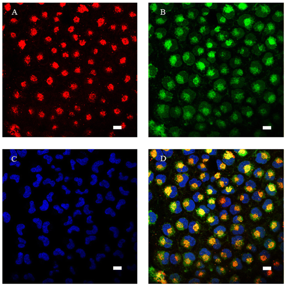
Confocal microscopy was performed to investigate the intracellular localization. In agreement with the DNA interaction studies, none of the complexes showed intranuclear localization in A549 cells, as shown in Fig. 10–13. When 16 μM 2a-Cl complex was incubated with A549 cells, it showed fast cellular internalization, localizing predominantly in lysosomes (Fig. 10), as has been determined by a calculated Pearson correlated coefficient of 0.79. 2a-Cl also localizes to a lesser extent in the cytoplasm, but not in mitochondria (Fig. S24 and S25†). However, although 2a-Cl was not detected inside the mitochondria, it ends up causing them damage. Thus, one-hour incubation with 16 μM 2a-Cl complex induced mitochondrial swelling and the loss of mitochondrial membrane potential was noticed after 30 min (Fig. 11), which has been associated with cell death either by apoptosis or necrosis, depending on the particular biological setting. Accordingly as shown before, this complex effectively alters the NADH/NAD+ pair, which can produce ROS, thus altering mitochondrial functions and cell death.
 |
| | Fig. 10 Laser confocal microscopy images of live A549 cells incubated with 16 μM 2a-Cl complex for 1 hour. Cells were stained with specific intracellular markers. A. LysoTracker (lysosomal marker, λex 543 nm) in red. B. 2a-Cl (λex 405 nm) in green. C. Hoechst (nuclei marker, λex 405 nm) in blue. D. Merged image showing colocalization of 2a-Cl with LysoTracker in lysosomes (yellow). Scale bar = 10 μm. | |
 |
| | Fig. 11 Laser confocal microscopy images of live A549 cells and loss of mitochondrial membrane potential (MMP) after incubation with 2a-Cl complex. (A and B) Cells were stained with specific intracellular markers: red: MitoTracker (mitochondrial marker, λex 633 nm) and blue: Hoechst (nuclei marker, λex 405 nm). (A) Control cells. (B) Cells incubated with 16 μM 2a-Cl complex for 1 hour. Scale bar = 10 μm. (C) Loss of MMP after 30 min of incubation with 16 μM 2a-Cl complex or 10 μM FCCP. Control indicates untreated cells. MMP values represent the mean ± standard error from three different experiments performed in quadruplicate. *p < 0.05 (Mann–Whitney U test for comparing two groups). | |
 |
| | Fig. 12 Laser confocal microscopy images of live A549 cells incubated with 32 μM 2b-PF6 and 2c-PF6 complexes for up to 24 hours. (A) Cells exposed to 32 μM 2b-PF6 for 1 hour (λex 405 nm). (B) Cells exposed to 32 μM 2b-PF6 for 24 hours (λex 405 nm). (C) Cells exposed to 32 μM 2c-PF6 for 1 hour (λex 405 nm). (D) Cells exposed to 32 μM 2c-PF6 for 24 hours (λex 405 nm). Scale bar = 20 μm. | |
 |
| | Fig. 13 Laser confocal microscopy images of live A549 cells incubated with 45 μM 3a-PF6 compound for up to 20 minutes. Top pictures: A549 cells incubated with 3a-PF6 stimulated with a 405 nm laser. Bottom pictures: A549 cells incubated with 3a-PF6 and labelled with MitoTracker stimulated with 405 and 633 nm lasers. Green: 3a-PF6; magenta: MitoTracker. Scale bar = 20 μm. | |
Complexes 2b-PF6 and 2c-PF6 were also internalized by A549 cells when they were incubated at 32 μM for 1 hour (Fig. 12A and C) or 24 hours (Fig. 12B and D). In agreement with their cytotoxicity results presented in Table 3 and Fig. S22,† the incubation of A549 cells with complex 2b-PF6 for 24 hours resulted in extended cell death while 2c-PF6 complex showed less toxicity for the same time.
Interestingly, when we incubated A549 cells with 45 μM of complex 3a-PF6, and followed the cells by confocal microscopy, we observed an unexpected behavior. Cells subjected to 405 nm laser stimulation showed increased toxicity compared to adjacent cells not subjected to the laser stimulation (Fig. 13). We did not observe laser-induced toxicity in A549 cells incubated with any of the complexes 2-Cl. However, 405 nm laser stimulation (from the confocal microscope) not only increased the toxicity of the compound but also increased its fluorescence (central region of pictures in Fig. 13). When we incubated the cells with 3a-PF6 and the mitochondrial marker MitoTracker, we observed loss of mitochondrial membrane potential and mitochondrial swelling (Fig. 13) only when we stimulated the cells with the 405 and the 633 lasers. We found that incubation of cells with 3a-PF6 and MitoTracker and stimulation with 633 nm laser did not damage the mitochondria or induced toxicity. Therefore, we conclude that 3a-PF6 can be excited using 405 nm irradiation increasing its fluorescence and inducing mitochondrial toxicity, possibly by inducing ROS formation (Table 4), leading to cell death. Mitochondria play an important role in many cellular operations, such as the generation of energy, maintaining intracellular redox balance and metabolism and, reports on cyclometalated iridium complexes causing mitochondria mediated dysfunction and apoptosis by several factors including reactive oxygen species (ROS) have been previously reported.96
Conclusion
We present here a series of novel Ir(III) cyclometalated cationic complexes incorporating different substituents in the position 3 of the 2,4-diflurophenylpyridinate cyclometalated group (H, dfppy; HCO, CHO-dfppy and COOH, COOH-dfppy) and using N,N′-dibutyl-2,2′-bipyridine-4,4′-dicarboxamide (dbbpy) as the diimine ligand with chloride (2-Cl) or PF6− ions (2-PF6) as counter anions, together with [Ir(dfppy)2(H2dcbpy)]PF6 (3a-PF6), to evaluate the influence of altering the position of the substituents on the optical properties and bioactivity of the complexes. All complexes have been fully characterized by 1H, 13C{1H} and 19F NMR spectroscopy, HMRS, and elemental analysis. In addition, the structure of complex 2a-Cl and of the cation [Ir(COOH-dfppy)2(dbbpy)]+ (from the structure of 2c-PO2F2) were confirmed by X-ray diffraction studies. Crystals of 2a-Cl reveal that this complex and, likely also complexes 2b,c-Cl, forms an ionic pair in which the Cl− establishes strong hydrogen bonding interactions with four H donors of the chelating dbbpy ligand, also supported by an NCI theoretical study. These complexes exhibit bright phosphorescence and a fine tuning of their emission colour can be achieved by modifying the cyclometalated or the substituents of the ancillary diimine ligand. DFT and TD-DFT calculations indicate that all complexes emit from a mixed 3ML′CT (Ir → N^N)/3LL′CT (C^N → N^N) excited state. The complexes can act as 1O2 sensitizers as suggested by the remarkable emission quenching observed in aerated DMSO solutions for all of them and confirmed and calculated in complexes 2b-PF6 and 3a-PF6.
The cytotoxic activity of the new complexes 2 and 3a has been evaluated against two different human tumour cell lines (A549, lung carcinoma and HeLa, cervix carcinoma) and the nontumoral BEAS-2B (bronchial epithelium) cell line. Only the most lipophilic compound 2a-Cl has shown remarkable activity in A549 and HeLa cells, significantly better than cisplatin in the same cells. Complexes 2b-PF6 and 3a-PF6 exhibited moderate activity, whereas 2b,c-Cl and 2a-PF6 did not show cytotoxicity. The binding experiments with the pBR322 plasmid DNA as well as confocal intracellular localization studies revealed that the interaction with nuclear DNA does not seem to be the anticancer mechanism. Complex 2a-Cl mainly localizes in lysosomes, but it causes mitochondrial damage. Its cytotoxic activity could be in part attributed to the production of ROS generated by intracellular imbalance of the NADH/NAD+ pair, as suggested by the observed oxidation of NADH in the presence of complex 2a-Cl.
Moreover, these complexes were photo-cytotoxic agents. Thus, a significant increase of antiproliferative activity was observed for complexes 2b-Cl and 2b-PF6, and, particularly, for 3a-PF6, upon short irradiation times, which has been related to their singlet oxygen generation ability in cancer cells.
Conflicts of interest
There are no conflicts to declare.
Acknowledgements
This work was supported by the Spanish Ministerio de Ciencia e Innovación (Project PID2019-109742GB-I00) funded by MCIN/AIE/10.13039/501100011033, by “ERDF A way of making Europe”, by the “European Union, and by the ADER (Gobierno de La Rioja; Project 2017-I-IDD-00031). G. M. is grateful to UR for a PhD grant. We are grateful to E. Alfaro-Arnedo for her technical help.
References
- M. Fanelli, M. Formica, V. Fusi, L. Giorgi, M. Micheloni and P. Paoli, Coord. Chem. Rev., 2016, 310, 41 CrossRef CAS.
- V. Brabec, O. Hrabina and J. Kasparkova, Coord. Chem. Rev., 2017, 351, 2 CrossRef CAS.
- B. J. Pages, K. B. Garbutcheon-Singh and J. R. Aldrich-Wright, Eur. J. Inorg. Chem., 2017, 1613 CrossRef CAS.
- N. J. Wheate, S. Walker, G. E. Craig and R. Oun, Dalton Trans., 2010, 39, 8113 RSC.
- W.-P. To, T. Zou, R. W.-Y. Sun and C.-M. Che, Philos. Trans. R. Soc., A, 2013, 371, 20120126 CrossRef PubMed.
- T. C. Johnstone, K. Suntharalingam and S. J. Lippard, Chem. Rev., 2016, 116, 3436 CrossRef CAS PubMed.
- X. Wang, X. Wang and Z. Guo, Acc. Chem. Res., 2015, 48, 2622 CrossRef CAS PubMed.
- Q. Cheng and Y. Liu, Wiley Interdiscip. Rev.: Nanomed. Nanobiotechnol., 2017, 9, e1410 CrossRef PubMed.
- L. G. Marcu, Pharmaceuticals, 2022, 15, 255 CrossRef CAS PubMed.
- S. A. Aldossary, Biomed. Pharmacol. J., 2019, 12, 7 CAS.
- A. Khoury, K. M. Deo and J. R. Aldrich-Wright, J. Inorg. Biochem., 2020, 207, 111070 CrossRef CAS PubMed.
- C. Yu, Z. Wang, Z. Sun, L. Zhang, W. Zhang, Y. Xu and J.-J. Zhang, J. Med. Chem., 2020, 63, 13397 CrossRef CAS PubMed.
- E. Ortega, G. Vigueras, F. J. Ballester and J. Ruiz, Coord. Chem. Rev., 2021, 446, 214129 CrossRef CAS.
- L. Feng, M. Gao, D. Tao, Q. Chen, H. Wang, Z. Dong, M. Chen and Z. Liu, Adv. Funct. Mater., 2016, 26, 2207 CrossRef CAS.
- L. He, K. Xiong, L. Wang, R. Guan, Y. Chen, L. Ji and H. Chao, Chem. Commun., 2021, 57, 8308 RSC.
- P.-Y. Ho, C.-L. Ho and W.-Y. Wong, Coord. Chem. Rev., 2020, 413, 213267 CrossRef CAS.
- Z. Yang, G. Jiang, Z. Xu, S. Zhao and W. Liu, Coord. Chem. Rev., 2020, 423, 213492 CrossRef CAS.
- U. Das, B. Kar, S. Pete and P. Paira, Dalton Trans., 2021, 50, 11259 RSC.
- N. Nayeem and M. Contel, Eur. J. Chem., 2021, 27, 8891 CrossRef CAS PubMed.
- P. Sudhindra, S. A. Sharma, N. Roy, P. Moharana and P. Paira, Polyhedron, 2020, 192, 114827 CrossRef CAS.
- C. C. Konkankit, S. C. Marker, K. M. Knopf and J. J. Wilson, Dalton Trans., 2018, 47, 9934 RSC.
- Y. Liu, Y. Wang, S. Song and H. Zhang, Chem. Sci., 2021, 12, 12234 RSC.
- S. Pete, N. Roy and P. Paira, Inorg. Chim. Acta, 2021, 517, 120184 CrossRef CAS.
- G.-X. Xu, E. C.-L. Mak and K. K.-W. Lo, Inorg. Chem. Front., 2021, 8, 4553 RSC.
- E. Zafon, I. Echevarría, S. Barrabés, B. R. Manzano, F. A. Jalón, A. M. Rodríguez, A. Massaguer and G. Espino, Dalton Trans., 2022, 51, 111 RSC.
- A. Bonfiglio, C. McCartin, U. Carrillo, C. Cebrian, P. C. Gros, S. Fournel, A. Kichler, C. Daniel and M. Mauro, Eur. J. Inorg. Chem., 2021, 2021, 1551 CrossRef CAS.
- Z. Liu and P. J. Sadler, Acc. Chem. Res., 2014, 47, 1174 CrossRef CAS PubMed.
- X.-D. Song, X. Kong, S.-F. He, J.-X. Chen, J. Sun, B.-B. Chen, J.-W. Zhao and Z.-W. Mao, Eur. J. Med. Chem., 2017, 138, 246 CrossRef CAS PubMed.
- G. Li, H. Liu, R. Feng, T.-S. Kang, W. Wang, C.-N. Ko, C.-Y. Wong, M. Ye, D.-L. Ma, J.-B. Wan and C.-H. Leung, Redox Biol., 2021, 48, 102129 CrossRef CAS PubMed.
- W.-Y. Zhang, F. Du, M. He, L. Bai, Y.-Y. Gu, L.-L. Yang and Y.-J. Liu, Eur. J. Med. Chem., 2019, 178, 390 CrossRef CAS PubMed.
- M.-M. Wang, X.-L. Xue, X.-X. Sheng, Y. Su, Y.-Q. Kong, Y. Qian, J.-C. Bao, Z. Su and H.-K. Liu, RSC Adv., 2020, 10, 5392 RSC.
- B.-B. Chen, N.-L. Pan, J.-X. Liao, M.-Y. Huang, D.-C. Jiang, J.-J. Wang, H.-J. Qiu, J.-X. Chen, L. Li and J. Sun, J. Inorg. Biochem., 2021, 219, 111450 CrossRef CAS PubMed.
- N. Roy, U. Sen, S. R. Chaudhuri, V. Muthukumar, P. Moharana, P. Paira, B. Bose, A. Gauthaman and A. Moorthy, Dalton Trans., 2021, 50, 2268 RSC.
- S. Shaikh, Y. Wang, F. ur Rehman, H. Jiang and X. Wang, Coord. Chem. Rev., 2020, 416, 213344 CrossRef CAS.
- X. Liu, K. Li, L. Shi, H. Zhang, Y.-H. Liu, H.-Y. Wang, N. Wang and X.-Q. Yu, Chem. Commun., 2021, 57, 2265 RSC.
- P. K.-K. Leung, L. C.-C. Lee, H. H.-Y. Yeung, K.-W. Io and K. K.-W. Lo, Chem. Commun., 2021, 57, 4914 RSC.
- J. Hao, H. Zhang, L. Tian, L. Yang, Y. Zhou, Y. Zhang, Y. Liu and D. Xing, J. Inorg. Biochem., 2021, 221, 111465 CrossRef CAS PubMed.
- S. Lee and W.-S. Han, Inorg. Chem. Front., 2020, 7, 2396 RSC.
- A. F. Henwood and E. Zysman-Colman, Chem. Commun., 2017, 53, 807 RSC.
- H. Huang, S. Banerjee and P. J. Sadler, ChemBioChem, 2018, 19, 1574 CrossRef CAS PubMed.
- M. R. Schreier, X. Guo, B. R. Pfund, Y. Okamoto, T. R. Ward, C. Kerzig and O. S. Wenger, Acc. Chem. Res., 2022, 55, 1290 CrossRef CAS PubMed.
- N. Okamura, T. Nakamura, S. Yagi, T. Maeda, H. Nakazumi, H. Fujiwara and S. Koseki, RSC Adv., 2016, 6, 51435 RSC.
- A. Kimyonok, B. Domercq, A. Haldi, J.-Y. Cho, J. R. Carlise, X.-Y. Wang, L. E. Hayden, S. C. Jones, S. Barlow, S. R. Marder, B. Kippelen and M. Weck, Chem. Mater., 2007, 19, 5602 CrossRef CAS.
- C. Sahin, A. Goren, S. Demir and M. S. Cavus, New J. Chem., 2018, 42, 2979 RSC.
- F. Lafolet, S. Welter, Z. Popović and L. De Cola, J. Mater. Chem., 2005, 15, 2820 RSC.
- Y. Zhou, W. Li, Y. Liu and M. Zhou, ChemPlusChem, 2013, 78, 413 CrossRef CAS.
- J. B. Waern, C. Desmarets, L.-M. Chamoreau, H. Amouri, A. Barbieri, C. Sabatini, B. Ventura and F. Barigelletti, Inorg. Chem., 2008, 47, 3340 CrossRef CAS PubMed.
- S. Bettington, M. Tavasli, M. R. Bryce, A. S. Batsanov, A. L. Thompson, H. A. Al Attar, F. B. Dias and A. P. Monkman, J. Mater. Chem., 2006, 16, 1046 RSC.
- C. Lorenzo-Aparicio, M. G. Gallego, C. R. de Arellano and M. A. Sierra, Dalton Trans., 2022, 51, 5138 RSC.
- B. Orwat, M. J. Oh, M. Zaranek, M. Kubicki, R. Januszewski and I. Kownacki, Inorg. Chem., 2020, 59, 9163 CrossRef CAS PubMed.
- E. Martìnez-Vollbert, C. Ciambrone, W. Lafargue-Dit-Hauret, C. Latouche, F. Loiseau and P.-H. Lanoë, Inorg. Chem., 2022, 61, 3033 CrossRef PubMed.
- S. A. Fitzgerald, H. Y. Otaif, Christopher E. Elgar, N. Sawicka, P. N. Horton, S. J. Coles, J. M. Beames and S. J. A. Pope, Inorg. Chem., 2021, 60, 15467–15484 CrossRef CAS PubMed.
- M. Martínez-Junquera, E. Lalinde, M. T. Moreno, E. Alfaro-Arnedo, I. P. López, I. M. Larráyoz and J. G. Pichel, Dalton Trans., 2021, 50, 4539 RSC.
- M. Martínez-Alonso, P. Sanz, P. Ortega, G. Espino, F. A. Jalon, M. Martin, A. M. Rodríguez, J. A. Lopez, C. Tejel and B. R. Manzano, Inorg. Chem., 2020, 59, 14171 CrossRef PubMed.
- V. Nemec, K. Lisac, N. Bedeković, L. Fotović, V. Stilinović and D. J. C. Cinčić, CrystEngComm, 2021, 23, 3063 RSC.
- G. R. Desiraju, Acc. Chem. Res., 2002, 35, 565 CrossRef CAS PubMed.
- S. Bhattacharjee and S. Bhattacharya, Chem. Commun., 2015, 51, 7019 RSC.
- L. M. Cavinato, G. Millán, J. Fernández-Cestau, E. Fresta, E. Lalinde, J. R. Berenguer and R. D. Costa, Adv. Funct. Mater., 2022, 2201975 CrossRef CAS.
- M. Rico-Santacruz, Á. E. Sepúlveda, C. Ezquerro, E. Serrano, E. Lalinde, J. R. Berenguer and J. García-Martínez, Appl. Catal., B, 2017, 200, 93 CrossRef CAS.
- I. Echevarría, E. Zafon, S. Barrabés, M. Á. Martínez, S. Ramos-Gómez, N. Ortega, B. R. Manzano, F. A. Jalón, R. Quesada and G. Espino, J. Inorg. Biochem., 2022, 231, 111790 CrossRef PubMed.
- R. D. Costa, E. Orti, H. J. Bolink, F. Monti, G. Accorsi and N. Armaroli, Angew. Chem., Int. Ed., 2012, 51, 8178 CrossRef CAS PubMed.
- R. Lara, G. Millan, M. T. Moreno, E. Lalinde, E. Alfaro-Arnedo, I. P. Lopez, I. M. Larrayoz and J. G. Pichel, Chem. – Eur. J., 2021, 27, 15757 CrossRef CAS PubMed.
- R. H. Young, K. Wehrly and R. L. Martin, J. Am. Chem. Soc., 1971, 93, 5774 CrossRef CAS.
- Y. Lu, R. Conway-Kenny, J. Wang, X. Cui, J. Zhao and S. M. Draper, Dalton Trans., 2018, 47, 8585 RSC.
- O. A. Peña-Morán, M. L. Villarreal, L. Álvarez-Berber, A. Meneses-Acosta and V. Rodríguez-López, Molecules, 2016, 21, 1013 CrossRef PubMed.
- G. Millan, N. Gimenez, R. Lara, J. R. Berenguer, M. T. Moreno, E. Lalinde, E. Alfaro-Arnedo, I. P. Lopez, S. Pineiro-Hermida and J. G. Pichel, Inorg. Chem., 2019, 58, 1657 CrossRef CAS PubMed.
- J. R. Berenguer, J. G. Pichel, N. Gimenez, E. Lalinde, M. T. Moreno and S. Pineiro-Hermida, Dalton Trans., 2015, 44, 18839 RSC.
- E. Lalinde, R. Lara, I. P. Lopez, M. T. Moreno, E. Alfaro-Arnedo, J. G. Pichel and S. Pineiro-Hermida, Chem. – Eur. J., 2018, 24, 2440 CrossRef CAS PubMed.
- F.-X. Wang, M.-H. Chen, X.-Y. Hu, R.-R. Ye, C.-P. Tan, L.-N. Ji and Z.-W. Mao, Sci. Rep., 2016, 6, 38954 CrossRef CAS PubMed.
- R. B. Badisa, S. F. Darling-Reed, P. Joseph, J. S. Cooperwood, L. M. Latinwo and C. B. Goodman, Anticancer Res., 2009, 29, 2993 CAS.
- J. A. Valderrama, V. Delgado, S. Sepúlveda, J. Benites, C. Theoduloz, P. Buc Calderon and G. G. Muccioli, Molecules, 2016, 21, 1199 CrossRef PubMed.
- G. I. Davou, N. Chuwang, U. Essien, T. Choji, B. Echeonwu and M. Lugos, Int. Res. J. Med. Med. Sci., 2019, 7, 40 CrossRef CAS.
- J. Chen, J. Wang, Y. Deng, T. Wang, T. Miao, C. Li, X. Cai, Y. Liu, J. Henri and L. Chen, Bioinorg. Chem. Appl., 2020, 2020, 8890950 Search PubMed.
- M. S. Costa, Y. G. Gonçalves, B. C. Borges, M. J. B. Silva, M. K. Amstalden, T. R. Costa, L. M. G. Antunes, R. S. Rodrigues, V. D. M. Rodrigues, E. de Faria Franca, M. A. P. Zoia, T. G.de Araújo, L. R. Goulart, G. Von Poelhsitz and K. A. G. Yoneyama, Sci. Rep., 2020, 10, 15410 CrossRef CAS PubMed.
- C. Li, K.-W. Ip, W.-L. Man, D. Song, M.-L. He, S.-M. Yiu, T.-C. Lau and G. Zhu, Chem. Sci., 2017, 8, 6865 RSC.
- E. Petruzzella, R. Sirota, I. Solazzo, V. Gandin and D. Gibson, Chem. Sci., 2018, 9, 4299 RSC.
- P. Wiji Prasetyaningrum, A. Bahtiar and H. Hayun, Sci. Pharm., 2018, 86, 25 CrossRef PubMed.
- N. Pantelić, B. B. Zmejkovski, D. D. Marković, J. M. Vujić, T. P. Stanojković, T. J. Sabo and G. N. Kaluđerović, Metals, 2016, 6, 226 CrossRef.
- A. Zamora, G. Vigueras, V. Rodríguez, M. D. Santana and J. Ruiz, Coord. Chem. Rev., 2018, 360, 34 CrossRef CAS.
- C. Caporale and M. Massi, Coord. Chem. Rev., 2018, 363, 71 CrossRef CAS.
- T. C. Pham, V.-N. Nguyen, Y. Choi, S. Lee and J. Yoon, Chem. Rev., 2021, 121, 13454 CrossRef CAS PubMed.
- B. Kar, U. Das, N. Roy and P. Paira, Coord. Chem. Rev., 2023, 474, 214860 CrossRef CAS.
- D. Wei, Y. Huang, B. Wang, L. Ma, J. Karges and H. Xiao, Angew. Chem., Int. Ed., 2022, 61, e202201486 CAS.
- G. De Soricellis, F. Fagnani, A. Colombo, C. Dragonetti and D. Roberto, Inorg. Chim. Acta, 2022, 541, 121082 CrossRef CAS.
- R. Guan, L. Xie, L. Ji and H. Chao, Eur. J. Inorg. Chem., 2020, 2020, 3978 CrossRef CAS.
- D. Ashen-Garry and M. Selke, Photochem. Photobiol., 2014, 90, 257 CrossRef CAS PubMed.
- Y. Wu, S. Li, Y. Chen, W. He and Z. Guo, Chem. Sci., 2022, 13, 5085 RSC.
- J. Yellol, S. A. Perez, A. Buceta, G. Yellol, A. Donaire, P. Szumlas, P. J. Bednarski, G. Makhloufi, C. Janiak and A. Espinosa, J. Med. Chem., 2015, 58, 7310 CrossRef CAS PubMed.
- Z. Liu, I. Romero-Canelón, B. Qamar, J. M. Hearn, A. Habtemariam, N. P. Barry, A. M. Pizarro, G. J. Clarkson and P. J. Sadler, Angew. Chem., 2014, 126, 4022 CrossRef.
- H. Huang, S. Banerjee, K. Qiu, P. Zhang, O. Blacque, T. Malcomson, M. J. Paterson, G. J. Clarkson, M. Staniforth and V. G. Stavros, Nat. Chem., 2019, 11, 1041 CrossRef CAS PubMed.
- C. Huang, C. Liang, T. Sadhukhan, S. Banerjee, Z. Fan, T. Li, Z. Zhu, P. Zhang, K. Raghavachari and H. Huang, Angew. Chem., 2021, 133, 9560 CrossRef.
- Z. Fan, J. Xie, T. Sadhukhan, C. Liang, C. Huang, W. Li, T. Li, P. Zhang, S. Banerjee and K. Raghavachari, Chem. – Eur. J., 2022, 28, e202103346 CAS.
- X. Liu, X. He, X. Zhang, Y. Wang, J. Liu, X. Hao, Y. Zhang, X. A. Yuan, L. Tian and Z. Liu, ChemBioChem, 2019, 20, 2767 CrossRef CAS PubMed.
- M. Frik, J. Jiménez, V. Vasilevski, M. Carreira, A. De Almeida, E. Gascón, F. Benoit, M. Sanaú, A. Casini and M. Contel, Inorg. Chem. Front., 2014, 1, 231 RSC.
- G. L. Cohen, W. R. Bauer, J. K. Barton and S. J. Lippard, Science, 1979, 203, 1014 CrossRef CAS PubMed.
- R. L. Panchangam, R. N. Rao, M. M. Balamurali, T. B. Hingamire, D. Shanmugam, V. Manickam and K. Chanda, Inorg. Chem., 2021, 60, 17593 CrossRef CAS PubMed.
Footnotes |
| † Electronic supplementary information (ESI) available: Experimental section, tables and figures giving structural, spectroscopic, photophysical, and theoretical data, and biological studies for the compounds. CCDC 2231729 and 2231730. For ESI and crystallographic data in CIF or other electronic format see DOI: https://doi.org/10.1039/d3dt00028a |
| ‡ These authors have contributed equally to this work. |
|
| This journal is © The Royal Society of Chemistry 2023 |
Click here to see how this site uses Cookies. View our privacy policy here. 
 Open Access Article
 This Open Access Article is licensed under a Creative Commons Attribution-Non Commercial 3.0 Unported Licence
 ab,
Icíar P.
López
c,
Cintia
Ezquerro
a,
Jesús R.
Berenguer
ab,
Icíar P.
López
c,
Cintia
Ezquerro
a,
Jesús R.
Berenguer