 Open Access Article
Creative Commons Attribution 3.0 Unported Licence
Correction: Time-resolved infra-red studies of photo-excited porphyrins in the presence of nucleic acids and in HeLa tumour cells: insights into binding site and electron transfer dynamics
Páraic M.
Keane
*ab,
Clara
Zehe
c,
Fergus E.
Poynton
ad,
Sandra A.
Bright
ad,
Sandra
Estayalo-Adrián
ad,
Stephen J.
Devereux
c,
Paul M.
Donaldson
e,
Igor V.
Sazanovich
e,
Michael
Towrie
e,
Stanley W.
Botchway
e,
Christine J.
Cardin
b,
D. Clive
Williams
d,
Thorfinnur
Gunnlaugsson
ad,
Conor
Long
*f,
John M.
Kelly
*a and
Susan J.
Quinn
*c
aSchool of Chemistry, Trinity College Dublin, Dublin 2, Ireland. E-mail: keanepa@tcd.ie; jmkelly@tcd.ie
bSchool of Chemistry, University of Reading, Whiteknights, Reading RG6 6AD, UK
cSchool of Chemistry, University College Dublin, Dublin 4, Ireland. E-mail: susan.quinn@ucd.ie
dTrinity Biomedical Sciences Institute, The University of Dublin, Pearse St, Dublin 2, Ireland
eSTFC Central Laser Facility, Research Complex at Harwell, Rutherford Appleton Laboratory, Didcot OX11 0QX, UK
fSchool of Chemical Sciences, Dublin City University, Dublin 9, Ireland. E-mail: conor.long@dcu.ie
First published on 18th August 2023
Abstract
Correction for ‘Time-resolved infra-red studies of photo-excited porphyrins in the presence of nucleic acids and in HeLa tumour cells: insights into binding site and electron transfer dynamics’ by Páraic M. Keane et al., Phys. Chem. Chem. Phys., 2022, 24, 27524–27531, https://doi.org/10.1039/D2CP04604K.
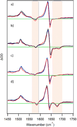
The TRIR spectrum given in panel b of Fig. 2 in the published version of the manuscript is incorrect. The correct figure is shown below.
The Royal Society of Chemistry apologises for these errors and any consequent inconvenience to authors and readers.
| This journal is © the Owner Societies 2023 |

