
Liquid crystal random lasers
Guangyin
Qu
a,
Xiaojuan
Zhang
a,
Siqi
Li
a,
Liang
Lu
a,
Jiangang
Gao
c,
Benli
Yu
a,
Si
Wu
 *b,
Qijin
Zhang
*b and
Zhijia
Hu
*a
*b,
Qijin
Zhang
*b and
Zhijia
Hu
*a
aKey Laboratory of Opto-Electronic Information Acquisition and Manipulation of Ministry of Education, Information Materials and Intelligent Sensing Laboratory of Anhui Province, School of Physics and Optoelectronic Engineering, Anhui University, Hefei 230601, China. E-mail: zhijiahu@ahu.edu.cn
bCAS Key Laboratory of Soft Matter Chemistry, Department of Polymer Science and Engineering, University of Science and Technology of China, Hefei 230026, China. E-mail: siwu@ustc.edu.cn; zqjm@ustc.edu.cn
cDepartment of Polymeric Materials and Engineering, School of Biological and Chemical Engineering, Anhui Polytechnic University, Wuhu, 241000, Anhui, China
First published on 30th November 2022
Abstract
The enthusiasm for research on liquid crystal random lasers (LCRLs) is driven by their unusual optical properties and promising potential for broad applications in manufacturing, communications, medicine and entertainment. From this perspective, we will summarize the most attractive advances in the development of LCRLs in the last decade and propose future prospects. This article will begin with a fundamental description of LCRLs, including the principle of laser generation and a description of LC substances. Then, we spend several chapters on the lasing performance control methods of LCRLs, including random lasing wavelength, threshold, and polarization properties. In addition, we analyze how the LC chiral agent structures, LC core–shell structures and new light-amplifying materials affect the design of LCRL devices. In the last chapter, we discuss the application of LCRLs in 3D displays, information encryption, biochemical sensing and other optoelectronics devices and finally end the perspective with LCRLs’ likely directions in future research.
 Si Wu | Si Wu is a full professor at the University of Science and Technology of China (USTC), Hefei, China. He studied polymer science at USTC and obtained bachelor's degree in 2005. He was supported by the joint doctoral promotion programme working at USTC and the Max Planck Institute for Polymer Research (MPIP), Mainz, Germany. After obtained PhD in 2010, he worked as a postdoctoral fellow at MPIP and became a group leader in 2012. He headed a research group at MPIP from 2012 to 2018. In 2018, he joined USTC as a full professor. His research focuses on photo responsive polymers. |
 Zhijia Hu | Zhijia Hu received the PhD degree in chemistry with the University of Science and Technology of China, Hefei, China, in 2014. He is currently a Professor in School of Physics and Optoelectronics Engineering, Anhui University, Hefei, China. His current research interests include fiber optics, fiber sensors, random lasers, and polymer optical fibers. From 2017 to 2019, he was a Marie Curie Research Fellow in Aston University, Birmingham, UK. |
Introduction
Lasers have been applied in various fields, such as science, technology, economy, military and social development. In 1917, Einstein1 put forward a new concept on the basis of quantum theory: in the interaction between matter and radiation field, the atoms or molecules constituting the matter can produce stimulated emission or absorption of photons under the excitation of photons. This proposal showed that if the number of atoms (molecules) constituting a substance can be reversed according to the thermal equilibrium (Boltzmann) distribution of energy levels, it is possible to achieve light amplification by stimulated emission of radiation (LASER). In the early 1950s, the application of electronics and microwave technology proposed pushing the development of radio technology from microwave to light wave. Therefore, there is a need for an oscillator that can generate controlled light waves similar to a microwave oscillator, namely, a laser. In the conventional laser structure, the resonator plays a key role. However, there is such a laser, which does not have a resonator and mainly depends on the multiple scattering of photons in the scattering medium to achieve sufficient gain to generate the laser. This type of laser is called a random laser (RL). A liquid crystal random laser (LCRL) is an RL with a liquid crystal as the scattering medium.In 1968, Letokhov2 theoretically predicted the existence of RLs in scattering media. To verify the prediction, Lawandy et al.3 used a pulsed laser with 530 nm to pump a methanol solution doped with rhodamine 640 dye and TiO2 nanoparticles and observed a narrow-band emission peak that is similar to a laser. Subsequently, Wiersma and Lagendijk4 formally proposed the concept of RL.
The RL radiation utilizes multiple scattering in disordered gain media and laser gain media to provide optical feedback amplification. The RL does not need an external resonator, and its radiation characteristics are closely related to the disordered medium. In traditional laser theory, scattering is harmful to the laser and should be avoided as much as possible. This is because scattering causes photons to escape from the lasing mode, resulting in a decrease in the quality factor of laser resonators. In the RL system, however, scattering plays a positive role. Multiple scattering increases the path length or dwell time of light in the active medium, and thus enhances light amplification by stimulated emission. The recurrent light scattering provides coherent feedback for lasing oscillation. There are two kinds of feedback: one is intensity or energy feedback and the other is field or amplitude feedback.5 The former feedback is incoherent and nonresonant and can boost incoherent RLs, and the latter is coherent and resonant and can boost coherent RLs.6
Conventional lasers need a resonant cavity such as mirrors to produce coherent feedback (Fig. 1(a)). However, RLs do not have a resonant cavity, and their feedback comes from the multiple scattering of photons among disordered scattering particles (Fig. 1(b)). The photons that are spontaneously emitted undergo multiple scattering before exiting the gain media. During the photons traveling in the gain media, additional photons are generated through the stimulated emission. However, the incoherent feedback provided by scattering only returns part of the photons to the gain media but not to the original position and thus any spatial resonance is absent. When the scattered light is trapped to form a closed loop in the disordered nanostructures, the scattering can provide coherent feedback. Its main characteristic is the laser spikes in the spectral emission, which is the fundamental difference between the features of an RL with coherent feedback and that with incoherent feedback, as shown in Fig. 1(c).
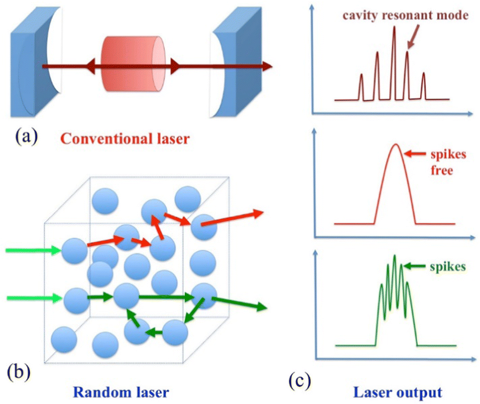 | ||
| Fig. 1 Working principle of lasing in a cavity and random media. (a) A conventional laser cavity; (b) an RL cavity illustrating the incoherent feedback (red arrows) and coherent feedback (green arrows); (c) illustration of spectral outputs of a conventional laser and an RL. The spikes free correspond to incoherent feedback, while the coherent feedback is recognized by its spiky signature. Reproduced with permission from ref. 6. Copyright 2015 Elsevier Ltd. | ||
In view of the wide application prospects of RLs, numerous scholars have reported a large number of theoretical and experimental studies on the fabrication of RL systems to control the lasing output. Liquid crystals (LCs), heavily scattering materials, are used in RL research due to their countless special physical and optical properties. As a special optoelectronic material and strong scattering medium, the LC has crystal anisotropy, which can produce birefringence, Bragg reflection, diffraction and optical rotation effects similar to crystals. It can also generate thermo-optic, electro-optic or magneto-optic effects under the action of an external field.
LC is a state of matter between liquid and crystal, and has both the fluidity of liquid and the anisotropy of crystal (Fig. 2(a)). Commonly, LCs are divided into two categories, lyotropic and thermotropic LCs, according to the formation conditions of LCs. Thermotropic LCs are divided into three categories according to the arrangement structure of LC molecules: smectic phase, nematic phase, and cholesteric phase (Fig. 2(b)).
 | ||
| Fig. 2 (a) The organization of rod-shaped molecules in a crystal, LC, and liquid. (b) The organization of rod-shaped molecules in different LC phases. Reproduced with permission from ref. 19. Copyright 2016 American Chemical Society. | ||
In general, in the cholesteric phase, molecules assigned to consecutive layers are slightly rotated relative to the previous phase. After a certain distance of the pitch P, their orientation is once again the same. The whole pattern is periodic, and if the size of the pitch is on the order of light wavelength scale, the cholesteric phase can exhibit a photonic stop band. Occasionally, cholesteric LCs are called chiral nematic. A perfectly oriented slab of cholesteric LC is a multifunctional material: it is at the same time a reflector, a notch filter, a polarizer, and an optical rotator. In addition, cholesteric LC molecules exhibit dielectric anisotropy, magnetic anisotropy and optical refractive index anisotropy under the action of an external field. Based on the special optical properties of cholesteric LCs, various optical devices can be made, such as LC lasers,7,8 light-controlled switches and displays,9–14 and photonic crystals.15–18
In the smectic phase, the molecules are organized in layers. With dependence on intra- and interlayer correlation among the molecules, different smectic phases have been identified. However, the most common smectic phases are smectic A and smectic C. In the smectic A phase, the molecules are upright in the layers, i.e., the director is parallel to the layer normal. In the smectic C phase, the molecules are tilted, i.e., the director makes a finite angle with the layer normal, and the tilt angle is temperature-dependent.19 The smectic LC has steep viscosity, and the molecules are not easy to rotate, that is, the response speed is low. Therefore, it is considered unsuitable for display devices. At present, the chiral smectic C phase, the ferroelectric phase20,21 among the various smectic phases, has attracted widespread interest. Ferroelectric LCs have excellent characteristics of high speed (microsecond level) and ultrahigh-density memory devices that nematic LCs do not have, which have been extensively studied in recent years.
Compared with other LC phases, the alignment of rod-like molecular centers of gravity in nematic LCs is disordered. They exhibit no pattern in the alignment of molecules. In application, compared with smectic LC, each molecule of nematic LC easily moves freely along the tall axis direction, so the viscosity is modest and the fluidity is rich.22 The arrangement and movement of nematic LC molecules are relatively free, and they are very sensitive to external effects, so they are widely used.23 In addition, nematic and cholesteric LCs can be converted into each other, and adding optically active material (racemic material) to nematic LCs (cholesteric LCs) will form a cholesteric phase (nematic phase).
Blue-phase liquid crystals (BPLCs) are a class of particular mesophases of chiral nematics in which the director axes self-assemble into cylinders comprising double-twist helices. In order of emergence with increasing temperature, the phases are frequently designated BPI, BPII, and BPIII. For BPI, the structure composed of a double-twist cylinder is cubic body-centered, for BPII is simple cubic, and BPIII is thought to be more likely amorphous. BPs are optically active and isotropic, exhibiting Bragg reflection due to their periodic structures. These characteristics, combined with the capability to shift the photonic band gap (PBG) in response to external perturbations, make BPs fascinating materials for applications such as tunable lasers, where laser emission can be realized in three orthogonal directions and can be obtained simultaneously using the three-dimensional PBG.
The direction of the optical axis of the LC molecules can alter with the external conditions, i.e., temperature and electric field, which causes a variety of distinct “texture” and optical properties. The LC can undergo phase transitions at different temperatures. The shift in the alignment direction of the LC molecules leads to the distribution difference of the refractive index. When an electric field is applied to the LC, the alignment direction of the LC molecules changes. Based on these characteristics, LCs can be used as disorder media to control RLs.
In the LCRL, LC is as the disordered scattering medium. By changing the LC phase or adjusting the orientation or arrangement of LC molecules, the anisotropic distribution (or disorder degree) of LC can be controlled, and then the RL radiation characteristics, e.g., lasing wavelength, polarization and threshold, can be tuned.
Control of the random lasing wavelength
When LC is used as a scattering medium, the disorder degree of the system and the orientation of dye molecules can be adjusted by taking advantage of the perfect responsive features of LC. Therefore, the LCRL optical properties can be controlled, which can considerably expand the LCRL application range. The tunable lasing wavelength of cholesteric LC lasers is proposed by controlling the reflection band of cholesteric LCs within the excitation fluorescence spectral range. There are various ways to achieve tunable LCRL, e.g., varying temperature,24,25 voltage,26–30 composition or cell thickness,31,32 mechanical strain in cross-linked cholesteric LC elastomers,33–35 or using reversible photochemical reactions.36,37Hu et al.38 reported a wavelength-tuned LCRL caused by the combined effects of multiple scattering and bandgap control of LCs. By rotating the infrared absorbing material on the side of the LC cell, the tunable random lasing wavelength range of the 80 nm RL from 500 to 700 nm under infrared light irradiation was observed. Interestingly, when the authors doped fluorescent dyes and heavily doped chiral reagents into the LC system, they demonstrated wavelength-tuned RLs rather than side-band lasers due to the combined effect of multiscattering of defective planar texture of cholesteric LC and band-gap control (Fig. 3(a)). Fig. 3(b) shows the transmission spectra for the prepared cholesteric LC cells at different temperatures. The band gap exhibits a redshift effect with decreasing temperature. Fig. 3(c) shows the RL spectra of cholesteric LCs under different NIR (850 nm) irradiation times. In addition, Zhang et al.39 reported a sunlight-fuelled, full-wavelength-tunable (Fig. 3(d)), and chirality-invertible superstructure to delicately manipulate the light wave, where the superstructure was obtained by doping molecular motors into the LC host. With the increase in the intensity of UV irradiation from 0.069 to 0.125 mW cm−2, the lasing wavelength can be steadily tuned to redshift from 569 to 705 nm at a fixed pumped energy of 2.9 μJ per pulse. The reversion of the circular polarization is obtained by changing the stable molecular motor state to an unstable state after the enhancement of the UV intensity. The lasing wavelength can be tuned from 790 to 600 nm with increasing UV irradiation intensity from 0.729 to 1.175 mW cm−2 (Fig. 3(e)).
 | ||
| Fig. 3 (a) Schematic of the band-gap-tailored RL. (b) Transmission spectra of cholesteric LC as a function of temperature. (c) Random lasing spectra of LCs in the cholesteric state at different NIR (850 nm) irradiation times. Reproduced with permission from ref. 38. Copyright 2018 Chinese Laser Press. (d) Measured reflection PBG spectra from the near-UV band to the NIR band for the right- and left-handed helical superstructures. (e) Wide-band tunability of the lasing emission. Reproduced with permission from ref. 39. Copyright 2022 Wiley-VCH GmbH. (f) Schematic of the LC–polymer composite laser with a symmetric sandwich structure in which a dye-doped nematic LC (DDNLC) layer is sandwiched between two polymer-stabilized cholesteric LC (PSCLC) layers. (g) Schematic diagram of the PBG-broadening mechanism of the PSCLC in the presence of one dc voltage: before polymerization, no voltage was applied (V = 0) after polymerization, and the applied voltage > threshold voltage (V > Vth) after polymerization. Reproduced with permission from ref. 40. Copyright 2020 American Chemical Society. | ||
Lin et al.40 demonstrated for the first time an electrically tunable LC-polymer composite laser with a symmetric sandwich structure. This structure includes two identical polymer-stabilized cholesteric LC (PSCLC) layers, and a dye-doped nematic LC (DDNLC) layer sandwiched between them. The PSCLC and DDNLC layers act as distributed Bragg reflectors and half-wave plates with a gain medium, respectively. When the same voltage is applied to the two PSCLC layers, the pitch gradients are simultaneously formed through the two layers because of the electrically induced ion-concentration gradients (Fig. 3(g)). When the voltage increases, the pitch gradients increase, thereby leading to the expansion of the cell PBG and a blueshift of the lasing wavelength. The sandwich cell PBG can expand from 55 to over 170 nm, and the tuning range of the lasing wavelength is as tall as 70 nm (Fig. 3(f)).
Control of the random lasing threshold and efficiency
The threshold is an important index to measure the performance parameters of the laser. In the practical application of the laser, the lower the threshold is, the better. To obtain low threshold lasing emission, there are some factors that should be considered: (I) defect structure. In LCRL systems, a scattering medium is required to provide optical feedback to obtain random lasing. The introduction of defective modes in LCRL systems can enhance the scattering and disorder of the system medium.41 Masanori et al.42 constructed twisted defects from composite films composed of two photopolymerized cholesteric LC (PCLC) layers. Low-threshold random lasing was observed in dye-doped dual-composite PCLC films. Schmidtke et al.41 obtained an exceptionally low threshold through defect mode lasing using a phase-jump structure.(II) The excitation condition, i.e., excitation by circularly polarized light (CPL). It is well known that the introduction of enantiomerically pure chiral compounds or groups into a molecular system induces the chiroptical properties of optical rotatory dispersion and circular dichroism in the ground state. Cholesteric LCs have the potential to form supramolecular helical assemblages because of their intrinsic twisting abilities. Selective reflection of light is the most significant property exhibited by these helical cholesteric LC structures. For instance, when linearly polarized light propagates into a right-handed cholesteric LC cell along the helical axis, right-handed CPL and left-handed CPL are reflected and transmitted. By choosing CPL, the threshold energy in cholesteric LC lasing cells can be obtained. Furumi et al.43 claimed that the CPL excitation with the opposite handedness to the molecular helical sense of the cholesteric LC helix obtains lower threshold power than CPL excitation with the same handedness. In contrast to this, Matsuhisa et al.44 reported CP photoexcitation with the same handedness to obtain a lower threshold power when the excitation wavelength coincides with the edge of the photonic bandgap. This is because the photons stay for a longer time at the edge of the band gap, which is the same conclusion predicted by Belyakov.45
(III) Cholesteric LC materials, i.e., use of extremely birefringent cholesteric LCs. As the value of birefringence Δn increases, the PBG width increases. A CLC cell with a broader PBG width has more pronounced and closely spaced multiple internal reflections near the PBG band edge, resulting in a higher density of states value and lower threshold energy. Chee et al.46 prepared cholesteric LC cells by utilizing three kinds of nematic LC materials with different optical birefringence anisotropy. The results showed that the use of LC cells with an elevated refractive index can reduce the laser threshold energy by more than 2 times.
(IV) Extremely performance dye molecules. Practically all the LCRLs thus far investigated have utilized commercial laser dyes. It is obvious that dyes with higher luminous efficiency (elevated molar absorption coefficient and strong quantum yield) have to be used to increase energy efficiency and decrease the threshold. Uchimura et al.47 synthesized a high-performance dye with elevated luminescence efficiency and extreme solubility in cholesteric LCs. The use of such efficient dyes enabled decreasing lasing thresholds in dye-doped distributed feedback cholesteric LC lasers. The threshold is reduced to one-twentieth of the value for using DCM dye.
(V) Strong multiscattering feedback. Numerous kinds of materials, such as nanoparticles, LC, and even the solution of pure laser dye, can provide the scattering mechanism.48 RLs have no resonant cavity, whose feedback comes from multiple scattering of photons between disordered scattering particles. Strong scattering and great gain are necessary conditions for generating RL radiation. To this end, by enhancing the scattering of the system can lower the lasing threshold. Liu et al.49 fabricated all-organic quasicrystals by spin-coating conjugated polymer poly(2-methoxy-5-(2′-ethyl-hexyloxy)-p-phenylenevinylene films with a thickness of 80 nm on the substrate surface. Compared with conventional dye-doped quasicrystal microcavity lasers, the threshold was reduced by more than 20 times due to strong scattering. Wan et al.50 reported a plasmon-enhanced low-threshold random lasing from dye-doped nematic LCs with titanium nitride (TiN) nanoparticles in capillary tubes. The threshold decreased to 2.37 μJ per pulse with increasing TiN nanoparticle number density from 5.613 × 10¹⁰ mL−1 to 5.314 × 10¹¹ mL−1.
Control of the random lasing polarization state
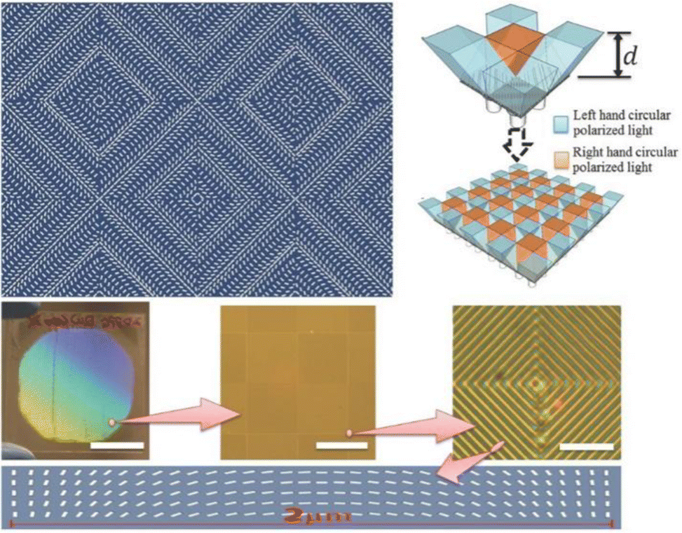
The emission polarization from conventional lasers is commonly controlled by polarizing elements such as Brewster windows or Lyot filters. Polarized laser emission is crucial for practical applications. The polarization of light includes linear polarization, elliptical polarization, and circular polarization. To date, there are three strategies to realize polarized random lasing, i.e., anisotropic scattering,51 anisotropic absorption,52 and anisotropic waveguides.53 The polarization characteristics of LC-based RLs commonly depend on the orientation direction of LC molecules. The methods for fabricating LC molecules orientation mainly include rubbing orientation and stress orientation. Based on the optical anisotropy of nematic LC and the plasticity of polymer mesh, the orientation of LC molecules can be realized by applying stress, such as shear, stretching and compression, and then the intensity and polarization degree of LCRL can be controlled.Yao et al.54 researched LCRL polarization based on dye-doped nematic LCs. The experimental results showed that the output LCRL is linearly polarized light, and its polarization direction is related to the maximum scattering direction and the LC director direction. Chen et al.55 observed and studied in detail bidirectional LCRL emission from an active twisted nematic LC, where the polarization states were asymmetric (nonparallel to each other). In such a laser, the LC acts as a random distributed feedback cavity with an inherently built-in polarization rotator (Fig. 4(a)). Lu et al.56 fabricated a polarization converter and obtained random lasing emission with a vector beam by means of photocontrolled orientation. The obtained converter itself can be directly used as a polarization mask to realize the vector light field in a simple experimental setup, as shown in Fig. 4(b). A converter with P = 1 and ϕ0 = 0 was used to fabricate a q-plate with q = 1. The obtained q-plate and resultant optical vortex are presented in Fig. 4(c) and (d), respectively. Huang et al.57 realized the polarized LCRL emission and found that the waveguide effect reduces the transmission loss of polarized LCRLs whose polarization directions are parallel to the LC molecules (Fig. 4(e)). Yang et al.58 designed and successfully synthesized a different types of carbon polymer dot LCs (CPDLCs) (Fig. 4(f)) with excellent LC properties and fluorescence efficiency. The CPDLCs can be used to fabricate linearly polarized devices. The resulting linearly polarized dichroic ratio (N) and polarization ratio (ρ) are 2.59 and 0.44, respectively (Fig. 4(f)). Du et al.59 successfully designed and fabricated a complex ordered LC polymer film, which was further developed into a holographic polarizer with 90%+ transmittance. Given that the transmittance of conventional absorptive and reflective film polarizers was limited to approximately 45%, the holographic polarizer has great potential to improve the optical efficiency of polarizing optical systems. As presented in Fig. 5, each unit of the polarizer was composed of four LC polymer polarization grating (PG) domains with all grating vectors pointing to common centers. To develop the structured PG into a holographic polarizer, a patterned quarter-wave plate (QWP) with two domains was assembled at this given distance. The function of the patterned QWP was to convert the circular polarization to linear polarization.
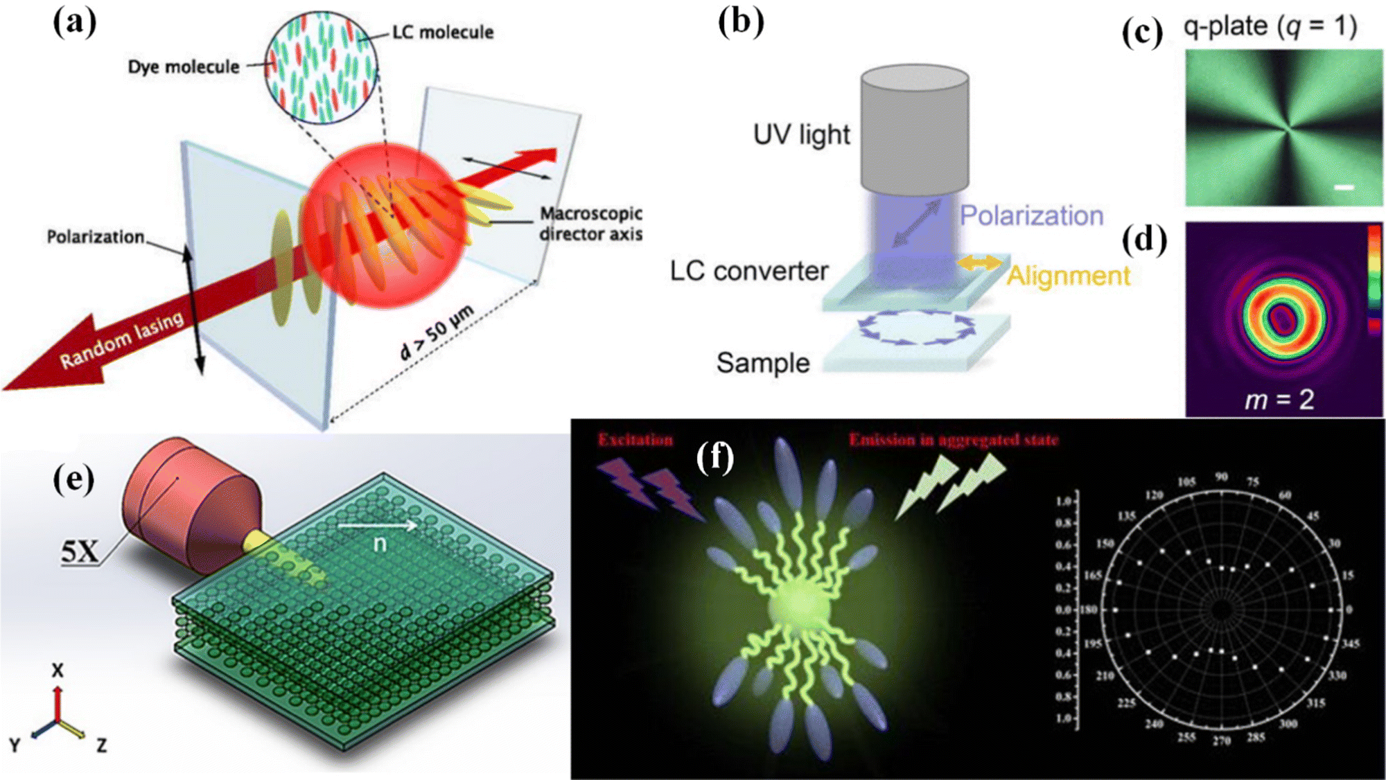 | ||
| Fig. 4 (a) Schematic diagram of RL emission from NPDDLC. Reproduced with permission from ref. 55. Rights managed by AIP Publishing. (b) Vector light experimental setup. (c) Micrograph with P = 1 and Φ0 = 0 converters. (d) Optical vortex captured by CCD. Reproduced with permission from ref. 56. Rights managed by AIP Publishing. (e) Cell configuration and LCM orientation. Reproduced with permission from ref. 57. Copyright 2021 Yunxi Huang et al., published by De Gruyter. (f) Molecular structure of CPD-LCs. Normalized PL intensity of the sample at 370 nm with different polarization angles. Reproduced with permission from ref. 58. Copyright 2021 American Chemical Society. | ||
 | ||
| Fig. 5 The alignment structure of the complex-ordered LC polymer film shows the schematics of the director distribution and the microscopic image of the experimental sample. Reproduced with permission from ref. 59. Copyright 2015 Wiley-VCH Verlag GmbH & Co. KGaA, Weinheim. | ||
The nanoscale LC director distribution comprises polymerizable LC molecules with nanoscale spatially varying directors, which function as a grating that diffracts incident nonpolarized light into left-hand circularly and right-hand circularly polarized light. The diffraction orders of left-handed CPL and right-handed CPL are −1 and +1, respectively, and the light of these two diffraction orders propagates in different directions due to different diffraction angles. This makes it possible to separate the light of the two polarization states within a certain range from the distance grating. This distance is determined by the size of the microdomain and nanodomain. Because the transmittance of conventional absorption- and reflection-type polarizers is less than 50%, such a demonstrated holographic polarizer shows great potential to double the optical efficiency of polarizing optical systems.
The design of the LC material
The design of LC chiral molecules
Chiral molecules, which are omnipresent in nature, feature nonsuperimposed mirror image molecular structures, resulting in a certain handedness determined by the corresponding enantiomer. The introduction of a stereocenter or axial chirality into a molecule produces a remarkable helical superstructure configuration due to the facility of molecular self-organization and the delicate interplay of intra- and intermolecular interactions. The molecular rearrangement driven by a phase transition forms certain spatial structures with molecules regularly located at equilibrium positions, which is referred to as superstructure, such as the typical liquid crystalline 1D periodic cholesteric phase. Cholesteric LCs commonly exhibit typical self-organization characteristics, which promote the formation of elegant helical superstructures or supra-molecular assemblies in chiral environments. Such elegant helices, which are commonly established in mesogenic soft materials via molecular self-organization, have structural parameters with a scale comparable to that of the wavelength of light, thus exhibiting typical optical chirality effects and enabling diverse applications in chiro-optics.60–62 This elegant helical superstructure is usually accompanied by a specific optical activity, that is, the polarization of linearly polarized light is rotated according to the handedness of the chiral medium. This phenomenon is mainly attributed to the difference between the optical indices of refraction of right-handed and left-handed CPL, leading to distinct and prominent chiral dependencies of propagation, transmission, reflection, and absorption, that is, chiral optical properties.63 Periodic helical superstructures of cholesteric LCs can selectively reflect CPL at specific wavelengths that match their helical pitch lengths.Li et al.64 developed the first example of a 1,2-dithienyldicyanoethene-based visible-light-driven chiral fluorescent molecular switch, which exhibits reversible photoisomerization in both organic solvent and LC media. This distinct chiral molecular (Fig. 6(a)) switch exhibits superior thermal and photochemical stability. The induced emissive cholesteric phase, fabricated using this visible-light-driven chiral molecular switch, exhibits reversible circularly polarized reflection and large variation in the intensity of its circularly polarized fluorescence upon visible-light irradiation at two wavelengths. Yu et al.65 demonstrated delicate patterns of three primary colors, red, green, and blue (RGB), with a black background based on piecewise reflection tuning of cholesteric LCs induced by a newly designed light-responsive tristable chirality switch (Fig. 6(b) and (c)). When a chiral molecule is doped into E7, the chiral switch immediately induced the optically tunable helical superstructures and endowed the resultant cholesteric LC system with two adjacent and continuous tuning periods of the reflection, which exhibited piecewise control of the reflection wavelength. By irradiation with 470 and 530 nm light, the selective reflections were tuned in the visible spectra including three primary RGB reflection colors. Subsequently, Li et al.66 designed and synthesized two novel light-driven chiral fluorescent molecular switches (Fig. 6(d)), which can exhibit unprecedented reversible Z/E photoisomerization behavior and tunable fluorescence intensity in isotropic and anisotropic media. Cholesteric LCs fabricated using these original fluorescent molecular switches as chiral dopants exhibited reversible tunable reflection color tuning spanning the visible and infrared regions of the spectra (Fig. 6(e)).
 | ||
| Fig. 6 (a) Light-driven chiral fluorescent molecular switch that can be isomerized using visible light at two wavelengths. Reproduced with permission from ref. 64. Copyright 2019 Wiley-VCH Verlag GmbH & Co. KGaA, Weinheim. (b) Chemical structure of the photoresponsive tristable chiral switch containing Azo near the chiral center and F-Azo far away from the chiral center. The moiety in the dashed box is an important intermediate named M4. (c) Schematic illustration showing the optimized molecular structures of the three configurations of the tristable chiral switch. Reproduced with permission from ref. 65. Copyright 2017 Wiley-VCH Verlag GmbH & Co. KGaA, Weinheim. (d) Chemical structures and reversible photoisomerization process of light-driven chiral fluorescent molecular switches MS-1 and MS-2. (e) Schematic illustration of the self-organized helical superstructures in the cholesteric LCs tuned by visible/UV light, reflecting different wavelengths, and fluorescing with different intensities when excited by blue light. Reproduced with permission from ref. 66. Copyright 2017 Wiley-VCH Verlag GmbH & Co. KGaA, Weinheim. | ||
The design of LC droplets and microshell structures
Typically, cholesteric LCs are investigated in planar geometries for both fundamental scientific studies and device applications. Recently, the special phenomena and properties arising from a planar geometry, such as in droplets and microshells, have been explored.67–70 These studies demonstrated that the impact and range of LC applicability can be considerably enhanced by confining LCs into spherical geometries rather than traditional thin films.Yu et al.71 designed geminate anticounterfeit labels by programming fluorescent cholesteric LC microdroplets with an adjustable reflection color and stable fluorescence properties in microfluidics (Fig. 7(a)). Photon-induced upconversion (UC) is a typical anti-Stokes process that converts two or more low-energy photons into one high-energy photon, which can be achieved by triplet fusion or triplet–triplet annihilation (TTA). Reichmanis et al.72 reported oil/water/oil/water triple emulsion droplets containing ultrathin double shells and active TTA-UC cores (Fig. 7(b)). Photonic shells consist of cholesteric LCs with periodic helical structures. Combined with planar anchoring at the LC boundary, the shell can serve as a resonant cavity for TTA-UC emission and enable UC spectral tuning under low power density excitation. The cholesteric LC shell can be stabilized by introducing polymerizable mesogens in the LC host. Due to the spherical symmetry of the microcapsules, the spontaneous emission of delayed fluorescence is amplified omnidirectionally at the stopband edge. Lee et al.73 used double-shell encapsulated cholesteric LC to design a capsule laser resonator (Fig. 7(c)) to simultaneously achieve high air stability, wavelength and intensity tunability, and lasing-direction controllability. In particular, cholesteric LCs in a granular format provide omnidirectional lasing, which is promising as a point light source. As shown in Fig. 7(d), Lee et al.74 designed and fabricated triple emulsion droplets with opposite chirality in a cholesteric LC core and cholesteric LC shell. The microcapsules exhibit structural colors induced by the two-photon band gap, resulting in a rich and diverse color palette on the optical palette. In addition, the microcapsules can switch the colors from either core or shell depending on the selection of light-handedness. Subsequently, Chen et al.75 investigated triple emulsion cholesteric LC droplets with integrated optical cavities. By combining gain dyes and cholesteric LCs into the core and shell, a dual-gain laser with variable modes can be flexibly configured. The distributed feedback (DFB) mode, formed by the feedback from the self-assembled helix periodic structure of cholesteric LCs, and the whispering gallery (WG) mode are selectively excited by controlling the spatial coupling between the pump beam and the droplet with gain. Using the advantages of dual gain and controllable laser, it is expected to develop a dual wavelength proportional thermometer prototype with self-calibration capability. Lee et al.76 used a microfluidics approach to integrate an ultrathin alignment layer into microcapsules to separate the cholesteric LC core and the elastomeric solid membrane using triple-emulsion drops as the templates (Fig. 7(e)). The cholesteric LC cores enclosed by aqueous shells exhibited striking reflection colors owing to the radial orientation of the helical axes caused by planar alignment at the interface (Fig. 7(f)). Temperature-dependent shifts in the structural colors of the cholesteric LCs render the microcapsules useful for injectable and dispersible colorimetric thermometers. Recently, Park et al.77 created cholesteric LC multishells with multiple stop bands by liquid–liquid phase separation (LLPS) in a simple but extremely controllable manner (Fig. 7(g)). With an optimum combination of ternary mixtures E7 LC, tetrahydrofuran (THF), and propylene glycol (PG), multiple-emulsion drops were produced in a very controlled manner. Interestingly, the concentration of the chiral dopant decreased from the outermost to the innermost cholesteric LC drop due to nonuniform partitioning during LLPS, which resulted in multiple stopbands. Therefore, the photonic multishells showed multiple structural colors. In addition, dye-doped multishells provided band-edge lasing at two different wavelengths.
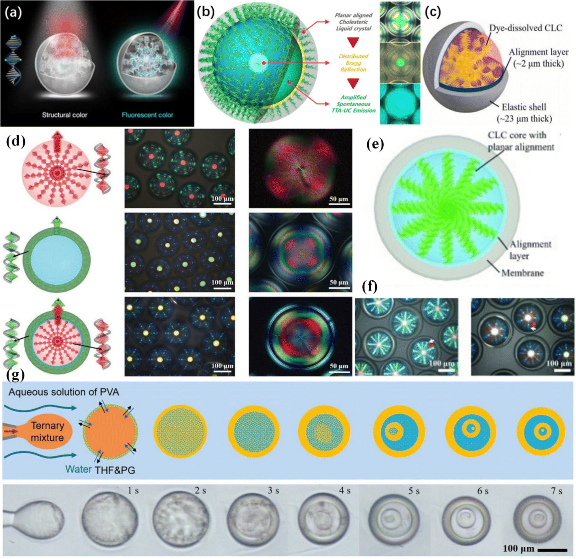 | ||
| Fig. 7 (a) Schematic illustration showing fluorescent cholesteric LC microdroplets (two-tone inks) combining structural color under white light and fluorescent color upon UV irradiation. Reproduced with permission from ref. 71. Copyright 2021, Yu et al. (b) Distributed Bragg reflection from an O/W/O/W triple-emulsion microcapsule with ultrathin double shells and an active TTA-UC core. Reproduced with permission from ref. 72. Copyright 2017 American Chemical Society. (c) Schematic of a cholesteric LC droplet in an elastic capsule separated by an aqueous alignment layer. Reproduced with permission from ref.73. Copyright 2018 The Authors. (d) Schematic diagram with the selection of right-handed light, left-handed light, and no selection. OM images of the core–shell capsules in reflection mode with the selection of right-handed light, left-handed light, and no selection. OM image in transmission with cross-polarization. Reproduced with permission from ref. 74. Copyright 2017 Wiley-VCH Verlag GmbH & Co. KGaA, Weinheim. (e) Schematic representation of a double-shell microcapsule with a cholesteric LC core with radially aligned helices. (f) OM images of cholesteric LC microcapsules with three different bandgap positions taken in reflection mode without polarizers. Reproduced with permission from ref. 76. Copyright 2015 Wiley-VCH Verlag GmbH & Co. KGaA, Weinheim. (g) Schematic and microscopic images of fabricating multiple concentric cholesteric LC shells through one-step emulsification. Reproduced with permission from ref. 77. Copyright 2020 Wiley-VCH Verlag GmbH & Co. KGaA, Weinheim. | ||
Compatibility of LCs and gain materials
Due to the disadvantages of chemical and optical instability, strong photothermal effects and triplet intrinsic three-level systems as the gain medium of LC lasers, doped fluorescent dyes limit the development of commercial applications of LC lasers.78–81 Studies have shown that optical gain materials of organic hybrid materials exhibit perfect amplified spontaneous emission and lasing properties, such as inorganic perovskites and colloidal quantum dots.82–86 By improving the mutual solubility of LCs and inorganic gain materials, LCRLs with better performance have been obtained.Chen et al.87 demonstrated an all-inorganic perovskite quantum dot (AIPQD)-doped cholesteric LC (AIPQDs-CLC) laser with greatly flexible tunability and long-term stability (Fig. 8(a)). AIPQD materials are efficient optical gain media in cholesteric LC planar structure optical resonator structures. The line width and energy threshold of the lasing emission from the AIPQD-CLC laser can be as narrow as 0.20 nm and decreased to 0.15 μJ per pulse, respectively. The AIPQD-CLC laser can retain approximately 87% of its initial laser efficiency after being stored at room temperature and 60% extreme humidity for half a year. Wang et al.88 developed an LCRL using high-concentration ZnCdSeS/ZnS alloy quantum dot-doped polymer dispersed LCs (QD-PDLCs) (Fig. 8(b)). The optimization of the threshold, linewidth and intensity of QD-PDLC RL was achieved by changing the LC concentration. The optimized QD-PDLC showed great stability. The laser output remained at 92% of the original emission intensity after 15 days of laser excitation (3 h per day). Satapathy et al.89 reported the rapidly electrically switching anisotropic photoluminescence of nanosoft composites composed of nematic LCs and cesium-lead halide perovskite quantum cubes. The LC material employed has a positive dielectric anisotropy, and as expected for such materials, an applied electric field applies a positive torque on the director n, allowing field-controlled orientation of the LC molecules. Thus, the LC molecules initially oriented in the plane of the substrate (along the X direction in the reference coordinate notation shown in Fig. 8(c)) could be reoriented to the historic direction (Z direction, also the excitation beam direction) by the application of an electric field. Application of an AC electric field of minor amplitude and its consequent coupling to the LC director was employed to continuously vary the magnitude of the green emission. These studies are important to obtain backlight sources with narrow full-width-at-half-maximum (FWHM) and polarized emission for achieving high-quality low-cost LC displays.
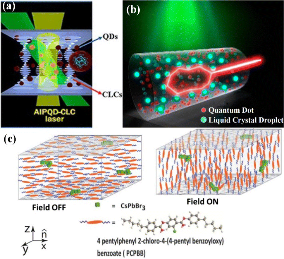 | ||
| Fig. 8 (a) Schematic illustration of the development of a closed-loop path of light via recurrent scattering inside the QD-PDLC. Reproduced with permission from ref. 87. Copyright 2018 American Chemical Society. (b) Schematic diagram of the AIPQD-CLC experiment. Reproduced with permission from ref. 88. Copyright 2020 American Chemical Society. (c) Illustration schematically showing the arrangement of the LC [4-pentylphenyl 2-chloro-4-(4-pentyl benoyloxy)benzoate or PCPBB] molecules and the QCs (CsPbBr3) forming a linear assembly. Reproduced with permission from ref. 89. Copyright 2019 Wiley-VCH Verlag GmbH & Co. KGaA, Weinheim. | ||
Application of LCRLs
Display
Structural color is a rare coloration originating from physical interactions between light and periodic submicrometer structures of matter.90,91 The assembled cholesteric LCs have periodic arrangements of twisted layers consisting of rod-like molecules or nano-objects aligned in parallel. They form the most representative structural color analogs since they recapitulate the self-organized helical architecture abundant in nature.92,93 As one of the chromogenic materials, responsive photonic crystals, composed of periodically structured materials with different refractive indices, exhibit tunable structural colors.94,95Cholesteric LCs have been investigated advantageously in creating rewritable photonic paper. Yu et al.96 reported a different strategy to write, erase and, importantly, tune the colors by developing unique light-driven cholesteric LCs. This LC has a self-organized helical superstructure with two structural elements: the pitch length (lattice constant) and the helical axis (reconfiguration) (Fig. 9(a)). The reconfiguration of the helical axis provides two high-contrast optical states for writing and erasing by pressure and current, while precise optical control of the pitch length facilitates local color tuning. Distinct multicolor patterns were mechanically written, optically tuned, and electrically erased on rewritable photonic paper in a programmable manner (Fig. 9(b)). This inkless photonic paper has great potential to improve user experience, opening a way to record, program and memorize complex images through visual color information and user-interactive display technologies.
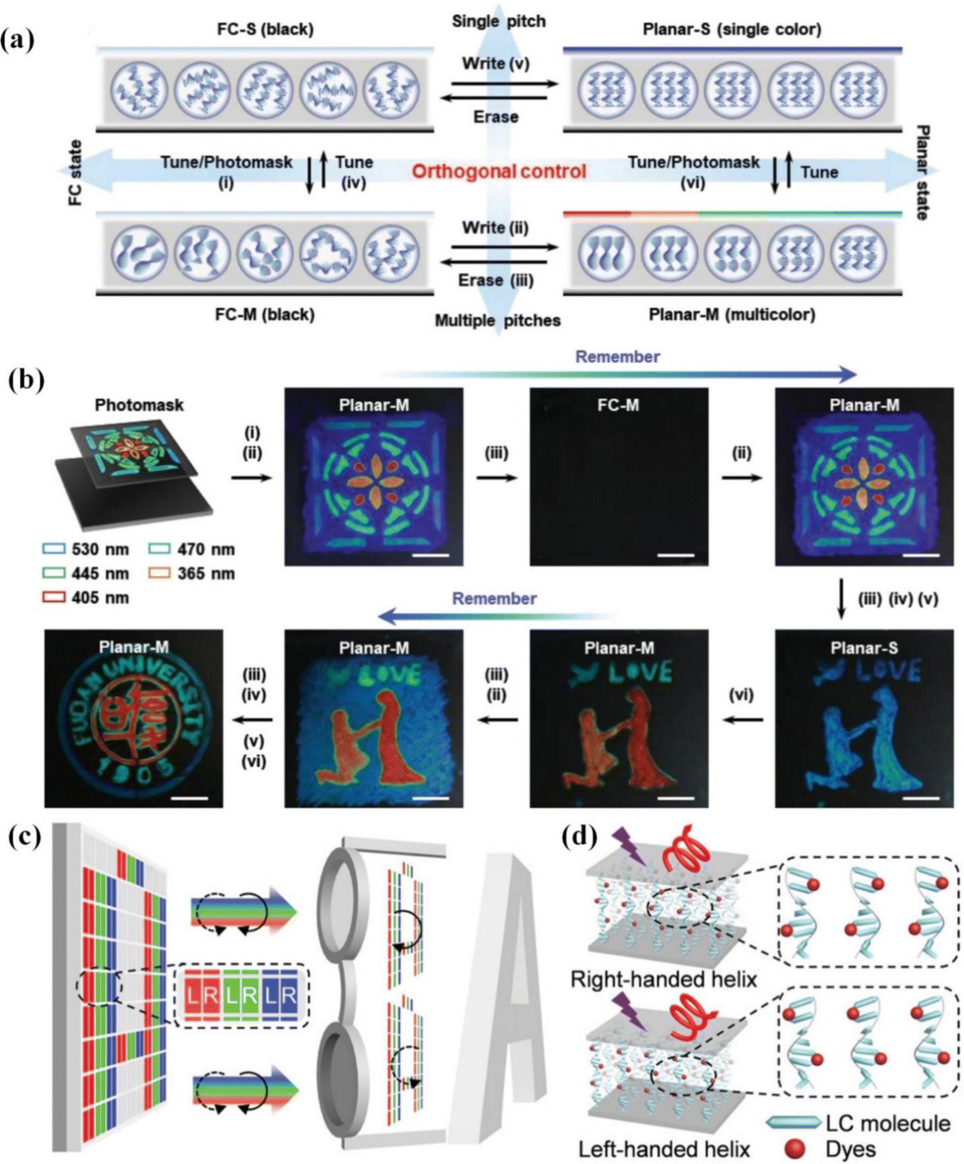 | ||
| Fig. 9 (a) Schematic illustration showing the orthogonal control of the optical states (helical axis) and structural colors (pitch) in the photonic paper. (b) Sequential photographs showing the distinct multicolor patterns recorded on the photonic paper by optical tuning, mechanical writing, and electrical erasure. Reproduced with permission from ref. 96. Copyright 2022 Wiley-VCH GmbH. (c) Schematic illustration of 3D laser displays based on CP microlaser arrays. (d) PBG-induced CPL emissions in dye-doped cholesteric LCs. Reproduced with permission from ref. 102. Copyright 2021 Wiley-VCH GmbH. | ||
In general, stereoscopic displays are realized through a linear-polarization-multiplexed approach, which present full-color images with orthogonal polarization states projecting into the left and right eyes, respectively, thereby rendering a compelling 3D sensation for the viewers. Cholesteric LCs, with the capability of assembling into right-handed or left-handed helical superstructures, enable the generation of intense CPL for their fantastic light manipulation capabilities.97,98
It seems that LCs would be promising candidates for the fabrication of pixelated laser display panels. However, the realization of such panels has been hindered by the difficulties in fabricating miniaturized individual LC lasers and patterning them into periodic red-green-blue pixel matrices.99,100 To solve this, Zhao et al.101 proposed a strategy of microtemplate-assisted inkjet printing to construct full-color laser display panels composed of pixelated LC microlaser arrays. Using the prepared LC microlaser pixel matrices as display panels, they achieved full-color laser displays.
Zhao et al.102 further used these printed cholesteric LC red-green-blue (RGB) microlaser arrays as display panels and demonstrated proof-of-concept full-color 3D laser displays by delivering images with orthogonal CP laser emissions into one's left and right eyes (Fig. 9(c)). These results provide valuable inspiration for the development of 3D laser display technology. The excellent circular polarization characteristic of cholesteric LCs enabled ultrahigh circular polarization degree values (gem = 1.6) of the CP laser emissions. As depicted in Fig. 9(d), thanks to the Bragg reflection and circular dichroism properties brought by their periodic helical superstructures, the propagation of right-handed CPL with wavelengths located in the PBG is forbidden in right-handed CLCs, leading to selective reflection of R-CPL, whereas left-handed CPL is transmitted, and vice versa. However, each adjacent RGB microlaser constituted an individual display pixel, which enabled full-color tunable CP lasing by properly adjusting the manner of excitation. The outstanding performance of full-color CP laser arrays opens a new avenue for the innovation of device architecture in next-generation 3D displays.
Information encryption
With the rapid development of photonic information technology, the need for high-density and high-security confidential labels has become increasingly urgent and has aroused great interest.103–105 Currently, the forefront of next-generation anti-counterfeiting technologies focuses on designing security materials that are able to offer numerous optical states to convey distinct information. LC materials, with their natural photonic crystal superstructure and excellent doping flexibility, are potential candidates for the development of narrow-bandwidth lasers13 that allow cryptographic applications as cryptographic primitives. Furthermore, as flexible organic laser systems, LCs have excellent processability and can be fabricated into ordered microlaser arrays, providing opportunities for high-throughput parallel encryption.106Zhao et al.107 fabricated LC microlaser arrays by a microtemplate-assisted inkjet printing method. The helical pitch of the LC superstructure was changed by the photoisomerization of chiral dopants, thereby realizing wavelength regulation of the LC microlaser. On this basis, using the wavelength of the LC microlaser as the main code, the encryption and authentication of the optically tunable information of the LC microlaser array was realized. Information was encrypted by utilizing LC microlasers as cryptographic primitives for information encryption, whose wavelengths represented binary codes. According to the ASCII binary code, any character can be converted into a standard 8-bit binary code sequence; for example, “01010101” was the capital letter “U”. Therefore, specific raw information consisting of multiple English characters “UCAS” can be efficiently encrypted in a series of LC microlasers, where the two laser wavelengths represent “0” and “1”, respectively. Next, photoresponsive and photoinactive chiral dopants were added into the LC matrix to prepare Ink 1 and Ink 2, respectively. The original information was encoded into an 8 × 4 LC microlaser array by inkjet printing. As shown in Fig. 10(a), the real information can only be obtained after UV irradiation and can be rehidden by visible light irradiation after UV irradiation. These results provide a feasible platform for the realization of microlaser-based cryptographic primitives in high-security information protection and anti-counterfeiting applications. Yu et al.71 constructed a two-color ink by replacing the pigment color of the luminescent material with the structural color of the photonic crystal “coating”. The fluorescent cholesteric LC (FCLC) was formed into monodisperse droplets using capillary microfluidic technology. These structurally defined microdroplets fabricated by a capillary microfluidic technique contributed to different but intact messages of both reflective and fluorescent patterns in the geminate labels. To test the authors’ hypothesis about improving the level of anti-counterfeiting, a two-color “anti-counterfeiting label” was also created using two-tone ink and demonstrated according to the schematic diagrams shown in Fig. 10(b). With this strategy, the geminate labels were hidden in colorful packaging of the goods by programming the structural color of the microdroplets, which were difficult to recognize under white light. However, the specific patterns encrypted in the fluorescent state of the labels will appear upon UV irradiation. This two-tone ink opens new opportunities for the combination of two fundamental optical features (reflection and fluorescence) and has enormous potential to considerably enhance the anti-counterfeiting level of security labels, providing a versatile platform for encryption and protection of valuable authentic information in anti-counterfeiting technology.
 | ||
Fig. 10 (a) Concept of information encryption with patterned LC microlasers serving as cryptographic primitives. Information authentication and hiding processes on an LC microlaser array. Reproduced from ref. 107. Copyright 2020 AAAS. (b) Encryption and decryption of an enhanced-security-level geminate label created by eight kinds of microdroplets in a “pixelated” array (21 × 21). The fluorescent QR code of “LC” is concealed behind a colorful reflective “Christmas tree”, which can be decrypted by smartphones upon UV irradiation. Each pixel is 2.0 mm × 2.0 mm × 400 μm, and the distance between two pixels is 0.5![[thin space (1/6-em)]](https://www.rsc.org/images/entities/char_2009.gif) mm. Reproduced with permission from ref. 71. Copyright 2021, Yu et al. mm. Reproduced with permission from ref. 71. Copyright 2021, Yu et al. | ||
Biochemical sensing
The unique combination of LC responsiveness to the environment and the striking optical effects that allow the rapid visualization of this response facilitates the use of LCs in biochemical applications.The LC-based sensor, recently proposed by Duan et al.,108 has a great sensitivity to urine molecules and can detect a molecular concentration as low as 0.1 mM (Fig. 11(a)). The system enabled quantitative and sensitive detection in real-time processing with much higher resolution (detection limit) than the previously described urea detector. These advantages were the result of acid-doped 5CB LC and WGM laser technology. The enzymatic reaction between urea and urease produced hydroxide ions. Deprotonation and self-assembly of stearic acid occurred at the aqueous/LC interface with increasing pH, promoting the reorientation of LC molecules. The detectable shift of WGM lasing spectra related to the configuration transition of LC microdroplets can be used as an indicator of enzymatic reaction, and this allows detection of urea molecules by the sensor at a concentration of only 0.1 mM. This property makes such systems the best candidates for monitoring enzymatic reactions (Fig. 11(b)).
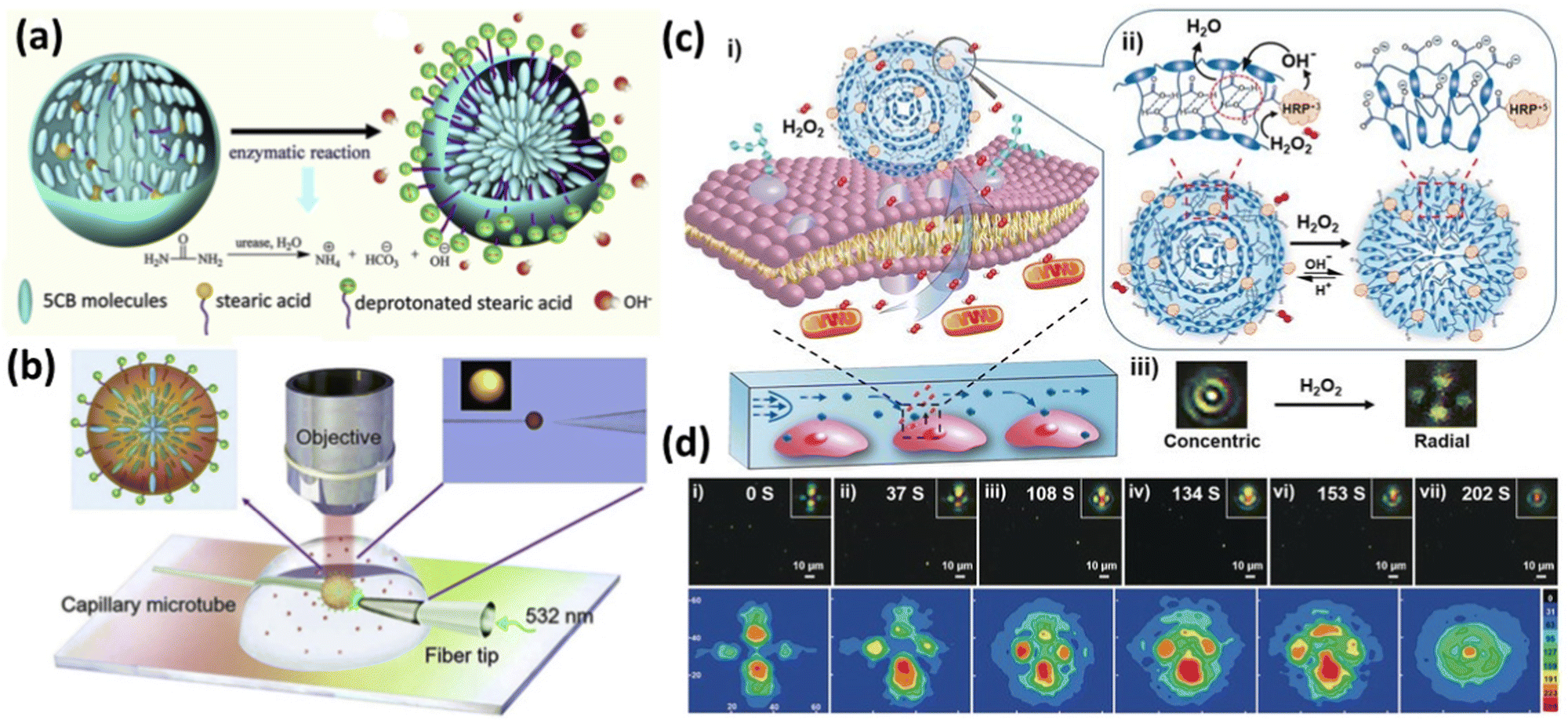 | ||
| Fig. 11 (a) Schematic illustration of the structural transition of stearic acid-doped 5CB microdroplets from planar anchoring to homeotropic anchoring. (b) Schematic diagram of the WGM lasing experimental setup. The 532 nm pump light was guided to the surface of the 5CB microdroplet via a fiber tip. The detection of H2O2 released from a single living cell by the LC elastomer microdroplet immobilized on the cell membrane. Reproduced with permission from ref. 108. Copyright 2019 Elsevier B.V. All rights reserved. (c) Schematic illustration of the experimental system and (d) POM images of reversible (radial-to-concentric) configuration after exposure to H2O2 (1.2 mM) at pH ¼ 6.7, and corresponding intensity distribution data of the LCEM. Reproduced with permission from ref. 109. Copyright 2020 Wiley-VCH Verlag GmbH & Co. KGaA, Weinheim. | ||
Decorating LCs with stimuli-responsive materials builds a bridge between LCs and biochemical interactions. The arrangement of LC molecules is particularly sensitive. These properties make LCs highly attractive for the development of biosensors that enable high-sensitivity and label-free detection of biomarkers at the water/liquid phase interface. Herein, Li et al.109 synthesized LC elastomer microspheres functionalized with horse-radish peroxidase (LCEM-HRP), which can be immobilized directly on the cell membrane to monitor the real-time release of H2O2 at the single-cell level (Fig. 11(c)). The amount of H2O2 released by cells was indirectly reflected by the deformation level of LCEM-HRP. The estimated detection sensitivity was 2.2 × 10⁻⁷ μm for a detection time of 10 min. LCEM-HRP provided a new method for in situ real-time imaging of H2O2-releasing enzymes in living cells and can serve as the basis for finding more advanced chemical probes for imaging various signaling molecules in the cellular microenvironment (Fig. 11(d)).
Summary and outlook
In recent years, LCRLs have achieved rapid and significant development in both science and technology. In particular, significant progress has been made in enhancing the performance and extending the functionality of LCRLs by designing the composition and structure of LC materials. The performance and threshold of LCRLs have been improved. Various control methods (electricity, magnetism, heat) are adopted, which drive the practical applications of LCRLs. This perspective summarizes the generation principles of LCRLs. The progress of performance optimization of LCRLs, including low laser threshold, wide-range wavelength regulation, polarization control, material design and modification, and applications including 3D display and information encryption, are mainly introduced.Although great progress has been made in the research of LCRLs, there are still some challenges. For the application of LCRLs to practical scenarios in the future, it is necessary to focus on promoting the research in the following aspects:
Fast response LCs
With the future development of nanophotonic circuits/chips with multiple functions, rapid or ultrafast responses of LC-based devices are necessary. At present, most LC-based devices mainly adopt the electric field driving method to achieve millisecond-level responses. Liu et al.110 achieved a millisecond response time by randomly confining microscale LC droplets in a polymer matrix. To obtain faster speeds, people can choose some special LCs, such as ferroelectric LCs,21,111,112 which mostly have a response time of microseconds.Multi-function or multi-control Integration
It would be great for a single device to perform multiple functions. LC devices look promising for a variety of functions. For example, azo dye-doped LCs and azobenzene LCs96,113–118 can respond not only to electric fields but also to light, which allows us to control them more freely.LC microphotonics
The properties of photon generation, transmission, and redirection will be the subject of future research in integrated microphotonics. Electrons can be accelerated and redirected by the force of an electric field, whereas photons cannot be accelerated or stopped by an external field. In this way, we need to control the flow of photons by means of the interaction of matter and photons. Fluorescence is a highly powerful and quick phenomenon of this sort, and it can be designed in terms of both chemistry and material properties. In the future, fluorescence control in LC-integrated microphotonics will most likely be used to realize rapid and possibly even ultrafast circuits based on self-organized LCs and other forms of soft matter.119Sensors
The orientation of LC molecules is easily affected by external forces, and the changes in color and brightness of LC can also be observed by polarized light microscopy. Therefore, LC sensing is expected to develop into an inexpensive, sensitive, and portable detection method. Decades of development of LC biosensors108,120 have yielded fruitful results in biological and chemical analysis, but more work needs to be done from experimental research to practical application. Due to the influence of factors such as LC film thickness, temperature, and reaction time, the stability and reproducibility of LC biosensors are worth investigating. In addition, there is still much room for exploration of various innovative LC-based biosensors, such as exploiting the electrical, optoelectronic, and dielectric properties of LC to develop novel sensors.Conflicts of interest
The authors declare no competing financial interest.Acknowledgements
This work was financially supported by National Natural Science Foundation of China (No. 51973204, 12174002, 11874012, 52073268 and 52120105004), Innovation project for the Returned Overseas Scholars of Anhui Province (2021LCX011); Key Research and Development Plan of Anhui Province (202104a05020059); The University Synergy Innovation Program of Anhui Province (GXXT-2020-052); Hefei Municipal Natural Science Foundation (No. 2021013), Anhui Provincial Natural Science Foundation (No. 1908085MB38) and the Fundamental Research Funds for the Central Universities (WK3450000006, WK2060190102); Anhui Project (Grant No. Z010118167).References
- A. Siegman and H. Weichel, Am. J. Phys., 1974, 42, 521–523 CrossRef.
- V. Letokhov, Sov. Phys. JETP, 1968, 26, 835–840 Search PubMed.
- N. M. Lawandy, R. Balachandran, A. Gomes and E. Sauvain, Nature, 1994, 368, 436–438 CrossRef.
- D. S. Wiersma, M. V. Albada and A. Lagendijk, Nature, 1995, 373, 203–204 CrossRef CAS.
- H. Cao, Wave Random Media., 2003, vol. 13, p. R1 Search PubMed.
- F. Luan, B. Gu, A. S. Gomes, K.-T. Yong, S. Wen and P. N. Prasad, Nano Today, 2015, 10, 168–192 CrossRef CAS.
- V. Kopp, B. Fan, H. Vithana and A. Genack, Opt. Lett., 1998, 23, 1707–1709 CrossRef CAS.
- P. Palffy-Muhoray and B. Taheri, Opt. Lett., 2001, 26, 804–806 Search PubMed.
- R. S. Zola, H. K. Bisoyi, H. Wang, A. M. Urbas, T. J. Bunning and Q. Li, Adv. Mater., 2019, 31, 1806172 Search PubMed.
- Y. Wang and Q. Li, Adv. Mater., 2012, 24, 1926–1945 CrossRef CAS PubMed.
- H. K. Bisoyi and Q. Li, Chem. Res., 2014, 47, 3184–3195 CrossRef CAS.
- M.-C. Choi, Y. Kim and C.-S. Ha, Prog. Polym. Sci., 2008, 33, 581–630 CrossRef CAS.
- H. Coles and S. Morris, Nat. Photonics, 2010, 4, 676–685 CrossRef CAS.
- P. Chaudhari, J. Lacey, J. Doyle, E. Galligan, S.-C. A. Lien, A. Callegari, G. Hougham, N. D. Lang, P. S. Andry and R. John, Nature, 2001, 411, 56–59 CrossRef CAS.
- A. Blanco, E. Chomski, S. Grabtchak, M. Ibisate, S. John, S. W. Leonard, C. Lopez, F. Meseguer, H. Miguez and J. P. Mondia, Nature, 2000, 405, 437–440 CrossRef CAS PubMed.
- A. C. Arsenault, D. P. Puzzo, I. Manners and G. A. Ozin, Nat. Photonics, 2007, 1, 468–472 CrossRef CAS.
- W. Cao, A. Munoz, P. Palffy-Muhoray and B. Taheri, Nat. Mater., 2002, 1, 111–113 CrossRef CAS PubMed.
- T. T. Larsen, A. Bjarklev, D. S. Hermann and J. Broeng, Opt. Express, 2003, 11, 2589–2596 CrossRef CAS PubMed.
- H. K. Bisoyi and Q. Li, Chem. Rev., 2016, 116, 15089–15166 CrossRef CAS PubMed.
- D. Miyajima, F. Araoka, H. Takezoe, J. Kim, K. Kato, M. Takata and T. Aida, Science, 2012, 336, 209–213 CrossRef CAS PubMed.
- D. M. Walba, E. Korblova, R. Shao, J. E. Maclennan, D. R. Link, M. A. Glaser and N. A. Clark, Science, 2000, 288, 2181–2184 CrossRef CAS PubMed.
- D. R. Link, G. Natale, R. Shao, J. E. Maclennan, N. A. Clark, E. Korblova and D. M. Walba, Science, 1997, 278, 1924–1927 CAS.
- M. Mitov, Adv. Mater., 2012, 24, 6260–6276 CrossRef CAS PubMed.
- S. Morris, A. Ford, C. Gillespie, M. Pivnenko, O. Hadeler and H. Coles, J. Soc. Inf. Disp., 2006, 14, 565–573 CrossRef CAS.
- A. Chanishvili, G. Chilaya, G. Petriashvili, R. Barberi, R. Bartolino, G. Cipparrone, A. Mazzulla, R. Gimenez, L. Oriol and M. Pinol, Appl. Phys. Lett., 2005, 86, 051107 CrossRef.
- Q. Song, L. Liu, L. Xu, Y. Wu and Z. Wang, Opt. Lett., 2009, 34, 298–300 CAS.
- B. Maune, M. Lončar, J. Witzens, M. Hochberg, T. Baehr-Jones, D. Psaltis, A. Scherer and Y. Qiu, Appl. Phys. Lett., 2004, 85, 360–362 CrossRef CAS.
- R. Liao, X. Zhan, X. Xu, Y. Liu, F. Wang and D. Luo, Liq. Cryst., 2020, 47, 715–722 CrossRef CAS.
- H. Lu, L. Yang, L. Xia, J. Kong, M. Xu, J. Zhu, L. Qiu and Z. Hu, Liq. Crys., 2021, 48, 255–262 CrossRef CAS.
- J. Xiang, A. Varanytsia, F. Minkowski, D. A. Paterson, J. M. Storey, C. T. Imrie, O. D. Lavrentovich and P. Palffy-Muhoray, Proc. Natl. Acad. Sci. U. S. A., 2016, 113, 12925–12928 CrossRef CAS PubMed.
- C.-T. Wang and T.-H. Lin, Opt. Express, 2008, 16, 18334–18339 CrossRef CAS PubMed.
- K. Sonoyama, Y. Takanishi, K. Ishikawa and H. Takezoe, Jpn. J. Appl. Phys., 2007, 46, L874 CrossRef CAS.
- A. Varanytsia, H. Nagai, K. Urayama and P. Palffy-Muhoray, Sci. Rep., 2015, 5, 1–8 Search PubMed.
- J. Schmidtke, S. Kniesel and H. Finkelmann, Macromolecules, 2005, 38, 1357–1363 CrossRef CAS.
- H. Finkelmann, S. T. Kim, A. Munoz, P. Palffy-Muhoray and B. Taheri, Adv. Mater., 2001, 13, 1069–1072 CrossRef CAS.
- A. Chanishvili, G. Chilaya, G. Petriashvili, R. Barberi, R. Bartolino, G. Cipparrone, A. Mazzulla and L. Oriol, Appl. Phys. Lett., 2003, 83, 5353–5355 CrossRef CAS.
- S. Furumi, S. Yokoyama, A. Otomo and S. Mashiko, Appl. Phys. Lett., 2004, 84, 2491–2493 CrossRef CAS.
- H. Lu, J. Xing, C. Wei, J. Xia, J. Sha, Y. Ding, G. Zhang, K. Xie, L. Qiu and Z. Hu, Photonics. Res., 2018, 6, 390–395 CrossRef CAS.
- J. Zhang, F. Wang, S. Ghafoor, H. Wang, W. Zhang, K. Xie, R. Cheng, J. Yan, L. Niu and P. Wang, Adv. Opt. Mater., 2022, 10, 2200426 CrossRef CAS.
- J.-D. Lin, Y.-S. Zhang, J.-Y. Lee, T.-S. Mo, H.-C. Yeh and C.-R. Lee, Macromolecules, 2020, 53, 913–921 CrossRef CAS.
- J. Schmidtke, W. Stille and H. Finkelmann, Phys. Rev. Lett., 2003, 90, 083902 CrossRef.
- M. Ozaki, R. Ozaki, T. Matsui and K. Yoshino, Jpn. J. Appl. Phys., 2003, 42, L472 CrossRef CAS.
- S. Furumi and Y. Sakka, Adv. Mater., 2006, 18, 775–780 CAS.
- Y. Matsuhisa, Y. Huang, Y. Zhou, S.-T. Wu, R. Ozaki, Y. Takao, A. Fujii and M. Ozaki, Appl. Phys. Lett., 2007, 90, 091114 CrossRef.
- V. Belyakov, Mol. Cryst. Liq. Cryst., 2006, 453, 43–69 CrossRef CAS.
- M. G. Chee, M. H. Song, D. Kim, H. Takezoe and I. J. Chung, Jpn. J. Appl. Phys., 2007, 46, L437 CrossRef CAS.
- M. Uchimura, Y. Watanabe, F. Araoka, J. Watanabe, H. Takezoe and Gi Konishi, Adv. Mater., 2010, 22, 4473–4478 CrossRef CAS PubMed.
- L. Ye, Z. Yin, C. Zhao, C. Hou, Y. Wang, Y. Cui and Y. Lu, J. Mod. Opt., 2013, 60, 1607–1611 CrossRef CAS.
- Z. Liu, R. Chen, Y. Liu, X. Zhang, X. Sun, W. Huang and D. Luo, Opt. Express, 2017, 25, 21519–21525 CrossRef CAS PubMed.
- Y. Wan, Y. An and L. Deng, Sci. Rep., 2017, 7, 1–8 CrossRef PubMed.
- Y. Ni, L. Gao, A. Miroshnichenko and C. Qiu, Opt. Express, 2013, 21, 8091–8100 CrossRef CAS PubMed.
- C. Wu, P. Tsay, H. Cheng and S. Bai, J. Appl. Phys., 2004, 95, 417–423 CrossRef CAS.
- E. S. Leong, S. F. Yu, A. Abiyasa and S. P. Lau, Appl. Phys. Lett., 2006, 88, 091116 CrossRef.
- F. Yao, W. Zhou, H. Bian, Y. Zhang, Y. Pei, X. Sun and Z. Lv, Opt. Lett., 2013, 38, 1557–1559 CrossRef PubMed.
- C.-W. Chen, H.-P. Huang, H.-C. Jau, C.-Y. Wang, C.-W. Wu and T.-H. Lin, J. Appl. Phys., 2017, 121, 033102 CrossRef.
- P. Chen, W. Ji, B.-Y. Wei, W. Hu, V. Chigrinov and Y.-Q. Lu, Appl. Phys. Lett., 2015, 107, 241102 CrossRef.
- Y. Huang, X. Zhang, B. Yu, J. Ma, K. Xie, S. Cheng, J. Zhang and Z. Hu, Nanophotonics, 2021, 10, 3541–3547 CrossRef.
- Q. Yang, J.-C. Zhu, Z.-X. Li, X.-S. Chen, Y.-X. Jiang, Z.-W. Luo, P. Wang and H.-L. Xie, ACS Appl. Mater. Interfaces, 2021, 13, 26522–26532 CrossRef CAS PubMed.
- T. Du, F. Fan, A. M. W. Tam, J. Sun, V. G. Chigrinov and H. Sing Kwok, Adv. Mater., 2015, 27, 7191–7195 CrossRef CAS PubMed.
- E. Yashima, N. Ousaka, D. Taura, K. Shimomura, T. Ikai and K. Maeda, Chem. Rev., 2016, 116, 13752–13990 CrossRef CAS PubMed.
- Z. Yu, H. K. Bisoyi, X. M. Chen, Z. Z. Nie, M. Wang, H. Yang and Q. Li, Angew. Chem., Int. Ed., 2022, 134, e202200466 Search PubMed.
- H. Yu and T. Ikeda, Adv. Mater., 2011, 23, 2149–2180 CrossRef CAS PubMed.
- Z. G. Zheng, Y. Q. Lu and Q. Li, Adv. Mater., 2020, 32, 1905318 CrossRef CAS PubMed.
- J. Li, H. K. Bisoyi, S. Lin, J. Guo and Q. Li, Angew. Chem., Int. Ed., 2019, 58, 16052–16056 CrossRef CAS PubMed.
- L. Qin, W. Gu, J. Wei and Y. Yu, Adv. Mater., 2018, 30, 1704941 CrossRef PubMed.
- J. Li, H. K. Bisoyi, J. Tian, J. Guo and Q. Li, Adv. Mater., 2019, 31, 1807751 CrossRef PubMed.
- I.-H. Lin, D. S. Miller, P. J. Bertics, C. J. Murphy, J. J. De Pablo and N. L. Abbott, Science, 2011, 332, 1297–1300 CrossRef CAS PubMed.
- Y. Uchida, Y. Takanishi and J. Yamamoto, Adv. Mater., 2013, 25, 3234–3237 CrossRef CAS PubMed.
- J. Noh, H.-L. Liang, I. Drevensek-Olenik and J. P. Lagerwall, J. Mater. Chem. C, 2014, 2, 806–810 RSC.
- L. Chen, Y. Li, J. Fan, H. K. Bisoyi, D. A. Weitz and Q. Li, Adv. Opt. Mater., 2014, 2, 845–848 CrossRef CAS.
- L. Qin, X. Liu, K. He, G. Yu, H. Yuan, M. Xu, F. Li and Y. Yu, Nat. Commun., 2021, 12, 1–9 CrossRef PubMed.
- J.-H. Kang, S.-H. Kim, A. Fernandez-Nieves and E. Reichmanis, J. Am. Chem. Soc., 2017, 139, 5708–5711 CrossRef CAS PubMed.
- S. S. Lee, J. B. Kim, Y. H. Kim and S.-H. Kim, Sci. Adv., 2018, 4, eaat8276 CrossRef PubMed.
- S. S. Lee, H. J. Seo, Y. H. Kim and S. H. Kim, Adv. Mater., 2017, 29, 1606894 CrossRef PubMed.
- K.-J. Che, Y.-J. Yang, Y.-L. Lin, Y.-W. Shan, Y.-H. Ge, S.-S. Li, L.-J. Chen and C. J. Yang, Lab Chip, 2019, 19, 3116–3122 RSC.
- S. S. Lee, S. K. Kim, J. C. Won, Y. H. Kim and S. H. Kim, Angew. Chem., Int. Ed., 2015, 127, 15481–15485 CrossRef.
- S. Park, S. S. Lee and S. H. Kim, Adv. Mater., 2020, 32, 2002166 CrossRef CAS PubMed.
- X. Chen, F. Zhang, Y. Ge, L. Shi, S. Huang, J. Tang, Z. Lv, L. Zhang, B. Zou and H. Zhong, Adv. Funct. Mater., 2018, 28, 1706567 CrossRef.
- L. Chen, B. Li, C. Zhang, X. Huang, X. Wang and M. Xiao, Nano Lett., 2018, 18, 2074–2080 CrossRef CAS PubMed.
- C. Yin, L. Chen, N. Song, Y. Lv, F. Hu, C. Sun, W. Y. William, C. Zhang, X. Wang and Y. Zhang, Phys. Rev. Lett., 2017, 119, 026401 CrossRef PubMed.
- M. A. Becker, R. Vaxenburg, G. Nedelcu, P. C. Sercel, A. Shabaev, M. J. Mehl, J. G. Michopoulos, S. G. Lambrakos, N. Bernstein and J. L. Lyons, Nature, 2018, 553, 189–193 CrossRef CAS PubMed.
- Q. Zhang, S. T. Ha, X. Liu, T. C. Sum and Q. Xiong, Nano Lett., 2014, 14, 5995–6001 CrossRef CAS PubMed.
- J. Ortega, C. L. Folcia and J. Etxebarria, Materials, 2018, 11, 5 CrossRef PubMed.
- J. Pan, S. P. Sarmah, B. Murali, I. Dursun, W. Peng, M. R. Parida, J. Liu, L. Sinatra, N. Alyami and C. Zhao, J. Phys. Chem. Lett., 2015, 6, 5027–5033 CrossRef CAS PubMed.
- X. Dai, Y. Deng, X. Peng and Y. Jin, Adv. Mater., 2017, 29, 1607022 CrossRef PubMed.
- M. Yu, M. H. Saeed, S. Zhang, H. Wei, Y. Gao, C. Zou, L. Zhang and H. Yang, Adv. Funct. Mater., 2022, 32, 2109472 CrossRef CAS.
- L.-J. Chen, J.-H. Dai, J.-D. Lin, T.-S. Mo, H.-P. Lin, H.-C. Yeh, Y.-C. Chuang, S.-A. Jiang and C.-R. Lee, ACS Appl. Mater. Interfaces, 2018, 10, 33307–33315 CrossRef CAS PubMed.
- Z. Wang, M. Cao, G. Shao, Z. Zhang, H. Yu, Y. Chen, Y. Zhang, Y. Li, B. Xu and Y. Wang, J. Phys. Chem. Lett., 2020, 11, 767–774 CrossRef CAS PubMed.
- P. Satapathy, P. K. Santra, A. Haque, C. Yelamaggad, S. Das and S. K. Prasad, Adv. Opt. Mater., 2019, 7, 1801408 CrossRef.
- M. M. Ito, A. H. Gibbons, D. Qin, D. Yamamoto, H. Jiang, D. Yamaguchi, K. Tanaka and E. Sivaniah, Nature, 2019, 570, 363–367 CrossRef CAS PubMed.
- I. C. Cuthill, W. L. Allen, K. Arbuckle, B. Caspers, G. Chaplin, M. E. Hauber, G. E. Hill, N. G. Jablonski, C. D. Jiggins and A. Kelber, Science, 2017, 357, eaan0221 CrossRef PubMed.
- D.-Y. Guo, C.-W. Chen, C.-C. Li, H.-C. Jau, K.-H. Lin, T.-M. Feng, C.-T. Wang, T. J. Bunning, I. C. Khoo and T.-H. Lin, Nat. Mater., 2020, 19, 94–101 CrossRef CAS PubMed.
- W. Zou, X. Lin and E. M. Terentjev, Adv. Mater., 2021, 33, 2101955 CrossRef CAS PubMed.
- R. Chen, D. Feng, G. Chen, X. Chen and W. Hong, Adv. Funct. Mater., 2021, 31, 2009916 CrossRef CAS.
- H. S. Kang, J. Lee, S. M. Cho, T. H. Park, M. J. Kim, C. Park, S. W. Lee, K. L. Kim, D. Y. Ryu and J. Huh, Adv. Mater., 2017, 29, 1700084 CrossRef PubMed.
- S. Cui, L. Qin, X. Liu and Y. Yu, Adv. Opt. Mater., 2022, 2102108 CrossRef CAS.
- H. Zheng, W. Li, W. Li, X. Wang, Z. Tang, S. X. A. Zhang and Y. Xu, Adv. Mater., 2018, 30, 1705948 CrossRef PubMed.
- S. Chen, D. Katsis, A. Schmid, J. Mastrangelo, T. Tsutsui and T. Blanton, Nature, 1999, 397, 506–508 CrossRef CAS.
- S. M. Morris, P. J. Hands and S. Findeisen-Tandel, Opt. Express, 2008, 16, 18827–18837 CrossRef CAS PubMed.
- S. J. Woltman and G. P. Crawford, J. Soc. Inf. Disp., 2007, 15, 559–564 CAS.
- F. F. Xu, Y. J. Li, Y. Lv, H. Dong, X. Lin, K. Wang, J. Yao and Y. S. Zhao, CCS Chem., 2020, 2, 369–375 CrossRef CAS.
- X. Zhan, F. F. Xu, Z. Zhou, Y. Yan, J. Yao and Y. S. Zhao, Adv. Mater., 2021, 33, 2104418 CAS.
- M. Irie, T. Fukaminato, T. Sasaki, N. Tamai and T. Kawai, Nature, 2002, 420, 759–760 CrossRef CAS PubMed.
- T. Sarkar, K. Selvakumar, L. Motiei and D. Margulies, Nat. Commun., 2016, 7, 1–9 Search PubMed.
- Y. Liu, S. Liang, C. Yuan, A. Best, M. Kappl, K. Koynov, H. J. Butt and S. Wu, Adv. Funct. Mater., 2021, 31, 2103908 CrossRef CAS.
- G. Strangi, V. Barna, R. Caputo, A. De Luca, C. Versace, N. Scaramuzza, C. Umeton, R. Bartolino and G. N. Price, Phys. Rev. Lett., 2005, 94, 063903 CrossRef PubMed.
- F.-F. Xu, Z.-L. Gong, Y.-W. Zhong, J. Yao and Y. S. Zhao, Research, 2020, 2020, 6539431 CrossRef CAS PubMed.
- R. Duan, Y. Li, B. Shi, H. Li and J. Yang, Talanta, 2020, 209, 120513 CrossRef CAS PubMed.
- W. Li, M. Khan, L. Lin, Q. Zhang, S. Feng, Z. Wu and J. M. Lin, Angew. Chem., Int. Ed., 2020, 132, 9368–9373 CrossRef.
- Y. Liu and X. Sun, Appl. Phys. Lett., 2007, 90, 191118 CrossRef.
- S. Aya, P. Salamon, D. A. Paterson, J. M. Storey, C. T. Imrie, F. Araoka, A. Jákli and Á. Buka, Adv. Mater. Interfaces, 2019, 6, 1802032 CrossRef.
- J. Li, H. Nishikawa, J. Kougo, J. Zhou, S. Dai, W. Tang, X. Zhao, Y. Hisai, M. Huang and S. Aya, Sci. Adv., 2021, 7, eabf5047 CrossRef CAS PubMed.
- T. Ikeda and O. Tsutsumi, Science, 1995, 268, 1873–1875 CrossRef CAS PubMed.
- X. Pang, Ja Lv, C. Zhu, L. Qin and Y. Yu, Adv. Mater., 2019, 31, 1904224 CrossRef CAS PubMed.
- Z. Zhang, M. Chen, I. Schneider, Y. Liu, S. Liang, S. Sun, K. Koynov, H.-Jr Butt and S. Wu, Macromolecules, 2020, 53, 8562–8569 CrossRef CAS.
- Y. Liu, S. Sun and S. Wu, Chin. J. Chem., 2020, 38, 1019–1022 CrossRef CAS.
- M. Chen, B. Yao, M. Kappl, S. Liu, J. Yuan, R. Berger, F. Zhang, H. J. Butt, Y. Liu and S. Wu, Adv. Funct. Mater., 2020, 30, 1906752 CrossRef CAS.
- W. C. Xu, S. Sun and S. Wu, Angew. Chem., Int. Ed., 2019, 58, 9712–9740 CrossRef CAS PubMed.
- I. Muševič, Liq. Cryst. Rev., 2016, 4, 1–34 CrossRef.
- R. Duan, Y. Li, H. Li and J. Yang, Biomed. Opt. Express, 2019, 10, 6073–6083 CrossRef CAS PubMed.
| This journal is © the Owner Societies 2023 |