
 Open Access Article
 This Open Access Article is licensed under a Creative Commons Attribution-Non Commercial 3.0 Unported Licence
Comment on “Structural transition and superconductivity in hydrothermally synthesized FeX (X = S, Se)” by U. Pachmayr, N. Fehn and D. Johrendt, Chem. Commun., 2016, 52, 194
Alberto
Martinelli

CNR-SPIN Corso Perrone 24, I-16152 Genova, Italy. E-mail: alberto.martinelli@spin.cnr.it
First published on 16th May 2023
Abstract
The occurrence of triclinic structural modification of β-FeSe was reported in the literature; in particular this polymorph was claimed to be observed at low temperature in samples prepared by hydrothermal synthesis. The establishment of triclinic symmetry was argued based on peculiar features characterizing some selected X-ray powder diffraction lines. Using high-resolution synchrotron X-ray powder diffraction, the same features were observed for an aged β-FeSe sample prepared by a solid state synthesis technique. Moreover, by refining the anisotropic microstrain parameters it was demonstrated that the diffraction pattern can be fairly fitted by applying the well-recognized orthorhombic structural model adopted for the low temperature phase of β-FeSe. This result indicates that the occurrence of the triclinic polymorph for β-FeSe can be ruled out.
Introduction
The class of materials referred to as Fe-based superconductors have attracted many research studies since the discovery of superconducting transition temperatures up to Tc = 26 K in LaFeAs(O1−xFx).1 Among all the compounds belonging to this class, β-FeSe (Tc ∼ 8 K) is characterized by the simplest crystal structure and chemical composition. At room temperature it crystallizes in the tetragonal P4/nmm space group, but on cooling a structural transition takes place (Ts ranging from 100 K down to 70 K2–5); the symmetry is thus reduced down to the orthorhombic Cmme space group. This compound is generally synthesized by means of a solid state reaction at high temperature under vacuum or an inert atmosphere. Interestingly, Pachmayr et al.6 reported the hydrothermal synthesis of β-FeSe; samples prepared in this way are not superconducting and, more relevant, exhibit a triclinic (P![[1 with combining macron]](https://www.rsc.org/images/entities/char_0031_0304.gif) space group) crystal structure below 60 K.
space group) crystal structure below 60 K.
In this article we report the structural properties of an aged β-FeSe sample synthesised using a standard solid state reaction. It is demonstrated that the X-ray powder diffraction patterns collected at low temperature for this sample exhibit the same structural features observed for the hydrothermally synthesized β-FeSe reported in ref. 6. This result questions the real existence of a triclinic polymorph of β-FeSe. As a matter of fact, the orthorhombic Cmme structural model accurately accounts for the observed diffraction pattern when anisotropic strain parameters are included in calculations during Rietveld refinement.
Experimental
Differently to the sample of Pachmayr et al.6 (synthesized using a hydrothermal technique), the β-FeSe sample investigated in the present work was prepared by a solid state reaction, annealing stoichiometric amounts of pure powdered elements in an evacuated silica ampoule at high temperature.7 In particular, the sample was prepared about 4 years before the structural analysis. During these years the sample was stored in a glovebox under an Ar atmosphere, but it was exposed several times to air for different kinds of analyses and characterization. Each of these analytical sessions in air unavoidably caused the sample to absorb oxygen and moisture. These absorbed impurities cannot be removed by simply storing the sample under argon; conversely, they can trigger oxygen diffusion inward of the sample particles, thus affecting the structural and physical properties of β-FeSe. The sample was analyzed by synchrotron X-ray powder diffraction (XRPD) analysis at the high-intensity–high-resolution ID22 beamline of ESRF using a wavelength λ = 0.3543 Å.8 XRPD data were collected in the thermal range of 10–150 K. Structural refinements were carried out according to the Rietveld method9 using the program FullProf; in particular, a file describing the instrumental resolution function (obtained by analysing a standard LaB6 sample) and a Thompson–Cox–Hastings pseudo-Voigt convoluted with axial divergence asymmetry function were used during calculations. In the final cycle, the following parameters were refined: the scale factor; the zero point of detector; the background; the unit cell parameters; the atomic site coordinates not constrained by symmetry; the atomic displacement parameters; and the anisotropic microstrain parameters.Results and discussion
Structural and microstructural analysis
Rietveld refinement reveals that the sample is mainly constituted of tetragonal (P4/nmm) β-FeSe; the hexagonal δ-FeSe polymorph is also observed as a secondary phase (∼7 wt%; Fig. 1).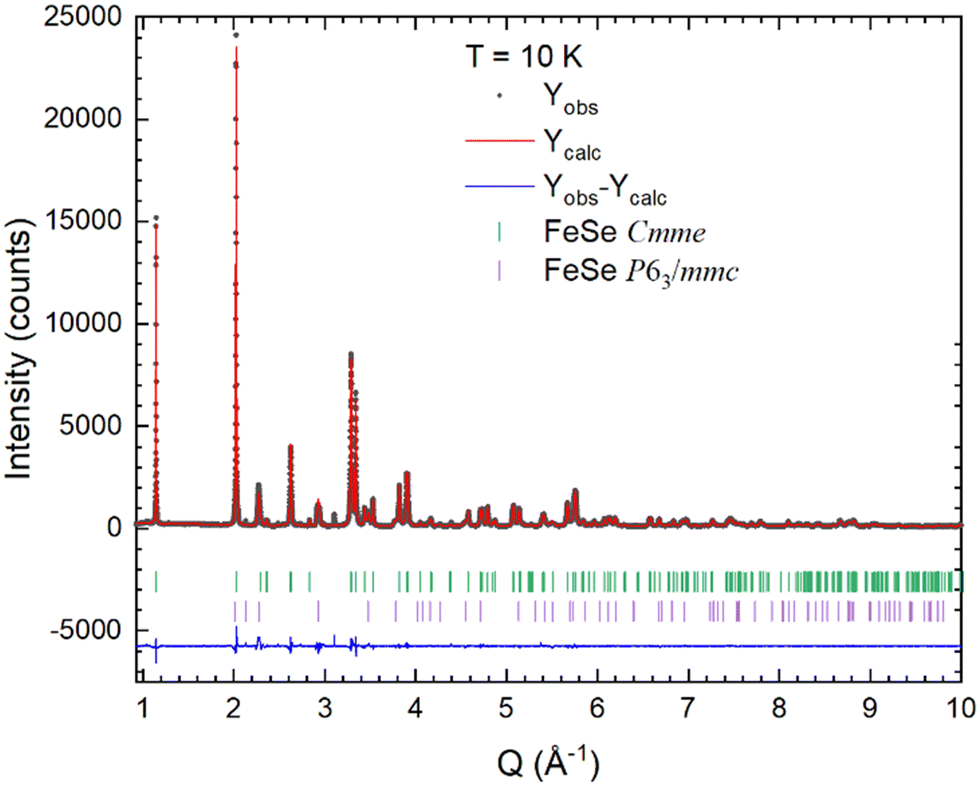 | ||
| Fig. 1 Rietveld refinement plot obtained by using data collected at 10 K and applying the Cmme structural model for β-FeSe. | ||
The P4/nmm → Cmme structural transition taking place in β-FeSe can be detected on cooling by the selective splitting affecting the Bragg peaks with strong components in the ab plane. Fig. 2 shows the evolution of the tetragonal 220 diffraction line on cooling. At lower temperature a clear separation of the orthorhombic 400 and 040 lines is not observed; rather, an asymmetric broadening takes place below 50 K. These same features were also observed for β-FeSe samples synthesized using hydrothermal methods. In particular, the asymmetric splitting of the tetragonal 220 diffraction line was related to the occurrence of a triclinic polymorph (P space group) growing below ∼60 K.6
 | ||
| Fig. 2 Thermal evolution of the diffraction lines at ∼4.72 Å−1 (full symbols: tetragonal 220 diffraction line; open symbols: orthorhombic 400 and 040 lines). | ||
In order to ascertain its real occurrence, the triclinic P structural model was tested and compared with the Cmme one using the data collected at 10 K. As a matter of fact, both structural models provide a symmetric splitting of the peak (Fig. 3, panels at the centre and on the left); hence the asymmetric shape of the peak cannot be related to a decrease of the crystal symmetry down to the triclinic system. Regardless, significantly better RBragg and Rfactor factors are obtained using the orthorhombic structural model that should be therefore preferred. Furthermore, the observed asymmetric splitting can be properly modelled by including the anisotropic microstrain parameters in the orthorhombic structural model during refinement (Fig. 3, panel on the right). Remarkably, the calculated diffraction pattern for the triclinic P structural model is characterized by the presence of several very faint diffraction lines that are missing for the orthorhombic model and that are not actually observed in the experimental data. Fig. 4 shows a magnification of the XRPD pattern at 10 K in the Q-region where some of the triclinic reflections should be observed, but they are actually missing.
 | ||
| Fig. 3 Fitting of the Bragg peak at ∼4.72 Å−1 using the triclinic (panel on the left), orthorhombic (panel at the centre) and orthorhombic with anisotropic strain parameter (panel on the right) structural models (XRPD data collected at 10 K). | ||
 | ||
| Fig. 4 XRPD data collected at 10 K compared with the diffraction patterns calculated for both Cmme and P structural models; faint diffraction lines (arrowed) are calculated for the monoclinic patterns that are not actually observed in the experimental data. | ||
In this context it is worth noting that in the sample prepared using hydrothermal synthesis the conductive properties were fully suppressed; conversely, in our aged sample superconductivity is still present, although with the transition temperature strongly decreased, down to ∼3 K.
Structural data obtained after refinement at 10 K are listed in Table 1, whereas Fig. 5 shows the thermal evolution of the in-plane lattice parameters for the aged β-FeSe sample; the phase fields of the tetragonal and orthorhombic phases are separated at Ts.
| Lattice parameters (Å) | ||
|---|---|---|
| A | b | C |
| 5.3170(1) | 5.3278(1) | 5.4810(1) |
| Atomic positions | ||||
|---|---|---|---|---|
| Atom | Wyckoff site | x | y | Z |
| Fe | 4g | 0 | 3/4 | 0.2336(1) |
| Se | 4b | 1/4 | 0 | 1/2 |
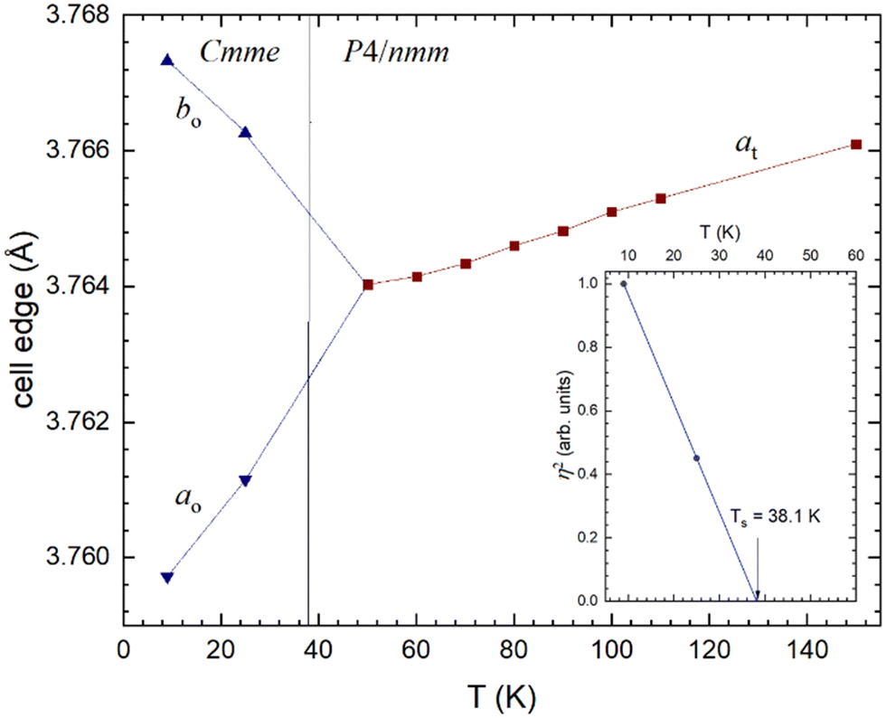 | ||
| Fig. 5 In-plane lattice parameters (expressed in terms of the primitive unit-cell) as a function of temperature through the P4/nmm → Cmme structural transition (t and o subscripts stand for tetragonal and orthorhombic, respectively). The inset shows the temperature-dependence of the squared value of the order parameter η2 for the structural transition. | ||
Remarkably, the anisotropy of the microstrain broadening can reveal the anisotropy of intrinsic properties of the inspected material. In particular, this shows that structural microstrain in the tetragonal phase remains constant along the main tetragonal crystallographic directions on cooling, but increases significantly along [hh0]; this is the same direction of the nesting wave-vector between the electron and hole pockets. This behaviour is in fair agreement with previous analyses of LnFeAsO phases (Ln: lanthanide)10–14 where the desymmetrisation P4/nmm → Cmme is observed. As a matter of fact, an in-plane 4-fold tensor surface develops on cooling (Fig. 6), whose anisotropy is fully consistent with the microstrain expected for a 4/mmm → mmm point group transition.15 These results clearly demonstrate that the low temperature phase crystallizes with a Cmme symmetry even though the peak at ∼4.72 Å−1 appears with asymmetric broadening rather than with resolved peak splitting. As a consequence, the occurrence of the triclinic P polymorph can be ruled out.
 | ||
| Fig. 6 Thermal dependence of the structural strain through the P4/nmm → Cmme structural transition; the upper panel shows the corresponding tensor isosurfaces at selected T (viewed along [001]) representing the microstrain broadening characterizing the tetragonal phase as the structural transition is approached. | ||
Group-theoretical analysis
The possible occurrence of the P polymorph of β-FeSe would have remarkable consequences from the physical point of view. In fact, in both P4/nmm → Cmme and P4/nmm → P desymmetrisations reported for β-FeSe a group–subgroup relationship is preserved between the high temperature–high symmetry form, crystallizing in the generic G space group and the low temperature–low symmetry one, crystallizing in the generic H space subgroup. Nonetheless, relevant features should differentiate these transitions if both would occur.
According to the Landau theory of phase transitions, condensation of one or more collective structural degrees of freedom that transform according to a single irreducible representation (IR) of the space group G can drive the structural distortion causing the symmetry breaking. In practice the low symmetry form is equivalent to the high symmetry form, but distorted according to one or more specific modes, i.e. collective atomic displacements fulfilling specific symmetry properties.
For the P4/nmm → Cmme transformation of β-FeSe the proper primary IR is B2g; as for all the LnFeAsO (Ln: lanthanide) compounds, the displacive B2g mode is not active at the 2a and 2c Wyckoff sites16 occupied by Fe and Se atoms in the P4/nmm structure of β-FeSe. This result agrees with theoretical calculations pointing to an electronically driven instability (spin, charge or orbital degrees of freedom).17
Conversely, the P4/nmm → P transition should be driven by the primary Eg mode, a structural degree of freedom that is active at both the 2a and 2c Wyckoff sites in the P4/nmm structure of β-FeSe. The formation of the hypothetical P polymorph deserves a further consideration. In fact, the identity matrix relating the cell edges of the P4/nmm and P unit cell imposes a P4/nmm → Pmmn → P21/m → P group–subgroup relationship. This is the same group–subgroup relationship observed for the P4/nmm → P21/m structural transition in Fe1+yTe.16 Notwithstanding, in Fe1+yTe the structural transition is driven by spin interaction, as clearly demonstrated by the coincident magnetic and structural transitions7 as well as by the correlated displacement of Fe atoms with ferromagnetic spin alignment (as sketched in Fig. 7). It is evident that as the nearest neighbouring Fe atoms are ferromagnetically aligned, their bond distance decreases; conversely, for antiferromagnetic coupling Fe atoms move away. It can be thus concluded that the shifting of Fe atoms (required for freezing the primary Eg mode and thus producing the P symmetry) can be obtained only by the occurrence of magnetic interactions, as in Fe1+yTe where an antiferromagnetic structure occurs. Conversely, in β-FeSe no magnetic structure develops with structural transition (whatever desymmetrisation is considered) and hence the freezing of the primary Eg mode can be ruled out.
 | ||
| Fig. 7 Primary displacement pattern (on the left) and spin ordering (on the right) characterizing the Fe-substructure in the P21/m structure of Fe1+yTe. | ||
Spontaneous strain analysis
Following the same approach reported already applied for other Fe-based superconductor materials,11,13 the dependence on temperature of the spontaneous strain was evaluated. By definition, spontaneous strain corresponds to the structural distortion solely induced by the desymmetrisation. It consists of up to six independent components, forming a symmetric second-rank tensor, which are subject to the constraints of symmetry.18 In particular, the appropriate symmetry-breaking component of the spontaneous strain for the P4/nmm → Cmme structural transition is e6 that in its general formulation corresponds to: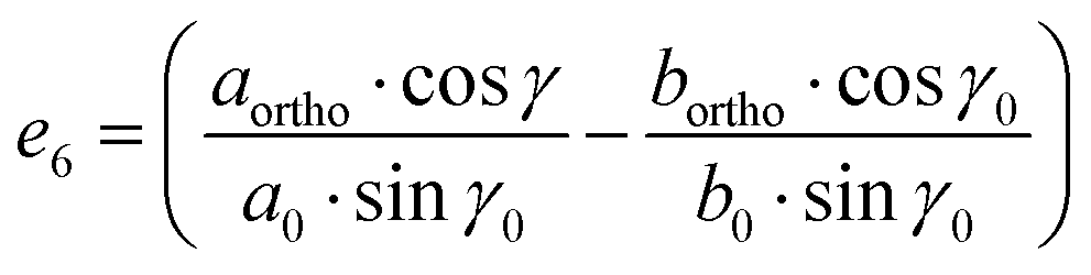 | (1) |
 | (2) |
transition (∼60 K).6
Conclusions
The detrimental effect of oxygen poisoning on both structural and superconductive properties of β-FeSe is a well-known phenomenon, as reported in several investigations.3,19,20,21 In particular, a small amount of oxygen poisoning was demonstrated to affect both structural and superconductive properties.21 Their corruption observed in both samples prepared by hydrothermal synthesis and aged after the solid state reaction can thus be reliably ascribed to oxygen poisoning, mediated by the reaction environment in the first case and by the ambient atmosphere in the second case. This oxygen poisoning determines both the electronic doping of the system as well as structural disordering at the local scale, as evidenced by pair distribution function analysis.20 It can be thus concluded that the occurrence of the non-superconductive P polymorph of β-FeSe can be excluded, on the basis of structural, microstructural and conductive analyses.
Conflicts of interest
There are no conflicts to declare.Acknowledgements
This work was carried out with the support of the European Synchrotron Radiation Facility, (proposal HC-4366; DOI: http://10.15151/ESRF-ES-445193642); A. M. acknowledges A. Fitch (ESRF-ID22) for his kind assistance during data collection at ID22 and G. Lamura (CNR-SPIN) for magnetization measurements.References
- H. Yanagi, T. Kamiya, Y. Kamihara, H. Hiramatsu, M. Hirano and H. Hosono, J. Am. Chem. Soc., 2008, 130, 3296 CrossRef PubMed.
- S. Margadonna, Y. Takabayashi, M. T. McDonald, K. Kasperkiewicz, Y. Mizuguchi, Y. Takano, A. N. Fitch, E. Suard and K. Prassides, Chem. Commun., 2008, 5607–5609 RSC.
- E. Pomjakushina, K. Conder, V. Pomjakushin, M. Bendele and R. Khasanov, Phys. Rev. B, 2009, 80, 024517 CrossRef.
- T. M. McQueen, A. J. Williams, P. W. Stephens, J. Tao, Y. Zhu, V. Ksenofontov, F. Casper, C. Felser and R. J. Cava, Phys. Rev. Lett., 2009, 103, 057002 CrossRef CAS PubMed.
- R. Khasanov, M. Bendele, K. Conder, H. Keller, E. Pomjakushina and V. Pomjakushin, New J. Phys., 2010, 12, 073024 CrossRef.
- U. Pachmayr, N. Fehn and D. Johrendt, Chem. Commun., 2016, 52, 194 RSC.
- A. Martinelli, A. Palenzona, M. Tropeano, C. Ferdeghini, M. Putti, M. R. Cimberle, T. D. Nguyen, M. Affronte and C. Ritter, Phys. Rev. B: Condens. Matter Mater. Phys., 2010, 81, 094115 CrossRef.
- A. Martinelli and A. Fitch, 2024, Searching for incommensurability in the FeSe structure at low temperature [Data set], European Synchrotron Radiation Facility DOI:10.15151/ESRF-ES-445193642.
- R. A. Young, IUCr Monographs on Crystallography, in The Rietveld Method, ed. R. A. Young, Oxford University Press, Oxford, 1993, vol. 5 Search PubMed.
- A. Martinelli, A. Palenzona, M. Putti and C. Ferdeghini, Phys. Rev. B: Condens. Matter Mater. Phys., 2012, 85, 224534 CrossRef.
- A. Martinelli, A. Palenzona, I. Pallecchi, C. Ferdeghini, M. Putti, S. Sanna, C. Curfs and C. Ritter, J. Phys.: Condens. Matter, 2013, 25, 395701 CrossRef CAS PubMed.
- A. Martinelli, Supercond. Sci. Technol., 2019, 32, 015014 CrossRef.
- A. Martinelli, P. Manfrinetti, A. Provino, C. Ritter and C. Ferdeghini, J. Phys.: Condens. Matter, 2019, 31, 064001 CrossRef CAS PubMed.
- A. Martinelli, P. Carretta, M. Moroni and S. Sanna, Phys. Rev. B, 2021, 103, 014518 CrossRef CAS.
- A. Leineweber, Z. Kristallogr., 2011, 226, 905 CrossRef CAS.
- A. Martinelli, J. Phys.: Condens. Matter, 2013, 25, 125703 CrossRef CAS PubMed.
- R. Fernandes, A. Chubukov and J. Schmalian, Nat. Phys., 2014, 10, 97 Search PubMed.
- M. A. Carpenter, E. K. H. Salje and A. Graeme-Barber, Eur. J. Mineral., 1998, 10, 621 CrossRef CAS.
- T. M. McQueen, Q. Huang, V. Ksenofontov, C. Felser, Q. Xu, H. Zandbergen, Y. S. Hor, J. Allred, A. J. Williams, D. Qu, J. Checkelsky, N. P. Ong and R. J. Cava, Phys. Rev. B: Condens. Matter Mater. Phys., 2009, 79, 014522 CrossRef.
- J. T. Greenfield, S. Kamali, K. Lee and K. Kovnir, Chem. Mater., 2015, 27, 588 CrossRef CAS.
- B. Rasche, M. Yang, L. Nikonow, J. F. K. Cooper, C. A. Murray, S. J. Day, K. Kleiner, S. J. Clarke and R. G. Compton, Angew. Chem., Int. Ed., 2019, 58, 15401 CrossRef CAS PubMed.
| This journal is © The Royal Society of Chemistry 2023 |