A Zr-MOF nanoflower sensor and its mixed-matrix membrane for the highly sensitive detection of nitroaromatics

Hui Xu *a, Fangyuan Zhong b, Faqiang Chen a, Tian-Xiang Luan c, Peizhou Li *c, Shiqing Xu *a and Junkuo Gao *b
aKey Laboratory of Rare Earth Optoelectronic Materials and Devices of Zhejiang Province, Institute of Optoelectronic Materials and Devices, Collage of Optical and Electronic Technology, China Jiliang University, Hangzhou 310018, People's Republic of China. E-mail: huixu@cjlu.edu.cn; shiqingxu75@163.com
bInstitute of Functional Porous Materials, The Key laboratory of Advanced Textile Materials and Manufacturing Technology of Ministry of Education, School of Materials Science and Engineering, Zhejiang Sci-Tech University, Hangzhou 310018, P. R. China. E-mail: jkgao@zstu.edu.cn
cSchool of Chemistry and Chemical Engineering, Shandong University, No. 27 Shanda South Road, Ji'nan, 250100, People's Republic of China. E-mail: pzli@sdu.edu.cn

Received 7th March 2022 , Accepted 21st April 2022

First published on 21st April 2022


Abstract

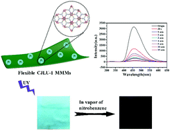
A new luminescent metal–organic framework nanoflower material CJLU-1 (Zr63-O)43-OH)4(OH)6(TCA)2(H2O)6) (H3TCA = tri-carboxylic acids 4,4′,4′′-nitrilotribenzoic acid) has been realized for the highly sensitive sensing of nitroaromatic molecules with fast response. CJLU-1 consists of six-connected Zr6 clusters and three-connected TCA ligands, and forms a 2D layered porous structure. The intrinsic 2D layered crystal structure and the optimal synthesis method create an ordered nanoflower structure constructed using ultrathin nanosheets. The highly dispersive nature and the controllable nanoflower structure with highly accessible active sites on the surface enable them to have full contact with the targeted analytes, which leads to superior sensing performance, with a high detection limit of 0.362 μM (≈83 ppb) toward 2,4,6-trinitrophenol (PA), strong anti-interference and a fast response within seconds. Moreover, a novel sensing platform, CJLU-1 mixed-matrix membranes (MMMs), is established by hybridization of the CJLU-1 nanoflower and cellulose acetate polymer, and exhibits desirable vapor sensing towards nitroaromatic molecules. This work contributes to the development of controllable nanostructured MOF probes and MOF-based MMMs as a novel sensing platform with superior mechanical strength, flexibility and sensitivity towards practical chemical sensing applications.


Introduction

Metal–organic framework materials (MOFs) are porous crystalline materials assembled from metal ions/metal clusters and organic ligands, and have shown many potential applications including gas storage and separation, heterogeneous catalysis, drug delivery, luminescence etc.1–7 The differential recognition/binding sites, tunable pore sizes and functionalized pore surfaces have made MOFs important sensing materials.8–10 An efficient luminescent sensing material requires high sensitivity, high selectivity, anti-interference, fast response time etc.11,12 Among them, the realization of high sensitivity requires sensing materials to be in close contact with analytes.13–15 The porous nature of MOFs facilitates adsorbing and pre-concentrating the analytes, increasing the possibility of host–guest interactions and improving the sensitivity.11,16,17 Moreover, the controllable nanostructure is also an important factor to improve the sensitivity. The unique nanostructure of MOFs endows them with larger surface area, more exposed functional sites, increasing the feasibility of adequate contact with the analytes and improved sensitivity.18–22 Two-dimensional (2-D) MOF nanosheets, an emerging type of 2D material that possess good dispersibility, large surface areas and abundant accessible sites, have shown great potential in luminescence sensing.23–31 There are some pioneering studies showing the superior performance of 2D MOF nanosheets in the construction of fluorescent sensors.32–35 However, work on MOFs with controllable nanostructures for luminescence sensing is still relatively scarce.

On the other hand, currently the sensing work of MOFs is mainly based on MOF powder or nanoscale MOFs dispersed in liquid media, which suffers from inconvenience in the recycling.9 And for further application, MOF samples need to be fabricated into thin films or membranes.36,37 MOF-based mixed-matrix membranes (MMMs) have been well demonstrated to be promising candidates. The MOF-based MMMs combine the superior properties of novel porous MOFs as well as flexibility and easy processability of traditional polymeric membranes.38–40 Compared with the pure MOF thin films, which usually have to be delicately grown on substrates, MOF-based MMMs exhibit superior mechanical properties, more flexibility, easier processability and lower cost. Currently, the applications of MOF-based MMMs are evaluated in gas separation, pervaporation, nanofiltration, and sensors.41–44 However, the studies of MOF-based MMMs for luminescence sensing are still at the early stage, and the use of MOF-based MMMs for nitroaromatic sensing has not been established yet.45 By incorporating luminescent MOFs into polymeric membranes, MOF-based MMMs exhibit efficient luminescence properties.46,47 The combination of structurally designable MOFs and flexible porous polymeric membranes offers unique and effective integration in specific sensing applications. Furthermore, MOF particles are uniformly distributed and closely enwrapped in the polymeric matrix, which enables good gas permeation flux and facilitates a full contact between MOF particles and analytes to enhance the detection sensitivity.48

Herein, we innovatively explored a new nanoflower-like CJLU-1 MOF (Zr63-O)43-OH)4(OH)6(TCA)2(H2O)6) (H3TCA= tri-carboxylic acids 4,4′,4′′-nitrilotribenzoic acid) luminescent sensing material with a hexanuclear Zr cluster and a 2D porous crystal structure, and the nanoflowers were constructed using two-dimensional ultrathin MOF nanosheets. By utilizing the strong electron donating ability of the nitrogen atom in the center of the triphenylamine ligand and uniform nanoflower structure, the CJLU-1 nanoflower material exhibited superior sensing properties towards electron deficient nitroaromatics with ppb-level sensitivity. Furthermore, the CJLU-1 nanoflower material was incorporated into a cellulose acetate matrix to prepare hybrid sensing membranes (CJLU-1 MMMs) (as shown in Scheme 1). The CJLU-1 MMMs inherited porosity and sensing functionality from the MOF as well as the flexibility and easy processability from the cellulose acetate matrix, and exhibited efficient and recyclable detection of nitroaromatics.


image file: d2tc00920j-s1.tif
Scheme 1 Schematic illustration of the preparation of CJLU-1 MMMs.

Results and discussion

The reaction of ZrCl4 with the tritopic carboxylate ligand H3TCA49 in DMF and HCl formed a new porous Zr-MOF CJLU-1. Interestingly, the in situ hydrothermal method easily yielded high-quality CJLU-1 nanoflowers. However the crystal size of the synthesized Zr-MOF sample was too small, making it very difficult to directly determine the crystal structure from single-crystal diffraction measurements. Therefore, powder X-day diffraction analysis and structure simulation were utilized to elucidate the MOF structure. Based on the nature of the triple symmetry of the TCA ligand and the geometry of the commonly formed hexanuclear Zr6 cluster as well as the results of PXRD measurements, a (3,6)-connected framework in kgd topology with an eclipsed AA stacking model was built using the Materials Studio software package (Fig. 1). The several relatively intense peaks at 5.93°, 10.16°, 21.12°, and 26.79° observed from the experimental PXRD pattern of the synthesized Zr-MOF were assigned to the diffractions of (100), (110), (130), and (401) planes, respectively. After Pawley refinements (Fig. 1), CJLU-1 was indexed to a trigonal P[3 with combining macron] space group with cell parameters a = b = 17.1740 Å, c = 7.0100 Å, α = β = 90°, γ = 120° and satisfactory residual values Rp = 6.25% and Rwp = 8.63%. CJLU-1 is a two-dimensional (2D) layered framework. Six Zr atoms are joined into a six connected Zr63-O)43-OH)4(OH)6 octahedron subunit, and then the Zr6-octahedron clusters are connected by TCA ligands to form a 2D layered porous structure (Fig. 1b and Fig. S1, ESI). The Zr6-octahedron cluster is bridged by 4 μ3-O and 4 μ3-OH groups to form an octahedron subunit, which are further linked with six TCA ligands. CJLU-1 has one-dimensional pores of about 3.5 × 4.0 Å2 along the c axis and a pore volume of 0.205 cm3 g−1. The comparison of the PXRD patterns between the experimental one and the one generated from the simulated structure reveals that they are consistent with each other, which also confirmed the phase purity of the synthesized sample of CJLU-1 (Fig. S2, ESI). The intrinsic 2D layered crystal structure and the optimal synthesis method create ordered nanoflower structures constructed using ultrathin nanosheets. The SEM and TEM measurements reveal that the obtained CJLU-1 exhibits uniform nanoflower morphologies with a typical diameter of 3 μm. The nanoflowers are constructed using dozens of interpenetrated ultrathin 2D nanosheets with smooth surfaces. The lateral dimension of the nanosheets reached up to micrometers and their thicknesses were less than 10 nm (Fig. 2).
image file: d2tc00920j-f1.tif
Fig. 1 (a) The PXRD patterns from the experimental measurements of the synthesized MOF (black curve) and the simulated structure in a (3,6)-connected framework with an eclipsed AA stacking model. (b) Simulated frameworks constructed using the TCA ligands and hexanuclear Zr6 clusters.

image file: d2tc00920j-f2.tif
Fig. 2 (a and b) The SEM images of CJLU-1; (c and d) the TEM images of CJLU-1.

The above nanoflower feature of CJLU-1 encouraged us to examine its potential application in luminescence sensing. The solid state CJLU-1 exhibits photoluminescence (PL) properties with a fluorescence peak at 454 nm (Fig. S3, ESI), which is slightly blue-shifted from the fluorescence peak of the free ligand H3TCA located at 458 nm (Fig. S3, ESI). The PL properties of CJLU-1 could be attributed to the fluorescence of ligand H3TCA in the framework. The CJLU-1 nanoflower material can be easily dispersed in ethanol to form a uniform suspension solution, and exhibits a strong PL emission peak located at 454 nm (Fig. S4, ESI).

The strong emission of the CJLU-1 nanoflower material encouraged us to examine its potential application in liquid-phase fluorescence detection of nitroaromatic molecules. The addition of a small amount of organic small molecules (5 × 10⁻⁶ mol, the amount of PA is 2.7 × 10⁻⁶ mol) has different effects on the luminescence intensity of the dispersed nanoflower in ethanol (Fig. 3c). Analytes, such as methanol, water, phenol, acetone, toluene, aniline, and N,N-dimethylformamide (DMF), do not substantially affect the luminescence intensity of CJLU-1 nanoflower ethanol solution, while the nitroaromatic molecules have a significant quenching effect on CJLU-1 nanoflowers, indicating that the CJLU-1 nanoflower material can be used to detect small amounts of nitroaromatic molecules. It is interesting that the CJLU-1 nanoflower material shows different quenching effects for different nitroaromatic molecules. The quenching effect of 2,4,6-trinitrophenol (PA) and p-nitrophenol (PNP) on MOF solution is significantly higher than that of nitrobenzene (NB), 2,4-dinitrotoluene (2,4-DNT) and 2,6-dinitrotoluene (2,6-DNT) (Fig. 3c). The quenching effect of different nitroaromatic molecules decreased in the order PA > PNP > NB > 2,4-DNT >2,6-DNT. In particular, PA has the strongest quenching effect on CJLU-1 nanoflower solution. The addition of only 2.65 μM PA can efficiently quench the nanoflowers’ fluorescence; the addition of 7.86 μM PA can quench the intensity to less than half of the original fluorescence intensity; and the addition of 25.3 μM PA can almost completely quench the nanoflowers’ fluorescence (Fig. 3a). The CJLU-1 nanoflower material exhibits superior sensing properties toward PA, with the detection limit reaching 0.362 μM (≈83 ppb) (Fig. S9, ESI), clearly indicating the excellent potential of the CJLU-1 controllable nanostructure approach in the precise detection of PA. The highly dispersive nature and the ultrathin nanosheets inside provide highly accessible active sites on their surface, which leads to adequate contact with the targeted nitroaromatic molecules and high detection sensitivity. Noticeably, the CJLU-1 nanoflower material displays a linear response with PA concentration in the low concentration range. The quenching efficiency of PA can be quantitatively explained by using the Stern–Volmer equation. The relationship between (I0/I − 1) and the concentration of PA was calculated using the Stern–Volmer equation (I0/I = 1+ Ksv[M]) and plotting the S–V plots, where I0 and I are the fluorescence intensities of CJLU-1 nanoflowers and CJLU-1 nanoflower material with PA at a concentration of [M], and Ksv is the quenching coefficient. As shown in Fig. 3b, the estimated Stern–Volmer constant is as high as 2.60 × 10⁵ M−1.


image file: d2tc00920j-f3.tif
Fig. 3 (a) The PL spectrum of CJLU-1 nanoflowers with different amounts of PA; (b) S–V plots of PA; inset: the picture above is a photo of CJLU-1 ethanol solution before and after adding 50 μL of 1.22% PA, under nature light and UV light at 365 nm, respectively; (c) the 454 nm emission intensity of CJLU-1 ethanol solution with 2.5 × 10⁻³ M different small organic molecule (the concentration of PA was 1.3 × 10⁻³ M); and (d) the emission intensity of CJLU-1 ethanol solution with 2.5 × 10⁻³ M nitro-aromatic molecules (the concentration of PA was 1.30 × 10⁻³ M) in the presence of 2.5 × 10⁻³ M other small organic molecules.

To gain more understanding of the detection selectivity towards nitroaromatic molecules in the presence of other analytes, the anti-interference experiment was carried out. The result shows that the presence of other analytes (such as toluene, aniline, water, phenol, acetone, DMF and MeOH) basically has little interference on the luminescence intensity of the CJLU-1 nanoflower ethanol solution with nitroaromatic molecules, indicating the high nitroaromatic molecule selectivity of CJLU-1 in the concurrent presence of other analytes (Fig. 3d).

In addition, the fluorescence intensity of CJLU-1 at 455 nm was measured as a function of time after the addition of 2.65 × 10⁻⁴ M PA. As shown in Fig. S10 (ESI), the fluorescence intensity was completely quenched after the addition of PA within 20 seconds, and kept almost the same for the remaining time, indicating the ability of fast-response detection of PA. The dispersible nature and highly exposed surface of the MOF nanoflower materials have enabled a close contact between the MOF nanoflowers and the organic molecules, which leads to fast response of luminescence sensing, thus providing a promising strategy to implement MOF nanoflowers as fast-response sensing materials.

The luminescence of the CJLU-1 nanoflower results from the fluorescence of the coordinated ligand H3TCA in the framework. The addition of the nitroaromatics resulted in the fluorescence quenching of CJLU-1, while the powder XRD patterns of CJLU-1 with the addition of a large amount (10−3 M) of nitroaromatics are identical with that of the original one (Fig. S11, ESI), indicating that the crystal structure of CJLU-1 remained stable in different chemical environments. The FT-IR spectra of CJLU-1 immersed in different nitroaromatic solutions were consistent with those of the original CJLU-1, indicating that there was no coordination with the analytes and MOFs (Fig. S12, ESI). The quenching effect of the nitroaromatics on CJLU-1 was evaluated by the fluorescence decay time of CJLU-1. As shown in Fig. S13 (ESI) and Table S2, (ESI) the nitroaromatics had no significant effect on the fluorescence lifetime of CJLU-1, demonstrating that there is no coordination between CJLU-1 and the analytes.

Based on previous pioneering work on MOF-based sensing materials for nitroaromatic detection,50 the quenching mechanism process mainly includes photoinduced electron transfer (PET) and fluorescence resonance energy transfer (FRET). The electronic properties of the MOFs are crucial to their sensing behaviors. Considering the electron-withdrawing feature of the nitroaromatic molecules, the rational design of electron donating MOFs can effectively promote the electron transfer between the MOFs and the targeted nitroaromatic molecules, and a similar mechanism has been widely applied in previous work.11,51 The nitrogen atom in the center of the triphenylamine ligand has strong electron donating ability, thus we speculate that the excited electrons at the conductive band of the CJLU-1 nanoflowers will easily migrate to the LUMO of the nitroaromatic molecules upon excitation and then followed a non-radiative relaxation, leading to a fluorescence quenching process. Besides, compared with other nitroaromatics, the absorption spectra of the PA and PNP effectively overlap with the emission spectrum of CJLU-1 (Fig. S14, ESI), which facilitates the energy transfer between the analytes and MOFs, and induces a dramatic decrease in the fluorescence intensity of CJLU-1.

For practical application, CJLU-1-based MMMs were fabricated. MOF-based MMMs utilize MOFs as inorganic filler components in polymer membranes, and preserve not only the porosity, chemical tunability, and functionality of the MOFs, but also stand in the form of a stable, flexible, freestanding membrane. Cellulose acetate is widely utilized as a polymer base material for the membrane because of its biocompatibility, nontoxicity and low cost features. Also the presence of acidic and carbonyl groups in the cellulose acetate structure helps in binding with functional additives, and improves mixed matrix membranes (MMMs) performance.52 The CJLU-1 MOF-based MMMs can be readily prepared by the combination of MOFs into the acetone/DMF solution of cellulose acetate to form casting solution. After casting onto a glass plate using a wiper and being immersed into deionized water and dried at room temperature, freestanding CJLU-1 MOF based MMMs can be obtained.

The delaminated CJLU-1 MMM is free-standing, mechanically robust, pliable, and free of macroscopic defects (Fig. 4a and b). The morphology of CJLU-1 MMMs was observed by SEM using a pure cellulose acetate membrane as a reference (Fig. 4d–i). At low magnifications, both the pure cellulose acetate membrane and the CJLU-1 MMMs exhibit smooth and neat surfaces, and the MOF particles are uniformly distributed in CJLU-1 MMMs (Fig. 4d and f). And at high magnifications, the pure cellulose acetate membrane exhibits crosslinking and macro-porous morphology (Fig. 4e). For the CJLU-1 MMMs, the CJLU-1 nanoflowers are closely embedded and partially exposed in the cellulose acetate matrix (Fig. 4g), which provides both strong MOF–polymer adhesion and adequate MOF–environment contact, and is very important for further sensing application. The cross-sectional SEM also indicates the smooth surface and the macro-porous feature of the CJLU-1 MMMs, and the thickness of the membrane was approximately 40 μm (Fig. 4h and i).


image file: d2tc00920j-f4.tif
Fig. 4 (a and b) Different views of CJLU-1 MMMs under mechanical stress; (c) PXRD of CJLU-1, CJLU-1 MMMs and cellulose acetate membrane; (d and e) SEM images of the cellulose acetate membrane; (f and g) SEM images of CJLU-1 MMMs; and (h and i) SEM images of the cross section of CJLU-1 MMMs.

The macro-porous feature of the membrane facilitates the diffusion of the analytes and helps in the sufficient contact between the analytes and MOF both inside and on the surface of the membrane, and thus improves the sensing performance.

As shown in Fig. 4c, PXRD of CJLU-1 MMMs was carried out. The pure cellulose acetate membrane was amorphous, evidenced by a broad peak between 2θ of 15 and 30°.53–55 With the addition of CJLU-1, the characteristic peak of CJLU-1 appears. Because of the strong characteristic peak intensity of CJLU-1, the diffraction peak of the cellulose acetate membrane is almost negligible. These results indicate that the crystallinity and structural features of CJLU-1 remain well in the cellulose acetate membrane. In addition, the loading of the cellulose acetate membrane based on MOFs was studied by thermogravimetric analysis, and the loading of CJLU-1 was calculated to be as high as 39.53% (Fig. S15, ESI). The N2 adsorption–desorption isotherms of CJLU-1, CJLU-1 MMMs and the pure cellulose acetate membrane at 77 K were obtained to confirm their specific surface areas. The BET of CJLU-1, CJLU-1 MMMs and pure cellulose acetate membrane was calculated to be 674.5019 m2 g−1, 110.9820 m2 g−1 and 13.9501 m2 g−1, respectively (Fig. S16–S18, ESI). These results indicated that the addition of CJLU-1 increased the specific surface area of the cellulose acetate membrane.

The fabricated CJLU-1 MMMs inherited the strong fluorescence of CJLU-1 and exhibited blue fluorescence emission located at 454 nm (Fig. 5c and Fig. S19, ESI). To demonstrate the feasibility of the fabricated MMMs as an effective sensing platform, CJLU-1 MMMs were employed to detect nitroaromatic molecules in the vapor phase. Impressive fluorescence quenching was observed upon exposing nitroaromatic molecules, such as nitrobenzene, 2-nitrotoluene and PNP (Fig. S20–S22, ESI). The fluorescence intensity of CJLU-1 MMMs significantly decreased when they were exposed to nitrobenzene vapor for a very short time (30 s). And the fluorescence intensity gradually decreased with time, when the CJLU-1 MMMs were exposed to nitrobenzene vapor for 5 min, the quenching efficiency (defined as (I0I)/I0 × 100) reached 95% (Fig. 5a and b). After the CJLU-1 MMMs were exposed to nitrobenzene vapor for 30 min, the white CJLU-1 MMMs changed to light yellow, and the membrane's fluorescence was darkened from blue under 365 nm ultraviolet light irradiation (Fig. 5c). While for PNP, the fluorescence intensity showed a less quenching efficiency of 25% for a 60 min exposure time, which is due to its lower vapor pressure and therefore limited interaction with MMMs, reducing its impacts (Fig. S20, ESI). For PA, the liquid detection experiment was carried out due to the very low concentration of the original PA solution (0.122% PA aqueous solution). The fluorescence intensity significantly decreases after the addition of 50 μL PA solution onto the membrane (Fig. S22, ESI). In addition, after the addition of a drop of liquid such as 2,4-DNT, 2,6-DNT, NB, PNP and PA with certain concentration, the fluorescence colors of the CJLU-1 MMMs darken from blue for a different degree under the illumination of 365 nm ultraviolet light (Fig. 5d), highlighting the straightforward and facile detection of nitroaromatic molecules by the MOF MMM approach.


image file: d2tc00920j-f5.tif
Fig. 5 (a) The emission of CJLU-1 MMMs upon exposure to nitrobenzene vapor for different time periods; (b) quenching efficiency profile of CJLU-1 MMMs upon exposure of NB as a function of time; (c) photo under nature light and 365 nm UV light of CJLU-1 MMMs before and after exposure to NB for 30 min; and (d) fluorescence photos of CJLU-1 MMMs after addition of 5 μL 0.1 M of nitroaromatics (the concentration of PA was 5.32 × 10⁻³ M) under 365 nm UV light.

To further investigate the practical utility of CJLU-1 MMMs, the recovery and reproducibility of the membrane were determined. The fluorescence intensity of CJLU-1 MMMs at 454 nm was quenched upon exposure to nitrobenzene vapor for 5 min, then the CJLU-1 MMMs were immersed in ethanol for 5 min to wash off nitrobenzene on the membrane and dried naturally in air, and the fluorescence intensity of the CJLU-1 MMMs can be recovered (Fig. 6). Even when the CJLU-1 MMMs were reused for five cycles, the recovered fluorescence intensity can largely remain stable (Fig. 6). The superior reversible sensing performances demonstrate the feasibility of the CJLU-1 MMMs for practical sensing applications.


image file: d2tc00920j-f6.tif
Fig. 6 The reversibility of the CJLU-1 MMM test.

Experimental

Synthesis of CJLU-1 nanoflowers

4,4′,4′′-Nitrilotribenzoic acid (H3TCA) (94.25 mg, 0.250 mmol) and ZrCl4 (0.0863 g, 0.370 mmol) were added to 10 mL of DMF in a 50 mL Teflon-lined stainless-steel autoclave, and after the mixture was dissolved, then concentrated HCl (10.75 mL, 100 equiv.) was added to the above mixture. After sonication for 10 minutes, the mixture was heated at 120 °C for 2 days. Then, the temperature of the mixture dropped to room temperature and it was washed with DMF and methanol several times, respectively. And then, white powder was obtained.

Organic small molecule sensing

For an organic small molecule sensing experiment, 10 mg sample powder was added to 100 mL ethanol and sonicated for 20 minutes to obtain a suspension of CJLU-1. The luminescence sensing experiment was carried out by adding 50 μL of DMF, acetone, phenol, H2O, aniline, toluene, NB, 2,4-DNT, 2,6-DNT, PNP (p-nitrophenol) and 1.22% PA (among them, the concentration of 1.22% PA was 5.32 × 10⁻² M) with a concentration of 0.1 M to 2 mL suspension of CJLU-1, respectively.

Fabrication of CJLU-1 MMMs

200 mg of cellulose acetate was added to 1.8 g of acetone/DMF (mass/mass = 2/1), and sonication was carried out for 5 hours to dissolve the cellulose acetate to obtain casting solution. And then 120 mg of CJLU-1 was added to the above casting solution to form a homogeneous solution by sonication. Then, the casting solution with CJLU-1 was cast onto a glass plate using a wiper and immersed in deionized water to separate the composite film from the glass plate. After drying at room temperature, 1 × 1 cm2 was cut from CJLU-1 MMMs for the sensing test.

Vapor sensing of nitroaromatics

Take nitrobenzene for example; 2 mL of nitrobenzene was placed in a 20 mL sample vial, which was placed in a closed reagent bottle and allowed to stand for a few days to bring the nitrobenzene to equilibrium vapor pressure. A 1 × 1 cm2CJLU-1 MMM was placed above the reagent bottle to expose it to nitrobenzene vapor. After a certain period of time, the slide was removed from the reagent bottle and its emission spectrum was collected immediately. The experimental details of other nitroaromatics are provide in the ESI.

Conclusions

To summarize, we have targeted a new luminescent flower-like metal-organic framework composed of ultra-thin nanosheet material CJLU-1 (Zr63-O)43-OH)4(OH)6(TCA)2(H2O)6) (H3TCA = tri-carboxylic acids 4,4′,4′′-nitrilotribenzoic acid) for the highly sensitive sensing of nitroaromatic molecules with fast response. The excellent luminescence sensing performance of the nanoflower material mainly comes from the rational design of an electron-donating triphenylamine ligand and the nanoflower structure with highly accessible active sites on the surface. The CJLU-1 nanoflower material shows a high detection limit of 0.362 μM (≈83 ppb) toward PA, strong anti-interference and a fast response within seconds. Moreover, a novel sensing platform was established by hybridization of the CJLU-1 nanoflower and cellulose acetate polymer into MMMs, and exhibits effective and reversible vapor sensing towards nitroaromatic molecules. This work suggests a promising route to develop novel nanostructured MOF probes and MOF-based MMMs as a nitroaromatic sensing platform with superior mechanical strength, flexibility and sensitivity towards practical sensing applications.

Conflicts of interest

There are no conflicts to declare.

Acknowledgements

This work was supported by the Natural Science Foundation of Zhejiang Province (No. LY19E020007 and LY20E020001), the National Natural Science Foundation of China (51672251) and the Fundamental Research Funds for the Provincial Universities of Zhejiang.

Notes and references

  1. Y. Cui, Y. Yue, G. Qian and B. Chen, Chem. Rev., 2012, 112, 1126–1162 CrossRef CAS PubMed.
  2. M. Ding, R. W. Flaig, H.-L. Jiang and O. M. Yaghi, Chem. Soc. Rev., 2019, 48, 2783–2828 RSC.
  3. D. J. Wales, J. Grand, V. P. Ting, R. D. Burke, K. J. Edler, C. R. Bowen, S. Mintova and A. D. Burrows, Chem. Soc. Rev., 2015, 44, 4290–4321 RSC.
  4. S.-N. Zhao, X.-Z. Song, S.-Y. Song and H.-J. Zhang, Coord. Chem. Rev., 2017, 337, 80–96 CrossRef CAS.
  5. J. Gao, X. Qian, R. Lin, R. Krishna, H. Wu, W. Zhou and B. Chen, Angew. Chem., Int. Ed., 2020, 59, 4396–4400 CrossRef CAS PubMed.
  6. H. Xu, S. Sommer, N. L. B. Nyborg, J. Gao and B. B. Iversen, Chem. – Eur. J., 2019, 25, 2051–2058 CrossRef CAS PubMed.
  7. J. Gao, Q. Huang, Y.-Q. Lan and B. Chen, Adv. Energy Sustainability Res., 2021, 2, 2100033 CrossRef.
  8. L. E. Kreno, K. Leong, O. K. Farha, M. Allendorf, R. P. Van Duyne and J. T. Hupp, Chem. Rev., 2012, 112, 1105–1125 CrossRef CAS PubMed.
  9. Y. Zhang, S. Yuan, G. Day, X. Wang, X. Yang and H.-C. Zhou, Coord. Chem. Rev., 2018, 354, 28–45 CrossRef CAS.
  10. L. Wang, H. Xu, J. Gao, J. Yao and Q. Zhang, Coord. Chem. Rev., 2019, 398, 213016 CrossRef.
  11. W. P. Lustig, S. Mukherjee, N. D. Rudd, A. V. Desai, J. Li and S. K. Ghosh, Chem. Soc. Rev., 2017, 46, 3242–3285 RSC.
  12. W.-M. He, Z. Zhou, Z. Han, S. Li, Z. Zhou, L.-F. Ma and S.-Q. Zang, Angew. Chem., Int. Ed., 2021, 60, 8505–8509 CrossRef CAS PubMed.
  13. J. Dong, A. K. Tummanapelli, X. Li, S. Ying, H. Hirao and D. Zhao, Chem. Mater., 2016, 28, 7889–7897 CrossRef CAS.
  14. Y. Guo, X. Feng, T. Han, S. Wang, Z. Lin, Y. Dong and B. Wang, J. Am. Chem. Soc., 2014, 136, 15485–15488 CrossRef CAS PubMed.
  15. L. Wang, J. Gao and J. Xu, Sens. Actuators, B, 2019, 293, 71–82 CrossRef CAS.
  16. E. A. Dolgopolova, A. M. Rice, C. R. Martin and N. B. Shustova, Chem. Soc. Rev., 2018, 47, 4710–4728 RSC.
  17. I. Stassen, N. Burtch, A. Talin, P. Falcaro, M. Allendorf and R. Ameloot, Chem. Soc. Rev., 2017, 46, 3185–3241 RSC.
  18. M. Zhao, Y. Wang, Q. Ma, Y. Huang, X. Zhang, J. Ping, Z. Zhang, Q. Lu, Y. Yu and H. Xu, Adv. Mater., 2015, 27, 7372–7378 CrossRef CAS PubMed.
  19. R. Xu, Y. Wang, X. Duan, K. Lu, D. Micheroni, A. Hu and W. Lin, J. Am. Chem. Soc., 2016, 138, 2158–2161 CrossRef CAS PubMed.
  20. Y.-Z. Li, Z.-H. Fu and G. Xu, Coord. Chem. Rev., 2019, 388, 79–106 CrossRef CAS.
  21. S. Gbadamasi, M. Mohiuddin, V. Krishnamurthi, R. Verma, M. W. Khan, S. Pathak, K. Kalantar-Zadeh and N. Mahmood, Chem. Soc. Rev., 2021, 50, 4684–4729 RSC.
  22. H. Xu, Y. Dong, Y. Wu, W. Ren, T. Zhao, S. Wang and J. Gao, J. Solid State Chem., 2018, 258, 441–446 CrossRef CAS.
  23. M. Zhao, Y. Huang, Y. Peng, Z. Huang, Q. Ma and H. Zhang, Chem. Soc. Rev., 2018, 47, 6267–6295 RSC.
  24. H. Xu, J. Gao, X. Qian, J. Wang, H. He, Y. Cui, Y. Yang, Z. Wang and G. Qian, J. Mater. Chem. A, 2016, 4, 10900–10905 RSC.
  25. J. Dong, K. Zhang, X. Li, Y. Qian, H. Zhu, D. Yuan, Q.-H. Xu, J. Jiang and D. Zhao, Nat. Commun., 2017, 8, 1–14 CrossRef CAS PubMed.
  26. A. K. Chaudhari, H. J. Kim, I. Han and J. C. Tan, Adv. Mater., 2017, 29, 1701463 CrossRef PubMed.
  27. X. Wang, Z. Jiang, C. Yang, S. Zhen, C. Huang and Y. Li, J. Hazard. Mater., 2022, 423, 126978 CrossRef CAS PubMed.
  28. F.-L. Li, P. Wang, X. Huang, D. J. Young, H.-F. Wang, P. Braunstein and J.-P. Lang, Angew. Chem., Int. Ed., 2019, 58, 7051–7056 CrossRef CAS PubMed.
  29. G. Lan, Z. Li, S. S. Veroneau, Y.-Y. Zhu, Z. Xu, C. Wang and W. Lin, J. Am. Chem. Soc., 2018, 140, 12369–12373 CrossRef CAS PubMed.
  30. Y. Ding, Y.-P. Chen, X. Zhang, L. Chen, Z. Dong, H.-L. Jiang, H. Xu and H.-C. Zhou, J. Am. Chem. Soc., 2017, 139, 9136–9139 CrossRef CAS PubMed.
  31. Y. Li, M. Lu, Y. Wu, Q. Ji, H. Xu, J. Gao, G. Qian and Q. Zhang, J. Mater. Chem. A, 2020, 8, 18215–18219 RSC.
  32. L. Cao, Z. Lin, W. Shi, Z. Wang, C. Zhang, X. Hu, C. Wang and W. Lin, J. Am. Chem. Soc., 2017, 139, 7020–7029 CrossRef CAS PubMed.
  33. T. Singha Mahapatra, A. Dey, H. Singh, S. S. Hossain, A. K. Mandal and A. Das, Chem. Sci., 2020, 11, 1032–1042 RSC.
  34. V. K. Maka, A. Mukhopadhyay, G. Savitha and J. N. Moorthy, Nanoscale, 2018, 10, 22389–22399 RSC.
  35. C.-X. Yu, F.-L. Hu, J.-G. Song, J.-L. Zhang, S.-S. Liu, B.-X. Wang, H. Meng, L.-L. Liu and L.-F. Ma, Sens. Actuators, B, 2020, 310, 127819 CrossRef CAS.
  36. A. Betard and R. A. Fischer, Chem. Rev., 2012, 112, 1055–1083 CrossRef CAS PubMed.
  37. M. S. Denny, J. C. Moreton, L. Benz and S. M. Cohen, Nat. Rev. Mater., 2016, 1, 1–17 Search PubMed.
  38. T. Kitao, Y. Zhang, S. Kitagawa, B. Wang and T. Uemura, Chem. Soc. Rev., 2017, 46, 3108–3133 RSC.
  39. J. Dechnik, J. Gascon, C. J. Doonan, C. Janiak and C. J. Sumby, Angew. Chem., Int. Ed., 2017, 56, 9292–9310 CrossRef CAS PubMed.
  40. L. Xiang, L. Sheng, C. Wang, L. Zhang, Y. Pan and Y. Li, Adv. Mater., 2017, 29, 1606999 CrossRef PubMed.
  41. B. Seoane, J. Coronas, I. Gascon, M. E. Benavides, O. Karvan, J. Caro, F. Kapteijn and J. Gascon, Chem. Soc. Rev., 2015, 44, 2421–2454 RSC.
  42. Y. Peng, Y. Li, Y. Ban and W. Yang, Angew. Chem., Int. Ed., 2017, 56, 9757–9761 CrossRef CAS PubMed.
  43. T. Feng, Y. Ye, X. Liu, H. Cui, Z. Li, Y. Zhang, B. Liang, H. Li and B. Chen, Angew. Chem., Int. Ed., 2020, 59, 21752–21757 CrossRef CAS PubMed.
  44. Y. Ding, Y. Lu, K. Yu, S. Wang, D. Zhao and B. Chen, Adv. Opt. Mater., 2021, 9, 2100945 CrossRef CAS.
  45. M. Kalaj, K. C. Bentz, S. Ayala Jr, J. M. Palomba, K. S. Barcus, Y. Katayama and S. M. Cohen, Chem. Rev., 2020, 120, 8267–8302 CrossRef CAS PubMed.
  46. T.-T. Li, L. Liu, M.-L. Gao and Z.-B. Han, Chem. Commun., 2019, 55, 4941–4944 RSC.
  47. Q.-Y. Li, Y.-A. Li, Q. Guan, W.-Y. Li, X.-J. Dong and Y.-B. Dong, Inorg. Chem., 2019, 58, 9890–9896 CrossRef CAS PubMed.
  48. X. Zhang, Q. Zhang, D. Yue, J. Zhang, J. Wang, B. Li, Y. Yang, Y. Cui and G. Qian, Small, 2018, 14, 1801563 CrossRef PubMed.
  49. J. Gao, Y. Cai, X. Qian, P. Liu, H. Wu, W. Zhou, D.-X. Liu, L. Li, R.-B. Lin and B. Chen, Angew. Chem., Int. Ed., 2021, 60, 20400–20406 CrossRef CAS PubMed.
  50. Z. Hu, B. J. Deibert and J. Li, Chem. Soc. Rev., 2014, 43, 5815–5840 RSC.
  51. S. S. Nagarkar, B. Joarder, A. K. Chaudhari, S. Mukherjee and S. K. Ghosh, Angew. Chem., Int. Ed., 2013, 52, 2881–2885 CrossRef CAS PubMed.
  52. L. Chu, X. Zhang, W. Niu, S. Wu, W. Ma, B. Tang and S. Zhang, J. Mater. Chem. C, 2019, 7, 7411–7417 RSC.
  53. W. Ren, J. Gao, C. Lei, Y. Xie, Y. Cai, Q. Ni and J. Yao, Chem. Eng. J., 2018, 349, 766–774 CrossRef CAS.
  54. S. Wang, F. Li, X. Dai, C. Wang, X. Lv, G. I. Waterhouse, H. Fan and S. Ai, J. Hazard. Mater., 2020, 384, 121417 CrossRef CAS PubMed.
  55. Y. Wu, Y. Xie, F. Zhong, J. Gao and J. Yao, Microporous Mesoporous Mater., 2020, 306, 110386 CrossRef CAS.

Footnote

Electronic supplementary information (ESI) available: Supporting figures. See DOI: https://doi.org/10.1039/d2tc00920j

This journal is © The Royal Society of Chemistry 2022