 Open Access Article
Creative Commons Attribution 3.0 Unported Licence
Correction: Rational design of Fe3O4@C nanoparticles for simultaneous bimodal imaging and chemo-photothermal therapy in vitro and in vivo
Qinghe
Han
a,
Xiaodong
Wang
b,
Zhiqiang
Sun
c,
Xiaofei
Xu
a,
Longhai
Jin
a,
Lixin
Qiao
a and
Qinghai
Yuan
*a
aRadiology Department, The Second Hospital of Jilin University, Changchun 130041, P. R. China. E-mail: yuanqinghai123@sina.com
bDigestive Endoscopic Department, The Second Hospital of Jilin University, Changchun 130041, P. R. China
cDepartment of Interventional Center, Jilin Provincial Cancer Hospital, Changchun, Jilin 130012, P. R. China
First published on 24th January 2022
Abstract
Correction for ‘Rational design of Fe3O4@C nanoparticles for simultaneous bimodal imaging and chemo-photothermal therapy in vitro and in vivo’ by Qinghe Han et al., J. Mater. Chem. B, 2018, 6, 5443–5450, DOI: 10.1039/C8TB01184B.
The authors regret errors in several TEM, histological and fluorescence images in their Journal of Materials Chemistry B article and the corresponding Supplementary Information.
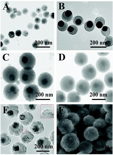
The original version of Fig. 1D contained two identical nanoparticles as the authors intended to include an inset of a typical nanoparticle and later decided to use the expanded image only, without removing the inset.
The correct version of Fig. 1 is shown here. The corrected version of Fig. 1 contains new data as the original data is no longer available.
The original version of Fig. 2E contained overlapping sections in panels a and c. The correct version of Fig. 2E is shown here. The corrected version of Fig. 2E contains new data as the original data is no longer available.
The original version of Fig. S10 was included in error and was a duplicate of the original Fig. 1E but with three nanoparticles removed. This occurred because these nanoparticles were removed to be used as insets and the image inadvertently saved after the nanoparticles had been removed. Fig. S10 in the Supplementary Information has been updated accordingly.
The Control and NPs panels for the Lung in the original version of Fig. S15 are duplicates and the original version of Fig. S16 was also included in error. Fig. S15 and S16 in the Supplementary Information have been corrected and updated accordingly.
An independent expert has viewed the corrected images and has concluded that they are consistent with the discussions and conclusions presented.
The Royal Society of Chemistry apologises for these errors and any consequent inconvenience to authors and readers.
| This journal is © The Royal Society of Chemistry 2022 |


