Recent progress of nanomedicine in secreted phospholipase A2 as a potential therapeutic target

Diya Shi , Congshu Feng , Jinhai Xie , Xi Zhang , HongLian Dai and Lesan Yan *
State Key Laboratory of Advanced Technology for Materials Synthesis and Processing, Biomedical Materials and Engineering Research Center of Hubei Province, Wuhan University of Technology, Wuhan 430070, China. E-mail: lsyan@whut.edu.cn

Received 21st March 2022 , Accepted 5th May 2022

First published on 6th June 2022


Abstract

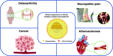
Overexpressed secretory phospholipase A2 (sPLA2) is found in many inflammatory diseases and various types of cancer. sPLA2 can catalyze the hydrolysis of phospholipid sn-2 ester bonds to lysophosphatidylcholine and free fatty acids, and its catalytic substrate and downstream products mediate a series of cascade reactions and inflammatory responses. Furthermore, different subtypes of sPLA2 can participate in different physiological processes by driving unique lipid pathways. Recently, many diseases have not been treated by appropriate chemotherapy methods due to low bioavailability and severe side effects of clinically available small-molecule drugs. Therefore, they have great development prospects of revealing the therapeutic mechanism of sPLA2 and use sPLA2 as a potential therapeutic target for designing and exploring new drugs and their delivery systems. Notably, the emergence of nanomedicines in recent years provides a practical and innovative means for overcoming the challenges associated with chemotherapy. With these considerations in mind, this paper systematically reviews recent studies on nanomedicines targeting sPLA2 overexpression in various diseases during the past few years.


image file: d2tb00608a-p1.tif

Diya Shi

Diya Shi received her BS degree in Biomedical Engineering from Chongqing University in 2019. She is currently a master student in the Department of Materials Science and Engineering at Wuhan University of Technology under the supervision of Prof. Lesan Yan. Her research interest is mainly focused on stimuli-responsive drug delivery systems based on phospholipase.

image file: d2tb00608a-p2.tif

Lesan Yan

Lesan Yan received his PhD in Polymer Chemistry and Physics from the Changchun Institute of Applied Chemistry, Chinese Academy of Sciences, in 2013. From 2013 to 2019, he worked as a postdoctoral fellow at Johns Hopkins University and University of Pennsylvania. Since 2019, he has been a professor at the State Key Laboratory of Advanced Technology for Materials Synthesis and Processing, Wuhan University of Technology. His research interests are focused on biodegradable polymer biomaterials, mainly based on polyesters, polypeptides, and polycarbonates, and their applications in nanomedicines and molecular imaging.


1. Introduction

Phospholipase A2 (PLA2) is a superfamily of enzymes with phospholipid decomposition activity.1 According to their structure and function, phospholipases are mainly divided into four categories:2,3 cytosolic phospholipase A2 (cPLA2), secreted phospholipase A2 (sPLA2), lipoprotein-associated phospholipase A2 (Lp-PLA2), and calcium-independent phospholipase A2 (iPLA2).

Secreted PLA2 was the first type of PLA2 enzyme to be identified. It is a small molecular weight secreted protein with molecular weights of 14–18 kDa and linked by 6–8 disulfide bonds.3 So far, the human genome has encoded 9 sPLA2 genes, all of which are named in the form of disulfide bonds and the order of discovery, showing different subtypes in human life activities.4 sPLA2 generally has an N-terminal signal peptide, which is related to the initiation of some functions. Protein folding structures may be different between different subtypes, but all sPLA2s have an active site slot of ∼15 Å depth. When the enzyme is transferred from the aqueous phase to the surface of the phospholipid membrane, the protein surface region surrounding the opening of the active site slot is in direct contact with the membrane, forming a water/membrane interface.5 The catalysis activity of sPLA2 requires the assistance of Ca2+ and is controlled by the composition, morphology, and physicochemical properties of the phospholipid membrane. sPLA2 generally doesn’t hydrolyze individual phospholipid molecules in solution but is only activated at the water/membrane interface. As a result, the enzyme must be adsorbed at the substrate membrane interface to hydrolyze phospholipids. In addition, the surface properties of the bound membrane, and the inhibition and degradation processes that result from binding to specific proteins will also affect the function of the enzyme.5,6

sPLA2s can catalyze the hydrolysis of the sn-2 ester bond in glycerophospholipids, releasing free fatty acids such as arachidonic acid (AA) and lysophospholipids (Fig. 1).5 Different subtypes of sPLA2 participate in different physiological processes or diseases by driving unique lipid pathways. For example, sPLA2-X in the airways of asthma patients plays an important role in promoting infection,7 while sPLA2-IIA has strong antibacterial activity in vivo by the degradation of bacterial membranes.8 sPLA2 can also modulate the systemic metabolic state by regulating lipoprotein phospholipids. Some sPLA2s even participate in the immune processes, and some may lead to severe obesity.9,10 Moreover, phospholipid hydrolysates are involved in tissue and nerve injury, as well as a series of inflammation and neurological disorders. Under pathological conditions, sPLA2 can be induced by multiple cascades and effector molecules. Many inflammatory pains, such as disc herniation, disc degeneration, atherosclerosis, osteoarthritis, and neuropathic pain, are associated with sPLA2. sPLA2 is also actively overexpressed in many types of cancer, including lung cancer,11,12 prostate cancer,13,14 and ovarian cancer.15 In addition, the hydrolysate-free fatty acid AA can be converted into a variety of precursors of eicosanoids, such as prostaglandins (PGs), leukotrienes (LTs), etc. Eicosanoids are involved in many physiological and pathological activities, such as sleep regulation, immune response, inflammation, and pain perception, by binding to specific G-protein coupled receptors.4,16 Therefore, sPLA2 has great potential both as the enzyme itself involved in regulating life activities and as the starting node of arachidonic acid metabolism.


image file: d2tb00608a-f1.tif
Fig. 1 The hydrolysis mechanism of sPLA2.

As we know, small-molecule therapeutic drugs often suffer from a short half-life in blood circulation and low bioavailability after intravenous administration in vivo. In contrast, nanomedicine emerged in recent years not only can improve the solubility and stability of hydrophobic drugs, and prolong the blood circulation time, but also can overcome several limitations, such as nonspecific systemic distribution, blood–brain barrier, etc. Therefore, the drug can be delivered to the lesion efficiently to exert its effectiveness.17,18 In addition, nanomedicine can realize the synergistic effect by co-delivery of multiple drugs, thereby improving the therapeutic efficacy and solving the drug resistance during the long-term use of a single drug. By adjusting the composition, structure, and other characteristics of the nanocarrier, the drug release rate can also be controlled, the blood drug concentration can be maintained at the therapeutic window level, and the drug can be targeted delivered to specific organs, tissues and even cells at a predictable rate and mechanism.19 Therefore, exploring potential drug targets and developing the targeted delivery of nanomedicine have become extremely promising and challenging tasks.

Based on these considerations, this paper reviews the latest progress of sPLA2 as a potential therapeutic target, discusses the mechanism of action of sPLA2 in different diseases, as well as its feasibility and importance as a drug target, and focuses on the recent progress of nanomedicine targeting sPLA2 in the treatment of inflammatory diseases and cancers.

2. Recent progress of nanomedicine targeting sPLA2 in the treatment of different diseases

2.1 Osteoarthritis

Osteoarthritis (OA), which occurs mostly in people over 65 years old, is one of the most common joint diseases in clinics with high morbidity and high disability rate. It shows symptoms of chronic pain, joint instability, stiffness, joint deformity, and narrowing of joint space.20 OA in young people is usually caused by a sports injury. After the injury, due to insufficient blood supply to articular cartilage and inefficient metabolic activity, the cartilage gradually degenerates and eventually causes OA.21 OA at the cellular level causes decreased mitochondrial respiration activity under a hypoxic environment, insufficient ATP production, more reactive oxygen species (ROS) and nitric oxide (NO) produced by chondrocytes and synoviocytes, and the disruption of the redox balance in vivo.22 In addition, many factors can affect OA. For example, aging will lead to cumulative DNA damage, which increases the expression of p16INK4a, a cyclin-dependent kinase inhibitor related to the cell cycle, and accelerates the rate of cellular aging.23 Obesity increases the loading on knee joints and secrete adipokines, leading to low-grade systemic inflammation.24 Since the middle of the 20th century, the incidence rate of OA has doubled due to the decrease in exercise and the increased population of obese people.25 According to the studies of genome-wide association screens (GWAS), more than 80 gene mutations have been related to the pathogenesis of OA.24 Different people have different clinical symptoms in OA, which indicates that its pathogenesis is influenced by a variety of genetic and environmental factors.

The sPLA2 family is one of the powerful inflammatory mediators involved in the development of OA. Through the analysis of AA and oleic acid release, sPLA2 can be induced by pro-inflammatory cytokines and free radicals under pathological conditions, and can participate in the development of OA through multiple cascades.26 Zhai et al.27 performed targeted metabolomic analysis on the collected clinical serum samples and evaluated the cartilage volume loss in the time course of 24 months by magnetic resonance imaging (MRI). The results suggested that the ratio of serum lysophosphatidylcholine (LysoPC) to phosphatidylcholine (PC) was associated with cartilage volume loss and markers of joint degradation, which could be used to predict the risk of knee OA. It also revealed that sPLA2 can catalyze the hydrolysis of phospholipids, which is exactly the reason that promotes the conversion of PC to lysoPC. Although there was no difference in the plasma sPLA2 content between OA patients and healthy controls, the enzyme was indeed highly expressed in OA chondrocytes under the stimulation of pro-inflammatory factors such as IL-1b, TNF, and IL-6.26,28 The ratio of LysoPC to PC can be easily measured in blood and can be used as an auxiliary means for clinicians to predict and diagnose knee OA.

In the clinic, cartilage is one of the earliest deteriorated tissues in the joint to develop into OA. It releases proteoglycan degradation products and damage-associated molecular patterns (DAMPs) into the joint space.29 The synovium is the connective tissue that connects the joint space, which maintains low-level inflammation by secreting pro-inflammatory cytokines and forms a positive feedback pathway, thus disrupting the self-healing balance of the joint.30 In fact, during intra-articular injection in patients with OA, it is very difficult for drugs to penetrate the extracellular matrix (ECM) into the joint and act on the lesions because the articular cartilage has no blood vessels and is surrounded by a dense negatively charged ECM. Even worse, the synovial fluid continuously flushes small-molecule drugs away, allowing them to be cleared through capillaries and drainage lymphatic vessels. All these factors make treating chondro-arthritis extremely challenging.31,32

Currently, the treatments for OA mainly focus on alleviating pain, suppressing the inflammatory response, and maintaining daily function. Nonsteroidal anti-inflammatory drugs (NSAIDs), as inhibitors of the cyclooxygenase (COX), are used in the OA treatment, but they will cause serious side effects on the gastrointestinal tract, kidneys, and cardiovascular system. For patients with NSAIDs intolerance, opioids often used may also cause side effects such as nausea, vomiting, dizziness, and constipation.33 Through the study of OA pathogenesis, it was found that using sPLA2 as a potential drug target, combined with the advantages of nanomedicine in vivo, may be able to improve the bioavailability of drugs, alleviate the OA symptoms and even reverse the process of OA.

As mentioned above, sPLA2 is usually present at a low level in healthy knee tissue, but it is highly expressed and active in synovial fluid and articular cartilage of OA patients. Therefore, when nanomedicine is used for intra-articular treatment, sPLA2 can be considered as the therapeutic target. By modifying its surface functional groups, nanomedicine can enhance the penetration of the ECM, thereby more effectively transporting sPLA2 inhibitors to the joint to increase the concentration of the inhibitor, and finally improve the therapeutic efficacy by inhibiting the sPLA2 activity. In this regard, Wei et al.34 designed an sPLA2 inhibitor-loaded nanoparticles (sPLA2i-NPs) (Fig. 2). Using the special amphiphilic properties of phospholipids, the lipid and inhibitors were self-assembled into micelles. After sPLA2i-NPs and free rhodamine were incubated with bovine cartilage tissue for a different time in vitro, the confocal fluorescence images of cartilage sections were obtained. The results showed that the fluorescence signal of the sPLA2i-NPs group doped with cationic DOTAP increased over time, indicating that the cationic DOTAP indeed helped the sPLA2i-NPs penetrate to the deep region of the cartilage. Using sPLA2i-NPs (DOTAP+) for the subsequent experiment, it was found that nanoparticles could remain in the knee joint for a longer time than free ICG. As sPLA2 is located in the deep cartilage tissue, it can enhance the deep cartilage penetration of nanoparticles and prolong the retention time in the knee joint by adjusting the surface charge, so that drugs promoted sufficient inhibition of sPLA2. Studies in the mouse model of OA also showed that local delivery of sPLA2i-NPs could attenuate the progression of OA, block joint injury, and relieve pain, compared with sPLA2i or nanoparticles alone. Because OA is a chronic disease, long-term treatment is required to further prove the safety of nanomedicine. However, the nanocarrier can improve the drug delivery efficiency, which undoubtedly provides a new idea for the treatment of OA.


image file: d2tb00608a-f2.tif
Fig. 2 (A) Schematic diagram of the sPLA2 inhibitor-loaded nanoparticle; (B) zeta potential of sPLA2i-NPs with/without cationic lipid DOTAP; (C) the response of sPLA2i-NPs to sPLA2 enzyme by a fluorescence spectrometer; (D) IVIS image of healthy and OA mouse knee joints after injection over 28 days; (E) semiquantitative analysis of time-course fluorescent radiant efficiency within healthy and OA mouse knee joints; (F) semiquantitative analysis of the AUC based on the fluorescence intensity profiles; (G) the design of sPLA2i-NP treatment for the destabilization of the medial meniscus (DMM)-induced OA mice; (H) Von Frey assay was performed after surgery; (I) IHC staining of the tibial articular cartilage at 2 months after surgery. Reproduced with permission.34 Copyright 2021, Science.

2.2 Spinal cord injury and neuropathic pain

Neuropathic pain is a frequent complication of spinal cord injury (SCI). After the initial core injury induced by mechanical force, the functional adaptability of the nervous system is poor, and it usually develops to the secondary injury stage through a series of complex cell networks, such as the regulation of synaptic remodeling and connection by microglia.35,36 During this period, a large number of nerve cells undergo apoptosis, resulting in the interruption of the neural circuit surrounding the injury core.37 Both spinal cord injury and nerve root compression can cause painful nerve damage, and some even produce secondary neuroinflammatory responses at the injury site and distal to the spinal cord. Glial cells promote the production of proinflammatory cytokines and chemokines, which trigger the hyperexcitability of spinal cord neurons, and play important roles in initiating and maintaining pain.38,39 It has been reported that the PLA2 family is involved in the process of spinal neuroimmune regulation of pain, and contributes to the production of downstream inflammatory proteins.40 In addition, phospholipid hydrolysates can also act as secondary signaling molecules to enhance the spinal cord inflammatory cascade.

To clarify the role of sPLA2 in spinal cord injury, Yang et al.41 confirmed the high-level expression of sPLA2-III in the brainstem, spinal cord, and cerebral neocortex by RT-PCR and western blot. Moreover, the electron microscopy results of the spinal cord and cerebral neocortex revealed that sPLA2-III was mainly located in dendrites or dendritic spines and played an important role in neuronal differentiation, growth, and signaling. Quartino et al.42 characterized myelin tissue by small-angle X-ray scattering (SAXS). The experimental results showed that the different sPLA2-treated myelin retained the lamellar structure, but the separation between the myelin membranes increased and the myelin was in an unstable state, which was easily affected by other factors. Herein, myelin is the phospholipid outside of neurons that helps neurons transmit signals. This insulating lipid will degrade and becomes ineffective due to the central nervous system dysfunction. The upregulation of sPLA2 activity accompanied by inflammation undoubtedly accelerates this process.

It is known that elevated phospholipase can promote the production of bioactive lipid mediators, thereby aggravating spinal inflammation and enhancing nociceptive signals.43 Neuropathic pain is the result of crosstalk mechanisms of multiple PLA2 subtypes, and sPLA2 and cPLA2 are expressed by immune activation simultaneously in the early stages of nerve injury. Tanaka et al.44 found that intra-spinal injection of viral-miRNA targeting sPLA2-III could significantly reduce the expression of sPLA2-III in the spinal cord, and substantially eliminate hyperalgesia after 14 days of treatment. Intrathecal treatment with antisense oligodeoxynucleotide or siRNA targeting sPLA2-III was able to reverse established thermal hyperalgesia. Using a well-established model of pain associated with nerve root compression, Kartha et al.45 investigated how sPLA2 in the spinal cord mediates the process of maintaining pain after nerve injury. The results showed that the level of sPLA2 was upregulated within 4 hours in spinal cord injury, and immediate inhibition could prevent the occurrence of pain and spinal cord hyperexcitability. 7 days after injury, the expression of sPLA2 in the spinal cord is still elevated. Inhibition of sPLA2 at this time can reduce the proportion of a wide dynamic range (WDR) neurons, abolish behavioral sensitivity, reduce neuronal discharge in the spinal cord, and restore the phenotype distribution and induced activity of spinal cord neurons to sham levels. In conclusion, many studies have confirmed the role of spinal cord sPLA2 in maintaining pain and central sensitization after nerve injury, providing experimental evidence for sPLA2 as a therapeutic target for the treatment of neuropathic pain.

After nerve injury, the expression of sPLA2 is elevated in the central spinal cord and peripheral dorsal root ganglion (DRG), accompanied by robust inflammatory and oxidative stress responses. Kartha et al.46 attempted to apply the COX-2 inhibitor, meloxicam, for pre-treatment before nerve root injury. After nerve root compression, the meloxicam pre-treated group showed alleviated pain and decreased activation of spinal microglia and astrocytes on day 7. Compared with the control group, meloxicam reduced the level of sPLA2 in the spinal cord and inhibited the oxidative stress response. If the sPLA2 inhibitor thioetheramide-PC (TEA-PC) was injected immediately after injury, it can effectively prevent the decrease of paw withdrawal threshold (PWT) on the first day after injury and the increase of large dynamic range spinal cord neurons caused by nerve root injury. In other words, the use of sPLA2 inhibitors can prevent mechanical ectopic pain and attenuate the hyperexcitability of the dorsal horn.47 Besides, Chen et al.40 investigated the role of CHEC-9, a broad-spectrum inhibitor of sPLA2, in neurodegeneration. Through the analysis of neuronal cells (SY5Y) treated with CHEC-9, it was found that CHEC-9 can inhibit the inflammatory cascades induced by sPLA2 and promote the survival of neuronal cells without promoting differentiation. In addition to the direct protection of neuron cells, CHEC-9 can also independently inhibit immune cell responses and play a neuroprotective role. This indicates that spinal sPLA2 may be related to the excitatory mechanism of early spinal neurons, and drugs can inhibit pain and subsequent inflammation by preventing the increase of sPLA2.

In the clinic, the systemic delivery of small molecule drugs has the problem of high blood clearance efficiency. If the drug is delivered directly to the spinal cord through intrathecal injection, it will cause harm to the uninjured spinal cord. To address this issue, Kartha et al.48 developed sPLA2 inhibitor-loaded micelles (Fig. 3). In particular, phospholipids and hydrophobic superparamagnetic iron oxide (SPIO) nanoparticles were co-assembled to form micelles, and sPLA2 inhibitors were incorporated into the phospholipids layer, which not only improved the solubility of SPIOs but also led to the dissociation of micelles in the sPLA2 overexpressed region. Thus, the inhibitors and other drugs were directly released into the pain region. It was found that local administration immediately after nerve root compression in the rodent model could effectively reduce the expression of sPLA2 in the spinal cord, and prevent the development of pain. Compared with the control group without inhibitors, the PWT was significantly higher in the inhibitor-loaded group. Delayed intravenous administration can also eliminate pain and reduce the expression of sPLA2 in the spinal cord, but the pain threshold is lower than that in the sham surgery group. In addition, SPIOs can be used as an imaging agent to track micelles and monitor the pathological state of pain sites. Therefore, the sPLA2-responsive phospholipid micelles can be used as a theragnostic agent and a sustained pain relieving agent. Compared with traditional drugs, this drug-loaded nanoparticle demonstrates excellent performance and broad application prospects.


image file: d2tb00608a-f3.tif
Fig. 3 (A) Schematic diagrams of sPLA2 inhibitor-loaded micelles. (B) The local/intravenous administration in a nerve root compression rodent model; (C) ipsilateral paw withdrawal thresholds and (D) the withdrawal threshold in the contralateral paw change following local administration with sPLA2 inhibitor-loaded micelles after a painful neuropathic nerve root compression injury; (E) iron is detected in the injured nerve root after local administration; (F) ipsilateral claw withdrawal threshold and (G) contralateral claw withdrawal threshold change following repeated intravenous administration of sPLA2 inhibitor-loaded micelles after nerve root compression; (H) spinal sPLA2 expression is significantly decreased (*p = 0.038) in the superficial dorsal horn after treatment of micelles with the inhibitor; (I) peripheral sPLA2 expression in the DRG is also significantly (*p = 0.002) reduced after treatment with sPLA2 inhibitor-loaded micelles. Reproduced with permission.48 Copyright 2020, American Chemical Society.

2.3 Atherosclerosis

Cardiovascular disease (CVD) is one of the most common causes of death worldwide and often leads to myocardial dysfunction. The human body supplies oxygen and nutrients to the heart through blood flow to maintain normal life activities. However, when lipid metabolism is impaired, it accumulates in the arterial intima, accompanied by fibrous tissue hyperplasia and calcium deposition. After atherosclerosis or thrombosis, the blood vessels become narrow or even blocked, and the heart experiences an ischemic shock, which may develop into myocardial infarction. After acute myocardial infarction, timely reperfusion is the main treatment to control the injury, but reperfusion itself can also damage myocardial cells, known as reperfusion injury.49,50

Insufficient oxygen supply caused by ischemia leads to metabolic acidosis, increased Ca2+, and elevated levels of ROS.51 During this process, a large amount of calcium-dependent sPLA2 is released, accelerating the degradation of phospholipids to AA, and promoting the release of inflammatory factors through the cyclooxygenase (COX) pathway, leading to myocardial necrosis. Meanwhile, elevated ROS levels can cause mitochondrial dysfunction. Due to the decomposition of phospholipid in the mitochondrial inner membrane, energy-producing enzymes in the electron transport chain are destroyed, finally leading to myocardial necrosis, as shown in Fig. 4.52 Several studies have shown that the activity of sPLA2 increases during reperfusion and inhibition of the enzyme's activity can protect the heart.53,54 Thus, in the pathophysiology of ischemia-reperfusion injury, sPLA2 provides a promising target for the treatment of vascular remodeling injury.


image file: d2tb00608a-f4.tif
Fig. 4 The mechanism of sPLA2 in myocardial reperfusion injury. Reproduced with permission.52 Copyright 2017, Elsevier.

Multiple sPLA2 subtypes have been shown to be associated with atherosclerosis. For example, sPLA2-X can lead to reduced cell cholesterol efflux and vascular cell proliferation by modifying lipoprotein.55 PLA2G5 can effectively hydrolyze phospholipids in lipoproteins, the hydrolysate can promote the proliferation of smooth muscle cells and induce apoptosis, and finally aggravate atherosclerosis.56 Yano et al.57 constructed a sPLA2-V−/− mouse model by knocking out the sPLA2-V gene. Compared with wild-type mice, sPLA2-V−/− mice had decreased left ventricular dysfunction and a 44% reduction in myocardial infarct area after experiencing a myocardial ischemia-reperfusion injury. Studies had shown that there is an independent association between elevated levels of sPLA2-IIA and early atherosclerosis, and thus it can be served as a biomarker for cardiovascular disease.58 In addition, sPLA2-IIA can promote the formation of aggregated/fused low-density lipoprotein (LDL) particles. As a result, the cholesterol carried by excessive LDL after oxidation will accumulate on the arterial wall, thereby causing arteriosclerosis.59 The sPLA2-IIA can also increase the expression of COX-2 and ROS and exert pro-inflammatory signals through M-type receptors to promote the formation of atherosclerotic lesions.60

So far, many sPLA2 inhibitors have been developed to reduce the risk of myocardial injury caused by atherosclerosis. For example, A-001 is a kind of inhibitor of the sPLA2 enzyme, and varespladib (A-002) is an orally bioavailable prodrug of A-001. Fraser et al.61 found that A-001 can effectively inhibit sPLA2 enzymes with IC50 values as low as the nM range. Moreover, it was found that oral administration of A-002 for 16 weeks significantly alleviated aortic atherosclerosis in the standard ApoE−/− mouse model. This improvement was associated with a significant reduction in total cholesterol but no effect on body weight, suggesting that A-002 was effective in significantly reducing the formation of atherosclerosis.

According to the mechanism of action of sPLA2 in atherosclerosis, it was found that inhibition of sPLA2 or the corresponding targets of its downstream signaling cascades may be a new therapeutic method to prevent or treat cardiovascular diseases. Till now, the development of selective inhibitors of sPLA2 has been carried out successively, and several therapeutic agents targeting sPLA2 to treat atherosclerosis are under clinical trials or in the preclinical phase.62 However, there is no relevant report on nanomedicines to the best of our knowledge. Due to the significant advantages of nanomedicines in applications, introducing the concept of nanomedicine into the delivery of sPLA2 inhibitors for atherosclerosis treatment is expected to be a major development direction in the future.

2.4 Cancer

Malignant tumor is one of the most lethal disease conditions in the world, seriously endangering human life and health. As the place for tumor cells to survive, the tumor microenvironment (TME) is a complex integrated system, including the internal and external cellular environment during tumor occurrence, growth, and metastasis. It is mainly composed of tumor cells, stromal cells, extracellular matrix, and soluble substances, wherein cells and substances are in a process of dynamic change. Immunosuppressive cells, tumor-associated macrophages, and a large number of inflammatory factors, such as IL-6, IL-10, TGF-β, and matrix metalloproteinase (MMP), gather together in the tumor microenvironment to jointly promote the proliferation and metastasis of tumor cells.63 In addition, there are significant differences between tumor tissues and normal tissues. For example, tumor cells produce a large amount of lactic acid by glycolysis, leading to a weak acidic environment (pH 6.5–7.2) in the tumor site.64 There are also high levels of ROS65 and reducing substances such as glutathione (GSH)66 in the tumor microenvironment, resulting in the imbalance of redox states.

Due to abnormal cell growth during tumorigenesis, the expression of certain genes will be disordered, leading to the abnormal expression or activity of certain enzymes inside or outside tumor cells, such as MMP, α-amylase, and cathepsin B.67,68 Notably, sPLA2 has also been shown to be overexpressed in a variety of tumors. For example, it is 22-fold higher in prostate cancer than in normal tissues.69 Therefore, revealing the mechanism of sPLA2 in cancer, to explore sPLA2 as a potential therapeutic target for cancer theranostics, has become an important development direction. In recent years, some progress has been made. For example, Xu et al.70 found that the expression levels of SLNCR1, an oncogenic long non-coding RNA (lncRNA), and sPLA2 were both upregulated in tumor tissues of patients with non-small cell lung cancer (NSCLC). Moreover, the expression levels of lncRNA, SLNCR1 and sPLA2 were positively correlated in the plasma of NSCLC patients, while these are not observed in the healthy controls. Lee et al.71 revealed that the enzyme catalyzes the hydrolysis of phospholipids and induces the phosphorylation of protein kinases, thereby activating the mitogen-activated protein kinase (MAPK) pathway and leading to cPLA2 activation. The cPLA2 can also hydrolyze phospholipid membranes to AA, forming a positive feedback loop. The crosstalk mechanism of sPLA2 and cPLA2 leads to the massive production of AA, and the AA-induced downstream inflammatory pathway and downstream product prostaglandin E2 (PGE2) to promote the development of cancer. It is reported that the expression of intracellular adhesion molecule-1 (ICAM-1) is associated with the increase of advanced lung cancer and lymph node metastasis, while the signal transducer and activator of transcription 3 (STAT3) can promote invasion. Studies have shown that knockout of sPLA2-related genes can effectively reduce the phenotype of cancer stem cells (CSCs) in NSCLC, and reduce the secretion of PGE2 and ICAM-1. As a result, the STAT3 pathway is blocked, which attenuates the progress of cancer invasion and thus plays an anti-cancer effect.11,72

As we know, liposomes composed of phospholipids have a bilayered structure similar to cell membranes. Both the internal cavity and the bilayer are very suitable for drug delivery. It can not only improve the solubility and bioavailability of drugs, but also reduce the side effects on other organs. Based on the specific hydrolysis properties of sPLA2, Arouri et al.73 proposed to replace the acyl chain at the sn-2 position with anticancer drugs. The obtained prodrugs can self-assemble into liposomes due to their amphiphilic properties (Fig. 5). Once subjected to sPLA2, the sn-2 ester bond would be specifically hydrolyzed, and the conjugated anticancer drugs would be cleaved and released from liposomes. The enzymatic hydrolysis process was analyzed by mass spectrometry. It was found that the introduction of other lipid components, such as 1,2-dipalmitoyl-sn-glycero-3-phosphocholine (DPPC), could accelerate hydrolysis compared with the single prodrug liposome alone. As a result, the premixing of the prodrug with DPPC significantly improved the drug release from liposomes. The design undoubtedly provides a new idea for the development of sPLA2-related nanomedicines.


image file: d2tb00608a-f5.tif
Fig. 5 Schematic diagram of liposomes composed of DPPC and C6-RAR, and hydrolyzed by sPLA2 enzyme. Reproduced with permission.73 Copyright 2011, Elsevier.

Due to the specific hydrolysis ability of sPLA2, liposomes as nanocarriers for sPLA2-overexpressed cancer therapy have been extensively developed. Liposomal cisplatin is the first chemotherapeutic drug developed to exploit using a sPLA2-dependent release mechanism and it is undergoing phase II clinical trials, currently.74 In addition to the release of the conjugated drug at the sn-2 position, the encapsulated drug in the aqueous interior cavity of the liposome can also be released arising from the imbalance of hydrophilic and hydrophobic properties under the catalytic hydrolysis of sPLA2. Hansen et al.75 used a fluorescent dye for release experiments to verify the sPLA2-triggered liposome hydrolysis. It was found that temperature can lead to changes in the calcein release profile by affecting the activity of sPLA2. Furthermore, DPPC was completely degraded in the presence of sufficient sPLA2, and the release percentage was proportional to the DPPC concentration. Notably, when C16 alkyl chain phospholipids replaced C14 alkyl chain phospholipids, the final percentage of calcein released from liposomes was greatly reduced. Mock et al.76 compared the different roles of sPLA2 in the drug release and cellular uptake between sterically stabilized liposomes (SSL) and sPLA2 responsive liposomes (SPRL) by regulating components of liposomes. It was found that when liposomes encapsulated with 6-carboxyfluorescein (6-CF) were exposed to sPLA2, the release increased with time, but SPRL was more dependent on sPLA2. After treating different subtypes of prostate cancer cells with doxorubicin (DOX)-loaded SSL and SPRL, it was found that the intracellular drug level in the SPRL group was 1.5–2 times higher than that in the SSL group. The content of DOX in PC-3 cells was the highest, while it was lower in the LNCaP cells. This indicates that cellular uptake is related to liposomes and cell types, and SPRL may be more suitable for the treatment of sPLA2 overexpressed diseases.

Tagami et al.77 developed a complex liposome composed of DPPC and triblock copolymers (Poloxamer 188). DPPC/P188 liposomes remained stable when incubated at 37 °C for 60 minutes without sPLA2, but released ∼80% encapsulated calcein within 10 minutes in the presence of sPLA2. In addition, the triggered release of DOX from the interior of liposome under the catalysis of sPLA2 exhibited significantly enhanced cytotoxicity against A549 lung cancer cells. Ghavami et al.78 attempted to deliver octaarginine-peptide nucleic acid (r8-PNA) conjugates using liposomes (Fig. 6). Herein, PNA is a macromolecular antisense drug, which can be effectively encapsulated in SPRL, and needs to be taken up by cells to play its role. Under serum-free conditions, sPLA2 triggered the release of PNA. However, the release of PNA was inhibited in a serum-containing medium. This may be due to the adsorption of serum protein on the surface of liposomes to form protein coronas, which prevents sPLA2 from hydrolyzing the phospholipid components in liposomes.


image file: d2tb00608a-f6.tif
Fig. 6 sPLA2 response mechanism of r8-PNA-loaded liposomes in the presence or absence of serum. Reproduced with permission.78 Copyright 2020, American Chemical Society.

Ostrem et al.79 developed an oxaliplatin liposome (L-OHP) that was highly sensitive to sPLA2. In the presence of sPLA2, L-OHP doped with 20% cholesterol showed ∼80% platinum release. In addition, it was observed that the L-OHP exhibited enhanced anti-proliferative effects in vitro on sPLA2-secreting MT-3 cells. However, the mouse model injected with sPLA2-sensitive liposomes developed severe systemic toxicity within 3 days. Similarly, Pourhassan et al.80 compared liposomes with different sPLA2 sensitivities, where the controlled drug release rate could be realized by adjusting the liposomal composition (Fig. 7). After intravenous injection, liposomal oxaliplatin significantly inhibited tumor growth compared to the free drug. However, the antitumor efficacy of sPLA2 sensitive liposomes was not significantly improved compared with non-sensitive or low-sensitive liposomes. After multiple injections of sPLA2 sensitive liposomes, subcutaneous bleeding and multifocal liver necrosis were even observed in experimental mice. These results indicate that although sPLA2 sensitive liposomes can enhance the anti-tumor effect compared to free drugs, there remains a safety concern and the possibility of failure in clinical application. Therefore, there is still a long way to go to bring sPLA2-sensitive nanomedicines to the clinic.


image file: d2tb00608a-f7.tif
Fig. 7 (A) Schematic diagram of the mechanism of sPLA2 sensitive liposomes in vitro and in vivo; (B) the cytotoxicity of liposomes in vitro; (C) petechial cutaneous hemorrhages, along with multifocal hepatonecrotic lesions in vivo. Reproduced with permission.80 Copyright 2017, Elsevier.

3. Conclusions and prospects

sPLA2 with phospholipid catalytic activity is an important enzyme to maintain the normal physiological activities of cells. Many inflammation-related diseases and cancers often exhibit changes in sPLA2 expression and abnormalities in its downstream cascade metabolites. Numerous studies have shown that inhibition of sPLA2 can block the inflammatory cascade pathway and other related signaling pathways, to achieve the therapeutic effect of treating inflammation and cancer.

To make better use of sPLA2 and make it further play a crucial role in the treatment process, it is necessary to understand the mechanism of action of sPLA2 in related diseases. As mentioned earlier, sPLA2 is a general term for a class of enzymes that includes different subtypes. Some function as tumor suppressors, while some are positively correlated with tumorigenesis. Some form signaling pathways with phospholipid hydrolysates and their downstream active factors to enhance tumor invasiveness, while some form a positive feedback loop with cPLA2 to promote the inflammation process through a crosstalk mechanism. Therefore, it is necessary to further explore the role of sPLA2 in the pathogenic process, clarify the pathogenesis, and thus select the appropriate sPLA2 subtype as the target to speed up the development of innovative drugs. Besides, the development of nanocarriers needs to be further strengthened. Due to so many obstacles for small molecule drug delivery, nanomedicine shows great potential in vivo. It can not only overcome multiple physiological barriers during the delivery process but also realize targeted controlled drug release and even co-delivery of multiple drugs through the design and modification of nanocarriers. Among various nanomedicines, it is worth noting that sPLA2 has broad application prospects as a potential therapeutic target. For example, functional prodrugs could be designed according to the property of sPLA2 to specifically hydrolyze the sn-2 ester bond; the sPLA2 responsive carriers can be designed to load imaging agents to realize the detection, localization and tracking of lesions. In addition, sPLA2 responsive carriers can also be utilized to co-deliver multiple drugs to play a synergistic effect, to overcome the drug resistance caused by the long-term use of a single drug. At the same time, the formulation and preparation process needs to be continuously optimized to achieve high-precision controlled release and large-scale production, and ultimately safe and effective use in vivo.

Overall, we discussed various diseases associated with the abnormal expression of sPLA2, summarized and discussed the application of sPLA2 in osteoarthritis, neuropathic pain, atherosclerosis, and cancer therapy. Although numerous small-molecule drugs targeting sPLA2 have been developed, there is still no available clinical drug due to their limited efficacy and severe side effects. Recent progress suggests that nanomedicines using sPLA2 as a therapeutic target show great potential and advantages in clinical translation. Therefore, this review may provide insights and ideas for the development of nanomedicine using sPLA2 as a potential therapeutic target, to expand the research direction, and provide help for its clinical translation.

Author contributions

The manuscript was written through the contributions of all authors. All authors have given approval to the final version of the manuscript.

Conflicts of interest

There are no conflicts to declare.

Acknowledgements

The research was supported by the National Natural Science Foundation of China (No. 52003211), the Natural Science Foundation of Hubei Province (No. 2020CFB418), and the Fundamental Research Funds for the Central Universities (Wuhan University of Technology, No. 2021IVA096, 2021IVB035, 2021IVB071 and 2022IVB003).

References

  1. M. Murakami, Biochim. Biophys. Acta, Mol. Cell Biol. Lipids, 2019, 1864, 763–765 CrossRef CAS PubMed.
  2. M. Chiorazzo, PhD dissertation, University of Pennsylvania, 2016, p. 1653.
  3. C. S. Batsika, A. D. Gerogiannopoulou, C. Mantzourani, S. Vasilakaki and G. Kokotos, Expert Opin. Drug Discovery, 2021, 16, 1287–1305 CrossRef CAS PubMed.
  4. R. H. Schaloske and E. A. Dennis, Biochim. Biophys. Acta, 2006, 1761, 1246–1259 CrossRef CAS PubMed.
  5. G. Lambeau and M. H. Gelb, Annu. Rev. Biochem., 2008, 77, 495–520 CrossRef CAS PubMed.
  6. M. Schewe, P. F. Franken, A. Sacchetti, M. Schmitt, R. Joosten, R. Bottcher, M. E. van Royen, L. Jeammet, C. Payre, P. M. Scott, N. R. Webb, M. Gelb, R. T. Cormier, G. Lambeau and R. Fodde, Cell Stem Cell, 2016, 19, 38–51 CrossRef CAS.
  7. J. D. Nolin, R. C. Murphy, M. H. Gelb, W. A. Altemeier, W. R. Henderson, Jr. and T. S. Hallstrand, Biochim. Biophys. Acta, Mol. Cell Biol. Lipids, 2019, 1864, 827–837 CrossRef CAS.
  8. V. P. van Hensbergen, Y. Wu, N. M. van Sorge and L. Touqui, Trends Immunol., 2020, 41, 313–326 CrossRef CAS.
  9. E. Dore, C. Joly-Beauparlant, S. Morozumi, A. Mathieu, T. Levesque, I. Allaeys, A. C. Duchez, N. Cloutier, M. Leclercq, A. Bodein, C. Payre, C. Martin, A. Petit-Paitel, M. H. Gelb, M. Rangachari, M. Murakami, L. Davidovic, N. Flamand, M. Arita, G. Lambeau, A. Droit and E. Boilard, JCI Insight, 2022, 7, e152638 CrossRef PubMed.
  10. E. Leiguez, P. Motta, R. Maia Marques, B. Lomonte, S. V. Sampaio and C. Teixeira, Biomolecules, 2020, 10, 1593 CrossRef CAS PubMed.
  11. A. L. Halpern, P. D. Kohtz, J. Y. Rove, L. Ao, X. Meng, D. A. Fullerton and M. J. Weyant, Mol. Cell. Biochem., 2019, 456, 145–156 CrossRef CAS PubMed.
  12. C. Autilio, S. Shankar-Aguilera, A. Minucci, L. Touqui and D. De Luca, Am. J. Physiol.: Lung Cell. Mol. Physiol., 2019, 316, L498–L505 CrossRef CAS PubMed.
  13. S. Lu and Z. Dong, Int. J. Oncol., 2017, 50, 2113–2122 CrossRef CAS PubMed.
  14. A. S. Alnaim, M. W. Eggert, B. Nie, N. D. Quanch, S. L. Jasper, J. Davis, G. S. Barnett, B. S. Cummings, P. R. Panizzi and R. D. Arnold, Proceedings: AACR Annual Meeting, 2019, p. 2985.
  15. U. Ray, D. Roy, L. Jin, P. Thirusangu, J. Staub, Y. Xiao, E. Kalogera, A. E. Wahner Hendrickson, G. D. Cullen, K. Goergen, A. L. Oberg and V. Shridhar, J. Exp. Clin. Cancer Res., 2021, 40, 182 CrossRef CAS PubMed.
  16. M. Murakami, Y. Nakatani, G. I. Atsumi, K. Inoue and I. Kudo, Crit. Rev. Immunol., 2017, 37, 127–195 Search PubMed.
  17. A. Wicki, D. Witzigmann, V. Balasubramanian and J. Huwyler, J. Controlled Release, 2015, 200, 138–157 CrossRef CAS PubMed.
  18. K. K. Jain, Methods Mol. Biol., 2020, 2059, 1–54 CrossRef CAS PubMed.
  19. S. Adepu and S. Ramakrishna, Molecules, 2021, 26, 5905 CrossRef CAS PubMed.
  20. B. Xia, C. Di, J. Zhang, S. Hu, H. Jin and P. Tong, Calcif. Tissue Int., 2014, 95, 495–505 CrossRef CAS PubMed.
  21. W. Zhang, H. Ouyang, C. R. Dass and J. Xu, Bone Res., 2016, 4, 15040 CrossRef CAS PubMed.
  22. A. Mobasheri, M. P. Rayman, O. Gualillo, J. Sellam, P. van der Kraan and U. Fearon, Nat. Rev. Rheumatol., 2017, 13, 302–311 CrossRef CAS PubMed.
  23. R. F. Loeser, J. A. Collins and B. O. Diekman, Nat. Rev. Rheumatol., 2016, 12, 412–420 CrossRef CAS PubMed.
  24. D. Chen, J. Shen, W. Zhao, T. Wang, L. Han, J. L. Hamilton and H. J. Im, Bone Res, 2017, 5, 16044 CrossRef CAS PubMed.
  25. I. J. Wallace, S. Worthington, D. T. Felson, R. D. Jurmain, K. T. Wren, H. Maijanen, R. J. Woods and D. E. Lieberman, Proc. Natl. Acad. Sci. U. S. A., 2017, 114, 9332–9336 CrossRef CAS PubMed.
  26. L. Leistad, A. J. Feuerherm, A. Faxvaag and B. Johansen, Scand. J. Rheumatol., 2011, 40, 308–316 CrossRef CAS PubMed.
  27. G. Zhai, J. P. Pelletier, M. Liu, D. Aitken, E. Randell, P. Rahman, G. Jones and J. Martel-Pelletier, Sci. Rep., 2019, 9, 9648 CrossRef PubMed.
  28. W. Zhang, G. Sun, D. Aitken, S. Likhodii, M. Liu, G. Martin, A. Furey, E. Randell, P. Rahman, G. Jones and G. Zhai, Rheumatology, 2016, 55, 1566–1574 CrossRef CAS PubMed.
  29. J. H. Rosenberg, V. Rai, M. F. Dilisio and D. K. Agrawal, Mol. Cell. Biochem., 2017, 434, 171–179 CrossRef CAS PubMed.
  30. E. V. Medvedeva, E. A. Grebenik, S. N. Gornostaeva, V. I. Telpuhov, A. V. Lychagin, P. S. Timashev and A. S. Chagin, Int. J. Mol. Sci., 2018, 19, 2366 CrossRef PubMed.
  31. S. Dolati, S. Sadreddini, D. Rostamzadeh, M. Ahmadi, F. Jadidi-Niaragh and M. Yousefi, Biomed. Pharmacother., 2016, 80, 30–41 CrossRef CAS PubMed.
  32. Z. Peng, H. Sun, V. Bunpetch, Y. Koh, Y. Wen, D. Wu and H. Ouyang, Biomaterials, 2021, 268, 120555 CrossRef CAS.
  33. J. Martel-Pelletier, A. J. Barr, F. M. Cicuttini, P. G. Conaghan, C. Cooper, M. B. Goldring, S. R. Goldring, G. Jones, A. J. Teichtahl and J. P. Pelletier, Nat. Rev. Dis. Primers, 2016, 2, 16072 CrossRef PubMed.
  34. Y. Wei, L. Yan, L. Luo, T. Gui, B. Jang, A. Amirshaghaghi, T. You, A. Tsourkas, L. Qin and Z. Cheng, Sci. Adv., 2021, 7, eabe6374 CrossRef CAS PubMed.
  35. K. Inoue and M. Tsuda, Nat. Rev. Neurosci., 2018, 19, 138–152 CrossRef CAS PubMed.
  36. G. Chen, Y. Q. Zhang, Y. J. Qadri, C. N. Serhan and R. R. Ji, Neuron, 2018, 100, 1292–1311 CrossRef CAS PubMed.
  37. T. Yagami, Y. Yamamoto and H. Koma, Mol. Neurobiol., 2014, 49, 863–876 CrossRef CAS.
  38. C. Sommer, M. Leinders and N. Uceyler, Pain, 2018, 159, 595–602 CrossRef CAS.
  39. M. Matsuda, Y. Huh and R. R. Ji, J. Anesth., 2019, 33, 131–139 CrossRef PubMed.
  40. S. Chen, L. Yao and T. J. Cunningham, PLoS One, 2012, 7, e39257 CrossRef CAS PubMed.
  41. H. Yang, N. J. Siddiqi, A. S. Alhomida and W. Y. Ong, Neurochem. Res., 2013, 38, 753–760 CrossRef CAS PubMed.
  42. P. J. Yunes Quartino, J. M. Pusterla, V. M. Galvan Josa, G. D. Fidelio and R. G. Oliveira, Biochim. Biophys. Acta, 2016, 1858, 123–129 CrossRef CAS PubMed.
  43. H. Y. Zhou, S. R. Chen and H. L. Pan, Expert Rev. Clin. Pharmacol., 2011, 4, 379–388 CrossRef CAS PubMed.
  44. K. Tanaka, N. Dozono, H. Neyama, J. Nagai, R. Tsukahara, K. Nagayasu, S. Kaneko and H. Ueda, Biochem. Biophys. Res. Commun., 2021, 568, 167–173 CrossRef CAS PubMed.
  45. S. Kartha, P. Ghimire and B. A. Winkelstein, Mol. Pain, 2021, 17, 17448069211066221 CrossRef CAS.
  46. S. Kartha, C. L. Weisshaar, B. H. Philips and B. A. Winkelstein, Neuroscience, 2018, 388, 393–404 CrossRef CAS PubMed.
  47. J. C. Quindlen-Hotek, S. Kartha and B. A. Winkelstein, NeuroReport, 2020, 31, 1084–1089 CrossRef CAS PubMed.
  48. S. Kartha, L. Yan, M. E. Ita, A. Amirshaghaghi, L. Luo, Y. Wei, A. Tsourkas, B. A. Winkelstein and Z. Cheng, ACS Nano, 2020, 14, 8103–8115 CrossRef CAS.
  49. H. Zhou, Q. Ma, P. Zhu, J. Ren, R. J. Reiter and Y. Chen, J. Pineal Res., 2018, 64, e12471 CrossRef.
  50. D. J. Hausenloy and D. M. Yellon, J. Clin. Invest., 2013, 123, 92–100 CrossRef CAS PubMed.
  51. G. A. Kurian, R. Rajagopal, S. Vedantham and M. Rajesh, Oxid. Med. Cell. Longev., 2016, 1656450 Search PubMed.
  52. S. Ravindran and G. A. Kurian, Biomed. Pharmacother., 2017, 92, 7–16 CrossRef CAS.
  53. Q. Xie and D. Zhang, Int. Heart J., 2017, 58, 115–124 CrossRef CAS PubMed.
  54. Y. Wang, J. Wu, J. Zhu, C. Ding, W. Xu, H. Hao, J. Zhang, G. Wang and L. Cao, J. Ethnopharmacol., 2021, 277, 114223 CrossRef CAS PubMed.
  55. R. Atout, S. A. Karabina, S. Dollet, M. Carreras, C. Payre, P. Andre, G. Lambeau, V. Lotteau, E. Ninio and L. Perrin-Cocon, Atherosclerosis, 2012, 222, 367–374 CrossRef CAS PubMed.
  56. W. Pruzanski, J. Kopilov and A. Kuksis, Prostaglandins Other Lipid Mediat., 2016, 122, 64–68 CrossRef CAS PubMed.
  57. T. Yano, D. Fujioka, Y. Saito, T. Kobayashi, T. Nakamura, J. E. Obata, K. Kawabata, K. Watanabe, Y. Watanabe, H. Mishina, S. Tamaru and K. Kugiyama, Cardiovasc. Res., 2011, 90, 335–343 CrossRef CAS PubMed.
  58. C. Q. Sun, C. Y. Zhong, W. W. Sun, H. Xiao, P. Zhu, Y. Z. Lin, C. L. Zhang, H. Gao and Z. Y. Song, Sci. Rep., 2016, 6, 34929 CrossRef CAS PubMed.
  59. J. K. Hakala, K. Oorni, M. O. Pentikainen, E. Hurt-Camejo and P. T. Kovanen, Arterioscler., Thromb., Vasc. Biol., 2001, 21, 1053–1058 CrossRef CAS.
  60. W. Annema, J. F. de Boer, A. Dikkers, L. G. Dimova, M. van der Giet, S. J. L. Bakker and U. J. F. Tietge, J. Clin. Med., 2020, 9, 1282 CrossRef CAS PubMed.
  61. H. Fraser, C. Hislop, R. M. Christie, H. L. Rick, C. A. Reidy, M. L. Chouinard, P. I. Eacho, K. E. Gould and J. Trias, J. Cardiovasc. Pharmacol., 2009, 53, 60–65 CrossRef CAS PubMed.
  62. L. Knerr, F. Giordanetto, P. Nordberg, D. Pettersen, N. Selmi, H. G. Beisel, H. de la Motte, T. Olsson, T. D. J. Perkins, M. Herslof, A. Mansson, M. Dahlstrom, I. Starke, J. Broddefalk, G. Saarinen, F. Klingegard, E. Hurt-Camejo, B. Rosengren, J. Brengdahl, F. Jansen, M. Rohman, J. Sandmark, K. Hallberg, T. Akerud, R. G. Roth and M. Ahlqvist, ACS Med. Chem. Lett., 2018, 9, 594–599 CrossRef CAS PubMed.
  63. M. Yao, G. Brummer, D. Acevedo and N. Cheng, Adv. Cancer Res., 2016, 132, 265–367 CrossRef CAS PubMed.
  64. J. Z. Du, C. Q. Mao, Y. Y. Yuan, X. Z. Yang and J. Wang, Biotechnol. Adv., 2014, 32, 789–803 CrossRef CAS PubMed.
  65. F. Weinberg, N. Ramnath and D. Nagrath, Cancers, 2019, 11, 1191 CrossRef CAS.
  66. Z. Zhou, H. Wu, R. Yang, A. Xu, Q. Zhang, J. Dong, C. Qian and M. Sun, Sci. Adv., 2020, 6, eabc4373 CrossRef CAS.
  67. S. Quintero-Fabian, R. Arreola, E. Becerril-Villanueva, J. C. Torres-Romero, V. Arana-Argaez, J. Lara-Riegos, M. A. Ramirez-Camacho and M. E. Alvarez-Sanchez, Front. Oncol., 2019, 9, 1370 CrossRef.
  68. R. Cheng, F. H. Meng, C. Deng and Z. Y. Zhong, Nano Today, 2015, 10, 656–670 CrossRef CAS.
  69. J. R. Graff, B. W. Konicek, J. A. Deddens, M. Chedid, B. M. Hurst, B. Colligan, B. L. Neubauer, H. W. Carter and J. H. Carter, Clin. Cancer Res., 2001, 7, 3857–3861 CAS.
  70. W. Xu, Q. Xu, D. Kuang, Z. Wang, Q. Lu, Q. Lin, H. Wu and L. Chen, Mol. Med. Rep., 2019, 20, 2591–2596 CAS.
  71. S. Lee, D. Kim, J. Kang, E. Kim, W. Kim, H. Youn and B. Youn, Cell. Physiol. Biochem., 2017, 42, 1684–1700 CrossRef CAS PubMed.
  72. D. T. Bennett, X. S. Deng, J. A. Yu, M. T. Bell, D. C. Mauchley, X. Meng, T. B. Reece, D. A. Fullerton and M. J. Weyant, Ann. Thorac. Surg., 2014, 98, 439–445 CrossRef PubMed ; discussion 445–436.
  73. A. Arouri and O. G. Mouritsen, Eur. J. Pharm. Sci., 2012, 45, 408–420 CrossRef CAS PubMed.
  74. S. S. Jespersen, E. S. Stovgaard, D. Nielsen, T. D. Christensen, A. S. K. Buhl, I. J. Christensen and E. Balslev, Appl. Immunohistochem. Mol. Morphol., 2021, 29, e5–e9 CrossRef CAS.
  75. A. H. Hansen, O. G. Mouritsen and A. Arouri, Int. J. Pharm., 2015, 491, 49–57 CrossRef CAS PubMed.
  76. J. N. Mock, L. J. Costyn, S. L. Wilding, R. D. Arnold and B. S. Cummings, Integr. Biol., 2013, 5, 172–182 CrossRef CAS PubMed.
  77. T. Tagami, Y. Ando and T. Ozeki, Int. J. Pharm., 2017, 517, 35–41 CrossRef CAS PubMed.
  78. M. Ghavami, T. Shiraishi and P. E. Nielsen, ACS Appl Bio Mater., 2020, 3, 1018–1025 CrossRef CAS PubMed.
  79. R. G. Ostrem, L. Parhamifar, H. Pourhassan, G. Clergeaud, O. L. Nielsen, A. Kjaer, A. E. Hansen and T. L. Andresen, J. Controlled Release, 2017, 262, 212–221 CrossRef PubMed.
  80. H. Pourhassan, G. Clergeaud, A. E. Hansen, R. G. Ostrem, F. P. Fliedner, F. Melander, O. L. Nielsen, C. K. O'Sullivan, A. Kjaer and T. L. Andresen, J. Controlled Release, 2017, 261, 163–173 CrossRef CAS PubMed.

This journal is © The Royal Society of Chemistry 2022