Bioactive hydrogels based on polysaccharides and peptides for soft tissue wound management

Lihuang Wu , Yiyan He , Hongli Mao * and Zhongwei Gu *
Research Institute for Biomaterials, Tech Institute for Advanced Materials, College of Materials Science and Engineering, Nanjing Tech University, Nanjing 211816, China. E-mail: h.mao@njtech.edu.cn; zwgu1006@scu.edu.cn

Received 20th March 2022 , Accepted 13th April 2022

First published on 20th April 2022


Abstract

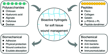
Due to their inherent and tunable biomechanical and biochemical performances, bioactive hydrogels based on polysaccharides and peptides have shown attractive potential for wound management. In this review, the recent progress of bioactive hydrogels prepared by polysaccharides and peptides for soft tissue wound management is overviewed. Meanwhile, we focus on the elaboration of the relationship between chemical structures and inherent bioactive functions of polysaccharides and peptides, as well as the strategies that are taken for achieving multiple wound repairing effects including hemostasis, adhesion, wound contraction and closure, anti-bacteria, anti-oxidation, immunomodulation, molecule delivery, etc. Some innovative and important works are well introduced as well. In the end, current study limitations, clinical unmet needs, and future directions are discussed.


image file: d2tb00591c-p1.tif

Hongli Mao

Dr Hongli Mao obtained his PhD in Materials Science and Engineering from the University of Tsukuba, Japan in July 2014. Then he began to work at RIKEN as a JSPS (Japan Society for the Promotion of Science) Postdoctoral Fellow. In March 2017, he moved to Nanjing, China, and has been a Professor at the College of Materials Science and Engineering, Nanjing Tech University since then. His main research interests include polymer biomaterials, tissue engineering, regenerative medicine, and 3D bioprinting.

1. Introduction

Wounds caused by surgery, trauma, burn, infection, diabetes, etc. have threatened human health for years.1–4 The treatment of wounds has become a severe clinical challenge and financial burden.5 Usually, wound repairing is a complicated biological process, including hemostasis, inflammation, proliferation, and remodeling.6 However, pathologically, it usually fails to progress following a normal pathway because problems such as infection, excessive inflammation, and secondary damage during wound recovery can lead to abnormal wound repairing.7,8 Clinical available treatments need superb surgical skills and are often, unfortunately, lead to unsatisfied results. Thus, innovative strategies are an urgent need to protect the wound sites and accelerate the wound repairing process.

Hydrogels own strong hygroscopicity, great biocompatibility, three-dimensional (3D) structure, tissue-like mechanical properties, and have become the most competitive materials in wound management.7 They enable the formation of a protective microenvironment for wound repairing once contact and infiltrate a defective tissue area. Therefore, hydrogels in the form of injectable liquid, prefabricated film, nanofibrous membrane, and 3D-printing construct can be used as wound dressings, bioadhesives, sutures, and tissue engineering scaffolds. They can perform biomechanical and biochemical functions in the wound repairing process, including physical protection, contaminated exudate absorption, damaged tissue adhesion and reconnection, hemostasis, molecule delivery and targeting, anti-bacteria, anti-oxidation, inflammation regulation, etc.7–10 A tremendous number of works have emerged in this field over the last decade, and many biopolymers were developed for hydrogels preparation. Polysaccharides and peptides are most commonly used as hydrogels matrix for their great hydrophilicity, biocompatibility, biodegradability, and inherent bioactive effects in wound management.8–10 Hydrogels prepared from polysaccharides and peptides can theoretically be used as ideal wound repairing platforms. This is because they are readily bioactive and can acquire multiple functions from a rational design and fabrication procedure. Thus, a comprehensive understanding of the intrinsic connection of chemical structure and bioactive effects of these biopolymers is favorable. The modification for acquiring superior multiple functions in hydrogels for wound repairing needs proper consideration and evaluation.

Herein, we provided a review of recently developed polysaccharides- and peptides-based hydrogels in wound management, with emphasis on the integrity of their molecular structures and inherent functionalities by materials categories (Fig. 1). Innovative strategies for achieving multiple bioactive effects based on the foundation of biochemical cues and nature inspirations are introduced. Although other researchers have previously made many reviews of close topics,3,6,7,9,11 we believe that this review can provide accessible knowledge and design ideas for future researchers.


image file: d2tb00591c-f1.tif
Fig. 1 Classification and summary of bioactive polysaccharides- and peptides-based hydrogels for wound management with chemical structures and inherent bioactive functions.

2. Polysaccharides-based hydrogels

Polysaccharides are one of the most widely sourced biopolymers from nature, including chitosan, hyaluronic acid, alginate, cellulose, dextran, etc.6,10 They are usually biodegradable and highly hydrophilic. Polysaccharides' unique water absorption and retention properties make them a great choice for preparing hydrogels matrix. In addition, abundant hydroxyl and carboxyl groups on the macromolecular chains provide feasible modification sites, thus further functionalization is available to overcome their defects.12

2.1. Chitosan

Chitosan has been widely used in the treatment of traumas and burns as gauzes or patches (Fig. 1) due to its inherent antibacterial, hemostatic and adhesive properties.13,14 It is obtained by deacetylation treatment of chitin from crustaceans and insects. After this, the amino groups are exposed on the chains (Fig. 1), making chitosan the only known alkaline polysaccharide with a positive charge in nature. The number of N-glucosamine units, defined as the degree of deacetylation (DDA), directly affects the biochemical performance of chitosan, including biodegradability and immunogenicity.3 Further chemical modification is often needed to prepare chitosan derivatives to enhance their solubility and reactivity, such as carboxymethyl chitosan, glycol chitosan, N-carboxyethyl chitosan, and N-(2-hydroxypropyl) chitosan.14

An important bioactive characteristic of chitosan is that it can act as a hemostatic agent to promote red blood cells and platelets aggregation, change hemodynamics and promote the formation of blood clots. The underlying mechanism is reported to be the cationic effect of the pendant amino group on chitosan or the Schiff base reaction between the amino group of chitosan and aldehyde group on the membrane glycoprotein of red blood cells.3 Inflammation at the wound sites can be accelerated by exposure to chitosan, and adhesion between chitosan and tissue may occur during the hemostatic process. Chitosan is one of the most successful commercial hemostatic wound dressings, typically available products include Celox®, HemCon®, and TraumaStat®, and it is reported to be the “star” material utilized in hydrogels for wound repairing.15

Chitosan usually acts as a cross-linker with aldehydes and diisocyanate functional biopolymers to prepare hydrogels.3 Many chemical modified methods have been taken on chitosan-based hydrogels to improve their hemostasis and adhesion performance, including catechol16–23 and pyrogallol24–26 decoration, amino27 and aldehyde28 modification, photoactive methacrylate17,29–33 and o-nitrobenzyl34 functionalization, etc. For example, Peng et al.35 fabricated a polyethyleneimine/polyacrylic acid/quaternized chitosan (PEI/PAA/QCS) powder through a freeze drying-grinding procedure with ultrafast self-gelling and wet adhesive properties (Fig. 2A). It was reported to form a pressure-resistant adhesive hydrogel barrier on contact with water and show instant and effective hemostasis against high-pressure arterial bleeding wounds of rats and non-compressible massive hemorrhage in porcine spleen and liver, as well as great wound-healing effects.


image file: d2tb00591c-f2.tif
Fig. 2 Chitosan-based hydrogels with multiple bioactive functions. (A) The polyethyleneimine/polyacrylic acid/quaternized chitosan (PEI/PAA/QCS) powder with ultrafast self-gelling and wet adhesive properties. The powder shows instant and effective hemostasis against high-pressure arterial bleeding wounds and non-compressible massive hemorrhage in the porcine spleen and liver. Reprinted from ref. 35 with permission. Copyright 2021 American Chemical Society. (B) A pH/glucose dual responsive multi-functional hydrogels system provides a local-specific drug dual-response release strategy for treating type II diabetic feet. Reprinted from ref. 23 with permission. Copyright 2022 American Chemical Society.

Antimicrobial activity can be easily obtained using chitosan-based hydrogels, although the exact mechanism remains obscure.36 It may also be suggested from the electrostatic interaction between the protonated amino of chitosan on wound sites and the electronegative microbial membrane. In this scenario, protonated amino can attract and recruit microorganisms, causing membrane damage that leads to bacterial death. Benefit from the strong hygroscopicity and abundant internal space, chitosan-based hydrogels can also absorb contaminants from wound effusion. Further antimicrobial-enhanced chemical modification (such as quaternary ammonium,30,35,37–43 alkylate44–46 and antibacterial peptide41,47) and physical encapsulation (such as antibiotics,31,32,45,47 polydopamine,38,42,48 graphene oxide,30,38,49 carbon nanotubes50 and other nanomaterials37,42,45,48,51) methods are also favorable.

By integrating the merits of the above-mentioned methods, one can prepare a multi-functional wound treatment platform. Recently a pH/glucose dual responsive multi-functional hydrogel based on phenylboronic acid/benzaldehyde bifunctional polyethylene glycol-co-poly(glycerol sebacic acid) and dihydrocaffeic acid/L-arginine grafted chitosan with metformin (Met) and graphene oxide (GO) incorporation was developed by Liang et al.23 In this system, Schiff base and phenylboronate ester were used to provide on-demand release of Met. It provided a local on-demand drug release strategy with an improving wound healing effect for treating type II diabetic feet (Fig. 2B). Therefore, in these considerations, the chitosan-based hydrogel is one of the best candidates in dealing with wounds.

2.2. Hyaluronic acid

Hyaluronic acid (HA) is an anionic polysaccharide consisting of D-glucuronic acid and N-acetyl-D-glucosamine repeating units (Fig. 1). It is widely found in the connective tissue of mammals and is one of the main components of the extracellular matrix (ECM).52 HA has a strong water-absorbing ability and great biocompatibility due to the abundant hydroxyl and carboxyl groups on the polymer chains. Its degradation products in vivo used to be considered to be nonimmunogenic.3 However, HA has been proven to have immunomodulatory effects in a molecular weight (size)-dependent manner with specifically high molecular weight (HMW) HA species have immunoprotective effects while the low molecular weight (LMW) one is a potent pro-inflammatory agent.53–56

Due to the lack of adhesive action, excessive swelling, and premature degradation upon contact with tissue, the mechanical properties of HA-based hydrogels are generally poor as wound repairing materials. These issues can be addressed by chemical modification of HA to improve the cross-linking degree of networks, and the interpenetration and entanglement of the polymer chains, including catechol57–63 and pyrogallol57,64 for mussel-inspired catechol chemistry through hydrogen-bond interaction and metal ions coordination, aldehydes from periodate oxidation of HA60,65–69 and photoactive o-nitrobenzyl70–73 for Schiff base reaction, methacrylate61,62,71,74–76 for photoinitiated free radical polymerization, phenylboronic acid77,78 for glucose-sensitive dynamic borate bond with polyphenol groups, etc.

Our group60 has designed a multiple cross-linked double-network using dopamine-functionalized oxidized HA (OHA-Dop), adipic acid dihydrazide-modified HA (HA-ADH), and aldehyde-terminated Pluronic F127 (AF127) as the backbones (Fig. 3A). The complex system provided strong adhesion, enhanced self-healing, and mechanical properties. Furthermore, recently we73 developed another dual-network hydrogels system based on azide-functionalized carboxymethyl chitosan and o-nitrobenzyl-modified HA (CMC-AZ/HA-NB) (Fig. 3B). Benefit from the o-nitrobenzyl group, polymer chains cross-linking based on photoactive Schiff base reaction was carried out without the cytotoxicity concern on the use of photoinitiators.


image file: d2tb00591c-f3.tif
Fig. 3 Hyaluronic acid (HA)-based hydrogels with enhanced mechanical properties, hemostatic and adhesive functions. (A) Hydrogels with multiple networks using dopamine-functionalized oxidized HA, adipic acid dihydrazide-modified HA, and aldehyde-terminated Pluronic F127. Reprinted from ref. 60 with permission. Copyright 2020 American Chemical Society. (B) Dual-network hydrogels developed by photoinitiator-free crosslinking based on azide-functionalized carboxymethyl chitosan and o-nitrobenzyl-modified HA. Reprinted from ref. 73 with permission. Copyright 2022 Elsevier. (C) A hemostatic adhesive hydrogel system using serotonin-conjugated HA with enhanced hemostasis by the mediation of serotonin and adhesive property via catechol chemistry. Reprinted from ref. 79 with permission. Copyright 2019 Royal Society of Chemistry. (D) A thiourea and catechol functional HA-based hydrogel system with pH-independent ultrafast gelation and promoted gastric ulcer healing by thiourea–quinone. Reprinted from ref. 63 with permission. Copyright 2020 American Association for the Advancement of Science.

In addition, bioinspired strategies are favourable for the development of hemostatic and adhesive HA-based hydrogels. For example, inspired by the blood clotting mediator in platelets, a hemostatic adhesive hydrogel system was prepared using serotonin-conjugated HA (Fig. 3C).79 It exhibited enhanced hemostasis by the blood clotting mediation of serotonin and adhesive property via catechol chemistry. Xu et al.63 designed a thiourea and catechol functional HA-based hydrogel with pH-independent ultrafast gelation and promoted gastric ulcer healing by thiourea–quinone reaction inspired by the action of minimizing the auto-oxidation of 3,4-dihydroxyphenylalanine (DOPA) in mussel foot proteins (Fig. 3D). The presence of reductive NCSN (“nitrogen–carbon–sulfur–nitrogen” in thiourea) groups effectively reduced the excessive oxidation of catechol. Thus, the hydrogels demonstrated preservation of the adhesion of catechol-conjugated hydrogels and instant protection in gastric wound models.

2.3. Alginate

Alginate is a naturally sourced anionic biopolymer extracted from brown algae.80 Structurally, it is a copolymer composed of β-D-mannuronic (M) and α-L-guluronic (G) residues in a consecutive or alternating way (Fig. 1). A typical method to prepare alginate hydrogels is by the coordination between G blocks of the polymer with divalent cations (e.g., Ca2+, Mg2+) in an interesting “egg-box” manner. Ca2+ cross-linked alginate hydrogels can release abundant Ca2+ through ion exchange with the blood, thus accelerating platelet aggregation and activating clotting cascades.3

However, alginate is bioinert and generally non-biodegradable in mammals due to the lack of active enzymes. There are feasible ways to degrade alginate hydrogels in vivo, for example, alginate lyase encapsulation81,82 for polymer chains cleavage or alginate partial ring-opening oxidation83–87 for hydrolytic instability promotion. In addition, dialdehydes groups are created on the open sites of alginate backbones during the oxidation process. This allows alginate to be cross-linked to other polymers with amine groups (e.g. chitosan,83 gelatin,84,86 collagen,88 other amines-decorated synthetic polymers85,87).

Alginate-based hydrogels have become one of the candidates for wound dressing and bioadhesives due to their abundant source, low price, and inherent hemostasis action. The mechanical, adhesive, and hemostatic properties of alginate-based hydrogels can get further improvement via elaborate fabrications. Our group83 has designed a double network hydrogel using collagen peptide-functionalized carboxymethyl chitosan (CMC-COP) and oxidized methacrylate sodium alginate (OMSA) (Fig. 4A). Enhanced mechanical and hemostatic properties were obtained in mouse models with full-thickness skin injuries due to the multiple internal cross-linking. In addition, fibroblast adhesion and proliferation were greatly improved benefiting from the grafting of collagen peptides. Zhang et al.89 engineered a platelet-rich plasma (PRP)/sodium alginate hydrogels system inspired by clinical used PRP gel (Fig. 4B). They found that Ca2+/thrombin can act as a gelation mediator in the PRR/alginate solution (via polymeric fibrin and alginate/Ca2+ “egg-box” networks). Thus, double-cross-linked hydrogels characterized by well-penetrated networks, improved mechanical property, and sustained growth factors release were obtained. The researchers suggested that this system may be desirable for clinical use.


image file: d2tb00591c-f4.tif
Fig. 4 (A) Double-network hydrogels using collagen peptide-functionalized carboxymethyl chitosan and oxidized methacrylate sodium alginate with improved mechanical and hemostatic properties and fibroblast adhesion and proliferation. Reprinted from ref. 83 with permission. Copyright 2021 Elsevier. (B) Engineered platelet-rich plasma (PRP) and sodium alginate hydrogels with well-penetrated networks, improved mechanical property and sustained growth factors release. Reprinted from ref. 89 with permission. Copyright 2021 Wiley. (C) Catechol-conjugated chitosan and aldehyde-modified cellulose nanocrystal cross-linking hydrogels with quick gel formation and good adhesion, self-healing, and blood cell coagulation. Reprinted from ref. 20 with permission. Copyright 2021 Wiley. (D) An interesting bioinspired spiral hydrogel bacterial cellulose fiber with great stretchability and high energy dissipation, as well as effective protection from further wound rupture. Reprinted from ref. 99 with permission. Copyright 2021 American Chemical Society.

2.4. Cellulose

Cellulose is the most abundant polysaccharide isolated from plant or bacterial cell walls. It consists of D-glucose units linked by β-(1,4) glycosidic bonds (Fig. 1), with high molecular regularity and hydroxyl groups that readily form inter- and intra-molecular hydrogen bonds, which results in high crystallinity and poor solubility.12,90 Cellulose derivatives with enhanced water/organic solvent solubility can be prepared by esterification or etherification of reactive hydroxyl groups on cellulose main chains, including methyl cellulose, carboxymethyl cellulose, hydroxyethyl cellulose, hydroxypropyl methyl cellulose, etc.90 However, native cellulose and its above-mentioned derivatives have limited bioactive activity in wound management.91 Instead, oxidized cellulose-based materials are reported to be excellent hemostatic agents.20,92–95 Cellulose-based hemostatic materials are also agreeable to be used in combination with other hemostatic biopolymers.20,93,94

Huang et al.20 developed catechol-conjugated chitosan (CHI-C) and aldehyde-modified cellulose nanocrystal (DACNC) cross-linking hydrogels (Fig. 4C). Quick gel formation and multiple functions (e.g., adhesion, self-healing, blood cell coagulation) were found in this system attributed to the existence of abundant catechol groups and aldehyde groups. CHI-C/DACNC hydrogel showed quick hemostasis with minimum blood loss and protection of bone regeneration on a rabbit ilium bone defect model and demonstrated potential clinical applications.

Significantly different from other polysaccharides, the mechanical properties of hydrogels prepared from cellulose and its derivatives tend to be impressively enhanced (compress and tensile strength >1 MPa91,96–99), especially from cellulose nanofibers (CNFs) and cellulose nanocrystals (CNCs) (Fig. 1).100–103 Cellulose hydrogels with high strength can also be directly prepared from some bacterial-sourced cellulose, which may provide biomechanical protection in wound caring. For example, recently an interesting bioinspired spiral hydrogel bacterial cellulose fiber (BHF) with great mechanical properties (Fig. 4D) was developed by Guan et al.99 Specifically, the bacterial cellulose hydrogel strip was made into a lotus-fiber-mimetic spiral structure using a deceptively simple but ingenious wet-twisting method, resulting in good stretchability and high energy dissipation. Thus, BHF was believed to be an ideal surgical suture on account of its excellent biomechanical behavior of absorbing energy from tissue deformation and effective protection of further wound rupture (Fig. 4D).

2.5. Dextran

Dextran is a bacterial-derived glucose polymer. It essentially consists of an α-(1,6)-linked D-glucose with some α-(1,3)-linked short side chains (Fig. 1) and has long been used as a volume expander in hypovolaemia due to its non-toxicity and strong water-binding ability and antithrombosis via the inhibition of platelets aggregation.104 Dextran is well developed in hydrogels for tissue engineering and drug delivery.104–106 Beyond these, it shows no bioactive behavior on defected tissues. The commonly documentary modification of it for wound repairing is the oxidation similar to other polysaccharides.107–111

According to our knowledge, the wound repairing potential of oxidized dextran-based composite hydrogel has been proposed as early as the last century.112 After that, a similar hydrogel system was developed by Giano et al.107 Its good tissue adhesion, hemostasis, and antibacterial properties were well demonstrated. Recently, some comparable or sophisticated systems with multi-functional wound repairing activities have been developed by other researchers.108–111 Further relevant discussions will not continue here, as many similar details have been covered in previous sections.

3. Peptides-based hydrogels

Peptides are widely found in living organisms and some of them can also be obtained by artificial synthesis. They usually have specific bioactive activities, including anti-bacteria, anti-inflammatory, tissue/cell adhesion, cell proliferation and migration promotion, etc.8,9,113 Documentary peptides used in bioactive hydrogels construction include collagen, gelatin, fibrin, various bioactive oligopeptide sequences, synthetic linear polypeptides, and peptide dendrimers, etc.8,9,114

3.1. Fibrin

The discussion of peptides-based hydrogels will start with fibrin because of their long history of use as tissue sealants. Commercial fibrin sealants are mainly composed of two components (fibrinogen and coagulation factor XIII, thrombin and Ca2+ concentrate) in separate containers (Fig. 1). Fibrin-based patches are also available. They essentially take a strategy to stop bleeding mimicking the final stage of the coagulation cascade, in which fibrinogen is cleaved into monomers by the action of thrombin and further cross-linked into fibrin clots by activated factors XIII (by thrombin).3

Fibrin sealants usually have great biocompatibility due to their human origin, but problems such as low adhesion to wet tissues, poor mechanical properties, and rapid degradation rate exist.115,116 The method of addition of other materials can remedy these problems and is already being applied to some products, such as collagen for providing enhanced cross-linking degree and mechanical support (TachoSil® patch), oxidized cellulose for improving adhesive and hemostatic properties (Evarrest® fibrin sealant patch) and aprotinin for reducing degradation rate (Tisseel® fibrin sealant), etc.3,117

Fibrin hydrogels have achieved great success in clinical practice. Recent studies have focused on their application as delivery vehicles for wound treatment.118–122 For example, recombinant vascular endothelial growth factor (VEGF) was combined to an α2-plasmin inhibitor-derived sequence that allowed fibrin hydrogels formation by Sacchi et al.118 This platform suggested a strategy for in vivo sustained delivering VEGF protein under the control of aprotinin and avoiding rapid body clearance, thus providing stable and safety angiogenesis in wound healing. Ouyang et al.121 developed sprayable black phosphorus (BP) and lidocaine (Lid) incorporated fibrin hydrogels based on conventional fibrinogen/thrombin formulation. The potential application of BP/Lid-based fibrin hydrogels was well demonstrated in diabetic ulcer (DU) treatment in the manner of near-infrared light (NIR) responsive microcirculatory blood flow acceleration, bacteria elimination, and pain relief.

3.2. Collagen and gelatin

Collagen-based hydrogel is another bioactive material commonly used in wound management based on natural proteins. It is characterized by a triple-helix structure (Fig. 1). Collagen is found primarily in the ECM of connective tissue in mammals. When blood vessels at a wound site are damaged, collagen is immediately exposed to the blood and activates platelets, leading to the aggregation of platelets and the formation of a clot in situ (primary hemostasis).123 Collagen is also reported to augment the coagulation cascade via the activation of coagulation factors and strengthen the platelets clot by forming cross-linked fibrin (secondary hemostasis).124 Thus, these have encouraged the development of collagen-based hemostasis and spawned a series of commercial products including Avitene®, Helistat®, Instat®, etc.

However, the inner immunogenicity risk of animal-derived collagen has aroused concerns, thus the focus of recent studies has shifted to gelatin (Fig. 1), an irreversibly denatured variant of collagen with a broad molecular weight range. Gelatin has also been found to have hemostatic effects similar to collagen. Gelatin-based hemostatic materials have been also well developed in clinical practice (e.g., GelFoam®, Surgifoam®, LifeSeal®, and Floseal®).

An advantage of incorporating gelatin into the hydrogel matrix is that it provides support for cell growth and proliferation, which may facilitate the regeneration of fibrous tissue and blood vessels.125–127 It is a common strategy to prepare hydrogels for wound treatment by Schiff base reaction between gelatin with oxidized polysaccharides.84,86,128–130 In addition, catechol130–134/pyrogallol135–137 are introduced into gelatin-based hydrogels for further adhesion enhancement, and methacrylate gelatin (GelMA) hydrogels are preferred for convenience in some cases.76,126,134,138–141 Building on the above methods, researchers have recently developed a series of excellent wound management strategies. For example, Huang et al.139 designed an octopus suction cup-mimicking wound patch with selective adhesive property and individualized design possibility (Fig. 5A). In this work, template-replication and mask-guided lithography were used to replicate individual wound sites, and VEGF-loaded GelMA hydrogel was used to provide adhesion and angiogenesis, thus accelerating wound healing. VEGF-loaded GelMA hydrogel has also been used as bioink of the custom-made handheld printer (Fig. 5B) designed by Nuutila et al.140 This intriguing device provided a convenient method that enabled rapid wound coverage by bioactive GelMA/VEGF hydrogels scaffolds, especially those complex, irregularly shaped defects. The in situ printed hydrogels showed a greater quality of healing including relieved wound contraction, less scar formation, and enhanced wound bed angiogenesis and neoepidermis formation on the porcine full-thickness wound model (Fig. 5B).


image file: d2tb00591c-f5.tif
Fig. 5 Developed wound repairing strategies based on GelMA hydrogels. (A) An octopus suction cup-mimicking VEGF-loaded GelMA hydrogels wound patch with selective adhesive property and individualized design possibility. Reprinted from ref. 139 with permission. Copyright 2021 Wiley. (B) A VEGF-loaded GelMA hydrogels-loaded custom-made handheld printer with rapid wound coverage, greater quality of angiogenesis, and neoepidermis formation. Reprinted from ref. 140 with permission. Copyright 2022 Elsevier.

3.3. Polypeptides and peptides-based polymers

Polypeptides and peptides-based polymers obtained from microbes and chemical procedures have been widely used in wound healing hydrogels. Microbial polypeptides, such as γ-poly(glutamic acid) (γ-PGA)25,67,142,143 and ε-poly(L-lysine) (ε-PL)66,142,144 are the most commonly used representative due to their high water-absorbing ability and abundant reactive sites (amino/carboxyl group) on polymer chains. As a polycationic polymer, ε-PL has broad-spectrum antibacterial activity.145 Teng et al. developed a glycopolypeptide hydrogels system based on catechol and glucose groups grafted ε-PL. The gel precursor solution could be cross-linked by FeCl3 and horseradish peroxidase (HRP)/H2O2 to form coordinated and covalent hydrogels, respectively. The two hydrogels (named R-Gels and V-Gels) showed tunable adhesion and microstructure with multiple functions in wound healing.

Peptide dendrimers and dendritic peptide polymers (Fig. 1) have also been introduced into wound management by our group95,146–148 and others149–154 and exhibited exceptional antibacterial and adhesive performance attributed to their highly symmetrical structure with multiple peripheral reactive groups. Precise design of dendrimers allows fine control of molecular structure and composition while providing multiple functions114,149 in wound repairing such as considerable mechanical enhancement for open wound contraction,95 immunomodulation,148,153 and bacteria elimination,95,154 due to the multivalent effect of dendrimers (Fig. 6A and B). In a previous work,95 we developed a biocompatible bioadhesive using oxidized polysaccharides and peptide dendrimers (OCMC/G3KP) with inherent hemostatic and antibacterial capacities (Fig. 6C). The utilization of oxidized carboxymethyl cellulose and dendritic polylysine allowed precise regulation of gelation time, degradation rate, and mechanical properties. Interesting, dendritic lysine in the hydrogels showed excellent broad-spectrum antibacterial ability due to their abundant protonated amines in the wound area. Therefore, we anticipate that this well-designed adhesive system will offer an innovative tool for developing multifunctional bioadhesives with the capability of meeting diverse clinical demands.


image file: d2tb00591c-f6.tif
Fig. 6 Well-designed peptides dendrimers and dendrimers-based hydrogels with diverse wound repairing functions. (A) Bioinspired designed mannose-decorated lysine dendrimers with proper macrophage polarization orchestration. Reprinted from ref. 148 with permission. Copyright 2021 Elsevier. (B) Bioinspired immunomodulatory peptide dendrimers with a composition similar to glatiramer acetate. Reprinted from ref. 149 with permission. Copyright 2021 Wiley. (C) Biocompatible tissue-adhesive hydrogels using oxidized polysaccharides and peptide dendrimers with inherent hemostatic and antibacterial capacities. Reprinted from ref. 95 with permission. Copyright 2019 American Chemical Society.

4. Conclusions and future perspectives

In summary, we provide a review of the research progress of polysaccharides- and peptides-based hydrogels in wound management, with emphasis on their composition- and structure-related bioactive functions, including hemostasis, adhesion, wound contraction and closure, anti-bacteria, anti-oxidation, immunomodulation, and molecule delivery. We expect this review will provide a solid understanding and design ideas for future researchers in the rational design of wound repairing hydrogels. Many efforts in the laboratory and industry have led to great success in the development of bioactive hydrogels for wound treatment. There are still many clinical unmet needs and challenges for further applications.

Despite successful clinical application, the wound repairing effect of polysaccharides and peptides-based hydrogels are usually compromised due to their inferior mechanical properties and the dynamic wound environment. Complex chemical modification and multiple component addition are often used for mechanical enhancement. Thus, a comprehensive biosafety evaluation of these materials is necessary during their application. It should be noted that the most frequently used murine models represent a significant acceleration in wound closure compared with human skin. This could cause frustrating clinical translation. In addition, issues including bleeding, bacteria, and inflammation, etc. during the wound repairing process must be carefully addressed from clinical needs.

Hemostasis and closure of an open site are the priority of wound treatment. Benefit from the inherent hemostatic properties of many polysaccharides and peptides (e.g., chitosan, Ca2+-crosslinked alginate, collagen, and gelatin), rapid hemostasis can be found in numerous reports. Whether these reported hydrogels can provide sufficient mechanical tension for quick wound contraction and closure, and prevent further bleeding needs careful consideration, particularly for bioadhesives. In addition, the relief and elimination of scar formation remain a clinical challenge.

The abuse of antibiotics has posed a threat to wound treatment. The development of innovative antibacterial hydrogels with introduced antimicrobial peptides, cationic polymers, photosensitizers, and nanomaterials with photodynamic therapy (PDT)/photothermal therapy (PTT) effects can be solutions to this problem, according to many previous preclinical studies. Besides, maintenance of the integrity of hydrogels covering the wound to prevent bacterial invasion is a consideration, and that can be addressed by self-healing hydrogels with reversible cross-linking (e.g., dynamic covalent bonds).

Inflammation revolution of wounds is key for tissue structure and function restoration. Some researchers have provided wound dressings with anti-inflammatory and antioxidant properties, and others have mentioned the importance of inflammation suppression during the wound repairing process. Since tissue regeneration is a complex and continuous biological process, hydrogels with a single inflammatory regulation function may not meet the therapeutic need of a dynamic wound environment and an ill-considered stuffing of single components can be possibly detrimental.

In addition, we believe that diagnosis and monitoring of wound progression are vital. They can provide valuable information for the on-demand delivery of bioactive agents, factors, and cells for wound repairing. However, frequent and long-term hospitalization for monitoring can be impractical due to the high financial burden and poor patient adherence. Some efforts on wound monitoring155–158 and responsive release of bioactive agents to wound sites23,66,76,159 have been made, but hydrogels with the integrity of sensing and on-demand delivery in response to a dynamic wound environment remain a challenge. In addition, the development of customized bioactive hydrogels in the treatment of individual wounds can be a breakthrough in the future. Prefabricated and injectable hydrogels in many current reports suffer from poor tissue adhesion and integration because an open wound usually appears in diverse sizes, shapes, topography, and pathology. 3D bioprinting thus has great potential in fabricating customized and complex hydrogels scaffolds for wound sites implantation.31,32,46,126 Moreover, recently several in situ bioprinting140,160–163 strategies have been developed to print bioinks directly at the defect sites. They can provide artificial skin substitutes that adapt to the dynamic surface deformations and improve wound repairing with promising clinical translational potential.

Although many efforts have been made in wound management, bioactive therapeutic hydrogel platforms consistent with wound repairing needs that are easy and safe for clinical use remain to be developed. We believe that the pathologically differentiated identity of acute and chronic wounds on different organs is a priority for developers. Thus, further progress may need collaboration between hospitals and laboratories for the understanding of practical clinical needs and the targeting of particular wound pathophysiology.

Conflicts of interest

There are no conflicts to declare.

Acknowledgements

The authors would like to acknowledge funding support by the National Key R&D Program of China (2020YFA0710800), the Key R&D Plan of Jiangsu Province (BE2018010-3), the National Natural Science Foundation of China (31800803 and 32071364), the Scientific Research Foundation for Talent Introduction of Nanjing Tech University (39803129 and 39803130), and the Priority Academic Program Development of Jiangsu Higher Education Institutions.

References

  1. G. M. Taboada, K. Yang, M. J. N. Pereira, S. S. Liu, Y. Hu, J. M. Karp, N. Artzi and Y. Lee, Nat. Rev. Mater., 2020, 5, 310–329 CrossRef.
  2. M. G. Jeschke, M. E. van Baar, M. A. Choudhry, K. K. Chung, N. S. Gibran and S. Logsetty, Nat. Rev. Dis. Primers, 2020, 6, 11 CrossRef PubMed.
  3. S. Nam and D. Mooney, Chem. Rev., 2021, 121, 11336–11384 CrossRef CAS PubMed.
  4. H. Wang, Z. Xu, M. Zhao, G. Liu and J. Wu, Biomater. Sci., 2021, 9, 1530–1546 RSC.
  5. S. R. Nussbaum, M. J. Carter, C. E. Fife, J. DaVanzo, R. Haught, M. Nusgart and D. Cartwright, Value Health, 2018, 21, 27–32 CrossRef PubMed.
  6. B. Guo, R. Dong, Y. Liang and M. Li, Nat. Rev. Chem., 2021, 5, 773–791 CrossRef CAS.
  7. Y. Liang, J. He and B. Guo, ACS Nano, 2021, 15, 12687–12722 CrossRef CAS PubMed.
  8. T. Guan, J. Li, C. Chen and Y. Liu, Adv. Sci., 2022, 9, 2104165 CrossRef CAS PubMed.
  9. T. Xu, Y. Tian, R. Zhang, B. Yu, H. Cong and Y. Shen, Appl. Mater. Today, 2021, 25, 101224 CrossRef.
  10. H. Hu and F. Xu, Biomater. Sci., 2020, 8, 2084–2101 RSC.
  11. J. Cheng, J. Liu, M. Li, Z. Liu, X. Wang, L. Zhang and Z. Wang, Front. Bioeng. Biotechnol., 2021, 9, 780187 CrossRef PubMed.
  12. X. Zhao, X. Chen, H. Yuk, S. Lin, X. Liu and G. Parada, Chem. Rev., 2021, 121, 4309–4372 CrossRef CAS PubMed.
  13. A. Moeini, P. Pedram, P. Makvandi, M. Malinconico and G. Gomez d'Ayala, Carbohydr. Polym., 2020, 233, 115839 CrossRef CAS PubMed.
  14. Y. Ou and M. Tian, J. Mater. Chem. B, 2021, 9, 7955–7971 RSC.
  15. D. A. Hickman, C. L. Pawlowski, U. D. S. Sekhon, J. Marks and A. Sen Gupta, Adv. Mater., 2018, 30, 1700859 CrossRef PubMed.
  16. S. Li, N. Chen, X. Li, Y. Li, Z. Xie, Z. Ma, J. Zhao, X. Hou and X. Yuan, Adv. Funct. Mater., 2020, 30, 2000130 CrossRef CAS.
  17. L. Wang, X. Zhang, K. Yang, Y. V. Fu, T. Xu, S. Li, D. Zhang, L.-N. Wang and C.-S. Lee, Adv. Funct. Mater., 2020, 30, 1904156 CrossRef CAS.
  18. D. Du, X. Chen, C. Shi, Z. Zhang, D. Shi, D. Kaneko, T. Kaneko and Z. Hua, Acta Biomater., 2021, 136, 223–232 CrossRef CAS PubMed.
  19. W. Fang, L. Yang, L. Hong and Q. Hu, Carbohydr. Polym., 2021, 271, 118428 CrossRef CAS PubMed.
  20. W. Huang, S. Cheng, X. Wang, Y. Zhang, L. Chen and L. Zhang, Adv. Funct. Mater., 2021, 31, 2009189 CrossRef CAS.
  21. F. Song, Y. Kong, C. Shao, Y. Cheng, J. Lu, Y. Tao, J. Du and H. Wang, Acta Biomater., 2021, 136, 170–183 CrossRef CAS PubMed.
  22. H. Li, X. Zhou, L. Luo, Q. Ding and S. Tang, Carbohydr. Polym., 2022, 281, 119039 CrossRef CAS PubMed.
  23. Y. Liang, M. Li, Y. Yang, L. Qiao, H. Xu and B. Guo, ACS Nano, 2022, 16, 3194–3207 CrossRef CAS PubMed.
  24. C. Chen, P. Zhou, C. Huang, R. Zeng, L. Yang, Z. Han, Y. Qu and C. Zhang, Carbohydr. Polym., 2021, 273, 118557 CrossRef CAS PubMed.
  25. K. Chen, Z. Wu, Y. Liu, Y. Yuan and C. Liu, Adv. Funct. Mater., 2021, 2109687 Search PubMed.
  26. C. Sun, X. Zeng, S. Zheng, Y. Wang, Z. Li, H. Zhang, L. Nie, Y. Zhang, Y. Zhao and X. Yang, Chem. Eng. J., 2022, 427, 130843 CrossRef CAS.
  27. J. Li, F. Yu, G. Chen, J. Liu, X. Li, B. Cheng, X. Mo, C. Chen and J. Pan, ACS Appl. Mater. Interfaces, 2020, 12, 2023–2038 CrossRef CAS PubMed.
  28. T. Hu, C. Chan, M.-Z. Lin, H. Bu, B. Liu and G.-B. Jiang, Adv. Healthcare Mater., 2021, 2102074 Search PubMed.
  29. W. Han, B. Zhou, K. Yang, X. Xiong, S. Luan, Y. Wang, Z. Xu, P. Lei, Z. Luo, J. Gao, Y. Zhan, G. Chen, L. Liang, R. Wang, S. Li and H. Xu, Bioact. Mater., 2020, 5, 768–778 CrossRef PubMed.
  30. Y. Liang, B. Chen, M. Li, J. He, Z. Yin and B. Guo, Biomacromolecules, 2020, 21, 1841–1852 CrossRef CAS PubMed.
  31. J. H. Teoh, A. Mozhi, V. Sunil, S. M. Tay, J. Fuh and C.-H. Wang, Adv. Funct. Mater., 2021, 31, 2105932 CrossRef CAS.
  32. J. H. Teoh, S. M. Tay, J. Fuh and C. Wang, J. Controlled Release, 2022, 341, 80–94 CrossRef CAS PubMed.
  33. Y. Yang, Y. Liang, J. Chen, X. Duan and B. Guo, Bioact. Mater., 2022, 8, 341–354 CrossRef CAS PubMed.
  34. Y. Ma, J. Yao, Q. Liu, T. Han, J. Zhao, X. Ma, Y. Tong, G. Jin, K. Qu, B. Li and F. Xu, Adv. Funct. Mater., 2020, 30, 2001820 CrossRef CAS.
  35. X. Peng, X. Xu, Y. Deng, X. Xie, L. Xu, X. Xu, W. Yuan, B. Yang, X. Yang, X. Xia, L. Duan and L. Bian, Adv. Funct. Mater., 2021, 31, 2102583 CrossRef CAS.
  36. P. Sahariah and M. Másson, Biomacromolecules, 2017, 18, 3846–3868 CrossRef CAS PubMed.
  37. D. Han, Y. Li, X. Liu, B. Li, Y. Han, Y. Zheng, K. W. K. Yeung, C. Li, Z. Cui, Y. Liang, Z. Li, S. Zhu, X. Wang and S. Wu, Chem. Eng. J., 2020, 396, 125194 CrossRef CAS.
  38. M. Li, Y. Liang, J. He, H. Zhang and B. Guo, Chem. Mater., 2020, 32, 9937–9953 CrossRef CAS.
  39. Y. Liang, Z. Li, Y. Huang, R. Yu and B. Guo, ACS Nano, 2021, 15, 7078–7093 CrossRef CAS PubMed.
  40. W. Liu, C. Yang, R. Gao, C. Zhang, W. Ou-Yang, Z. Feng, C. Zhang, X. Pan, P. Huang, D. Kong and W. Wang, Adv. Healthcare Mater., 2021, 10, 2101247 CrossRef CAS PubMed.
  41. W. Qiu, Q. Wang, M. Li, N. Li, X. Wang, J. Yu, F. Li and D. Wu, Composites, Part B, 2021, 227, 109402 CrossRef CAS.
  42. R. Yuan, N. Yang, S. Fan, Y. Huang, D. You, J. Wang, Q. Zhang, C. Chu, Z. Chen, L. Liu and L. Ge, Small, 2021, 17, 2103997 CrossRef CAS PubMed.
  43. S. Guo, M. Yao, D. Zhang, Y. He, R. Chang, Y. Ren and F. Guan, Adv. Healthcare Mater., 2022, 11, 2101808 CrossRef CAS PubMed.
  44. G. Chen, Y. Yu, X. Wu, G. Wang, J. Ren and Y. Zhao, Adv. Funct. Mater., 2018, 28, 1801386 CrossRef.
  45. N. Yang, M. Zhu, G. Xu, N. Liu and C. Yu, J. Mater. Chem. B, 2020, 8, 3908–3917 RSC.
  46. X. Du, L. Wu, H. Yan, Z. Jiang, S. Li, W. Li, Y. Bai, H. Wang, Z. Cheng, D. Kong, L. Wang and M. Zhu, Nat. Commun., 2021, 12, 4733 CrossRef CAS PubMed.
  47. Y. Huan, Q. Kong, Q. Tang, Y. Wang, H. Mou, R. Ying and C. Li, Carbohydr. Polym., 2022, 280, 119033 CrossRef CAS PubMed.
  48. Z. Ling, Z. Chen, J. Deng, Y. Wang, B. Yuan, X. Yang, H. Lin, J. Cao, X. Zhu and X. Zhang, Chem. Eng. J., 2021, 420, 130302 CrossRef CAS.
  49. L. Ma, X. Feng, H. Liang, K. Wang, Y. Song, L. Tan, B. Wang, R. Luo, Z. Liao, G. Li, X. Liu, S. Wu and C. Yang, Mater. Today, 2020, 36, 48–62 CrossRef CAS.
  50. J. He, M. Shi, Y. Liang and B. Guo, Chem. Eng. J., 2020, 394, 124888 CrossRef CAS.
  51. X. Sun, P. Jia, H. Zhang, M. Dong, J. Wang, L. Li, T. Bu, X. Wang, L. Wang, Q. Lu and J. Wang, Adv. Funct. Mater., 2022, 32, 2106572 CrossRef CAS.
  52. K. T. Dicker, L. A. Gurski, S. Pradhan-Bhatt, R. L. Witt, M. C. Farach-Carson and X. Jia, Acta Biomater., 2014, 10, 1558–1570 CrossRef CAS PubMed.
  53. C. M. McKee, M. B. Penno, M. Cowman, M. D. Burdick, R. M. Strieter, C. Bao and P. W. Noble, J. Clin. Invest., 1996, 98, 2403–2413 CrossRef CAS PubMed.
  54. D. Jiang, J. Liang, J. Fan, S. Yu, S. Chen, Y. Luo, G. D. Prestwich, M. M. Mascarenhas, H. G. Garg, D. A. Quinn, R. J. Homer, D. R. Goldstein, R. Bucala, P. J. Lee, R. Medzhitov and P. W. Noble, Nat. Med., 2005, 11, 1173–1179 CrossRef CAS PubMed.
  55. J. E. Rayahin, J. S. Buhrman, Y. Zhang, T. J. Koh and R. A. Gemeinhart, ACS Biomater. Sci. Eng., 2015, 1, 481–493 CrossRef CAS PubMed.
  56. A. G. Tavianatou, I. Caon, M. Franchi, Z. Piperigkou, D. Galesso and N. K. Karamanos, FEBS J., 2019, 286, 2883–2908 CrossRef CAS PubMed.
  57. J. Shin, S. Choi, J. H. Kim, J. H. Cho, Y. Jin, S. Kim, S. Min, S. K. Kim, D. Choi and S.-W. Cho, Adv. Funct. Mater., 2019, 29, 1903863 CrossRef CAS.
  58. X. Xia, X. Xu, B. Wang, D. Zhou, W. Zhang, X. Xie, H. Lai, J. Xue, A. Rai, Z. Li, X. Peng, P. Zhao, L. Bian and P. W.-Y. Chiu, Adv. Funct. Mater., 2021, 2109332 Search PubMed.
  59. Y. Zhang, H. Wu, P. Li, W. Liu, Y. Zhang and A. Dong, Adv. Healthcare Mater., 2022, 11, 2101722 CrossRef CAS PubMed.
  60. B. Yang, J. Song, Y. Jiang, M. Li, J. Wei, J. Qin, W. Peng, F. L. Lasaosa, Y. He, H. Mao, J. Yang and Z. Gu, ACS Appl. Mater. Interfaces, 2020, 12, 57782–57797 CrossRef CAS PubMed.
  61. C. Salzlechner, T. Haghighi, I. Huebscher, A. R. Walther, S. Schell, A. Gardner, G. Undt, R. M. P. da Silva, C. A. Dreiss, K. Fan and E. Gentleman, Adv. Healthcare Mater., 2020, 9, 1901134 CrossRef CAS PubMed.
  62. S. Zhang, J. Hou, Q. Yuan, P. Xin, H. Cheng, Z. Gu and J. Wu, Chem. Eng. J., 2020, 392, 123775 CrossRef CAS.
  63. X. Xu, X. Xia, K. Zhang, A. Rai, Z. Li, P. Zhao, K. Wei, L. Zou, B. Yang, W.-K. Wong, W.-Y. Chiu Philip and L. Bian, Sci. Transl. Med., 2020, 12, eaba8014 CrossRef CAS PubMed.
  64. S. Kim, K. Kim, B. S. Kim, Y. An, U. Lee, S. Lee, S. L. Kim, B. Kim and N. S. Hwang, Biomaterials, 2020, 242, 119905 CrossRef CAS PubMed.
  65. B. Hu, M. Gao, K. O. Boakye-Yiadom, W. Ho, W. Yu, X. Xu and X.-Q. Zhang, Bioact. Mater., 2021, 6, 4592–4606 CrossRef CAS PubMed.
  66. S. Shen, D. Fan, Y. Yuan, X. Ma, J. Zhao and J. Yang, Chem. Eng. J., 2021, 426, 130610 CrossRef CAS.
  67. R. Yang, J. Huang, W. Zhang, W. Xue, Y. Jiang, S. Li, X. Wu, H. Xu, J. Ren and B. Chi, Carbohydr. Polym., 2021, 273, 118607 CrossRef CAS PubMed.
  68. R. Yang, X. Liu, Y. Ren, W. Xue, S. Liu, P. Wang, M. Zhao, H. Xu and B. Chi, Acta Biomater., 2021, 127, 102–115 CrossRef CAS PubMed.
  69. L. Zhou, H. Zheng, Z. Liu, S. Wang, Z. Liu, F. Chen, H. Zhang, J. Kong, F. Zhou and Q. Zhang, ACS Nano, 2021, 15, 2468–2480 CrossRef CAS PubMed.
  70. Y. Hong, F. Zhou, Y. Hua, X. Zhang, C. Ni, D. Pan, Y. Zhang, D. Jiang, L. Yang, Q. Lin, Y. Zou, D. Yu, D. E. Arnot, X. Zou, L. Zhu, S. Zhang and H. Ouyang, Nat. Commun., 2019, 10, 2060 CrossRef PubMed.
  71. Y. Zhang, Y. Zheng, F. Shu, R. Zhou, B. Bao, S. Xiao, K. Li, Q. Lin, L. Zhu and Z. Xia, Carbohydr. Polym., 2022, 276, 118752 CrossRef CAS PubMed.
  72. W. Zhang, B. Bao, F. Jiang, Y. Zhang, R. Zhou, Y. Lu, S. Lin, Q. Lin, X. Jiang and L. Zhu, Adv. Mater., 2021, 33, 2105667 CrossRef CAS PubMed.
  73. H. Mao, S. Zhao, Y. He, M. Feng, L. Wu, Y. He and Z. Gu, Carbohydr. Polym., 2022, 285, 119254 CrossRef CAS PubMed.
  74. D. He and H. Li, Adv. Funct. Mater., 2020, 30, 2004709 CrossRef CAS.
  75. K. Gwon, W. I. Choi, S. Lee, J. S. Lee and J. H. Shin, Biomater. Sci., 2021, 9, 8160–8170 RSC.
  76. L. Wang, L. Sun, F. Bian, Y. Wang and Y. Zhao, ACS Nano, 2022, 16, 2640–2650 CrossRef CAS PubMed.
  77. W. Shi, Y. Kong, Y. Su, M. A. Kuss, X. Jiang, X. Li, J. Xie and B. Duan, J. Mater. Chem. B, 2021, 9, 7182–7195 RSC.
  78. Z. Xu, G. Liu, J. Huang and J. Wu, ACS Appl. Mater. Interfaces, 2022, 14, 7680–7689 CrossRef CAS PubMed.
  79. S. An, E. J. Jeon, J. Jeon and S.-W. Cho, Mater. Horiz., 2019, 6, 1169–1178 RSC.
  80. K. Y. Lee and D. J. Mooney, Prog. Polym. Sci., 2012, 37, 106–126 CrossRef CAS PubMed.
  81. X. Liu, Y. Zuo, J. Sun, Z. Guo, H. Fan and X. Zhang, Carbohydr. Polym., 2017, 178, 8–17 CrossRef CAS PubMed.
  82. K. T. Campbell, R. S. Stilhano and E. A. Silva, Biomaterials, 2018, 179, 109–121 CrossRef CAS PubMed.
  83. Y. He, Y. Li, Y. Sun, S. Zhao, M. Feng, G. Xu, H. Zhu, P. Ji, H. Mao, Y. He and Z. Gu, Carbohydr. Polym., 2021, 261, 117870 CrossRef CAS PubMed.
  84. Y. Xing, X. Qing, H. Xia, S. Hao, H. Zhu, Y. He, H. Mao and Z. Gu, Front. Chem., 2021, 9, 744099 CrossRef CAS PubMed.
  85. L. Gao, Y. Zhou, J. Peng, C. Xu, Q. Xu, M. Xing and J. Chang, NPG Asia Mater., 2019, 11, 66 CrossRef CAS.
  86. W. Ma, M. Zhou, W. Dong, S. Zhao, Y. Wang, J. Yao, Z. Liu, H. Han, D. Sun and M. Zhang, J. Mater. Chem. B, 2021, 9, 7492–7505 RSC.
  87. H. Zheng, S. Wang, F. Cheng, X. He, Z. Liu, W. Wang, L. Zhou and Q. Zhang, Chem. Eng. J., 2021, 424, 130148 CrossRef CAS.
  88. X. Feng, X. Zhang, S. Li, Y. Zheng, X. Shi, F. Li, S. Guo and J. Yang, Int. J. Biol. Macromol., 2020, 164, 626–637 CrossRef CAS PubMed.
  89. X. Zhang, D. Yao, W. Zhao, R. Zhang, B. Yu, G. Ma, Y. Li, D. Hao and F.-J. Xu, Adv. Funct. Mater., 2021, 31, 2009258 CrossRef CAS.
  90. C. Chang and L. Zhang, Carbohydr. Polym., 2011, 84, 40–53 CrossRef CAS.
  91. Z. Wang, W. Hu, Y. Du, Y. Xiao, X. Wang, S. Zhang, J. Wang and C. Mao, ACS Appl. Mater. Interfaces, 2020, 12, 13622–13633 CrossRef CAS PubMed.
  92. S. Zhang, J. Li, S. Chen, X. Zhang, J. Ma and J. He, Carbohydr. Polym., 2020, 230, 115585 CrossRef CAS PubMed.
  93. J. Shen, R. Chang, L. Chang, Y. Wang, K. Deng, D. Wang and J. Qin, Carbohydr. Polym., 2022, 281, 119052 CrossRef CAS PubMed.
  94. H. Yuan, L. Chen and F. F. Hong, ACS Appl. Mater. Interfaces, 2020, 12, 3382–3392 CrossRef CAS PubMed.
  95. H. Zhu, X. Mei, Y. He, H. Mao, W. Tang, R. Liu, J. Yang, K. Luo, Z. Gu and L. Zhou, ACS Appl. Mater. Interfaces, 2019, 12, 4241–4253 CrossRef PubMed.
  96. K. Luo, Y. Yang and Z. Shao, Adv. Funct. Mater., 2016, 26, 872–880 CrossRef CAS.
  97. D. Zhao, J. Huang, Y. Zhong, K. Li, L. Zhang and J. Cai, Adv. Funct. Mater., 2016, 26, 6279–6287 CrossRef CAS.
  98. W. Kong, C. Wang, C. Jia, Y. Kuang, G. Pastel, C. Chen, G. Chen, S. He, H. Huang, J. Zhang, S. Wang and L. Hu, Adv. Mater., 2018, 30, 1801934 CrossRef PubMed.
  99. Q.-F. Guan, Z.-M. Han, Y. Zhu, W.-L. Xu, H.-B. Yang, Z.-C. Ling, B.-B. Yan, K.-P. Yang, C.-H. Yin, H. Wu and S.-H. Yu, Nano Lett., 2021, 21, 952–958 CrossRef CAS PubMed.
  100. H. Du, W. Liu, M. Zhang, C. Si, X. Zhang and B. Li, Carbohydr. Polym., 2019, 209, 130–144 CrossRef CAS PubMed.
  101. E. Prince and E. Kumacheva, Nat. Rev. Mater., 2019, 4, 99–115 CrossRef.
  102. R. Ajdary, B. L. Tardy, B. D. Mattos, L. Bai and O. J. Rojas, Adv. Mater., 2021, 33, 2001085 CrossRef CAS PubMed.
  103. Q.-F. Guan, H.-B. Yang, Z.-M. Han, Z.-C. Ling, C.-H. Yin, K.-P. Yang, Y.-X. Zhao and S.-H. Yu, ACS Nano, 2021, 15, 7889–7898 CrossRef CAS PubMed.
  104. Q. Hu, Y. Lu and Y. Luo, Carbohydr. Polym., 2021, 264, 117999 CrossRef CAS PubMed.
  105. M. Jain, R. Rajan, S.-H. Hyon and K. Matsumura, Biomater. Sci., 2014, 2, 308–317 RSC.
  106. X. Wang, Z. Li, T. Shi, P. Zhao, K. An, C. Lin and H. Liu, Mater. Sci. Eng., C, 2017, 73, 21–30 CrossRef CAS PubMed.
  107. M. C. Giano, Z. Ibrahim, S. H. Medina, K. A. Sarhane, J. M. Christensen, Y. Yamada, G. Brandacher and J. P. Schneider, Nat. Commun., 2014, 5, 4095 CrossRef CAS PubMed.
  108. L. Zhou, F. Chen, Z. Hou, Y. Chen and X. Luo, Chem. Eng. J., 2021, 409, 128224 CrossRef CAS.
  109. S. Wu, Y. Yang, S. Wang, C. Dong, X. Zhang, R. Zhang and L. Yang, Carbohydr. Polym., 2022, 278, 118994 CrossRef CAS PubMed.
  110. Y. Wu, Y. Wang, L. Long, C. Hu, Q. Kong and Y. Wang, J. Controlled Release, 2022, 341, 147–165 CrossRef CAS PubMed.
  111. X. Yang, P. Li, W. Tang, S. Du, M. Yu, H. Lu, H. Tan and X. Xing, Carbohydr. Polym., 2021, 251, 117040 CrossRef CAS PubMed.
  112. J.-P. Draye, B. Delaey, A. Van de Voorde, A. Van Den Bulcke, B. De Reu and E. Schacht, Biomaterials, 1998, 19, 1677–1687 CrossRef CAS PubMed.
  113. N. Mookherjee, M. A. Anderson, H. P. Haagsman and D. J. Davidson, Nat. Rev. Drug Discovery, 2020, 19, 311–332 CrossRef CAS PubMed.
  114. H. Zhu, J. Tian, H. Mao and Z. Gu, Curr. Opin. Biomed. Eng., 2021, 18, 100271 CrossRef CAS.
  115. D. H. Sierra, A. W. Eberhardt and J. E. Lemons, J. Biomed. Mater. Res., 2002, 59, 1–11 CrossRef CAS PubMed.
  116. N. Annabi, Y.-N. Zhang, A. Assmann, S. Sani Ehsan, G. Cheng, D. Lassaletta Antonio, A. Vegh, B. Dehghani, U. Ruiz-Esparza Guillermo, X. Wang, S. Gangadharan, S. Weiss Anthony and A. Khademhosseini, Sci. Transl. Med., 2017, 9, eaai7466 CrossRef PubMed.
  117. A. Bal-Ozturk, B. Cecen, M. Avci-Adali, S. N. Topkaya, E. Alarcin, G. Yasayan, Y.-C. E. Li, B. Bulkurcuoglu, A. Akpek, H. Avci, K. Shi, S. R. Shin and S. Hassan, Nano Today, 2021, 36, 101049 CrossRef CAS PubMed.
  118. V. Sacchi, R. Mittermayr, J. Hartinger, M. Martino Mikaël, M. Lorentz Kristen, S. Wolbank, A. Hofmann, A. Largo Remo, S. Marschall Jeffrey, E. Groppa, R. Gianni-Barrera, M. Ehrbar, A. Hubbell Jeffrey, H. Redl and A. Banfi, Proc. Natl. Acad. Sci. U. S. A., 2014, 111, 6952–6957 CrossRef CAS PubMed.
  119. P. Heher, S. Mühleder, R. Mittermayr, H. Redl and P. Slezak, Adv. Drug Delivery Rev., 2018, 129, 134–147 CrossRef CAS PubMed.
  120. C. A. Joseph, C. W. McCarthy, A. G. Tyo, K. R. Hubbard, H. C. Fisher, J. A. Altscheffel, W. He, R. Pinnaratip, Y. Liu, B. P. Lee and R. M. Rajachar, ACS Biomater. Sci. Eng., 2019, 5, 959–969 CrossRef CAS PubMed.
  121. J. Ouyang, X. Ji, X. Zhang, C. Feng, Z. Tang, N. Kong, A. Xie, J. Wang, X. Sui, L. Deng, Y. Liu, S. Kim Jong, Y. Cao and W. Tao, Proc. Natl. Acad. Sci. U. S. A., 2020, 117, 28667–28677 CrossRef CAS PubMed.
  122. J. Tan, L. Li, H. Wang, L. Wei, X. Gao, Z. Zeng, S. Liu, Y. Fan, T. Liu and J. Chen, Mater. Sci. Eng., C, 2021, 121, 111749 CrossRef CAS PubMed.
  123. M. Hoffman, J. Thromb. Thrombolysis, 2003, 16, 17–20 CrossRef CAS PubMed.
  124. R. W. Farndale, J. J. Sixma, M. J. Barnes and P. G. De Groot, J. Thromb. Haemost., 2004, 2, 561–573 CrossRef CAS PubMed.
  125. M.-L. Kang, H.-S. Kim, J. You, S. Choi Young, B.-J. Kwon, H. Park Chan, W. Baek, S. Kim Min, J. Lee Yong, G.-I. Im, J.-K. Yoon, B. Lee Jung and H.-J. Sung, Sci. Adv., 2020, 6, eaay5413 CrossRef CAS PubMed.
  126. R. I. R. Ibanez, R. J. F. C. do Amaral, C. R. Simpson, S. M. Casey, R. L. Reis, A. P. Marques, C. M. Murphy and F. J. O'Brien, Adv. Funct. Mater., 2022, 32, 2109915 CrossRef CAS.
  127. L. S. Gomez-Aparicio, J. Bernáldez-Sarabia, T. A. Camacho-Villegas, P. H. Lugo-Fabres, N. E. Díaz-Martínez, E. Padilla-Camberos, A. Licea-Navarro and A. B. Castro-Ceseña, Biomater. Sci., 2021, 9, 726–744 RSC.
  128. L. Zhou, C. Dai, L. Fan, Y. Jiang, C. Liu, Z. Zhou, P. Guan, Y. Tian, J. Xing, X. Li, Y. Luo, P. Yu, C. Ning and G. Tan, Adv. Funct. Mater., 2021, 31, 2007457 CrossRef CAS.
  129. L. Fan, C. Xiao, P. Guan, Y. Zou, H. Wen, C. Liu, Y. Luo, G. Tan, Q. Wang, Y. Li, P. Yu, L. Zhou and C. Ning, Adv. Healthcare Mater., 2022, 11, 2101556 CrossRef CAS PubMed.
  130. Y. He, J. Leng, K. Li, K. Xu, C. Lin, Z. Yuan, R. Zhang, D. Wang, B. Tao, T. J. Huang and K. Cai, Biomaterials, 2021, 278, 121164 CrossRef CAS PubMed.
  131. T. Xie, J. Ding, X. Han, H. Jia, Y. Yang, S. Liang, W. Wang, W. Liu and W. Wang, Mater. Horiz., 2020, 7, 605–614 RSC.
  132. N. Gasek, H. E. Park, J. J. Uriarte, F. E. Uhl, R. A. Pouliot, A. Riveron, T. Moss, Z. Phillips, J. Louie, I. Sharma, B. Mohammed, J. Dearborn, P. C. Lee, T. Jensen, J. Garner, C. Finck and D. J. Weiss, Acta Biomater., 2021, 131, 222–235 CrossRef CAS PubMed.
  133. H. Zhang, X. Sun, J. Wang, Y. Zhang, M. Dong, T. Bu, L. Li, Y. Liu and L. Wang, Adv. Funct. Mater., 2021, 31, 2100093 CrossRef CAS.
  134. F. Zhou, Y. Yang, W. Zhang, S. Liu, A. B. Shaikh, L. Yang, Y. Lai, H. Ouyang and W. Zhu, Appl. Mater. Today, 2022, 26, 101290 CrossRef.
  135. B. Liu, Y. Wang, Y. Miao, X. Zhang, Z. Fan, G. Singh, X. Zhang, K. Xu, B. Li, Z. Hu and M. Xing, Biomaterials, 2018, 171, 83–96 CrossRef CAS PubMed.
  136. Z. Ahmadian, A. Correia, M. Hasany, P. Figueiredo, F. Dobakhti, M. R. Eskandari, S. H. Hosseini, R. Abiri, S. Khorshid, J. Hirvonen, H. A. Santos and M.-A. Shahbazi, Adv. Healthcare Mater., 2021, 10, 2001122 CrossRef CAS PubMed.
  137. C. Cao, N. Yang, Y. Zhao, D. Yang, Y. Hu, D. Yang, X. Song, W. Wang and X. Dong, Nano Today, 2021, 39, 101165 CrossRef CAS.
  138. X. Zhang, G. Chen, Y. Liu, L. Sun, L. Sun and Y. Zhao, ACS Nano, 2020, 14, 5901–5908 CrossRef CAS PubMed.
  139. R. Huang, X. Zhang, W. Li, L. Shang, H. Wang and Y. Zhao, Adv. Sci., 2021, 8, 2100201 CrossRef CAS PubMed.
  140. K. Nuutila, M. Samandari, Y. Endo, Y. Zhang, J. Quint, T. A. Schmidt, A. Tamayol and I. Sinha, Bioact. Mater., 2022, 8, 296–308 CrossRef CAS PubMed.
  141. K. Zhou, Y. Sun, J. Yang, H. Mao and Z. Gu, J. Mater. Chem. B, 2022, 10, 1897–1907 RSC.
  142. A. Sun, X. He, L. Li, T. Li, Q. Liu, X. Zhou, X. Ji, W. Li and Z. Qian, NPG Asia Mater., 2020, 12, 25 CrossRef CAS.
  143. L. Zhang, Y. Tai, X. Liu, Y. Liu, Y. Dong, Y. Liu, C. Yang, D. Kong, C. Qi, S. Wang and A. C. Midgley, Appl. Mater. Today, 2021, 25, 101186 CrossRef.
  144. L. Teng, Z. Shao, Q. Bai, X. Zhang, Y.-S. He, J. Lu, D. Zou, C. Feng and C.-M. Dong, Adv. Funct. Mater., 2021, 31, 2105628 CrossRef CAS.
  145. R. Wang, J. Li, W. Chen, T. Xu, S. Yun, Z. Xu, Z. Xu, T. Sato, B. Chi and H. Xu, Adv. Funct. Mater., 2017, 27, 1604894 CrossRef.
  146. W. Ma, X. Chen, L. Fu, J. Zhu, M. Fan, J. Chen, C. Yang, G. Yang, L. Wu, G. Mao, X. Yang, X. Mou, Z. Gu and X. Cai, ACS Appl. Mater. Interfaces, 2020, 12, 22479–22491 CrossRef CAS PubMed.
  147. G. Yang, M. Fan, J. Zhu, C. Ling, L. Wu, X. Zhang, M. Zhang, J. Li, Q. Yao, Z. Gu and X. Cai, Biomaterials, 2020, 255, 120155 CrossRef CAS PubMed.
  148. Y. Jiang, W. Zhao, S. Xu, J. Wei, F. L. Lasaosa, Y. He, H. Mao, R. M. Bolea Bailo, D. Kong and Z. Gu, Biomaterials, 2022, 280, 121323 CrossRef CAS PubMed.
  149. M. D. Konieczynska, J. C. Villa-Camacho, C. Ghobril, M. Perez-Viloria, K. M. Tevis, W. A. Blessing, A. Nazarian, E. K. Rodriguez and M. W. Grinstaff, Angew. Chem., Int. Ed., 2016, 55, 9984–9987 CrossRef CAS PubMed.
  150. X. Deng, X. Li, W. Chen, T. Zhao, W. Huang and H. Qian, Med. Chem. Res., 2017, 26, 580–586 CrossRef CAS.
  151. M. D. Konieczynska, J. C. Villa-Camacho, C. Ghobril, M. Perez-Viloria, W. A. Blessing, A. Nazarian, E. K. Rodriguez and M. W. Grinstaff, Mater. Horiz., 2017, 4, 222–227 RSC.
  152. X. Cai, J. Tian, J. Zhu, J. Chen, L. Li, C. Yang, J. Chen and D. Chen, Chem. Eng. J., 2021, 426, 131919 CrossRef CAS.
  153. D. Erzina, A. Capecchi, S. Javor and J.-L. Reymond, Angew. Chem., Int. Ed., 2021, 60, 26403–26408 CrossRef CAS PubMed.
  154. V. Patrulea, B.-H. Gan, K. Perron, X. Cai, P. Abdel-Sayed, E. Sublet, V. Ducret, N. P. Nerhot, L. A. Applegate, G. Borchard, J.-L. Reymond and O. Jordan, Carbohydr. Polym., 2022, 280, 119025 CrossRef CAS PubMed.
  155. H. Guo, M. Bai, Y. Zhu, X. Liu, S. Tian, Y. Long, Y. Ma, C. Wen, Q. Li, J. Yang and L. Zhang, Adv. Funct. Mater., 2021, 31, 2106406 CrossRef CAS.
  156. X. Shi and P. Wu, Small, 2021, 17, 2101220 CrossRef CAS PubMed.
  157. Z. Xiong, S. Achavananthadith, S. Lian, E. Madden Leigh, X. Ong Zi, W. Chua, V. Kalidasan, Z. Li, Z. Liu, P. Singh, H. Yang, P. Heussler Sascha, S. M. P. Kalaiselvi, B. H. Breese Mark, H. Yao, Y. Gao, K. Sanmugam, C. K. Tee Benjamin, P.-Y. Chen, W. Loke, T. Lim Chwee, H. Chiang Grace Shu, Y. Tan Boon, H. Li, L. Becker David and S. Ho John, Sci. Adv., 2021, 7, eabj1617 CrossRef CAS PubMed.
  158. N. Tang, R. Zhang, Y. Zheng, J. Wang, M. Khatib, X. Jiang, C. Zhou, R. Omar, W. Saliba, W. Wu, M. Yuan, D. Cui and H. Haick, Adv. Mater., 2022, 34, 2106842 CrossRef CAS PubMed.
  159. X. Qu, S. Wang, Y. Zhao, H. Huang, Q. Wang, J. Shao, W. Wang and X. Dong, Chem. Eng. J., 2021, 425, 131523 CrossRef CAS.
  160. W. Zhao and T. Xu, Biofabrication, 2020, 12, 045020 CrossRef CAS PubMed.
  161. R. Y. Cheng, G. Eylert, J.-M. Gariepy, S. He, H. Ahmad, Y. Gao, S. Priore, N. Hakimi, M. G. Jeschke and A. Günther, Biofabrication, 2020, 12, 025002 CrossRef CAS PubMed.
  162. Z. Zhu, S. Park Hyun and C. McAlpine Michael, Sci. Adv., 2020, 6, eaba5575 CrossRef PubMed.
  163. W. Zhao, H. Chen, Y. Zhang, D. Zhou, L. Liang, B. Liu and T. Xu, Bioeng. Transl. Med., 2022, e10303 Search PubMed.

This journal is © The Royal Society of Chemistry 2022