Molybdenum disulfide (MoS2)-based nanostructures for tissue engineering applications: prospects and challenges

Anuj Kumar *ab, Ankur Sood a and Sung Soo Han *ab
aSchool of Chemical Engineering, Yeungnam University, 280 Daehak-ro, Gyeongsan 38541, Korea. E-mail: anuj.budhera@gmail.com; sshan@yu.ac.kr
bInstitute of Cell Culture, Yeungnam University, 280 Daehak-ro, Gyeongsan 38541, Korea

Received 17th January 2022 , Accepted 20th February 2022

First published on 8th March 2022


Abstract

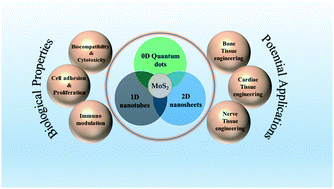
Molybdenum disulfide (MoS2) nanostructures have recently earned substantial thoughts from the scientific communities owing to their unique physicochemical, optical and electrical properties. Although MoS2 has been mostly highlighted for its industrial applications, its biological applicability has not been extensively explored. The introduction of nanotechnology in the field of tissue engineering has significantly contributed to human welfare by displaying advancement in tissue regeneration. Assimilation of MoS2 nanostructures into the polymer matrix has been considered a persuasive material of choice for futuristic tissue engineering applications. The current review provides a general discussion on the structural properties of different MoS2 nanostructures. Further, this article focuses on the interactions of MoS2 with biological systems in terms of its cellular toxicity, and biocompatibility along with its capability for cell proliferation, adhesion, and immunomodulation. The article continues to confer the utility of MoS2 nanostructure-based scaffolds for various tissue engineering applications. The article also highlights some emerging prospects and possibilities of the applicability of MoS2-based nanostructures in large organ tissue engineering. Finally, the article concludes with a brief annotation on the challenges and limitations that need to be overcome in order to make plentiful use of this wonderful material for tissue engineering applications.


image file: d2tb00131d-p1.tif

Anuj Kumar

Dr Anuj Kumar is an Assistant Professor (International Faculty Member)-Polymers & Biomaterials in the School of Chemical Engineering at the Yeungnam University (YU), South Korea. During this tenure, he has been awarded three sponsored research projects (two NRF projects as Principal Investigator (PI) and one Indo-Korea joint research project awarded jointly by MSIT-NRF Korea/DST India as Co-PI) for developing injectable and 3D printable hydrogels for soft and hard tissue regeneration. Prior to this position, he was Postdoctoral Research Associate in YU and Assistant Professor (Chemsitry) in DIT University, Dehradun (India). He received his PhD (Polymer Science and Engineering), MTech (Fibre Science and Technology), and MSc (Organic Chemistry) from Indian Institute of Technology Roorkee (IITR), Indian Institute of Technology Delhi (IITD), and Chaudhary Charan Singh University Meerut (CCSUM), India, respectively. He has authored more than 60 publications in internationally reputed journals, 1 book, and 10 book chapters. His research interests are focused on sustainable biomass, nanocellulose, biopolymers, hydrogels, design of biomaterials/nano-biomaterials, 3D bioprinting, microfluidics, tissue engineering, drug delivery systems, cancer therapy, wearable bioelectronics, etc.

image file: d2tb00131d-p2.tif

Ankur Sood

Dr Ankur Sood received his PhD in the field of drug delivery and imaging applications of nanoparticles from GGS Indraprastha University, New Delhi, India. He worked as a Research Associate at the Institute of Nano Science and Technology, Punjab, India. During this tenure he worked on polymer/metal nanocomposites and fluorescent gold clusters for biomedical applications. He worked as Senior Project Scientist at Indian Institute of Technology; Mandi, India where his work was focused on polymer nanoparticles based drug delivery systems. He was Postdoctoral Research Fellow in Yeungnam University, South Korea till February 2022 and currently he is working as an Assistant Professor (International Faculty Member) in School of Chemical Engineering at the Yeungnam University, South Korea. His current research is focused on 3D bioprinting, tissue engineering, and inorganic/organic nanoparticles for drug delivery and biosensing applications.

image file: d2tb00131d-p3.tif

Sung Soo Han

Dr Sung Soo Han is a Professor at the School of Chemical Engineering, Yeungnam University (YU), South Korea. He received his PhD, MS, and BS degrees from the Department of Polymer and Textile Engineeirng, Seoul National University (SNU), Seoul, South Korea. He has been awarded various NRF funded projects including one Indo-Korea joint research project awarded jointly by MSIT-NRF Korea/DST India as PI). He has published many research and review articles in internationally reputed journals. His research interests are focused on biomaterials, nanomaterials, tissue engineering, drug delivery, cancer therapy, bioprocess engineering, and artificial meat culture.


1. Introduction

The development of novel materials has widened the scope of their utility in numerous commercial applications. With the evolution of nanotechnology, the use of orthodox materials and production methods have been sidetracked and superseded with new methodologies, and novel engineered materials with remarkable features. It would not be an embellishment to exclaim this era as the epoch of nanomaterials. Nanomaterials offer distinctive properties compared to their counter bulk part, which makes them a unique and interesting class of materials.1

On a broader prospect, nanomaterials are classified as zero-(0D), one-(1D), two-(2D), and three-dimensional (3D) based on their shapes and structures.2 Until recently, 2D nanostructures and layered materials have marched rapidly and have lured the attention of many researchers around the world, as an elegant and efficient class of nanomaterials in a wide range of applications such as energy storage,3 transparent electrodes,4 wearable devices,5 cancer treatment,6 and biosensing.7 Since the inception of graphene in 2004, the first known 2D material, remarkable progress has been perceived on atomically thin 2D materials with diverse applications across various scientific turfs.8 Features like transparency, high electrical and thermal conductivity, high specific surface area and young's modulus have marked the success of this fascinating class of material.9 Owing to these stupendous properties along with the shortcoming of zero band gap, efforts have been bestowed to further explore 2D graphene analogues with semiconducting nature. In this regard, Transition Metal Dichalcogenides (TMD) with the general representation of MX2 (M as a transition metal atom (Mo, W, Ta, etc.)) and X is a chalcogen atom (S, Se, Te, etc.) have emerged as promising alternatives.10 Within a single layer of TMD, X–M–X atoms are bonded covalently in a hexagonally packed plane and such single layers are bound together by weak van der Waals forces. The TMD monolayer crystal offers distinctive properties compared to their bulk analogues due to the lack of a center of inversion, which allows admittance to a new degree of freedom, the k-valley index, and led to a new field of physics called valleytronics.11,12

TMD demonstrates a distinctive amalgamation of the direct band gap, resilient spin–orbital coupling, atomic-scale thickness, high conductivity along with promising electronic and mechanical properties. These features inscribe its attainment as a proficient material for applications in high-end electronics, energy harvesting, flexible and wearable electronics, DNA sequencing and personalized medicine.13 Among different layered TMDs, molybdenum disulfide (MoS2) has gained tremendous attention owing to its fascinating properties such as ease of synthesis, high catalytic properties, exceptional on/off ratio, satisfactory biocompatibility, and enormous direct band gap of 1.8 eV for the monolayer.14,15

MoS2 has achieved prime progress in the field of optoelectronics,4,16 energy storage17 and conversion,18 hydrogen evolution reaction (HER),19,20 and next-generation switches.21,22 Moreover, MoS2 layers are often associated with crystal-dependent fluorescence23 or the ability to quench fluorescence.24 In addition, thin nanosheets of MoS2 exhibit strong absorbance in the near-infrared region (NIR)25 along with efficiency for photothermal conversion.26 Also, the constituents of MoS2, i.e. molybdenum, and sulfur also play a key role in maintaining biological systems.27 All these characteristics along with the key attribute of MoS2 of having very high specific surface area makes imperative utilization of this fascinating material across diverse biomedical applications, which include but are not limited to drug delivery,28,29 antibacterial activity,30–32 biosensing,33,34 and theranostics.35,36

Humans have always envisaged the revamping of damaged organs and tissues with passable functioning, an aspiration, which is feasible using tissue engineering. In tissue engineering, cells/tissues, scaffolds, reinforcements, and growth factors are coalesced to invigorate or amend damaged or pathologically alimented tissues.37 Lately, a wide range of 2D nanostructures has been explored for their utility for tissue engineering applications.38 A comprehensive review by Liu et al. although focuses on the use of MoS2-based nanostructures for biomedical applications27 it has not highlighted its use in tissue engineering applications. Another review by Yadav et al. describes the application of 2D MoS2-based nanostructures specifically for biosensing, bioimaging and therapeutics.2 Moreover, the review of Nguyen et al. mainly focuses on the surface properties of 2D materials from biosensing to tissue engineering.39 However, to date, no dedicated review is published on the application of MoS2 nanostructures in tissue engineering applications. In the current review, we aim to explore the three main structural morphologies of MoS2, i.e. nanosheets, nanotubes, and quantum dots with their inherent properties. The review also focuses on the interaction of MoS2 nanostructures with biological systems. With this review, a spotlight is also shed on the utility of MoS2-based scaffolds for various tissue engineering applications. Finally, the review highlights some key shortcomings in using MoS2-based materials for tissue engineering applications that could be addressed to make this potential material a significant performer for next-generation tissue engineering applications.

2. Structure and properties of different forms of MoS2 nanostructures

The diverse electronic properties of TMDs result from the filling of the non-bonding d bands from group 4 to group 10 species.40 TMDs exhibit metallic properties, when the orbitals are partially occupied, whereas when the orbitals are fully occupied, TMDs display semiconducting properties. On the contrary, compared to the influence of metal atoms, chalcogen atoms exhibit a minor impact on the electronic structure; however, it is examined that upon broadening of the d bands, the band gap decreases by increasing the atomic number of the chalcogen.41

MoS2 has a 2D layered structure with each layer of thickness of about 0.65 nm, which are heaped upon each other to form bulk. Weak van der Waals forces clasp these layers together.42 MoS2 exists in three different crystal structures namely, a trigonal phase (1T), a hexagonal phase (2H), and a rhombohedral phase (3R). The letters in the representation correspond to the crystal structures whereas the digits represent the number of monolayers in the unit cell. The Mo–S coordination is octahedral in the IT phase, while the coordination is trigonal prismatic in the case of 2H and 3R phases, respectively.43 Naturally existing MoS2 crystals (e.g. molybdenite) mostly occur in the 2H phase, which is also the thermodynamically stable phase, while synthetic MoS2 mainly comprises 1T and 3R phases and are metastable in nature.44 The next section describes different forms of MoS2 nanostructures.

2.1. 2D MoS2 (nanosheets)

With the great success of graphene, researchers are motivated with an equivalent encouraging surge to develop alternate 2D materials that could result in the formation of atomic sheets with astonishing properties.45,46 Among all known 2D TMDs, MoS2 is one such material that is accompanied by a naturally layered structure, which makes it convenient to fabricate eminent quality 2D nanosheets without making much efforts during the synthesis process47,48 (Fig. 1(a)). The conversion process of MoS2 from bulk to single-layered nanosheets imparts direct bandgap, resulting in exceptional photoelectric properties.49 This significant bandgap transition is accompanied by strong fluorescence emission signals in the visible reason, which could be explored for numerous cost-effective biomedical applications such as biosensing and imaging.50 Owing to the large lateral dimensions, MoS2 nanosheets are easily dispersible in liquid/gaseous media. In addition, by engineering the edges of MoS2 nanosheets, to be terminated with either Mo or S atoms, the overall electrical performance of the nanosheets could be fine-tuned. These properties serve as vital prerequisites for many applications involving electrochemical biosensors.51 Many researchers have elucidated the unique properties of MoS2 nanosheets. Castellanos-Gomez et al. assessed the elastic properties of a suspended portion of MoS2 nanosheets (5–25 layered) using AFM and reported an exceptionally high Young's modulus (E[thin space (1/6-em)] = [thin space (1/6-em)]0.33[thin space (1/6-em)] ± [thin space (1/6-em)]0.07 TPa) for the nanosheets.52 Further, the stiffness and breaking strength of ultrathin MoS2 nanosheets were measured and reported by Bertolazzi et al.53 The enhancement in mechanical properties of 2D MoS2 could be explored as a reinforcement material for numerous tissue-engineering applications, especially for bone tissue engineering. Even though MoS2 nanosheets display a range of novel properties that are remarkably discrete from their bulk counterparts, the production of large-scale and defect-free atomic layers of MoS2 with desired thickness and control over substrates is still a challenging task.
image file: d2tb00131d-f1.tif
Fig. 1 Classification, electronic structure, and physical properties of (a) two-dimensional (2-D), (b) two-dimensional (1-D), and (c) zero-dimensional (0-D) materials. (DOS: Density of States). Reproduced with permission from.64 Copyright 2020 Elsevier.

2.2. 1D MoS2 (nanotubes)

2D TMD materials have gained considerable attention from the scientific community owing to their remarkable properties compared to their bulk analogues.54 Over the past decade, much of the research revolved around MoS2-based nanostructures, a key ambassador of the TMD family. As the dimension of MoS2-based nanostructures is reduced to a single or few layers, these materials tend to sustain their semiconducting nature and thereby uncover extraordinary physicochemical properties.55 Further modification in the structure of 2D TMDs is achieved by draping individual layers, resulting in the formation of chiral cylindrical-like structures known as 1D nanotubes56 (Fig. 1(b)).

1D nanomaterials embrace two out of the three dimensions in the nano regime, while the third dimension is in the micrometer range.57 In the year 2000, Seifert et al. proposed the existence of MoS2 nanotubes in mainly two forms, zigzag and armchair.58 They reported a narrow direct band gap associated with zigzag MoS2 nanotubes, while the armchair MoS2 nanotubes demonstrate a nonzero moderate direct gap. Moreover, the alteration in their structural arrangement has direct implications on their electrical properties.59 The cylindrical tube-like arrangement of MoS2 nanotubes serves as containers in which molecules/nanomaterials could be trapped. These properties could further be explored for numerous biomedical applications including biosensing, bioimaging and drug delivery.60,61 The bond length of MoS2 nanotubes is slightly more than MoS2 nanosheets. Further, MoS2 nanotubes share a similar band structure with carbon nanotubes with the exception that in the case of MoS2 nanotubes the bandgap augments as the diameter of the nanotubes increases.62 Efforts are being made to fabricate hybrids MoS2 nanotubes, which could offer the property of high mechanical strength that is a key requirement of bone tissue engineering. Also designing hollow tubes could be of high importance for nerve tissue engineering. MoS2 nanotubes could also be used as reinforcement for polymer scaffolds in order to improve their thermal and mechanical strength. Several key issues must be addressed if 1D MoS2 nanotubes are to be realized for their complete scientific and technological potential in the field of tissue engineering.

2.3. 0D MoS2 (quantum dots)

The size of quantum dots (QDs) is in the range of 2–10 nm. At these low dimensions, the quantity of atoms existing in the material reduces. This reduction in the number of atoms results in a significant drop in the number of overlapping energy levels, thereby increasing the energy gap between the valence band and the conduction band. The energy levels of the electrons in this state are not continuous but tend to form a discrete set of energy bands, a phenomenon known as quantum confinement. Due to quantum confinement and edge effect, the QDs present remarkable optical, and chemical properties63 (Fig. 1(c)).

Monolayer MoS2 QDs have become an important class of materials due to their outstanding properties that include abundant active edge sites, high specific surface area, exceptional electrical conductivity and photophysical properties along with strong hydrogen adsorption properties.65,66 These characteristics make it a potent material for applications such as electrocatalysts for hydrogen evolution reactions (HERs), and energy storage.67 MoS2 QDs comprise monolayered hollow closed nanostructures, with the smallest permitted unit “nanooctahedra” within the range of 3–8 nm33 and can be classified as n-type semiconductors as determined by Vikraman et al. by fabricating field-effect transistors (FETs) utilizing thin MoS2 QD layers.68 A decrease in the size of bulk material in the nano regime within the range of QDs tends to impart strong optical nonlinearity properties making QDs a favourable photoluminescence material.

The impact of size variation in MoS2 QDs is linked to their optical properties, which in turn can be tweaked by altering the size of MoS2 QDs. As the confinement energy is dependent on the size of the QD, the emission wavelength is precisely linked to the QD size. With the alteration in the size of QDs, the band gap changes, these results in altered emission and absorption of light. For a QD with less confinement, the size is bigger thereby reflecting color towards the reddish side, and the more the confinement, the lesser is the size of QDs, the bluish it is. The optical properties of MoS2 QDs also cover their ability for photoluminescence, which is significantly dependent on the excitation wavelength. It is well acknowledged in the literature that polydispersed MoS2 QDs exhibits significantly more excitation-dependent emission compared to monodispersed MoS2 QDs.69 This notion is remarkably dependent on the adsorption of oxygen atoms at the edges of QDs thus creating defects.70 The optical properties of MoS2 QDs are of significant importance in terms of biomedical applications such as real-time imaging,71 biosensing,72 drug delivery,73 and early cancer diagnosis.18 Even though MoS2 QDs are at a very early stage in the field of tissue engineering, the remarkable properties of this material have opened a new arena for innovative aspects in tissue engineering and regenerative medicines. Table 1 summarizes the different fabricating strategies for different MoS2 nanostructures.

Table 1 Fabrication strategies of different MoS2 morphology
S. no Morphology Processing method Exfoliating agent/solvent/substrate Features Ref.
1. Nanosheets Solvothermal method Heptamolybdate tetrahydrate, thiourea (NH2CSNH2), oleylamine Enhanced biological properties and cellular response 74
2. Nanosheets Supercritical hydrothermal process Molybdenum trioxide (MoO3), ascorbic acid (AA), formic acid (FA) MoS2 sheets with 6–7 layers, precise control over the phase and number of layers of MoS2, very fast process 75
3. Nanosheets Chemical vapour deposition (CVD) MoO3, sulphur (S) powder Large surface area (1 cm2), vertically aligned, thickness of ≤50 nm 76
4. Nanosheets Probe sonication method Bulk MoS2, poly (acrylic acid) High yield of approximately 100%, nanosheets with 5–6 layers, peroxidase mimicking activity 77
5. Nanosheets Microwave-assisted hydrothermal synthesis Sodium molybdate dehydrate, NH2CSNH2, cellulose paper Responsivity of 290 mA W−1, Detection up to 1.8 × 10⁹ Jones, external quantum efficiency of 37% 78
6. Nanosheets Heating-up approach 1-Dodecanethiol Thickness of approximately 100 nm, offers electrical bistability and NDR behaviour 79
7. Nanosheets Ultrasonication and chemical route Sodium molybdate hexahydrate, NH2CSNH2, polydopamine, carbon nanotubes Reversible capacity of 747.3 mA h g−1 and 512.4 mA h g−1 over 200 cycles at 200 mA g−1 for sodium-ion batteries (SIB) and lithium-ion batteries (LIB) after 100 cycles 80
8. Nanosheets Hydrothermal method MoO3, L-cysteine Reversible capacity of 1097 mA h g−1 at a current density of 50 mA g−1 after 25 cycles for LIB 81
9. Nanosheets CVD SiO2/Si substrate, MoO3, S element Uniform grain size reaching upto 146[thin space (1/6-em)]μm, precise control over intermediate product (MoO3−x) 82
10. Nanosheets Solid-state reaction method The figure of merit ∼2.52 × 10⁻¹⁵ esu cm, optical limiting (OL) effects with low threshold FOL ∼ 44 mJ cm−2 83
11. Nanotubes Catalyst-free vapour–gas–solid reaction H8N2MoS4, nitric acid, sodium dodecyly sulphate Diameter 30–200 nm, multiwall structure 84
12. Nanotubes Hydrothermal method (NH4)6Mo7O24·4H2O, ethylenediamine (EDA), L-cysteine Hollow tubes, rate cyclability (127 mA h g−1 at 200 mA g−1 after 100 cycles in the potassium ion battery) 85
13. Nanotubes Gas–solid reaction H8N2MoS4 Open ended tubes, outer diameter ∼25 nm, inner diameter is ∼12 nm 86
14. Nanotubes Hydrothermal method H8N2MoS4, EDA, hydrochloric acid Reversible capacity of 1327 mA h g−1 at 0.1 C and 850 mA h g−1 at 5 C after 300 cycles for LIH, reversible capacity of 480 mA h g−1 at 0.5 C after 200 cycles for SIB 87
15. Quantum dots Colloidal chemical route H8N2MoS4, oleyl amine Quantum yield of 4.4%, thickness of approximately 3 nm 69
16. Quantum dots Hydrothermal method H8N2MoS4 Blue fluorescent MoS2 QDs, narrow lateral size distribution, detection of hyaluronidase 72
17. Quantum dots Laser ablation Bulk MoS2 Fast, green, and one-step process to synthesize MoS2 QDs, High electrocatalytic activity for hydrogen evolution reactions (HERs), high yield of 36.73[thin space (1/6-em)]wt%, 65
18. Quantum dots Liquid nitrogen quenching & chemical routes Bulk MoS2, isopropanol Narrow lateral size distribution (1.41 nm), quantum yield of 5.06% 88
19. Quantum dots Pulsed Laser ablation Bulk MoS2 Average size 2–8.5 nm, high electrocatalytic activity for HERs, high aqueous dispersion stability 89
20. Quantum dots Electrochemical method Lithium bis-trifluoromethylsulphonylimide, n-type porous silicon (PS) Significant improvement in electroluminescence performance, enhancement in F band 90
21. Quantum dots Wet grinding and co-solvent sonication Bulk MoS2, N-methyl-2-pyrrolidone, 1,2-dichlorobenzene Quantum yield of 7 mg mL−1, average size range of 2–5 nm, blue luminescence upon UV excitation 91
22. Quantum dots CVD MoO3, S powder Single layered 2H–MoS2 structure, diameter of 4–7 nm, Density of 1011 cm−2 92
23. Quantum dots Hydrothermal method H8N2MoS4 Sensing of adenosine triphosphate, linearity for 0–200 μM for Fe3+ and 0–140 μM for ATP 93
24. Quantum dots Laser ablation method Bulk MoS2, n-hexane, n-butyllithium Diameter of 10 nm, retention time of 10 s 94
Miscellaneous
1. Thin films Polymer-assisted deposition, thermolysis Anhydrous ammonium tetrathiomolybdate (H8N2MoS4) linear-poly(ethylenimine) Uniform thickness (6 in), fast photoresponse (<1 ms), High on–off ratio (∼104) 95
2. Edge terminating films Sulfurization/selenization process SiO2/Si Vertically aligned layers, HER catalysis, 96
3. Single-crystal Hybrid thermolysis p-Si wafer High-quality p–n heterojunctions, generation of highly crystalline product 97
4. Films Chemical Vapour Deposition (CVD) Amorphous SiO2 substrate Highly crystalline, large area atomic layers 98
5. Metallic films Laser irradiation at low temperature Polyethylene terephthalate (PET) Fast response, controllable shape, ability for patterning 99


3. Cellular interactions of different forms MoS2 nanostructures

3.1. Biocompatibility and cellular toxicity

In order to fabricate a material with its utility for biological interactions, biocompatibility and cytotoxicity assessment play a crucial role. It is a well-documented notion that cell penetration and cleavage of active cell lines are the two main hurdles that restrict the clinical use of any nanomaterials.100,101 Recently, MoS2 nanostructures have been widely explored for their biological interactions with cell membranes due to the large surface area.102 The unique features of MoS2 nanostructures have supported its utility as a potent material for numerous biomedical applications103–105 (Fig. 2). Despite the reported compatibility of MoS2 nanostructures for biomedical applications, MoS2 nanomaterials have also been associated with the provocation of cellular membrane damage in different unicellular systems.
image file: d2tb00131d-f2.tif
Fig. 2 Features of MoS2-based nanostructures for cellular interactions.

In this regard, Chng et al. studied the dependency of the level of toxicity on the extent of exfoliation of MoS2. This work demonstrated that the lesser exfoliated MoS2 presents more level of toxicity.106 Through this work, the severity of the mechanical interaction between the cellular membrane and MoS2 nanostructures was assessed, which is accounted for the phospholipids extraction, which in turn destroys the integrity of the membrane, directing to cytoplasm leakage and finally cell death. Analogous work was also highlighted by Wu et al. in the case of E. coli and S. aureus. The study revealed the interactions between MoS2 nanosheets and membrane phospholipids supported by experimental and simulation-based data.107 The interaction of materials with the biological system has to undergo a series of events starting from adhesion followed by recognition and finally internalization. The first encounter of any material with the cell membrane decides its fate and journey. The studies highlighted above are concerned to access the toxicity of MoS2 nanosheets and their interaction with the phospholipids present in the cellular membrane. The results will have a significant impact on the utility of MoS2 nanostructures while designing scaffolds for tissue engineering applications.

Given the above context, investigation concerning biocompatibility assessment of MoS2 nanostructures is an essential matter. In this regard, Yang et al. reported the linkage of the antibacterial activity of chemically exfoliated 2D MoS2 nanosheets to the production of reactive oxygen species (ROS).108 Further, Shun et al. reported the enhancement in the antibacterial properties and cellular response upon modification of MoS2 with ultrafine-grained titanium.109 Along this line, Fan et al. demonstrated the capability of single-layered MoS2 nanosheet suspensions to offer photocatalytic antimicrobial activity upon exposure to visible light under the influence of an electron donor, ethylenediaminetetraacetic acid (EDTA).110 In this case, the EDTA is responsible for transferring electrons to MoS2, which results in increased light absorption and augmented the separation of photoelectron–hole pairs, which enhances the ROS yield, resulting in high antibacterial performance. Moreover, few studies have also reported enhancement of the photothermal performance of MoS2 under light irradiation,111 and ROS-independent oxidative stress, disturbing bacterial structure and vital functions112 as the reason for the antibacterial property of MoS2 based nanostructures.

The ability to impart antimicrobial properties makes MoS2 nanostructures a compelling candidate for wound healing and tissue engineering applications. Moreover, the work carried out by Teo et al. demonstrated the fact that TMDs are less toxic compared to their graphene analogues.113 The study also emphasized the non-suitability of MTT assay as the sole criteria to assess the cytotoxicity evaluation of nanomaterials.

The pulmonary hazard of 2D MoS2 compared to the aggregated MoS2 (Agg-MoS2) was studied by Wang et al. The data obtained from this study suggested the attenuation of toxicity levels under exfoliation for 2D MoS2 compared to Agg-MoS2.114 This data provide an important understanding towards the safety assessment of 2D MoS2 for biomedical applications. Further, Domi et al. studied the fate of commercially available colloidal solutions of MoS2 and studied their physicochemical and toxicological responses at in vitro levels.115 The study emphasized the importance of the biocompatibility of the MoS2 nanoparticles as a key factor affecting cellular interactions.

Further, the degradation of MoS2-based nanostructures could also present vital stats in terms of applications in biological systems. In this context, Wang et al. carried out a detailed study on the degradation pathway in biologically relevant media and presented a detailed analysis emphasizing the molecular oxygen-driven dissolution process of MoS2 nanosheets in biological media.116 It is well documented that MoS2 can be decomposed when exposed to hydrogen peroxide (H2O2) due to the change in the oxidation state from Mo(IV) to Mo(VI), mostly in the form of MoO3/MoO42− ions while releasing sulphur, which plays a central role in many biological activities and functions (e.g., a constituent of sulfur-containing amino acids, formation and activity of iron–sulfur proteins, etc.).117–119 Moreover, Kurapati et al. studied the degradation of MoS2 nanosheets at a physiological concentration of H2O2.120 In this study, the biodegradation of MoS2 materials has also been examined using enzyme models of peroxidase enzyme horseradish peroxidase (HRP), and human myeloperoxidase (MPO) under the exposure of low concentration of H2O2. An elementary understanding of the degradation mechanisms, followed pathway, and the performance of the resulting by-products of MoS2 nanostructures in biofluids is very crucial to further maximize the potential benefit that could be extracted from them for their application in the field of tissue engineering. In addition, the biodistribution and clearance behaviour of nanomaterials are vital for their utility in biomedicines/nanomedicines. To understand the clearance of PEGylated-MoS2, Hao et al. carried out a study, which depicted the accumulation of PEGylated MoS2 nanostructures mostly in reticuloendothelial systems (RES) organs such as liver and spleen both at in vitro and in vivo levels.

Although, the data for cytotoxicity evaluation of MoS2 nanostructures are minimal, the assessment of their biocompatibility along with stability and degradation upon interaction with the biological system could provide insights into the impact of MoS2 in the field of biomedical sciences. Understanding the interaction of MoS2 nanostructures at the cellular level could be of high importance to develop MoS2 reinforced scaffolds for tissue engineering applications.

3.2. Cellular adhesion and proliferation

2D nanomaterials have been receiving incessant consideration from the scientific communities due to their remarkable features of chemical functionality and structural anisotropy.121 Recently, MoS2 nanostructures have been extensively investigated for numerous applications due to their band gap tenability.122 Recently, these properties are being explored for photothermal therapies123,124 and applications concerning cellular interactions.125,126 In terms of cellular interaction, cell adhesion and proliferation plays a crucial role in getting adequate cellular responses. Understanding the mechanistic pathway concerning cell proliferation upon interaction with MoS2 could lead to different application-oriented strategies. A lot of new outcomes could be uncovered concerning photo-based therapies, early diagnosis, cancer treatment and especially tissue engineering applications, where the fate of the treatment process heavily depends on the cellular response, interaction, and proliferation. In this regard, Ke et al. fabricated a gold nanoparticles-modified monolayer MoS2 sensor, capable of rapidly detecting DNA molecules with high sensitivity, and selectivity.127 The study was based on the change in the dielectric environment of the MoS2-based probe upon the interaction with nucleosides of DNA, which in turn alters the band gap of MoS2. The sensitivity of this probe in real-time applications will certainly depend on the interaction of cells with the MoS2-based probe system. This work may have applicability while designing scaffolds for tissue engineering applications with real-time monitoring.

Further, Carrow et al. studied the interaction of 2D MoS2 and human mesenchymal stem cells (hMSCs) at the molecular level.128 Here, the synthesized exfoliate 2D MoS2 nanosheets were physicochemically evaluated using XRD, AFM, TEM, Raman spectroscopy and XPS. Further, it was reported that the photoluminescent intensity for exfoliated MoS2 decreased upon lithium (Li) intercalation; subsequently, the phase transition from semiconducting 2H to metallic 1T phase took place. The study highlighted the effect of protein adsorption on 2D MoS2 on cellular adhesion and internalization because of the formation of protein corona on MoS2 nanosheets (Fig. 3).


image file: d2tb00131d-f3.tif
Fig. 3 Physicochemical characterization of exfoliated 2D MoS2 nanosheets. (A) X-ray diffractogram of bulk and exfoliated MoS2, (B) atomic force microscopy (AFM) for confirming the 2D shape, (C) transmission electron microscopy (TEM) images of ultrathin MoS2 sheets along with electron diffraction pattern, (D) atomic composition of bulk and exfoliated MoS2 determined via elemental analysis, (E) X-ray photoelectron spectroscopy (XPS) analysis, (F) Raman spectroscopy, (G) photoluminescent measurements, (H) response to near infra-red (NIR) light was determined using an infra-red (IR) camera and change in temperature over time, (I) zeta potential and hydrodynamic size of exfoliated MoS2 in water, phosphate buffer saline (PBS), and media, and (J) 8-anilino-1-naphthalenesulfonic acid (ANS) assay shows protein structures are intact in the presence of exfoliated MoS2.128

Cellular adhesion is a crucial parameter in the area of wound healing and tissue repair as in the case of tissue engineering, the adhesion of cells to the external scaffolds directly affects the tissue repair process. In this regard, Seung et al. studied the self-healing and adhesion property of monolayer MoS2 and silicon oxide.129 The concept could have utility in designing novel MoS2-based scaffolds for tissue engineering applications. Further, the work carried out by Manish et al. sheds light upon the wetting characteristics by modulating the super-hydrophobic states of 2D MoS2 that could be achieved by controlling the atomic-level defects in MoS2 nanostructures.130 The modulation in atomic defects of MoS2 nanostructures is suggested to have direct implications on the cell adhesion characteristics that could be leveraged for tissue engineering and biomedical applications. However, Anna et al. evaluated the applicability of surfactant-free liquid-phase exfoliated 2D MoS2 as a platform for the treatment and detection of cancer.131 In order to further enhance the selectivity and efficiency of this 2D MoS2-based platform, the system was equipped with antigen–antibody binding modalities. The detection of cancer cells is based on the level of internalization efficacy of cancer cells towards a modified 2D MoS2-based detection system. Further, assessment of cellular proliferation could be a vital stat to drive this process for advancement.

The surface chemistry of a material plays a crucial role in tissue engineering applications. The type of bonds formed between the material and the cell surface dictates the efficiency of adhesion and in turn, affects cellular behaviour and proliferation. In order to utilize the MoS2 nanostructures-based scaffolds for tissue engineering applications, the surface functionality and modifications need to be carried out in a proficient manner. This will impart better control over cell adhesion, proliferation and differentiation, which is very essential for an effective tissue regeneration strategy.

3.3. Immunomodulatory properties

The recent surge in MoS2 nanostructures-based research has led to the utility of MoS2 in numerous fields ranging from cutting-edge electronic devices to biomedical applications. However, in order to evaluate the impact of MoS2 on human health, the necessity to understand the interaction of MoS2 with a biological system is crucial. When a tissue confronts adverse conditions (tissue damage, injury, autoimmune response, or infections), the defense system of our body tends to protect or minimize the impairment caused through a complex biological response known as inflammation.132 The complete process of inflammation involves a series of well-organized and dynamic responses comprising both cellular and vascular events with specific humoral secretions.133 Inflammatory response mainly includes cells of the immune systems and inflammatory modulators.134 The innate immune response of a body is governed by cytokines which include pro-inflammatory cytokines such as interleukin (IL)-1β (IL-1β), IL-6, IL-8, and tumour necrosis factor-α (TNF-α), and anti-inflammatory cytokines such as IL-10.135

Accompanied by its widespread applicability, the immunomodulatory response of MoS2 has also gained considerable attention owing to its utility in biomedical applications. In this regard, Moore et al. studied the inflammatory responses of different sized MoS2 flakes on various cell lines (monocyte, THP-1; inhalation, A549; ingestion, AGS), which were selected to represent different means of exposure.136 This work demonstrated the size-dependent response of macrophages with the smallest sized MoS2 flake exercising maximum cytokine upregulation. This size dependency of the macrophage response could be associated with the increased concentration of endotoxin on the surface of the nanomaterial. As the size decreases, the surface-to-volume ratio increases, thereby providing more surfaces for exposure. However, Gu et al. studied the underlying mechanism of MoS2-mediated immune response in detail.137 The work included both molecular dynamics simulation and flow cytometric experimentations to investigate the molecular mechanism of cytokines production by MoS2 and PEG-functionalized MoS2. Moreover, Han et al. reported the fabrication of nanoconjugates of MoS2 nanosheets functionalized with cytosine–phosphate–guanine (CpG) and PEG.138 The fabricated nanoconjugate was reported to have augmented the cellular uptake of pristine MoS2 and improved the production of pro-inflammatory cytokines leading to an efficient immune response. The study presented the capability to modulate immunotherapy-based cancer treatment. To further investigate the immunomodulating properties of MoS2, Jiang et al. fabricated MoS2–CuO hetero-nanocomposites loaded with bovine serum albumin and imiquimod (R837), an immunoadjuvant.139 In this study, it was demonstrated that MoS2–CuO nanocomposites were able to generate hydroxyl free radicals when exposed to overproduced hydrogen peroxide in tumours. The study also reported the ability of the designed nanocomposites for modulating immunotherapy-based cancer treatment. For a successful regeneration of damaged tissue, inflammation, repair and remodeling are the three main phases. The efficiency and time taken in crossing each phase are very crucial and decide the fate of the tissue regeneration process. While designing a scaffold for tissue engineering, assessment of the immunological process is very important. In terms of exploring MoS2-based scaffolds for tissue engineering, major emphasis could be given to designing immune-compatible scaffolds by incorporating external agents such as fibrinogen and interleukins.

Zhan and co-workers further investigated the immunostimulatory property of 2D MoS2. Their study reported the role of few-layered MoS2 nanosheets in enhancing dendritic cells (DC) maturation, migration and T cell elicitation.140 In this work, the migration ability of DCs was examined upon the introduction of MoS2 nanosheets (MSNs). It was also demonstrated that the exposure of MSNs increases the migration distance and velocity of DCs compared to non-treated controls. The work presented a detailed study on the important parameters of dendritic cells such as maturation, migration and T cell activation backed with statistical analysis. Activation and proliferation of CD4+ and CD8+ T cells were observed at in vivo levels, implicating immunomodulating effects of MoS2 nanosheets (Fig. 4). Moreover, Baimanov et al. studied the immunological responses of MoS2 nanosheets–protein corona complexes.141 They provided an insight into the pro-inflammatory effect of native MoS2 nanosheets and their complex with blood proteins and highlighted the contribution of blood proteins corona towards the enhancement of inflammatory responses of MoS2 nanosheets. All these studies revealed the immunomodulatory effect of MoS2. Assessment of the immunological response of MoS2 will provide important insights for the safety evaluation of MoS2 based nanostructures. This will further ensure the fabrication of rational MoS2-based designs to be used for tissue engineering applications.


image file: d2tb00131d-f4.tif
Fig. 4 MSNs improved the ex vivo movement and in vivo homing ability of DCs. Notes: (A) the ex vivo movement of DCs, (B) statistical data of the length and velocity of DC movement. (C) The in vivo homing of tissue-resident DCs, (D) statistical data of the homing percentage. N = five for each group. (E) Imaging of the overall distribution of intravenously injected Fluc+ DCs at 2 h. (F) The tissue accumulation of circulating DCs at 48 h after injection.140

4. MoS2-based nanostructures in tissue engineering

Tissue engineering is a dynamic field that deals with the regeneration of tissues/organs that are damaged or incapable of self-revival. In order to engineer a tissue construct, cells are usually seeded on scaffolds/biomaterials that imitate the extracellular matrix (ECM) and tissue microenvironment to aid in tissue development. Biodegradable polymers (synthetic and natural), nanocomposites, and porous scaffolds have been under constant investigation to be used for tissue engineering applications.142 Generally, the scaffolds designed using polymer composites/nanocomposites and biomaterials for tissue regeneration must possess high porosity that is helpful in inducing cell migration, nutrient transportation and tissue growth.143 However, in certain cases, the mechanical stability (in case of bone tissue engineering),144 and conducting nature (in case of neural and cardiac tissue engineering)145 of the designed scaffolds could also play a crucial role.

In this regard, MoS2-based nanostructures are being explored owing to their remarkable properties of high surface area/mass ratio, decent electrical conductivity, exceptional optical properties, and suitable biocompatibility.113 The application of femtosecond (fs) laser pulses to micropattern MoS2/acrylamide electrospun nanofibers was studied by Paula et al.146 The work reported the impact on the topography of composite nanofiber under the influence of laser pulse energy and scanning speed. The application of fs laser resulted in the formation of micropores while preserving the nature of the composite nanofibers. The proposed nanofibers could be utilized for their applicability in the field of tissue engineering due to the additional advantage of photoluminescence and enhanced mechanical strength upon strengthening with MoS2. Further, Chen et al. proposed CVD-grown monolayer MoS2 nanosheets to use as bioabsorbable electronics-based biosensors.147 The cytotoxicity and biocompatibility studies of the as-synthesized MoS2 nanosheets provided a set of parameters that could be vital in understanding the effectiveness of the synthesized nanosheets for biological systems. The study emphasized the enhancement of the dissolution rate of the monolayer MoS2 in PBS solution upon reduction of its grain size and intensification of intrinsic defects. This feature could be very important in terms of controlling the lifetime of the designed material and its usefulness as a scaffold for tissue engineering applications and wearable implants.

To further implicate the applicability of 2D MoS2 in tissue engineering, biosensors, and electrochemical electrodes, Sim et al. fabricated silk fibroin exfoliated MoS2 nanosheets with a high yield.148 The study presented a detailed investigation of silk fibroin-based MoS2 dispersions as a function of centrifugation speed, time taken for sonication, and initial concentrations of silk powder and solvent. In order to design effective MoS2 based scaffolds for tissue engineering applications, the yield and uniformity of MoS2 nanostructures are very important. Careful assessment and controlling of these parameters will result in the fabrication of scaffolds with enhanced immunomodulation and tissue regeneration ability.

MoS2 has also been mixed with other low dimensional nanomaterials to further improve the scaffold properties and impart a synergistic effect in terms of mechanical strength that is advantageous for tissue engineering applications. In a study by Feng et al., 0 D nanodiamond particles and MoS2 nanosheets were uniformly dispersed into poly (3-hydroxybutyrate-co-3-hydroxyvalerate) (PHBV) scaffolds using selective laser sintering. However, the synergistic effect of graphene oxide with MoS2 was studied by Wan et al. High mechanical strength was recorded for these reinforced and synthesized scaffolds and a detailed mechanism concerning their enhanced tensile and compressive strength was also reported with enhanced electrical conductivity and mechanical property. These studies have provided a new approach for designing scaffolds for various organ-specific tissue engineering applications. The proceeding section will further discuss a few specific tissue engineering aspects in detail.

4.1. Bone tissue engineering

Ailments related to the bone have global predominance since they can be occurred due to multiple causes, which include cancer, injuries, and accidents. Since its inception in the early 90s, bone tissue engineering has always been explored to fabricate inventive novel biological materials that could aid in bone regeneration and enhance the repairing process. Wang et al. explored the synergistic effect of MoS2 in both tissue engineering and tumor therapy by fabricating a bifunctional scaffold of MoS2 nanosheets grown on 3D-printed bioceramic scaffolds of akermanite (AKT) (Ca2MgSi2O7) in situ. It was also demonstrated that, upon irradiation, the temperature of the designed scaffold (MS-AKT) rapidly increased, which in turn decreased the viability of osteosarcoma cells and breast cancer cells, thereby inhibiting the growth of tumours in vivo. Further, MS-AKT scaffolds supported the cell adhesion, proliferation, osteogenic differentiation of bone mesenchymal stem cells, and encouraged bone regeneration in vivo. The fabrication method was a merger of 3D printing technology and the hydrothermal method.149 The ability of the designed scaffold to generate bone was also tested at in vitro and in vivo levels (Fig. 5(I)). Moreover, Wang et al. reported a novel scaffold of 2D MoS2–PLGA with 3D printed bioactive borosilicate glass as a platform for the synergistic treatment of bone repair and tumor therapy.150 The prepared scaffolds could stimulate proliferation and differentiation of rat bone mesenchymal stem cells and promoted bone repair for calvarial defects in the rat. With high photothermal efficiency, the designed scaffold has promising futuristic applications for bone cancer-related defects. All these studies assessed the ability of MoS2 nanostructure-based scaffolds to support and promote osteogenic differentiation and angiogenesis. Further, a hydrothermal method was employed by Zhang et al. to fabricate MoS2 for tissue engineering applications.151 In this work, the influence of MoS2 on the focal adhesion (FA) was detected using the immunofluorescent staining for detecting vinculin (adaptor protein forming FA). Apart from the focal adhesion formation, upregulation of osteogenic genes and cell proliferation was added advantage of the synthesized MoS2 nanostructures (Fig. 5(II)).
image file: d2tb00131d-f5.tif
Fig. 5 (I) Fracture morphologies of 0.2 MoS2-modified akermanite (MS-AKT) scaffolds at different magnifications (a–c), energy-dispersive spectrometer (EDS) elemental mapping of the cross-section of MoS2–AKT interface: all elements (d), element Mo (e), element S (f); EDS line scanning profiles of all elements (g), element Mo (h) and element S (i) from the interior to the strut surface of the scaffolds, which exhibited a step profile, confirming the elemental interpermeation between MoS2 and AKT.149 (II) Immunostaining of the focal adhesion protein vinculin. (a) Vinculin (red fluorescence) of MSCs grown on the flat substrate and the nanostructured MoS2. Scale bar: 25 m. The images are merged images of F-actin (green fluorescence), vinculin (red fluorescence) and DAPI (blue fluorescence). (b) Percentage of the vinculin expressed area against the cell spreading area. Reproduced with permission from.151 Copyright 2018 Elsevier.

The bone-forming ability of akermanite-based scaffolds has been reported by Huang et al. with beta-tricalcium phosphate (beta-TCP) as a control.152 Further, Liu et al. reported the effect of akermanite-based scaffolds on the proliferation, adhesion, and osteogenic differentiation of human adipose-derived stem cells.153 The application of MoS2 in the akermanite scaffold not only improved bone regeneration but also showed applicability in tumor reduction. This makes it a promising material for tumor-induced bone defects. Furthermore, Wu et al. combined the electrospinning technique with doping to fabricate polyacrylonitrile/MoS2 nanofibrous scaffolds.154 The designed scaffolds promoted cell proliferation and activity and offered excellent biocompatibility and osteogenic differentiation. The ability of MoS2 as an excellent reinforcement for bone tissue engineering was further explored by Awasthi et al. who fabricated polycaprolactone/zein (PZ) composite nanofibers supplemented with albumin induced exfoliated MoS2 nanosheets (PZM).155 The MoS2-reinforced scaffold offered improved wettability and mechanical properties compared to the pristine polycaprolactone/zein composite nanofibers. Improved calcium–phosphate deposition upon assessment with simulated body fluid (SBF) biomineralization and Alizarin Red S (ARS) test were added advantages associated with MoS2-reinforced composite nanofibers (Fig. 6).


image file: d2tb00131d-f6.tif
Fig. 6 ARS staining and alkaline phosphate (ALP) test results. (a) The digital images, (b) absorbance data, and (c) ALP activity. For ARS, the respective scaffolds were stained at (7 and 14) days. The ALP activity was also evaluated on (7 and 14 days). Reproduced with permission from ref. 155. Copyright 2020 Elsevier.

In order to tackle the in situ bacterial infection and promote osteointegration of orthopaedic implants, Yuan et al. fabricated MoS2/polydopamine/RGD peptide coated titanium implants. The work highlighted the capability of the modified titanium implants to kill bacteria with an efficiency of almost 92% upon NIR radiation exposure without causing damage to the normal tissue. The work could be applicable for reoccurring infections after surgery and could have significant potential for bone tissue engineering, as antibacterial and upregulation of genes responsible for osteogenesis. The capability of 2D MoS2-reinforced hydroxyapatite (HAP) scaffolds to provide osteogenic differentiation, proliferation, and bone regeneration at in vitro and in vivo levels was investigated by Yadav et al.156 The cells incubated with the designed scaffolds presented higher cell adhesion and proliferation along with enhanced alkaline phosphatase activity (ALP). The obtained results were also supported with increased levels of osteogenic markers and bone morphogenetic protein-2.

Moreover, for a successful bone tissue engineering outcome, enhancement in the osteogenic process is very important as it gives a clear indication of repair of bone deformations and injuries. The regeneration of bone without the aid of external growth factors further provides a promising outcome for a potent scaffold for tissue engineering applications. Apart from high porosity and pore interconnectivity, load-bearing efficiency is also very crucial while designing scaffolds. Further research could be implemented to study the load-bearing capacity of MoS2 based scaffolds.

4.2. Cardiac tissue engineering

The field of cardiac tissue engineering deals with the repair and regeneration of cardiac tissues, muscles and cells. Close coordination between the cardiac cells and the support system (scaffold) is required for effective cardiac tissue regeneration. The application of MoS2 nanosheets in cardiac tissue engineering was studied by Nazari et al. The study reported the fabrication of electrospun nanofibers of MoS2 nanosheets incorporated nylon 6.157 In this work, the fabricated MoS2 NPs were mostly in the shape of nanosheets with a size of approximately 400 nm with the presence of some sheet-like nanostructures with a size >2 μm. In addition, the mean diameter of nylon and nylon/MoS2 nanofibers was found to be approximately 182 and 161 nm, respectively (Fig. 7). The synthesized nanofibers offered high mechanical properties and enhanced electrical conductivity with improved cell proliferation and attachment. The study emphasized the ability of MoS2 to upregulate cardiac functional genes and induction of cardiogenic differentiation and maturation of mouse embryonic cardiac cells (mECCs). The electrical signaling response of MoS2-based scaffolds needs to be investigated in detail as this could open gates for the intense utilization of MoS2-based scaffolds for nerve and cardiac tissue engineering. Further, the application of MoS2 to enhance the mechanical, electrical, thermal and structural properties of chitosan was studied by Feng et al.158 The reinforcement effect and well dispersion state of MoS2 with chitosan were credited for the enhancement in the properties of pristine chitosan films. This work could be utilized to develop MoS2–chitosan scaffolds for providing necessary electrical stimulus that can be explored for cardiac tissue engineering applications. The regeneration of functional cardiac tissues with adequate response will have a significant impact in eradicating many ailments related to cardiovascular tissue damage. The properties of cardiogenic differentiation, cell adhesion, and proliferation should be the key parameters to be targeted while designing scaffolds for cardiac tissue beginning.
image file: d2tb00131d-f7.tif
Fig. 7 (a) Scanning electron microscopy (SEM) image of MoS2 nanosheets, (b and c) TEM images of MoS2 nanosheets, (d) Raman spectroscopy MoS2, (e) electrical conductivity of nylon and nylon/MoS2 scaffolds, (f and g) SEM micrograph of nylon and nylon/MoS2 nanofibers, (g–k) TEM micrograph of nylon/MoS2 nanofibers. Reproduced with permission from ref. 157. Copyright 2019 Wiley.

4.3. Neural tissue engineering

The cases of nerve injuries are on the rise and can be a result of accidents, trauma, ailment, and congenital defects. However, nerves with a gap smaller than 1 cm could be restored through the surgical process but large nerve injuries are difficult to fix.159,160 In order to meet this demand, designing biocompatible artificial cylindrical constructs imitating the structural and functional property of nerves has been intensively explored. In this regard, Taheri et al. developed lithium intercalated MoS2-coated cotton thread as a potent candidate for nerve tissue-engineering applications161 (Fig. 8). Apart from testing the biocompatibility and cytotoxicity of the designed scaffolds for multiple cell lines, the conductivity and permeability of the scaffolds were also evaluated. The work could impart an opportunity to fabricate biomimetic fibrillary construction of the ECM. In another study, Wang et al. investigated the application of PVDF–MoS2 scaffolds for promoting neural stem cell (NSC) differentiation with high efficiency and without including external growth factors.162 In this work, the immunofluorescent staining method was executed with precise neural markers, such as neuronal associated class III beta-tubulin protein (Tuj1) and glial fibrillary acidic protein (GFAP), to account for the degree of differentiation of NSCs into neural cell types. The study showed a positive effect that the nanostructured MTFs exhibit significantly positive effects on NSC proliferation and attachment with negligible cytotoxicity.
image file: d2tb00131d-f8.tif
Fig. 8 (A) Schematic diagram illustrating the workflow of pre-treating the thread with cationic polyacrylamide (CPAM) and preparing lithium-intercalated MoS2 nanoflake coated thread. (B) Depiction of lithium-ion intercalation of MoS2-thread to render the thread conductive, Reproduced with permission from ref. 161. Copyright 2019 American Chemical Society.

Good electrical conductivity and large surface area are tagged as added advantages associated with the designed scaffolds that help ion cell adhesion and proliferation. The field of MoS2 based neural tissue engineering is still under exploration and has a lot of potential. In this area, inspecting the simulation-based electrical signalling response of designed scaffolds will certainly have an edge in further developing improved scaffolds with remarkable properties. A detailed summary of the applications of MoS2-based nanostructures for different organ-specific tissue engineering is provided in Table 2.

Table 2 MoS2-based scaffolds for tissue engineering applications
S. No Nature of MoS2 Composition Scaffold type Processing method Cell line Targeted tissue Features Ref.
1. Quantum dots Polyamide Nanofibers scaffold Electrospinning General Threshold energy of nanofibrous membrane reduced from 3.5 to 1.8 nJ, uniform distribution of MoS2 in the membrane, micropatterning of pillars 146
2. Nanosheets Silk fibroin Nanosheets Freeze-dried General Significant output for scale up, thickness of 3–6 nm, high aqueous stability 148
3. Nanosheets Akermanite Scaffold 3D-printing technology and hydrothermal method Saos-2 cells, MDA-MB-231 cells Bone tissue engineering Supports cell attachment, proliferation and osteogenic differentiation of rBMSCs, induced bone regeneration at in vivo level 149
4. Nanosheets Bioactive borosilicate glass Scaffold 3D-printing technology MNNG/HOS cells Bone tissue engineering Retention up to 60 days, tumour therapy, stimulate differentiation of rBMSCs, upregulate osteogenic genes expression 150
5. Nanoflakes Fluorine doped tin oxide coated glass Scaffold Hydrothermal method rBMSCs Bone tissue engineering Formation of focal adhesion between cells and substrate, promoted osteogenesis 151
6. Nanofibers Polyacrylonitrile Composite nanofibers scaffold Electrospinning technology rBMSCs Bone tissue engineering Promote rBMSCs growth, proliferation and activity 154
7. Nanosheets Polycaprolactone, zein Composite nanofibers scaffold Albumin-induced exfoliation MC3T3-E1 Bone tissue engineering Enhanced cell attachment, proliferation and differentiation 155
8. Nanosheets Hydroxyapatite Nanocomposite scaffold Hydrothermal method MG-63 cells Bone tissue engineering In vitro and in vivo osteogenic differentiation, proliferation, rapid osteogenesis 156
9. Nanoflakes Nylon Nanofibers scaffold Electrospinning technology mECCs Cardiac tissue engineering Maturation and upregulation of cardiac functional genes, elongated morphology 157
10. Nanoflakes Chitosan Composite scaffolds Acidic exfoliation General Enhanced mechanical and thermal properties 158
11. Nanoflakes Cotton thread Thread scaffolds Exfoliation and Electrochemical method NG108-15, PC 12 Nerve tissue engineering Electrical conductivity of 9.4 × 101 S m−1, cell proliferation over a period of 3 days 161
Nanosheets Polyvinylidene fluoride Nanofibrous scaffold Hydrothermal method and electrospinning technology NSC Nerve tissue engineering Promote cell differentiation of NSC and neural maturation, 162


5. Conclusions and future prospective

MoS2-based nanostructures signify as one of the most promising and attractive materials for numerous applications ranging from the industrial segment to the biomedical field. The unique set of physicochemical properties associated with MoS2 nanostructures makes it an exceptional candidate for its widespread utility. The review discusses the different structural arrangements (2D, 1D, 0D) in which MoS2 could be perceived. The review also addressed the structural-based alterations in the properties of different MoS2-based nanostructural morphologies. To further investigate the compatibility of MoS2 with biological systems, comprehensive wisdom of their cellular interaction is very essential. Thus, the effect of nanostructured MoS2 on cellular toxicity, biocompatibility, proliferation, adhesion, and immunomodulation is thoroughly reviewed. This will render an approximation of the level of MoS2 that could be utilized for their applicability with biological systems. The review further presents a comprehensive overview of the exploitation of nanostructured MoS2 in tissue engineering applications with emphasis on bone, cardiac and neural tissue engineering. Further, the tissue-specific features of the fabricated MoS2-based scaffolds are thoroughly reviewed and analyzed to implicate their practicality in regenerating large organs as well.

Although, considerable progress is still underway in terms of the applicability of nanostructured MoS2-based scaffolds for tissue engineering applications, in-depth knowledge of the cellular interaction of MoS2 is deficient. Different forms and sizes of any material will give an altered response when exposed to a living system, which in turn will affect the anticipated application of any material. This unlocks new opportunities, which need to be explored to design effective MoS2-based scaffolds for tissue engineering applications. In addition, novel strategies to synthesize and functionalize uniform MoS2 nanostructures with substantial control over their size and properties will further aid in their widespread utility. However, in order to utilize MoS2-based scaffolds for large tissue regeneration such as skin, further refinement in fabricating process is required. To design such scaffolds, imitating ECM could not be the sole criteria but repair and improvement of diseased tissue ECM is a vital criterion to be handled for effectual tissue regeneration. To tackle this hurdle, additive manufacturing techniques could be explored for effectual MoS2-based scaffold production. An interdisciplinary approach with collaborative effort is necessary to see the clinical translation of nanostructured MoS2-based scaffolds for tissue engineering applications.

Data availability

The data that support the tables within this article and other findings of this study are available from the corresponding author upon reasonable request.

Author contributions

Anuj Kumar: conceptualization, methodology, investigation, writing – original draft, writing – review & editing, supervision, funding acquisition. Ankur Sood: methodology, investigation, software, writing – original draft. Sung Soo Han: writing – review & editing, supervision, funding acquisition.

Conflicts of interest

The authors declare that they have no known competing financial interests or personal relationships that could have appeared to influence the work reported in this paper.

Acknowledgements

This work was supported by the National Research Foundation of Korea (NRF) funded by the Ministry of Education, Science and Technology, grant numbers “2021R1I1A3059994, 2020K1A3A1A19088873, 2020R1A2C1012586, and 2020R1A6A1AA03044512”. Authors of this work thank the authors whose research or review articles have been cited in this review article.

References

  1. N. Baig, I. Kammakakam and W. Falath, Mater. Adv., 2021, 2, 1821–1871 RSC.
  2. V. Yadav, S. Roy, P. Singh, Z. Khan and A. Jaiswal, Small, 2019, 15, 1803706 CrossRef PubMed.
  3. K. Zhao, W. Zhu, S. Liu, X. Wei, G. Ye, Y. Su and Z. He, Nanoscale Adv., 2020, 2, 536–562 RSC.
  4. H. Park, D. S. Oh, K. J. Lee, D. Y. Jung, S. Lee, S. Yoo and S.-Y. Choi, ACS Appl. Mater. Interfaces, 2020, 12, 4749–4754 CrossRef CAS PubMed.
  5. D. Zhou, L. Zhao and B. Li, J. Energy Chem., 2021, 62, 27–42 CrossRef.
  6. W. Yin, L. Yan, J. Yu, G. Tian, L. Zhou, X. Zheng, X. Zhang, Y. Yong, J. Li, Z. Gu and Y. Zhao, ACS Nano, 2014, 8, 6922–6933 CrossRef CAS PubMed.
  7. A. Bolotsky, D. Butler, C. Dong, K. Gerace, N. R. Glavin, C. Muratore, J. A. Robinson and A. Ebrahimi, ACS Nano, 2019, 13, 9781–9810 CrossRef CAS PubMed.
  8. A. McCreary, O. Kazakova, D. Jariwala and Z. Y. Al Balushi, 2D Mater., 2020, 8, 013001 CrossRef.
  9. A. S. Sethulekshmi, J. S. Jayan, S. Appukuttan and K. Joseph, Phys. E, 2021, 132, 114716 CrossRef CAS.
  10. X. Yin, C. S. Tang, Y. Zheng, J. Gao, J. Wu, H. Zhang, M. Chhowalla, W. Chen and A. T. S. Wee, Chem. Soc. Rev., 2021, 50, 10087–10115 RSC.
  11. T. Cao, G. Wang, W. Han, H. Ye, C. Zhu, J. Shi, Q. Niu, P. Tan, E. Wang, B. Liu and J. Feng, Nat. Commun., 2012, 3, 887 CrossRef PubMed.
  12. F. Bussolotti, H. Kawai, Z. E. Ooi, V. Chellappan, D. Thian, A. L. C. Pang and K. E. J. Goh, Nano Futures, 2018, 2, 032001 CrossRef.
  13. S. Manzeli, D. Ovchinnikov, D. Pasquier, O. V. Yazyev and A. Kis, Nat. Rev. Mater., 2017, 2, 17033 CrossRef CAS.
  14. G. Zhang, H. Liu, J. Qu and J. Li, Energy Environ. Sci., 2016, 9, 1190–1209 RSC.
  15. J. Ryou, Y.-S. Kim, S. Kc and K. Cho, Sci. Rep., 2016, 6, 29184 CrossRef CAS PubMed.
  16. M. Timpel, G. Ligorio, A. Ghiami, L. Gavioli, E. Cavaliere, A. Chiappini, F. Rossi, L. Pasquali, F. Gärisch, E. J. W. List-Kratochvil, P. Nozar, A. Quaranta, R. Verucchi and M. V. Nardi, npj 2D Mater. Appl., 2021, 5, 64 CrossRef CAS.
  17. C. Liu, Y. Bai, Y. Zhao, H. Yao and H. Pang, Energy Storage Mater., 2020, 33, 470–502 CrossRef.
  18. Z. Yang, L. Zhu, C. Lv, R. Zhang, H. Wang, J. Wang and Q. Zhang, Mater. Chem. Front., 2021, 5, 5880–5896 RSC.
  19. W. Li, G. Liu, J. Li, Y. Wang, L. Ricardez-Sandoval, Y. Zhang and Z. Zhang, Appl. Surf. Sci., 2019, 498, 143869 CrossRef CAS.
  20. S. Niu, J. Cai and G. Wang, Nano Res., 2021, 14, 1985–2002 CrossRef CAS.
  21. W. Wang, G. N. Panin, X. Fu, L. Zhang, P. Ilanchezhiyan, V. O. Pelenovich, D. Fu and T. W. Kang, Sci. Rep., 2016, 6, 31224 CrossRef CAS PubMed.
  22. F. Wu, S. Si, P. Cao, W. Wei, X. Zhao, T. Shi, X. Zhang, J. Ma, R. Cao, L. Liao, T.-Y. Tseng and Q. Liu, Adv. Electron. Mater., 2019, 5, 1800747 CrossRef.
  23. N. Pallikkarathodi Mani, M. Ganiga and J. Cyriac, Analyst, 2018, 143, 1691–1698 RSC.
  24. L. Lan, D. Chen, Y. Yao, X. Peng, J. Wu, Y. Li, J. Ping and Y. Ying, ACS Appl. Mater. Interfaces, 2018, 10, 42009–42017 CrossRef CAS PubMed.
  25. S. Das, Y. Wang, Y. Dai, S. Li and Z. Sun, Light: Sci. Appl., 2021, 10, 27 CrossRef CAS PubMed.
  26. C. Fu, L. Tan, X. Ren, Q. Wu, H. Shao, J. Ren, Y. Zhao and X. Meng, Chem. Commun., 2018, 54, 13989–13992 RSC.
  27. T. Liu and Z. Liu, Adv. Healthcare Mater., 2018, 7, 1701158 CrossRef PubMed.
  28. K. Zhang, Y. Zhuang, W. Zhang, Y. Guo and X. Liu, Drug Delivery, 2020, 27, 909–916 CrossRef CAS PubMed.
  29. R. Deng, H. Yi, F. Fan, L. Fu, Y. Zeng, Y. Wang, Y. Li, Y. Liu, S. Ji and Y. Su, RSC Adv., 2016, 6, 77083–77092 RSC.
  30. M. Zhang, K. Wang, S. Zeng, Y. Xu, W. Nie, P. Chen and Y. Zhou, Chem. Eng. J., 2021, 411, 128517 CrossRef CAS.
  31. S. Roy, A. Mondal, V. Yadav, A. Sarkar, R. Banerjee, P. Sanpui and A. Jaiswal, ACS Appl. Bio Mater., 2019, 2, 2738–2755 CrossRef CAS PubMed.
  32. J. Li, J. Zheng, Y. Yu, Z. Su, L. Zhang and X. Chen, Nanotechnology, 2020, 31, 125101 CrossRef CAS PubMed.
  33. S. Barua, H. S. Dutta, S. Gogoi, R. Devi and R. Khan, ACS Appl. Nano Mater., 2018, 1, 2–25 CrossRef CAS.
  34. Z. Hu, R. Xu, S. Yu, J. Li and Z. Yang, Analyst, 2020, 145, 7864–7869 RSC.
  35. C. Song, C. Yang, F. Wang, D. Ding, Y. Gao, W. Guo, M. Yan, S. Liu and C. Guo, J. Mater. Chem. B, 2017, 5, 9015–9024 RSC.
  36. H. Jiang, Y. Du, L. Chen, M. Qian, Y. Yang, T. Huo, X. Yan, T. Ye, B. Han, Y. Wang and R. Huang, Int. J. Pharm., 2020, 586, 119606 CrossRef CAS PubMed.
  37. E. A. Makris, A. H. Gomoll, K. N. Malizos, J. C. Hu and K. A. Athanasiou, Nat. Rev. Rheumatol., 2015, 11, 21–34 CrossRef CAS PubMed.
  38. Y. Zheng, X. Hong, J. Wang, L. Feng, T. Fan, R. Guo and H. Zhang, Adv. Healthcare Mater., 2021, 10, 2001743 CrossRef CAS PubMed.
  39. E. P. Nguyen, C. de Carvalho Castro Silva and A. Merkoçi, Nanoscale, 2020, 12, 19043–19067 RSC.
  40. S. A. Han, R. Bhatia and S.-W. Kim, Nano Convergence, 2015, 2, 17 CrossRef.
  41. M. Chhowalla, H. S. Shin, G. Eda, L.-J. Li, K. P. Loh and H. Zhang, Nat. Chem., 2013, 5, 263–275 CrossRef PubMed.
  42. K. S. Novoselov, D. Jiang, F. Schedin, T. J. Booth, V. V. Khotkevich, S. V. Morozov and A. K. Geim, Proc. Natl. Acad. Sci. U. S. A., 2005, 102, 10451–10453 CrossRef CAS PubMed.
  43. W. Zhao, J. Pan, Y. Fang, X. Che, D. Wang, K. Bu and F. Huang, Chem. – Eur. J., 2018, 24, 15942–15954 CrossRef CAS PubMed.
  44. C. Ataca, H. Şahin and S. Ciraci, J. Phys. Chem. C, 2012, 116, 8983–8999 CrossRef CAS.
  45. W. Choi, N. Choudhary, G. H. Han, J. Park, D. Akinwande and Y. H. Lee, Mater. Today, 2017, 20, 116–130 CrossRef CAS.
  46. X. Duan, C. Wang, A. Pan, R. Yu and X. Duan, Chem. Soc. Rev., 2015, 44, 8859–8876 RSC.
  47. E. Canadell, A. LeBeuze, M. A. El Khalifa, R. Chevrel and M. H. Whangbo, J. Am. Chem. Soc., 1989, 111, 3778–3782 CrossRef CAS.
  48. H. Xu, J. Zhu, Q. Ma, J. Ma, H. Bai, L. Chen and S. Mu, Micromachines, 2021, 12, 240 CrossRef PubMed.
  49. A. Chaves, J. G. Azadani, H. Alsalman, D. R. da Costa, R. Frisenda, A. J. Chaves, S. H. Song, Y. D. Kim, D. He, J. Zhou, A. Castellanos-Gomez, F. M. Peeters, Z. Liu, C. L. Hinkle, S.-H. Oh, P. D. Ye, S. J. Koester, Y. H. Lee, P. Avouris, X. Wang and T. Low, npj 2D Mater. Appl., 2020, 4, 29 CrossRef CAS.
  50. H. D. Ha, D. J. Han, J. S. Choi, M. Park and T. S. Seo, Small, 2014, 10, 3858–3862 CrossRef CAS PubMed.
  51. K. Kalantar-zadeh and J. Z. Ou, ACS Sens., 2016, 1, 5–16 CrossRef CAS.
  52. A. Castellanos-Gomez, M. Poot, G. A. Steele, H. S. J. van der Zant, N. Agraït and G. Rubio-Bollinger, Adv. Mater., 2012, 24, 772–775 CrossRef CAS PubMed.
  53. S. Bertolazzi, J. Brivio and A. Kis, ACS Nano, 2011, 5, 9703–9709 CrossRef CAS PubMed.
  54. M. Liu, K. Hisama, Y. Zheng, M. Maruyama, S. Seo, A. Anisimov, T. Inoue, E. I. Kauppinen, S. Okada, S. Chiashi, R. Xiang and S. Maruyama, ACS Nano, 2021, 15, 8418–8426 CrossRef CAS PubMed.
  55. P. Chithaiah, S. Ghosh, A. Idelevich, L. Rovinsky, T. Livneh and A. Zak, ACS Nano, 2020, 14, 3004–3016 CrossRef CAS PubMed.
  56. R. Tenne, L. Margulis, M. Genut and G. Hodes, Nature, 1992, 360, 444–446 CrossRef CAS.
  57. M. Remskar, A. Mrzel, M. Virsek, M. Godec, M. Krause, A. Kolitsch, A. Singh and A. Seabaugh, Nanoscale Res. Lett., 2010, 6, 26 Search PubMed.
  58. G. Seifert, H. Terrones, M. Terrones, G. Jungnickel and T. Frauenheim, Phys. Rev. Lett., 2000, 85, 146–149 CrossRef CAS PubMed.
  59. M. Damnjanović, T. Vuković and I. Milošević, Isr. J. Chem., 2017, 57, 450–460 CrossRef.
  60. H.-U. Kim, H. Kim, C. Ahn, A. Kulkarni, M. Jeon, G. Y. Yeom, M.-H. Lee and T. Kim, RSC Adv., 2015, 5, 10134–10138 RSC.
  61. T. Liu, C. Wang, X. Gu, H. Gong, L. Cheng, X. Shi, L. Feng, B. Sun and Z. Liu, Adv. Mater., 2014, 26, 3433–3440 CrossRef CAS PubMed.
  62. M. Remškar, M. Viršek and A. Mrzel, Appl. Phys. Lett., 2009, 95, 133122 CrossRef.
  63. D. Bera, L. Qian, T.-K. Tseng and P. H. Holloway, Materials, 2010, 3, 2260–2345 CrossRef CAS.
  64. X. Tong, X. Zhan, D. Rawach, Z. Chen, G. Zhang and S. Sun, Prog. Nat. Sci.: Mater. Int., 2020, 30, 787–795 CrossRef CAS.
  65. B. Li, L. Jiang, X. Li, P. Ran, P. Zuo, A. Wang, L. Qu, Y. Zhao, Z. Cheng and Y. Lu, Sci. Rep., 2017, 7, 11182 CrossRef PubMed.
  66. Y. Hao, W. Su, L. Hou, X. Cui, S. Wang, P. Zhan, Y. Zou, L. Fan and J. Zheng, Sci. China Mater., 2020, 63, 1046–1053 CrossRef CAS.
  67. J. Benson, M. Li, S. Wang, P. Wang and P. Papakonstantinou, ACS Appl. Mater. Interfaces, 2015, 7, 14113–14122 CrossRef CAS PubMed.
  68. D. Vikraman, K. Akbar, S. Hussain, G. Yoo, J.-Y. Jang, S.-H. Chun, J. Jung and H. J. Park, Nano Energy, 2017, 35, 101–114 CrossRef CAS.
  69. H. Lin, C. Wang, J. Wu, Z. Xu, Y. Huang and C. Zhang, New J. Chem., 2015, 39, 8492–8497 RSC.
  70. H. Nan, Z. Wang, W. Wang, Z. Liang, Y. Lu, Q. Chen, D. He, P. Tan, F. Miao, X. Wang, J. Wang and Z. Ni, ACS Nano, 2014, 8, 5738–5745 CrossRef CAS PubMed.
  71. C. Sweet, A. Pramanik, S. Jones and P. C. Ray, ACS Omega, 2017, 2, 1826–1835 CrossRef CAS PubMed.
  72. W. Gu, Y. Yan, C. Zhang, C. Ding and Y. Xian, ACS Appl. Mater. Interfaces, 2016, 8, 11272–11279 CrossRef CAS PubMed.
  73. L. Liu, H. Jiang, J. Dong, W. Zhang, G. Dang, M. Yang, Y. Li, H. Chen, H. Ji and L. Dong, Colloids Surf., B, 2020, 185, 110590 CrossRef CAS PubMed.
  74. M. Yi and C. Zhang, RSC Adv., 2018, 8, 9564–9573 RSC.
  75. Y. Takahashi, Y. Nakayasu, K. Iwase, H. Kobayashi and I. Honma, Dalton Trans., 2020, 49, 9377–9384 RSC.
  76. G. Deokar, D. Vignaud, R. Arenal, P. Louette and J. F. Colomer, Nanotechnology, 2016, 27, 075604 CrossRef CAS PubMed.
  77. S. Thangudu, M. T. Lee and S. Rtimi, Catalysts, 2020, 10, 1009 CrossRef CAS.
  78. N. J. A. Cordeiro, C. Gaspar, M. J. d. Oliveira, D. Nunes, P. Barquinha, L. Pereira, E. Fortunato, R. Martins, E. Laureto and S. A. Lourenço, Appl. Sci., 2021, 11, 1234 CrossRef CAS.
  79. X. Li, A. Tang, J. Li, L. Guan, G. Dong and F. Teng, Nanoscale Res. Lett., 2016, 11, 171 CrossRef PubMed.
  80. H. Zhou, R. Zhang, S. Song, C. Xiao, G. Gao and S. Ding, ACS Appl. Energy Mater., 2018, 1, 5112–5118 CrossRef CAS.
  81. C. Perumal Veeramalai, F. Li, H. Xu, T. W. Kim and T. Guo, RSC Adv., 2015, 5, 57666–57670 RSC.
  82. Z. Tu, G. Li, X. Ni, L. Meng, S. Bai, X. Chen, J. Lou and Y. Qin, Appl. Phys. Lett., 2016, 109, 223101 CrossRef.
  83. R. Wei, H. Zhang, X. He, Z. Hu, X. Tian, Q. Xiao, Z. Chen and J. Qiu, Opt. Mater. Express, 2015, 5, 1807–1814 CrossRef CAS.
  84. S. S. Sinha, L. Yadgarov, S. B. Aliev, Y. Feldman, I. Pinkas, P. Chithaiah, S. Ghosh, A. Idelevich, A. Zak and R. Tenne, J. Phys. Chem. C, 2021, 125, 6324–6340 CrossRef CAS.
  85. Y. Zhong, D. Liu, L.-T. Wang, H.-g. Zhu and G. Hong, J. Colloid Interface Sci., 2020, 561, 593–600 CrossRef CAS PubMed.
  86. J. Chen, S.-L. Li, Q. Xu and K. Tanaka, Chem. Commun., 2002, 1722–1723 RSC.
  87. X. Zhang, X. Li, J. Liang, Y. Zhu and Y. Qian, Small, 2016, 12, 2484–2491 CrossRef CAS PubMed.
  88. M. Baby and K. Rajeev Kumar, Mater. Sci. Technol., 2019, 35, 1416–1427 CrossRef CAS.
  89. G. Pradhan, P. P. Dey, A. Khare and A. K. Sharma, J. Appl. Phys., 2021, 129, 025112 CrossRef CAS.
  90. M. Shrivastava, R. Kumari, M. R. Parra, P. Pandey, H. Siddiqui and F. Z. Haque, Opt. Mater., 2017, 73, 763–771 CrossRef CAS.
  91. J. Ali, G. U. Siddiqui, K. H. Choi, Y. Jang and K. Lee, J. Lumin., 2016, 169, 342–347 CrossRef.
  92. S. J. Park, S. W. Pak, D. Qiu, J. H. Kang, D. Y. Song and E. K. Kim, J. Lumin., 2017, 183, 62–67 CrossRef CAS.
  93. Y. Zhong and T. Yi, J. Mater. Chem. B, 2019, 7, 2549–2556 RSC.
  94. S.-J. An, D. Y. Park, C. Lee, S. Bang, D. A. Nguyen, S. H. Kim, H. Y. Kim, H. J. Jeong and M. S. Jeong, Appl. Surf. Sci., 2020, 511, 145507 CrossRef CAS.
  95. H. Yang, A. Giri, S. Moon, S. Shin, J.-M. Myoung and U. Jeong, Chem. Mater., 2017, 29, 5772–5776 CrossRef CAS.
  96. D. Kong, H. Wang, J. J. Cha, M. Pasta, K. J. Koski, J. Yao and Y. Cui, Nano Lett., 2013, 13, 1341–1347 CrossRef CAS PubMed.
  97. A. Hasani, Q. V. Le, M. Tekalgne, M.-J. Choi, T. H. Lee, H. W. Jang and S. Y. Kim, NPG Asia Mater., 2019, 11, 47 CrossRef CAS.
  98. Y.-H. Lee, X.-Q. Zhang, W. Zhang, M.-T. Chang, C.-T. Lin, K.-D. Chang, Y.-C. Yu, J. T.-W. Wang, C.-S. Chang, L.-J. Li and T.-W. Lin, Adv. Mater., 2012, 24, 2320–2325 CrossRef CAS PubMed.
  99. J. Jung, J. Lee, Y. Kim, H. Bark and C. Lee, J. Phys. D: Appl. Phys., 2019, 52, 18LT01 CrossRef CAS.
  100. S. Barua and S. Mitragotri, Nano Today, 2014, 9, 223–243 CrossRef CAS PubMed.
  101. P. Shah, T. N. Narayanan, C.-Z. Li and S. Alwarappan, Nanotechnology, 2015, 26, 315102 CrossRef PubMed.
  102. J. Jian, H. Chang and T. Xu, Materials, 2019, 12, 198 CrossRef CAS PubMed.
  103. F. Yin, T. Anderson, N. Panwar, K. Zhang, S. C. Tjin, B. K. Ng, H. S. Yoon, J. Qu and K. T. Yong, Nanotheranostics, 2018, 2, 371–386 CrossRef PubMed.
  104. Z. Sobańska, L. Zapór, M. Szparaga and M. Stępnik, Int. J. Occup. Med. Environ. Health, 2020, 33, 1–19 CrossRef PubMed.
  105. A. Murali, G. Lokhande, K. A. Deo, A. Brokesh and A. K. Gaharwar, Mater. Today, 2021, 50, 276–302 CrossRef CAS PubMed.
  106. E. L. K. Chng, Z. Sofer and M. Pumera, Nanoscale, 2014, 6, 14412–14418 RSC.
  107. R. Wu, X. Ou, R. Tian, J. Zhang, H. Jin, M. Dong, J. Li and L. Liu, Nanoscale, 2018, 10, 20162–20170 RSC.
  108. X. Yang, J. Li, T. Liang, C. Ma, Y. Zhang, H. Chen, N. Hanagata, H. Su and M. Xu, Nanoscale, 2014, 6, 10126–10133 RSC.
  109. M. H. Shin, S. M. Baek, A. V. Polyakov, I. P. Semenova, R. Z. Valiev, W. B. Hwang, S. K. Hahn and H. S. Kim, Sci. Rep., 2018, 8, 9907 CrossRef PubMed.
  110. J. Fan, Y. Li, H. N. Nguyen, Y. Yao and D. F. Rodrigues, Environ. Sci.: Nano, 2015, 2, 370–379 RSC.
  111. C. Wang, J. Li, X. Liu, Z. Cui, D.-F. Chen, Z. Li, Y. Liang, S. Zhu and S. Wu, Biomater. Sci., 2020, 8, 4216–4224 RSC.
  112. X. Yang, J. Li, T. Liang, C. Ma, Y. Zhang, H. Chen, N. Hanagata, H. Su and M. Xu, Nanoscale, 2014, 6, 10126–10133 RSC.
  113. W. Z. Teo, E. L. Chng, Z. Sofer and M. Pumera, Chem. – Eur. J., 2014, 20, 9627–9632 CrossRef CAS PubMed.
  114. X. Wang, N. D. Mansukhani, L. M. Guiney, Z. Ji, C. H. Chang, M. Wang, Y.-P. Liao, T.-B. Song, B. Sun, R. Li, T. Xia, M. C. Hersam and A. E. Nel, Small, 2015, 11, 5079–5087 CrossRef CAS PubMed.
  115. B. Domi, K. Bhorkar, C. Rumbo, L. Sygellou, S. N. Yannopoulos, R. Quesada and J. A. Tamayo-Ramos, Nanotechnology, 2020, 31, 445101 CrossRef CAS PubMed.
  116. Z. Wang, A. von dem Bussche, Y. Qiu, T. M. Valentin, K. Gion, A. B. Kane and R. H. Hurt, Environ. Sci. Technol., 2016, 50, 7208–7217 CrossRef CAS PubMed.
  117. L. Dong, S. Lin, L. Yang, J. Zhang, C. Yang, D. Yang and H. Lu, Chem. Commun., 2014, 50, 15936–15939 RSC.
  118. J. Hao, G. Song, T. Liu, X. Yi, K. Yang, L. Cheng and Z. Liu, Adv. Sci., 2017, 4, 1600160 CrossRef PubMed.
  119. K. Bazaka, I. Levchenko, J. W. M. Lim, O. Baranov, C. Corbella, S. Xu and M. Keidar, J. Phys. D: Appl. Phys., 2019, 52, 183001 CrossRef CAS.
  120. R. Kurapati, L. Muzi, A. P. R. de Garibay, J. Russier, D. Voiry, I. A. Vacchi, M. Chhowalla and A. Bianco, Adv. Funct. Mater., 2017, 27, 1605176 CrossRef.
  121. D. Voiry, A. Goswami, R. Kappera, C. e Silva Cde, D. Kaplan, T. Fujita, M. Chen, T. Asefa and M. Chhowalla, Nat. Chem., 2015, 7, 45–49 CrossRef CAS PubMed.
  122. Y. L. Huang, Y. Chen, W. Zhang, S. Y. Quek, C.-H. Chen, L.-J. Li, W.-T. Hsu, W.-H. Chang, Y. J. Zheng, W. Chen and A. T. S. Wee, Nat. Commun., 2015, 6, 6298 CrossRef PubMed.
  123. J. Shi, J. Li, Y. Wang, J. Cheng and C. Y. Zhang, J. Mater. Chem. B, 2020, 8, 5793–5807 RSC.
  124. A. Abareshi, M. Arshadi Pirlar and M. Houshiar, Mater. Res. Express, 2019, 6, 105050 CAS.
  125. Y. Zhang, W. Jiang, D. Feng, C. Wang, Y. Xu, Y. Shan, J. Wang, Z. Yin, H. Deng, X. Mi and N. Dai, Nanomaterials, 2021, 11, 545 CrossRef CAS PubMed.
  126. X. Zhu, X. Ji, N. Kong, Y. Chen, M. Mahmoudi, X. Xu, L. Ding, W. Tao, T. Cai, Y. Li, T. Gan, A. Barrett, Z. Bharwani, H. Chen and O. C. Farokhzad, ACS Nano, 2018, 12, 2922–2938 CrossRef CAS PubMed.
  127. K. Jin, L. Xie, Y. Tian and D. Liu, J. Phys. Chem. C, 2016, 120, 11204–11209 CrossRef CAS.
  128. J. K. Carrow, K. A. Singh, M. K. Jaiswal, A. Ramirez, G. Lokhande, A. T. Yeh, T. R. Sarkar, I. Singh and A. K. Gaharwar, Proc. Natl. Acad. Sci. U. S. A., 2020, 117, 13329–13338 CrossRef CAS PubMed.
  129. S. R. Na, Y. Kim, C. Lee, K. M. Liechti and J. W. Suk, Sci. Rep., 2017, 7, 14740 CrossRef PubMed.
  130. M. K. Jaiswal, K. A. Singh, G. Lokhande and A. K. Gaharwar, Chem. Commun., 2019, 55, 8772–8775 RSC.
  131. A. Kálosi, M. Labudová, A. Annušová, M. Benkovičová, M. Bodík, J. Kollár, M. Kotlár, P. Kasak, M. Jergel, S. Pastoreková, P. Siffalovic and E. Majkova, Biomater. Sci., 2020, 8, 1973–1980 RSC.
  132. V. Chiurchiù, A. Leuti and M. Maccarrone, Front. Immunol., 2018, 9, 38 CrossRef PubMed.
  133. L. A. Abdulkhaleq, M. A. Assi, R. Abdullah, M. Zamri-Saad, Y. H. Taufiq-Yap and M. N. M. Hezmee, Vet. World, 2018, 11, 627–635 CrossRef CAS PubMed.
  134. S. M. Lucas, N. J. Rothwell and R. M. Gibson, Br. J. Pharmacol., 2006, 147(suppl 1), S232–S240 CrossRef CAS PubMed.
  135. C. A. Howell, S. R. Sandeman, G. J. Phillips, A. W. Lloyd, J. G. Davies, S. V. Mikhalovsky, S. R. Tennison, A. P. Rawlinson, O. P. Kozynchenko, H. L. H. Owen, J. D. S. Gaylor, J. J. Rouse and J. M. Courtney, Biomaterials, 2006, 27, 5286–5291 CrossRef CAS PubMed.
  136. C. Moore, D. Movia, R. J. Smith, D. Hanlon, F. Lebre, E. C. Lavelle, H. J. Byrne, J. N. Coleman, Y. Volkov and J. McIntyre, 2D Mater., 2017, 4, 025065 CrossRef.
  137. Z. Gu, S. H. Chen, Z. Ding, W. Song, W. Wei, S. Liu, G. Ma and R. Zhou, Nanoscale, 2019, 11, 22293–22304 RSC.
  138. Q. Han, X. Wang, X. Jia, S. Cai, W. Liang, Y. Qin, R. Yang and C. Wang, Nanoscale, 2017, 9, 5927–5934 RSC.
  139. F. Jiang, B. Ding, S. Liang, Y. Zhao, Z. Cheng, B. Xing, P. Ma and J. Lin, Biomaterials, 2021, 268, 120545 CrossRef CAS PubMed.
  140. L. Deng, X. Pan, Y. Zhang, S. Sun, L. Lv, L. Gao, P. Ma, H. Ai, Q. Zhou, X. Wang and L. Zhan, Int. J. Nanomed., 2020, 15, 2971–2986 CrossRef CAS PubMed.
  141. D. Baimanov, J. Wu, R. Chu, R. Cai, B. Wang, M. Cao, Y. Tao, J. Liu, M. Guo, J. Wang, X. Yuan, C. Ji, Y. Zhao, W. Feng, L. Wang and C. Chen, ACS Nano, 2020, 14, 5529–5542 CrossRef CAS PubMed.
  142. G. Lalwani, A. M. Henslee, B. Farshid, P. Parmar, L. Lin, Y. X. Qin, F. K. Kasper, A. G. Mikos and B. Sitharaman, Acta Biomater., 2013, 9, 8365–8373 CrossRef CAS PubMed.
  143. A.-V. Do, B. Khorsand, S. M. Geary and A. K. Salem, Adv. Healthcare Mater., 2015, 4, 1742–1762 CrossRef CAS PubMed.
  144. M. Xu, H. Li, D. Zhai, J. Chang, S. Chen and C. Wu, J. Mater. Chem. B, 2015, 3, 3799–3809 RSC.
  145. A. Burnstine-Townley, Y. Eshel and N. Amdursky, Adv. Funct. Mater., 2020, 30, 1901369 CrossRef CAS.
  146. K. T. Paula, L. A. Mercante, R. Schneider, D. S. Correa and C. R. Mendonca, Photonics, 2019, 6, 3 CrossRef CAS.
  147. X. Chen, Y. J. Park, M. Kang, S.-K. Kang, J. Koo, S. M. Shinde, J. Shin, S. Jeon, G. Park, Y. Yan, M. R. MacEwan, W. Z. Ray, K.-M. Lee, J. A. Rogers and J.-H. Ahn, Nat. Commun., 2018, 9, 1690 CrossRef PubMed.
  148. H. Sim, J. Lee, B. Park, S. J. Kim, S. Kang, W. Ryu and S. C. Jun, Nano Res., 2016, 9, 1709–1722 CrossRef CAS.
  149. X. Wang, T. Li, H. Ma, D. Zhai, C. Jiang, J. Chang, J. Wang and C. Wu, NPG Asia Mater., 2017, 9, e376 CrossRef CAS.
  150. H. Wang, X. Zeng, L. Pang, H. Wang, B. Lin, Z. Deng, E. L. X. Qi, N. Miao, D. Wang, P. Huang, H. Hu and J. Li, Chem. Eng. J., 2020, 396, 125081 CrossRef CAS.
  151. X. Zhang, J. Nie, X. Yang, Z. Liu, W. Guo, J. Qiu, S. Wang, X. Yu, Y. Guan, H. Liu and L. Li, Appl. Mater. Today, 2018, 10, 164–172 CrossRef.
  152. Y. Huang, X. Jin, X. Zhang, H. Sun, J. Tu, T. Tang, J. Chang and K. Dai, Biomaterials, 2009, 30, 5041–5048 CrossRef CAS PubMed.
  153. Q. Liu, L. Cen, S. Yin, L. Chen, G. Liu, J. Chang and L. Cui, Biomaterials, 2008, 29, 4792–4799 CrossRef CAS PubMed.
  154. S. Wu, J. Wang, L. Jin, Y. Li and Z. Wang, ACS Appl. Nano Mater., 2018, 1, 337–343 CrossRef CAS.
  155. G. P. Awasthi, V. K. Kaliannagounder, B. Maharjan, J. Y. Lee, C. H. Park and C. S. Kim, Mater. Sci. Eng., C, 2020, 116, 111162 CrossRef CAS PubMed.
  156. U. Yadav, H. Mishra, V. Singh, S. Kashyap, A. Srivastava, S. Yadav and P. S. Saxena, ACS Biomater. Sci. Eng., 2019, 5, 4511–4521 CrossRef CAS PubMed.
  157. H. Nazari, A. Heirani-Tabasi, M. S. Alavijeh, Z. S. Jeshvaghani, E. Esmaeili, S. Hosseinzadeh, F. Mohabatpour, B. Taheri, S. H. A. Tafti and M. Soleimani, ChemistrySelect, 2019, 4, 11557–11563 CrossRef CAS.
  158. X. Feng, X. Wang, W. Xing, K. Zhou, L. Song and Y. Hu, Compos. Sci. Technol., 2014, 93, 76–82 CrossRef CAS.
  159. H. Fujimaki, K. Uchida, G. Inoue, M. Miyagi, N. Nemoto, T. Saku, Y. Isobe, K. Inage, O. Matsushita, S. Yagishita, J. Sato, S. Takano, Y. Sakuma, S. Ohtori, K. Takahashi and M. Takaso, J. Biomed. Mater. Res., Part A, 2017, 105, 8–14 CrossRef CAS PubMed.
  160. M. Nune, U. M. Krishnan and S. Sethuraman, Mater. Sci. Eng., C, 2016, 62, 329–337 CrossRef CAS PubMed.
  161. N. Sadeghi Taheri, Y. Wang, K. Berean, P. Chan and K. Kalantar Zadeh, ACS Appl. Nano Mater., 2019, 2, 2044–2053 CrossRef.
  162. S. Wang, J. Qiu, W. Guo, X. Yu, J. Nie, J. Zhang, X. Zhang, Z. Liu, X. Mou, L. Li and H. Liu, Adv. Biosyst., 2017, 1, e1600042 CrossRef PubMed.

Footnote

Equal contribution.

This journal is © The Royal Society of Chemistry 2022