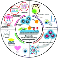
Recent progress and perspectives in applications of 2-methacryloyloxyethyl phosphorylcholine polymers in biodevices at small scales

Sasikarn Seetasang a and Yan Xu *abc
aDepartment of Chemical Engineering, Graduate School of Engineering, Osaka Prefecture University, Sakai, Osaka 599-8570, Japan. E-mail: xu@chemeng.osakafu-u.ac.jp
bJapan Science and Technology Agency (JST), PRESTO, Kawaguchi, Saitama 332-0012, Japan
cNanoSquare Research Institute, Research Center for the 21st Century, Organization for Research Promotion, Osaka Prefecture University, Sakai, Osaka 599-8570, Japan

Received 5th December 2021 , Accepted 26th January 2022

First published on 27th January 2022


Abstract

Bioinspired materials have attracted attention in a wide range of fields. Among these materials, a polymer family containing 2-methacryloyloxyethyl phosphorylcholine (MPC), which has a zwitterionic phosphorylcholine headgroup inspired by the structure of the cell membrane, has shown an outstanding ability to prevent nonspecific protein adsorption. This property makes MPC polymers excellent materials for the construction of biocompatible surfaces and interfaces with high antibiofouling performance for both macroscopic and microscopic applications. In this review, we summarize recent progress in the design, synthesis, and application of MPC polymers for biodevices with characteristic length scales ranging from millimeters to nanometers, with a focus on their applications in microfluidic devices, biosensors/bioprobes, artificial implants, and drug delivery systems. Finally, future perspectives and challenges in this field are discussed.


1. Introduction

Biocompatible polymers can be applied in biological systems and perform their desired functions without excessive adverse responses from the system. Natural and synthetic biocompatible polymers have been used for several decades in a broad range of fields such as chemistry, biology, bioengineering, medicine, pharmaceutical science, drug discovery, materials science, and environmental science. Among biocompatible polymers, a polymer family containing 2-methacryloyloxyethyl phosphorylcholine (MPC) has attracted attention for both fundamental studies and applications.1–6 MPC polymers are synthetic phospholipid polymers with zwitterionic phosphorylcholine (PC) head groups, which can form cell-membrane-like structures on various material surfaces.4,7,8 This special surface structure has a unique capacity to prevent nonspecific protein adsorption (NPA), and thus biofouling by the adsorption of biomolecules, cells, and other biological entities. This property is useful in applications involving biological entitles (e.g., proteins, other biomolecules, cells), biological samples (e.g., tear, saliva), contact with plasma and whole blood, and implantation in the body or other biological environments. Thus, various MPC polymers with different molecular architectures, such as random, block, graft, and charged structures, have been developed for a wide range of macroscopic and microscopic applications.9–15

Meanwhile, there is currently a trend to miniaturize biodevices that have a biocompatible property and use in biology and biomedicine, because small devices can be portable and simple to operate, provide high-throughput analysis, consume little energy, require less reagent use, and can be delivered or inserted into small target areas.16–20 The length of such biodevices usually range from millimeters to nanometers. However, because of their small sizes, fouling by biological entities (e.g., proteins, cells, other biomolecules) significantly decreases the functional surface area of the devices, resulting in remarkable changes in their properties and decreasing their performance. In the last decade, various MPC polymers have been designed and synthesized to resolve the critical biofouling issue in such small biodevices to improve their biocompatible property and they can be used in a wide range of applications. In particular, their development for microfluidic devices, biosensors/bioprobes, artificial implants, and drug delivery systems (DDSs) is quite active, because these areas show promise for purposes of bioanalysis, diagnosis, and therapy. Hence, these four types of biodevices with characteristic lengths ranging from millimeters to nanometers are the particular focus of this review. Accordingly, Fig. 1 summarizes current applications of MPC polymers in these four types of biodevices at small scales, including sensitivity improvement and cell manipulation in microfluidic devices, qualitative and quantitative analysis by biosensors/bioprobes, drug loading and release by extracellular or intercellular stimulation in DDSs, and the repair, replacement, or restoration of organs/tissues by artificial implants. This review describes recent progress in the design, synthesis, and application of MPC polymers as antibiofouling coatings on the surfaces of these typical small biodevices and their successful application in the related areas. Finally, future directions and the remaining challenges in the development of MPC polymers in biodevices at small scales are also discussed. Our article is not intended to be a comprehensive review of the entire field of MPC polymers but rather to motivate the expansion, improvement, and development of MPC polymers by highlighting some representative developments focused on biodevices at small scales with a wide application range.


image file: d1tb02675e-f1.tif
Fig. 1 Overview of applications of MPC-based materials in biodevices at small scales.

2. Microfluidic devices

Microfluidic devices, including lab-on-a-chip or micrototal analysis systems, have been developed to manipulate subnanoliters of fluids inside and/or around geometries with submillimeter scales for diverse applications in areas such as chemistry, biology, medicine, environmental science and materials science. Applications have been performed in the small spaces of microfluidic devices, where the surface area of the wall is small. Hence, controlling the surface is very important because the surface-area-to-volume ratio increases dramatically as the size of the functional space decreases. The biofouling of the wall is a significant effect that must be prevented, especially in applications involving proteins, cells, and other biological entities, as it can cause irregular fluid flow, clog the channel, or change the properties of functional surfaces, significantly degrading the performance. The use of MPC polymers is a powerful strategy to suppress biofouling and undesirable effects in microfluidic devices. The MPC polymers have been employed in microfluidic devices since the early stage of the development of microfluidic devices.21–24 They are used mainly for two purposes. One is to prevent biofouling which can reduce clogging and detection noise to enhance the analytical performance, including selectivity and sensitivity during quantitative analysis such as chip-based immunoassays,25–27 surface plasmon resonance (SPR),28–33 and separation techniques,34–37 The other is to realize specific functions such as cell purification,38,39 cell preservation,40–42 protein/cell-adhesive behavior,14,15,43–45 and fluidic manipulation.46,47

For early diagnosis of cancer by capturing and detecting circulating tumor cells (CTCs), Xiao et al. immobilized zwitterionic poly(2-methacryloyloxyethyl phosphorylcholine) (PMPC) on brominated polyethyleneimine/polyvinyl alcohol (PEI/PVA–Br) nanofibers via atomic transfer radical polymerization (ATRP).48 After that, folic acid (FA) ligands were attached to the PMPC-nanofibers by the redox-sensitive disulfide group (Fig. 2A). The PEI/PVA–PMPC–FA nanofibers on the surface of the microfluidic platform potentially prevented NPA and blood cell adhesion, making it possible to capture cancer cells with overexpressed FA receptors. Tris(2-carboxyethyl)phosphine (TCEP), an effective reducing agent for breaking disulfide bonds, was employed to release the FA-bound FA receptor from the PMPC before staining and quantitative counting under a fluorescence microscope. The developed MPC polymer-based material coupling with the microfluidic method exhibited potential to be applied in clinics to analyze CTCs from the whole blood of cancer patients without complicated sample pretreatment.


image file: d1tb02675e-f2.tif
Fig. 2 Representative applications of MPC polymers in microfluidic devices: (A) schematic of nanofibrous PEI/PVA alignment for capture and release of CTCs (reprinted with permission from ref. 48. Copyright © 2018, Royal Society of Chemistry), (B) schematic of a PMAMPC brush immobilized on gold-coated SPR chips for AVD determination in blood plasma to increase the detection sensitivity with a LOD at 1.5 nM (reproduced with permission from ref. 28. Copyright © 2012, American Chemical Society), (C) separation of iPSCs (spiked circles) from other cells (smooth circles) in an anti-SSEA-1/PMBN modified microchip (reproduced with permission from ref. 38. Copyright © 2017, American Chemical Society), (D) introduction of PMBV and PVA solution into a microchannel to form a hydrogel for spontaneous cell packaging and storage and the deformation of the developed hydrogel by D-fructose (reproduced with permission from ref. 40. Copyright © 2010, John Wiley and Sons, and ref. 42. Copyright © 2015, American Chemical Society), (E) variability of cells stored in the synthesized hydrogel in the microplate/microchannel at 4 °C and 25 °C (reproduced with permission from ref. 42. Copyright © 2015, American Chemical Society), and (F) cytotoxicity responses to saponin, Triton X-100, and methanol of cells preserved in the hydrogel on a microfluidic chip compared with that of cells in standard culture medium (reproduced with permission from ref. 40. Copyright © 2010, John Wiley and Sons).

MPC polymers have attracted attention for use as coatings on the microfluidic surfaces used in the SPR technique to analyze samples containing biomolecules, cells, and other biological entities. Akkahat et al. used the grafting-to method to immobilize thiol-terminated poly(MPC-co-methacrylic acid (MAA)) (referred to as PMAMPC–SH in the ref. 28) brushed on gold (Au)-coated SPR chips for avidin (AVD) determination in blood plasma (Fig. 2B).28 PMAMPC reduced NPA and thus enhanced the sensitivity and specificity of detection by improving the signal-to-noise ratio. The limit of detection (LOD) for AVD analysis was 1.5 nM, which is much lower than that of conventional SPR sensors that have a surface of a self-assembled monolayer of 11-mercaptoundecanoic acid (150 nM AVD).

In addition to quantitative analysis, MPC polymers have been applied for other specific purposes in microfluidic chips. For example, Otaka synthesized poly(MPC-co-n-butyl methacrylate (BMA)-co-p-nitrophenyl-oxycarbonyl poly(ethylene glycol) methacrylate (MEONP)) (referred to as PMBN in ref. 39) via radical polymerization followed by covalent bonding with antibodies against stage-specific embryonic antigen 1 (AbSSEA-1) on the surface of microchannels in microfluidic chips. The PMBN–AbSSEA-1-modified microchannel was used to purify pluripotent stem cells (iPSCs) in routine cell culture.39 The modified surface affected the difference in mobility between positive and negative SSEA-1 cells. The iPSCs (spiked circles, positive SSEA-1 cells) moved more slowly because of interactions with AbSSEA-1. In contrast, negative cells (smooth circles) and other impurity molecules moved faster because the PMBN–AbSSEA-1-modified surface prevented NPA (Fig. 2C). This approach provides an alternative purification method for separating target molecules from impurities in a stem cell culture solution.

Another interesting research study was reported by Xu et al. in which an MPC polymer-based hydrogel was prepared for cell preservation.40–42 Poly(MPC-co-BMA-co-p-vinylphenylboronic acid (VPBA)) (referred to as PMBV) was prepared via radical polymerization and then cross-linked with high average molecular-weight (high-MW) PVA to form a polymer network for storing L929 cells in microfluidic channels (Fig. 2D). The addition of the hydroxyl groups in a low-MW sugar solution such as D-fructose or D-galactose to the microchannel can break the boronic linkage and transform the hydrogel into a solution, resulting in cell release, as shown in the orange box in Fig. 2D. The prepared hydrogel performed well in the sustainable storage of cells under hypothermic conditions at 4 °C for 1 week and at 25 °C for at least 4 days. The viability of cells under typical cell culture conditions in the microchannel was better than that of cells on a microplate (Fig. 2E). The reasons were that the microchips prevented the evaporation of moisture in the hydrogel while maintaining gas exchange. The chip could reduce cell contamination from the external environment. In addition, the cytotoxicity responses of the cells preserved at 37 °C in the developed hydrogel on the chip and in the standard cell culture medium were compared. Fig. 2F shows that the response of the cells preserved in the developed hydrogel on the chip to toxins (saponin, Triton X-100, and methanol) was similar to that of cells in the standard cell culture medium. This work provides an alternative method of storing and delivering cells for external laboratory analysis while maintaining good cell function.

3. Biosensors and bioprobes

Biosensors and bioprobes are small biodevices used to measure targeted molecules and monitor targeted sites, respectively. The detection area of the devices binds to the targets before generating various types of signals (for example, changes in light intensity, light absorbance, potential, or heat) that are directly or indirectly proportional to the presence of the targets. The interferences resulting from the non-specific adsorption of biological entities is a critical issue impeding the use of the biosensors and bioprobes to obtain highly precise and accurate detection. The adsorption may cause severe problems, such as reductions in the surface area and specificity for reactions and decreased stability of functional groups. These problems would result in a low signal to noise ratio, insufficient output conversion, and poor analytical performance. MPC polymer coatings have been applied to modify the surface of biosensors/bioprobes to suppress biofouling and prevent undesirable behaviors. Generally, there are two primary applications of surface coatings of MPC polymers: (1) quantitative analysis of biological samples such as tears, serum, and urine,49–56 and (2) semi-quantitative or qualitative analysis for disease diagnosis.57–63

Several publications have reported the advantages of coating a standard electrode with MPC polymers, which can significantly increase binding selectivity and decrease noise during measurements because of their ability to prevent NPA.51,52,54,55,64 For instance, Li et al. synthesized poly(MPC-co-VPBA-co-vinylferrocene (VFC)) (referred to as PMVF in ref. 51) by radical polymerization. The prepared polymer was used to support NPA resistance and intermediary electron transfer. The PMVF solution was mixed with a glucose oxidase (GOx)–PVA solution to form a hydrogel, causing the enzyme to become stably accommodated in the ferrocene units of the hydrogel. Next, a photoreactive azide-unit pendented water-soluble PVA (AWP) solution was dropped onto an Au electrode before the PMVF/GOx–PVA hydrogel was spin-coated layer by layer (LBL) for glucose analysis (Fig. 3A). In a solution containing glucose, GOx catalyzed the oxidation of glucose leading to a decrease in the reduction current of the modified electrode. This study demonstrated a rapid, simple electrode-preparation approach to motivate the further development of enzyme electronic biosensors using different enzymes for specific analytes.


image file: d1tb02675e-f3.tif
Fig. 3 Representative applications of MPC polymers in biosensors and bioprobes: (A) LBL modification of the PMVF/(GOx)–PVA hydrogel on the surface of a gold electrode (reprinted with permission from ref. 51. Copyright © 2012, Elsevier), (B) glucose biosensor attached to a PDMS contact lens using a mixture of PMEH and GOx enzyme (reprinted with permission from ref. 50. Copyright © 2011, Elsevier), (C) schematic illustration of the binding between PMB-coated CNTs and an apolipoprotein complex with the receptor on adipose tissue (left) and NIR images of a mouse with (right, top) and without (right, bottom) the injection of PMB-coated CNTs (reprinted with permission from ref. 57. Copyright © 2017, Springer Nature), and (D) schematic illustration of drug release from TPP@PMM micelles for atherosclerosis theragnosis (reproduced with permission from ref. 61. Copyright © 2020, John Wiley and Sons).

The versatility of MPC polymers also makes them suitable for the quantitative analysis of various materials. For example, the Mitsubayashi group prepared poly(MPC-co-2-ethylhexyl methacrylate (EHMA)) (referred to as PMEH in ref. 50) via free radical polymerization using α,α′-azobisisobutyronitrile (AIBN) as an initiator. Then, they prepared a mixture of the polymer solution with GOx. The prepared mixture was dropped on a polydimethylsiloxane (PDMS) contact lens and cured at 80 °C to obtain a sensing electrode attached to the contact lens, as shown in Fig. 3B.49,50 The modified contact lens could analyze glucose in a tear (as in the aforementioned detection principle) without interference of NPA. Another example was reported by Pinyorospathum et al.,53 who synthesized thiol-terminated PMPC (referred to as PMPC–SH in ref. 53) by reversible addition–fragmentation chain-transfer (RAFT) polymerization. The PMPC–SH was modified on a screen-printed electrode-based paper with a gold nanoparticle (AuNPs) surface on the working electrode. The PMPC–SH self-assembled onto the working electrode via the interaction of the thiol group with gold. The modified MPC polymer on the paper helps to reduce NPA during the analysis of C-reactive protein (CRP) in a certified serum. A change in current was observed when adding CRP into the solution in the presence of calcium (Ca2+). This is due to the fact that CRP interacted with the PC group of PMPC and persisted on the surface of the electrode. The PMPC modified electrode exhibited selective binding with CRP without interference from bilirubin, myoglobin, or albumin, at a suitable pH and Ca2+ concentration. In addition to its biocompatibility, the MPC polymer itself can facilitate CRP-specific binding in the presence of Ca2+.4,29,65–68 These results exhibit promise for using the MPC polymers in biosensors for CRP determination.

MPC polymers have attracted attention for being used as surface coatings on bioprobes. The MPC-modified surface can resist NPA, enabling efficient semi-quantitative or qualitative analysis such as image analysis for disease diagnosis or for observation of an area inside the body without surgery.57–63 For example, Yudasaka et al. replaced sodium deoxycholate on the surface of single-walled carbon nanotubes (CNTs) by poly(MPC-co-BMA) (referred to as PMB) using ultrafiltration.69 The obtained PMB-coated CNT emits near-infrared (NIR) fluorescence at wavelengths of 1100–1400 nm.57 Apolipoproteins in blood were adsorbed on the PMB-coated CNTs before specific binding with apolipoprotein receptors on the capillary endothelial cells of brown adipose tissue (BAT), as illustrated schematically in Fig. 3C, left. This interaction resulted in a bright bioimage indicating capillary density in the BAT as observed using an NIR camera, as shown in Fig. 3C, right. This method provided a more precise image with less light scattering compared to images obtained using organic dyes. However, the adsorption mechanism between PMB and apolipoprotein is still unclear. This work may support the development of methods for the analysis of adipose tissues and other thermogenic tissues, which may provide a new therapeutic target for obesity-associated metabolic disorders. Another use of MPC polymers for qualitative sensing was reported by Ma et al., who synthesized a diblock copolymer of poly(MPC-b-2-methylthio ethanol methacrylate (MEMA)) (referred to as PMM in ref. 61) via RAFT polymerization. After that, a fluorophore that emits light by two-photon induction (TP) was bridged with prednisolone (Pred), an atherosclerosis theragnostic, via reactive oxygen species (ROS) responsive linkage (referred to as TPP in ref. 61). Finally, TPP was loaded into core–shell PMM via self-assembled micelles of PMM (referred to as TPP@PMM).61 The prepared micelles were biocompatible, with little NPA; thus, they circulated stably in the blood to deliver the dual-encapsulated fluorophore and Pred to an inflammatory atherosclerosis site. The two molecules were linked by β-cyclodextrin (CD), which is sensitive to ROS. Therefore, in the target area, the TPP@PMM micelles dissociated because of high ROS expression and high lipid levels; consequently, the CD bond was broken, and the drug was released (Fig. 3D). The two-photon aggregation-induced emission (AIE) of the active fluorophore successfully generated fluorescence emission in the infected tissue for image analysis. The practical application of this method was demonstrated by in vivo use on atherosclerosis-prone apolipoprotein E-deficient mice (referred to as ApoE−/− in ref. 61) for potential atherosclerosis theragnosis.

4. Artificial implants

Artificial implants are small biodevices used to repair, replace, or restore various body parts such as the hip and other joints, ocular lens, heart, breast, and teeth. Because implanted devices must be attached to or inserted into the body, they will inevitably come into contact with biological entities such as proteins and cells. Implantation usually causes nonspecific adsorption of proteins and cells, resulting in undesirable responses such as thrombus formation and capsular formation on the surface of the implants, which can cause infection and, in the worst cases, death. Thus, the surfaces of artificial implants should be modified to give them antibiofouling properties and thereby biocompatibility. Coating with MPC polymers is among the best strategies for this purpose. Moreover, the use of MPC polymers provides the additional benefit of enhancing the lubricant property of the devices because it has hydrated layers at the PC group.4

Several implanted devices for heart disease treatment have been developed, for example, valves,70–72 catheters,73–76 and stents.77–80 In this type of treatment, it is essential to reduce the adhesion of blood cells, which can induce thrombus formation and blood clogging. Copolymers of MPC and many kinds of other monomers via ATRP,81 free radical polymerization,70,77,78,82 and condensation reaction83,84etc., have been employed to obtain different structural architectures before coating on the cardiovascular devices allowing devices with an antifouling surface. A dip-coat approach with/without surface activation,77,82,83,85 or further polymerization79,86,87 are common methods used for the surface coating. Liu et al. prepared random copolymers of poly(MPC-co-lauryl methacrylate (LMA)-co-N-(3-aminopropyl) methacrylamide hydrochloride (APMA)) (referred to as PM(PCLA) in ref. 70) by free radical polymerization. The introduction of APMA increased the crosslinking strength with glutaraldehyde that was treated on the surface of porcine pericardium (PP), a heart valve leaflet (Fig. 4A, top). The average molecular weight (MW) of the polymers was adjusted, and the devices coated with PP–PM(PCLA)3 (MW 116[thin space (1/6-em)]868 g mol−1) exhibited remarkably reduced blood coagulation, as demonstrated in Fig. 4A, which shows thrombus formation on the valve (Fig. 4A, middle) and a scanning electron microscopy (SEM) image of platelet/blood cell adhesion on the surface of the material after 2 h circulation (Fig. 4A, bottom).


image file: d1tb02675e-f4.tif
Fig. 4 Representative applications of MPC polymers in artificial implants: (A) schematic illustration of NPA resistance on the MPC-modified surface of valve leaflets (top), illustrations of thrombus formation on the developed valves (middle), and SEM images of platelet and red blood cell adhesion on the surface of the developed valves (bottom) (reprinted with permission from ref. 70. Copyright © 2021, Elsevier), (B) capsule formation 6 months after silicone implantation in a Yorkshire pig (reproduced with permission from ref. 90. Copyright © 2020, American Chemical Society), (C) surface modification of Ti using a DMA–MPC copolymer to resist bacterial adhesion and increase lubrication (reproduced with permission from ref. 108. Copyright © 2019, American Chemical Society), (D) comparisons of lipid and protein adsorption on a hydrogel contact lens with/without PMAE modification (reproduced with permission from ref. 106. Copyright © 2021, American Chemical Society), and (E) interaction force of HSA on a contact lens (left) and coefficient of friction of contact lens with/without PMAE modification (right) (reprinted with permission from ref. 103. Copyright © 2021, Elsevier).

MPC polymers may also be applicable in artificial implants used in the beauty industry such as silicone for breast implants. PDMS is a common material of breast silicone that is initiated before covalent bonding with PMPC via photo- or heat-induced radical polymerization.88–91 Ham et al. grafted a nanolayer of PMPC on PDMS silicone by simply dipping the silicone in a mixture of MPC and ethylene glycol dimethacrylate, and irradiating the silicone with UV light. The covalently grafted PMPC silicone was implanted in a Yorkshire pig for 6 months and showed excellent anti-inflammation and anti-fibrous capsule behavior, as displayed in Fig. 4B.90 Kang et al. introduced heat-induced polymerization as an alternative to UV-induced polymerization.91 They immersed a silicone breast in an initiator mixture solution of benzoyl peroxide and dipentaerythritol penta-/hexa-acrylate in dichloromethane/acetone for 16 h at 70 °C. The results revealed that the silicone modified by heat-induced polymerization resulted in 45% less capsular formation, while the modified silicone produced by UV-induced polymerization gave ∼25% less capsular formation, when both modified silicones were compared with unmodified silicone. The heat-induced polymerization gave a thicker nanolayer PMPC-grafted surface; therefore, a better inhibition of capsular formation was obtained. However, both coating approaches reduced the incidence of infection by ∼20%.

In addition to the suppression of fouling, MPC polymers also play an additional role in increasing lubrication and softness, which enhances the performance of materials used in orthopedics92–98 and ophthalmology.99–107 Most of the grating methods used for coating MPC polymers on orthopedic devices is photo-induced radical polymerization followed by a few reports of thermal-induced radical polymerization94,96 or the dip-coat method.108 For instance, Liu et al. synthesized poly(MPC-co-dopamine methacrylamide (DMA)) (referred to as DMA–MPC in ref. 108) using free radical polymerization and coated the copolymer on a titanium alloy (Ti6Al4V) by simply immersing in the copolymer solution for 24 h. The DMA–MPC copolymer was successfully coated onto the Ti6A14V substrate through an ion–dipole interaction. After that the alloy was used as articular cartilage. Tribological tests revealed that the coefficient of friction (COF) decreased from 0.124 for the unmodified surface to 0.051 for the modified surface because the hydrated PC layers acted as a lubricant. Fig. 4C shows the surface modification procedure and highlights the two advantages of the DMA–MPC-modified surface: (1) bacterial inhibition, which reduces infection, and (2) lubricant formation, which improves the wear performance. In ophthalmology, the synthesis of MPC polymers via free radical polymerization, ATRP, or RAFT have been attempted before merging into interpenetration networks (IPN) such as poly(MPC-co-bis(trimethylsilyloxy)methylsilylpropyl glycerol methacrylate),99 2-hydroxyethyl methacrylate hydrogel,104 collagen networks,100,105,109,110 and silicone hydrogel,103,106,111,112 to create small optical devices. In addition, MPC polymers can be grafted on small optical devices by spin coating,113 plasma technology,107 and salinization114etc. The development not only provides an antifouling property, but also provides lubricant and soft properties of intraocular lenses (IOLs), contact lenses (CLs), and corneal etc. For example, Tan et al. synthesized poly(MPC-co-MAA) (referred to as P(MPC–MAA) in ref. 107) via free radical polymerization before coating the polymers on an IOL via air plasma treatment. Six weeks after cataract surgery, the modified lens exhibited remarkable suppression of anterior capsule opacification and inflammation while maintaining diopter, resolution, transmission, and bending and stretching properties. This work proved that the use of P(MPC–MAA) in IOLs could potentially provide better reconstruction and restoration of vision. Another example in the ophthalmologic field, in which MPC polymers are widely applied as surface coatings, is the CLs. The Ishihara research group reported synthesis of silicone hydrogel CLs by radical polymerization. The hydrogel was dipped into poly(methacrylic acid) solution to generate IPN before immersing into a mixture solution of polyaminoamide-epichlorohydrin (PAE) and poly(MPC-co-2-aminoethyl methacrylate (AEMA)) (referred to as PMA in ref. 106). This step allowed PAE and PMA to bind the substrate via am amide-bond.103,106 The prepared CLs strongly resisted the nonspecific adsorption of lipids and proteins. In addition, the softer surface and lower COF of the lens made it more comfortable to wear. Fig. 4D and E show the antifouling and lens friction properties of the modified CLs, respectively. The results show that the interaction force with human serum albumin (HSA), and COF values of the modified surface are significantly lower than the bare surface (Fig. 4E). The prepared MPC polymers as aforementioned examples have shown promise for increasing moisture at the surfaces of lenses and joints. Thus, the lubrication and softness are the properties that play a significant role in orthopedics and ophthalmology.

5. Drug delivery systems

DDSs are engineered small devices used to carry, deliver, and release therapeutic agents to specific sites inside the body. The NPA may induce failures in the delivery of therapeutic agents to the desired target sites, leading to drug degradation, cell toxicity, and unexpected side reactions. To resolve the problem, copolymers composed of MPC able to suppress NPA and other moieties allowing encapsulation, delivery and controlled release of therapeutic agents have been developed to form many kinds of drug carrier structures such as nanoparticles (NPs),115–121 dendrimers,122,123 micelles,124–129 polymersomes,130–132 and liposomes9,132,133 on micrometer to nanometer scales. The characteristics of the MPC moiety in MPC copolymers to suppress NPA can play a critical role in the improvement of the biocompatibility of the DDS by increasing blood circulation time, enhancing specific binding to the targeted cells, avoiding immune recognition and elimination, and reducing cell toxicity. In addition, the capability of the MPC moiety to enhance lubrication is also a favorable property in the development of DDSs. Meanwhile, MPC–copolymer-based drug carriers with responsive groups can afford controlled drug release and binding with the target sites under an extracellular or intracellular stimulus such as pH,134–137 temperature,138 an enzymatic reaction,122 a redox reaction,124,127,139–142 a competitive metal formation,143 light,129,144,145 or oscillation.146

Cai et al. reported the preparation of poly(MPC-b-ε-caprolactone (CL)) (referred to as PCL-ss-PMPC in the ref.,124 where -ss- denotes the disulfide bonds), via ring-opening polymerization (ROP) and ATRP. Then, they encapsulated doxorubicin (DOX), an anticancer drug into the PCL-ss-PMPC micelles using a dialysis method. The PMPC part allowed the PCL-ss-PMPC micelles to circulate for a long time in blood, accumulating in the cancer region before rapidly internalizing into tumor cells because of their biomimetic shells. The prepared micelles had the disulfide bonds as the reduction responsive linkage. Therefore, the remarkable difference in redox potential between the intracellular tumor cells and the extracellular environment after the addition of dithiothreitol, a reducing substance, triggered disulfide bond dissociation in the PCL-ss-PMPC micelles, resulting in DOX release, as shown in Fig. 5A.124 Treatment of tumors in rats using these micelles showed promising results, as the tumor volume decreased significantly. This innovative strategy could be used to create other biopolymeric drug carriers for effective cancer therapy.


image file: d1tb02675e-f5.tif
Fig. 5 Representative applications of MPC polymers containing different responsive groups in DDSs: (A) redox-responsive PCL-ss-PMPC polymeric micelles (reprinted with permission from ref. 124. Copyright © 2018, Elsevier), (B) pH-responsive PIC vesicles (reproduced with permission from ref. 137. Copyright © 2019, American Chemical Society), (C) competitive-metal-complex-formation-responsive Fe3+–curcumin/PMPC-b-PserA/NCPs (reproduced with permission from ref. 143. Copyright © 2021, American Chemical Society), (D) extracellular-oscillation-responsive PMPC/PEI/MSNPs (reprinted with permission from ref. 146. Copyright © 2018, Elsevier), and (E) visible-light-responsive bMSNPs–AZO/DS/CD–PMPC (top) and lubrication generated by the prepared NPs (bottom) (reprinted with permission from ref. 145. Copyright © 2021, Royal Society of Chemistry).

DDSs using MPC copolymers with pH-sensitive units have been widely developed. For example, Ohara et al. prepared a cationic copolymer of poly(MPC-b-3-(methacrylamidopropyl) trimethylammonium chloride (MATAC)) (referred to as P20M167 in ref. 137) and anionic copolymer of poly(MPC-b-sodium 6-acrylamidohexanoate (AaH)) (referred to as P20M190 in ref. 137) via RAFT radical polymerization. Polyion complex (PIC) vesicles were prepared by electrostatic interactions between the cationic (P20M167) and anionic (P20A190) copolymers, making it possible to encapsulate Texas red-labeled dextran (TD 70), a hydrophilic nonionic molecule, which was used as a model small molecule. The hydrophilic PC group of PIC vesicles played a role in NPA resistance and excellent biocompatibility allowing the PIC vesicles to enter into the targeted cell by endocytosis. After that, the PIC vesicles were dissociated under acidic conditions inside the lysosome because negative charges of PAaH were neutralized resulting in charge imbalance. On the other hand, P20M167 contributed to lysosome breakage, which released the TD70 molecules to the cytoplasm (Fig. 5B, bottom).137 Fluorescence images of TD70 in L292 cells revealed successful drug release, opening a path to further developments in the introduction of drugs into the cytoplasm without cytotoxicity.

The employment of MPC polymers in DDSs coupled with other trigger strategies for drug release are also of interest, although there are few publications. For example, Chen et al. prepared a diblock copolymer of poly(MPC-b-serinyl acrylate (serA)) (referred to as PMPC-b-PserA in ref. 115) by RAFT with 4,4′-azobis(4-cyanovaleric acid) (ACVA) as an initiator and mixed it with FeCl3 for self-assembly into nanoscale coordination polymers (NCPs) via metal complex formation of ferric ion (Fe3+) and PserA. Next, curcumin, an anticancer drug, was loaded by an oil-in-water emulsion method.115 The zwitterionic PC of PMPC-b-PserA/NCPs prolonged the drug circulation time and decreased the drug toxicity because of its hydrophilicity. To release the drugs, deferoxamine (DFO), an iron-chelating agent, was employed to capture Fe3+ from the PMPC-b-PserA/NCPs (Fig. 5C).143 The results showed promise for efficient drug delivery using PMPC-b-PserA/NCPs and release of anticancer drugs using competitive metal complex formation of two metal chelates (PserA and DFO). Scalcedo et al. modified PMPC on the surface of PEI-grafted core–shell Fe3O4@SiO2 mesoporous NPs (referred to as PEI/MSNPs) using glutaraldehyde as a crosslinking reagent to obtain PMPC/PEI/MSNPs. The prepared MSNPs were employed in ovarian chemotherapy to store TWIST-siRNA (a sensing molecule) and daunorubicin (an antitumor antibiotic). The particle size of the PMPC/PEI/MSNPs remained stable for 72 h compared with the uncoated PEI/MSNPs after incubation in bovine serum albumin (BSA) and fetal bovine serum (FBS) plasma proteins. The results proved the NPA resistant property which allowed the PMPC/PEI/MSNPs to survive from opsonization in the bloodstream and to be internalized by the cancer cell. After that, an external magnetic field was introduced to oscillate the magnetic MSNPs, resulting in heat generation, which facilitated the release of the encapsulated molecules from the silica pores (Fig. 5D).146 This method achieved in coating the PMPC on PEI/MSNPs to co-deliver sensing molecules and drugs for dual-function therapy, which may motivate future research on other multifunctional MPC polymer coupled drug carriers to obtain multiple effects in therapy. Although these two articles demonstrated effective drug delivery to the targeted cell by association of MPC polymers with interesting drug releasing strategies, the risk due to the disposal pathway of the metal residues after dissociation should be addressed. Zhang et al. conjugated 1,2-ethaneditiol-modified MPC (HS–MPC) on the surface of phosphoramidate (PAD) dendrimers by a thiol-Michael addition click reaction (referred to as PAD–MPC in ref. 122). The zwitterionic surface of dendrimers forms hydrated layers which can reduce NPA and extend the blood circulation time to increase tumor targeting. The modified dendrimers were designed to store DOX. The P–O ester bond in PAD–MPC was effectively hydrolyzed with the assistance of the phospholipase C (referred to as PLC) enzyme as a catalyst. Because breast cancer cells overproduce PLC, the drugs were released at the target cells.122 The dendrimers showed high cytotoxicity against tumors, as demonstrated by several results. Thus, the MPC polymer-modified dendrimers can be applied as a drug carrier using an enzyme as a stimulator in a drug-release strategy. The final example of MPC polymers in DDSs was coupled with light sensitive moieties and implemented by Zhao et al.145 They combined CD-modified PMPC (referred to as CD–PMPC in ref. 145) with azobenzene-modified mesoporous silica NPs (referred to as bMSNPs–AZO in the same reference) by supramolecular interaction to capture the anti-inflammatory drug diclofenac sodium (DS). The azobenzene in the synthesized NPs was partial trans-to-cis isomerized under visible light, resulting in drug release (Fig. 5E, top). Apart from the biocompatible property of PMPC, after light exposure, the robust hydration layer of the N+(CH3)3 and PO4 groups of CD–PMPC improved lubrication (Fig. 5E, bottom), which was indicated by the decreasing COF value in a tribological test. This property is the recent finding of PMPC ability that has been used in DDSs and can be useful in osteoarthritis therapy. Because this method uses visible light, which can reduce the damage to cells and penetrate cells more deeply than ultraviolet light,129,144,147 it may result in safer treatment.

6. Future perspectives

The selected progress highlighted in this review has proved that MPC polymers can offer excellent functions and performance in applications in biodevices at small scales. Based on the current progress, brilliant future perspectives of this field in both fundamental study and practical applications can be depicted as shown in Fig. 6, while many challenges towards these future perspectives need to be conquered as described in the following.
image file: d1tb02675e-f6.tif
Fig. 6 Future perspectives for MPC-based materials in biodevices at small scales.

(i) The extension of MPC functionality by not only enhancing the reported properties such as antibiofouling, lubrication, and direct penetration into the cell membrane without toxicity, but also developing other new properties, is expected to be important in some emerging fields using small biodevices such as nanorobotics, nano energy, space science, and quantum biology. The development of nanorobots for transporting, manipulating, transducing, actuating, and sensing molecules or subcellular entities in the body is a recently growing field.148–150 To reach this goal, it is necessary to suppress NPA to prolong the resident time of the nanorobots and to have energy sources for driving and controlling the nanorobots inside the body. Surface modification of the nanorobots using new MPC copolymers with additional functional moieties enabling us to induce energy conversion by taking advantage of the in vivo biological environment could play an essential role in this field. Such MPC polymers would not only guarantee the biocompatibility and working efficiency of the nanorobots in the body but also offer mechanisms to supply nano energy for tiny nanorobots by converting chemical energy inside the body into electric energy. In addition, the biocompatible and water-rich properties of miniaturized MPC hydrogels with other moieties to preserve nutrients without generating toxicity or to degrade itself into fertilizer may be an alternative hydro-absorbent to grow plants for astronauts during space missions, which is necessary especially when the space mission requires a long period of time to reach other planetary bodies.151 Lastly, the use of well-defined nanoscale MPC surfaces to eliminate quantum coherence resulting from nonspecific interactions between single molecules and the quantum material-based biosensors would be helpful to elucidate quantum coherences of certain biological interactions of interest in some physiological processes,152–155 finally contributing to the development of the emerging field of quantum biology.156

(ii) As further miniaturization to nanometer sizes is a trend in the development of biodevices, the surface modification of emerging nanoscale biodevices such as nanoneedles,157–159 nanowires160–162 and nanofluidic devices,163–172 provides new opportunities for the use of MPC-based materials. As nanodevices have ultrahigh surface-to-volume ratios, it is not easy to modify and functionalize the surface. In addition, the MPC polymers available for use in microdevices or open nanostructured surfaces may no longer be satisfactory for these nanodevices. The reason is that the dimensions of the polymer typically approach the length scales of these nanodevices, making it extremely difficult to handle polymers inside the nanospace. Hence, in such cases polymers with well-regulated short chain lengths are required. A proof-of-concept of this strategy has been done by Shinomiya et al.164 In their work, well-tailored thermoresponsive polymer brushes with well-regulated short chains were successfully self-assembled in tiny nanofluidic channels, enabling the active regulation of femtoliter-scale fluids. According to this principle, this strategy will also be effective for MPC polymers. Another alternative strategy is to consider other forms of MPC-based materials. For example, the direct use of MPC-derived monomers with special structures and functions may make it possible to avoid this issue,173 but comprehensive studies are necessary to maintain the high performance exhibited by the polymers.

(iii) The use of zwitterionic MPC polymers in biological applications has increased significantly because of their outstanding biocompatibility and biofouling resistance. However, it is necessary to understand the mechanism of NPA resistance by the PC group to promote the design of functional MPC polymers. The structure of the water layer around the PC groups has been considered to be key to the role of MPC polymers in the suppression of NPA. Hence, the investigation of the structure and behaviors of water in MPC polymers and their surfaces has been investigated by using techniques such as differential scanning calorimetry,174,175 attenuated total reflection infrared spectroscopy,176,177 and thermogravimetric analysis.178 The use of these approaches can provide information of the water layer from the bulk, to micrometer, or even nanometer scales, but still faces great difficulty in further extending to a single molecule or atomic level to fully understand the details of the role of the water molecule in the MPC polymers. A combination of these conventional techniques with small nanodevices allowing the confinement of single molecules would provide new approaches to reach this goal.163,164,170,179–183 Some preliminary efforts in this direction have been done by Xu et al. They reported in situ probing of the behavior of water confined in a nanofluidic device.183 Further application of the in situ probing technique to nanochannels with well-defined assembly of single MPC molecules would provide new insights into the mechanism of MPC polymer to suppress NPA.

(iv) The description of available polymers, including MPC polymers, is based mainly on average information such as average MW, average degree of polymerization, and the abundance of polymer sequences, which may vary depending on the synthesis pot. To fully explore the potential of MPC polymers, it is essential to develop methods that allow precise synthesis of MPC polymers with the desired sequence and properties that have not yet been obtained. The development of techniques enabling the manipulation of single monomer molecules in the liquid phase is necessary but still highly challenging. Nanodevices with nanoconfinement effects of single molecules would be potential tools to overcome the challenge.179,184,185 Novel mechanisms allowing free manipulation and arrangement of MPC monomers in a precise sequence could be expected by further coupling such nanodevices with other manipulation mechanisms such as optics, magnetics, electrostatics, and fluidics in the future.

7. Conclusions

The significant development of MPC polymers, which have been successfully used in biodevices at small scales ranging from millimeters to nanometers, has been demonstrated in a broad range of applications. Because of their outstanding antibiofouling properties, MPC polymers are potential materials for use in small biodevices in contact with biomolecules, cells, and other biological entities. Moreover, the copolymerization of MPC with other monomers affords a variety of functions for specific purposes in microfluidic devices, biosensors/bioprobes, artificial implants, and DDSs. Therefore, MPC polymers are a powerful material with many benefits, as summarized in this review. The challenges in acquiring a deeper understanding of the properties and extending the applications of MPC polymers are given and are waiting for researchers to explore.

Conflicts of interest

There are no conflicts to declare.

Acknowledgements

This work was partially supported by JSPS KAKENHI (Grant No. JP21H04640, JP20H00497, JP26706010, JP18H01848, and JP19KK0129), JST PRESTO (Grant No. JPMJPR18H5), MEXT KAKENHI (Grant No. JP21H05231, JP26107714, JP19H04678, and JP17H05468), the Asahi Glass Foundation, and the National Natural Science Foundation of China (NSFC) (Grant No. 21628501).

References

  1. K. Ishihara, R. Aragaki, T. Ueda, A. Watenabe and N. Nakabayashi, J. Biomed. Mater. Res., 1990, 24, 1069–1077 CrossRef CAS PubMed.
  2. K. Ishihara, T. Ueda and N. Nakabayashi, Polym. J., 2005, 22, 355–360 CrossRef.
  3. M. Kojima, K. Ishihara, A. Watanabe and N. Nakabayashi, Biomaterials, 1991, 12, 121–124 CrossRef CAS PubMed.
  4. T. Goda, K. Ishihara and Y. Miyahara, J. Appl. Polym. Sci., 2015, 132, 41766 CrossRef.
  5. K. Ishihara, M. Mu and T. Konno, J. Biomater. Sci., Polym. Ed., 2018, 29, 844–862 CrossRef CAS PubMed.
  6. K. Ishihara, H. Oda and T. Konno, Biomaterials, 2019, 230, 119628 CrossRef PubMed.
  7. K. Ishihara, J. Biomed. Mater. Res., Part A, 2019, 107, 933–943 CrossRef CAS PubMed.
  8. K. Ishihara, M. Mu, T. Konno, Y. Inoue and K. Fukazawa, J. Biomater. Sci., Polym. Ed., 2017, 28, 884–899 CrossRef CAS PubMed.
  9. S. Chatterjee and T. Ooya, ACS Appl. Polym. Mater., 2020, 2, 1909–1916 CrossRef CAS.
  10. A. Clark, M. E. Taylor, M. J. Panzer and P. Cebe, Thermochim. Acta, 2020, 691, 178710 CrossRef CAS.
  11. C. Kojima, R. Katayama, T. Lien Nguyen, Y. Oki, A. Tsujimoto, S.-I. Yusa, K. Shiraishi and A. Matsumoto, Eur. Polym. J., 2020, 136, 109932 CrossRef CAS.
  12. T. Masuda, Y. Hiraguchi, K. Kushiro, Y. Araki, T. Wada and M. Takai, Eur. Polym. J., 2020, 135, 109885 CrossRef CAS.
  13. T. Koga, T. Matsuoka, Y. Morita and N. Higashi, Mater. Adv., 2021, 2, 4068–4074 RSC.
  14. Y. Xu, M. Takai and K. Ishihara, Biomacromolecules, 2009, 10, 267–274 CrossRef CAS PubMed.
  15. Y. Xu, M. Takai and K. Ishihara, Biomaterials, 2009, 30, 4930–4938 CrossRef CAS PubMed.
  16. G. M. Whitesides, Nature, 2006, 442, 368–373 CrossRef CAS PubMed.
  17. T. Kaneta, W. Alahmad and P. Varanusupakul, Appl. Spectrosc. Rev., 2019, 54, 117–141 CrossRef.
  18. T. Kaneta, Chem. Rec., 2019, 19, 452–461 CrossRef CAS PubMed.
  19. S. Seetasang and T. Kaneta, Talanta, 2019, 204, 586–591 CrossRef CAS PubMed.
  20. I. Jeerapan, C. Moonla, P. Thavarungkul and P. Kanatharana, in Micro/Nanofluidics and Lab-on-Chip Based Emerging Technologies for Biomedical and Translational Research Applications – Part B, ed. A. Pandya and V. Singh, Academic Press, 2022, vol. 1 DOI:10.1016/bs.pmbts.2021.07.025.
  21. Y. Xu, M. Takai and K. Ishihara, Ann. Biomed. Eng., 2010, 38, 1938–1953 CrossRef PubMed.
  22. K. Ishihara, Y. Xu and T. Konno, Adv. Polym. Sci., 2011, 247, 141–165 CrossRef.
  23. Y. Xu, M. Takai and K. Ishihara, in Handbook of Modern Coating Technologies, ed. M. Aliofkhazraei, N. Ali, M. Chipara, N. B. Laidani and J. T. M. D. Hosson, Elsevier, 2021, 16, pp. 555–595 Search PubMed.
  24. K. Jang, K. Sato, Y. Tanaka, Y. Xu, M. Sato, T. Nakajima, K. Mawatari, T. Konno, K. Ishihara and T. Kitamori, Lab Chip, 2010, 10, 1937–1945 RSC.
  25. J. Park, S. Kurosawa, M. Takai and K. Ishihara, Colloids Surf., B, 2007, 55, 164–172 CrossRef CAS PubMed.
  26. N. Kameyama, S. Matsuda, O. Itano, A. Ito, T. Konno, T. Arai, K. Ishihara, M. Ueda and Y. Kitagawa, Cancer Biother. Radiopharm., 2011, 26, 697–704 CrossRef CAS PubMed.
  27. S. Chantasirichot and K. Ishihara, Biosens. Bioelectron., 2012, 38, 209–214 CrossRef CAS PubMed.
  28. P. Akkahat, S. Kiatkamjornwong, S. Yusa, V. P. Hoven and Y. Iwasaki, Langmuir, 2012, 28, 5872–5881 CrossRef CAS PubMed.
  29. Y. Kitayama and T. Takeuchi, Anal. Chem., 2014, 86, 5587–5594 CrossRef CAS PubMed.
  30. Y. Kamon, Y. Kitayama, A. N. Itakura, K. Fukazawa, K. Ishihara and T. Takeuchi, Phys. Chem. Chem. Phys., 2015, 17, 9951–9958 RSC.
  31. O. Wiarachai, T. Vilaivan, Y. Iwasaki and V. P. Hoven, Langmuir, 2016, 32, 1184–1194 CrossRef CAS PubMed.
  32. R. Matsuura, K. Tawa, Y. Kitayama and T. Takeuchi, Chem. Commun., 2016, 52, 3883–3886 RSC.
  33. T. Saeki, H. Sunayama, Y. Kitayama and T. Takeuchi, Langmuir, 2018, 35, 1320–1326 CrossRef PubMed.
  34. D. Qiu, F. Li, M. Zhang and J. Kang, Electrophoresis, 2016, 37, 1725–1732 CrossRef CAS PubMed.
  35. J. Cheng, X. Chen, Y. Cai, Y. He, Z. Chen, Z. Lin and L. Zhang, Electrophoresis, 2013, 34, 1189–1196 CrossRef CAS PubMed.
  36. X. Lei, J. Cui, S. Wang, T. Huang and X. Wu, J. Chromatogr. A, 2020, 1623, 461175 CrossRef CAS PubMed.
  37. K. Nii, K. Sueyoshi, K. Otsuka and M. Takai, Microfluid. Nanofluid., 2013, 14, 951–959 CrossRef CAS.
  38. A. Otaka, K. Kitagawa, T. Nakaoki, M. Hirata, K. Fukazawa, K. Ishihara, A. Mahara and T. Yamaoka, Langmuir, 2017, 33, 1576–1582 CrossRef CAS PubMed.
  39. A. Otaka, A. Mahara, K. Ishihara and T. Yamaoka, J. Micromech. Microeng., 2021, 31, 045012 CrossRef CAS.
  40. Y. Xu, K. Sato, K. Mawatari, T. Konno, K. Jang, K. Ishihara and T. Kitamori, Adv. Mater., 2010, 22, 3017–3021 CrossRef CAS PubMed.
  41. Y. Xu, K. Jang, T. Konno, K. Ishihara, K. Mawatari and T. Kitamori, Biomaterials, 2010, 31, 8839–8846 CrossRef CAS PubMed.
  42. Y. Xu, K. Mawatari, T. Konno, T. Kitamori and K. Ishihara, ACS Appl. Mater. Interfaces, 2015, 7, 23089–23097 CrossRef CAS PubMed.
  43. M. A. Jamiolkowski, J. R. Woolley, M. V. Kameneva, J. F. Antaki and W. R. Wagner, J. Biomed. Mater. Res., Part A, 2015, 103, 1303–1311 CrossRef PubMed.
  44. Y. Kozuka, Z. Lu, T. Masuda, S. Hara, T. Kasama, R. Miyake, N. Isu and M. Takai, J. Mater. Chem. B, 2021, 9, 4480–4487 RSC.
  45. K. Ishihara, M. Abe, K. Fukazawa, T. Konno, K. Ishihara, K. Fukazawa, M. Abe and T. Konno, Macromol. Biosci., 2021, 21, 2000341 CrossRef CAS PubMed.
  46. Y. Xu, M. Takai, T. Konno and K. Ishihara, Lab Chip, 2007, 7, 199–206 RSC.
  47. R. K. Jena and C. Y. Yue, Biomicrofluidics, 2012, 6, 012822 CrossRef PubMed.
  48. Y. Xiao, M. Wang, L. Lin, L. Du, M. Shen and X. Shi, Mater. Chem. Front., 2018, 2, 891–900 RSC.
  49. M. Chu, T. Shirai, D. Takahashi, T. Arakawa, H. Kudo, K. Sano, S. Sawada, K. Yano, Y. Iwasaki, K. Akiyoshi, M. Mochizuki and K. Mitsubayashi, Biomed. Microdevices, 2011, 13, 603–611 CrossRef CAS PubMed.
  50. M. X. Chu, K. Miyajima, D. Takahashi, T. Arakawa, K. Sano, S. I. Sawada, H. Kudo, Y. Iwasaki, K. Akiyoshi, M. Mochizuki and K. Mitsubayashi, Talanta, 2011, 83, 960–965 CrossRef CAS PubMed.
  51. Z. Li, T. Konno, M. Takai and K. Ishihara, Biosens. Bioelectron., 2012, 34, 191–196 CrossRef CAS PubMed.
  52. T. Goda, M. Tabata, M. Sanjoh, M. Uchimura, Y. Iwasaki and Y. Miyahara, Chem. Commun., 2013, 49, 8683–8685 RSC.
  53. C. Pinyorospathum, S. Chaiyo, P. Sae-ung, V. P. Hoven, P. Damsongsang, W. Siangproh and O. Chailapakul, Microchim. Acta, 2019, 186, 1–10 CrossRef CAS PubMed.
  54. T. Uen, K. Kushiro, H. Hibino and M. Takai, Sens. Actuators, B, 2020, 309, 127758 CrossRef CAS.
  55. T. Fukuma, S. Nishitani and T. Sakata, ECS Meet. Abstr., 2020, MA2020-01, 1399.
  56. T. Koshida, T. Arakawa, T. Gessei, D. Takahashi, H. Kudo, H. Saito, K. Yano and K. Mitsubayashi, Sens. Actuators, B, 2010, 146, 177–182 CrossRef CAS.
  57. M. Yudasaka, Y. Yomogida, M. Zhang, T. Tanaka, M. Nakahara, N. Kobayashi, Y. Okamatsu-Ogura, K. Machida, K. Ishihara, K. Saeki and H. Kataura, Sci. Rep., 2017, 7, 44760 CrossRef CAS PubMed.
  58. J. Hu, W. Zhuang, B. Ma, X. Su, T. Yu, G. Li, Y. Hu and Y. Wang, Bioconjugate Chem., 2018, 29, 1897–1910 CrossRef CAS PubMed.
  59. S. Yoneoka, Y. Nakagawa, K. Uto, K. Sakura, T. Tsukahara and M. Ebara, Sci. Technol. Adv. Mater., 2019, 20, 291–304 CrossRef CAS PubMed.
  60. J. Liu, Z. Xiong, J. Zhang, C. Peng, B. Klajnert-Maculewicz, M. Shen and X. Shi, ACS Appl. Mater. Interfaces, 2019, 11, 15212–15221 CrossRef CAS PubMed.
  61. B. Ma, H. Xu, W. Zhuang, Y. Wang, G. Li and Y. Wang, Small, 2020, 16, 2003253 CrossRef CAS PubMed.
  62. C. Kojima, T. Koda, T. Nariai, J. Ichihara, K. Sugiura and A. Matsumoto, Macromol. Biosci., 2021, 21, 2100170 CrossRef CAS PubMed.
  63. W. Zhuang, B. Ma, G. Liu, G. Li and Y. Wang, J. Appl. Polym. Sci., 2018, 135, 45651 CrossRef.
  64. K. Sato and T. Konno, Electroanalysis, 2020, 32, 898–901 CrossRef CAS.
  65. I. Y. Kitamura, S. Go, K. Hataoaka, Y. Kitamura, M. Shimada, K. Hatanaka, Y. Miyano, Y. Nishimune, M. Murakami and H. Shibata, Nature, 1979, 281, 155–157 CrossRef PubMed.
  66. M. K. Chang, C. J. Binder, M. Torzewski and J. L. Witztum, Proc. Natl. Acad. Sci. U. S. A., 2002, 99, 13043–13048 CrossRef CAS PubMed.
  67. T. Goda, P. Kjall, K. Ishihara, A. Richter-Dahlfors and Y. Miyahara, Adv. Healthcare Mater., 2014, 3, 1733–1738 CrossRef CAS PubMed.
  68. Y. Iwasaki, T. Kimura, M. Orisaka, H. Kawasaki, T. Goda and S. I. Yusa, Chem. Commun., 2014, 50, 5656–5658 RSC.
  69. Y. Yomogida, T. Tanaka, M. Zhang, M. Yudasaka, X. Wei and H. Kataura, Nat. Commun., 2016, 7, 1–8 Search PubMed.
  70. K. Liu, M. Li, F. Zhang, Y. Wang, C. Chen, Y. Wei, L. Yang, R. Luo and Y. Wang, Chem. Eng. J., 2021, 426, 131803 CrossRef CAS.
  71. G. Guo, W. Jin, L. Jin, L. Chen, Y. Lei and Y. Wang, J. Mater. Chem. B, 2019, 7, 1427–1434 RSC.
  72. Y. Kambe, A. Mahara, H. Tanaka, S. Kakinoki, K. Fukazawa, Y. Liu, M. Kyomoto, K. Minatoya, K. Ishihara and T. Yamaoka, J. Biomed. Mater. Res., Part A, 2019, 107, 1052–1063 CrossRef CAS PubMed.
  73. Y. Iida, K. Hongo, T. Onoda, Y. Kita, Y. Ishihara, N. Takabayashi, R. Kobayashi and T. Hiramatsu, Sci. Rep., 2021, 11, 1–6 CrossRef PubMed.
  74. S. Asif, K. Asawa, Y. Inoue, K. Ishihara, B. Lindell, R. Holmgren, B. Nilsson, A. Rydén, M. Jensen-Waern, Y. Teramura and K. N. Ekdahl, Macromol. Biosci., 2019, 19, 1800485 CrossRef PubMed.
  75. Y. Roth and D. Y. Lewitus, Polymers, 2020, 12, 1131 CrossRef CAS PubMed.
  76. L. Soletti, A. Nieponice, Y. Hong, S. H. Ye, J. J. Stankus, W. R. Wagner and D. A. Vorp, J. Biomed. Mater. Res., Part A, 2011, 96A, 436–448 CrossRef CAS PubMed.
  77. H. Il Kim, K. Ishihara, S. Lee, J. H. Seo, H. Y. Kim, D. Suh, M. U. Kim, T. Konno, M. Takai and J. S. Seo, Biomaterials, 2011, 32, 2241–2247 CrossRef PubMed.
  78. D. Lv, P. Li, L. Zhou, R. Wang, H. Chen, X. Li, Y. Zhao, J. Wang and N. Huang, React. Funct. Polym., 2021, 163, 104897 CrossRef CAS.
  79. W. Ma, P. Yang, J. Li, S. Li, P. Li, Y. Zhao and N. Huang, Appl. Surf. Sci., 2015, 349, 445–451 CrossRef CAS.
  80. Q. Zhong, J. Yan, X. Qian, T. Zhang, Z. Zhang and A. Li, Colloids Surf., B, 2014, 121, 238–247 CrossRef CAS PubMed.
  81. P. Li, Z. Luo, X. Li, R. Wang, H. Chen, Y. Zhao, J. Wang and N. Huang, Appl. Surf. Sci., 2020, 502, 144085 CrossRef CAS.
  82. Y. Yao, K. Fukazawa, W. Ma, K. Ishihara and N. Huang, Appl. Surf. Sci., 2012, 258, 5418–5423 CrossRef CAS.
  83. Y. Yao, K. Fukazawa, N. Huang and K. Ishihara, Colloids Surf., B, 2011, 88, 215–220 CrossRef CAS PubMed.
  84. W. Ma, P. Yang, Y. Zhao and N. Huang, Adv. Eng. Mater., 2018, 20, 1800624 CrossRef.
  85. S. H. Ye, C. A. Johnson, J. R. Woolley, H. Murata, L. J. Gamble, K. Ishihara and W. R. Wagner, Colloids Surf., B, 2010, 79, 357–364 CrossRef CAS PubMed.
  86. C. Wu, W. Chang, H. Qi, L. Long, J. Zhao, X. Yuan, Z. Li and X. Yang, J. Coat. Technol. Res., 2017, 14, 1127–1135 CrossRef CAS.
  87. T. Tateishi, M. Kyomoto, S. Kakinoki, T. Yamaoka and K. Ishihara, J. Biomed. Mater. Res., Part A, 2014, 102, 1342–1349 CrossRef PubMed.
  88. T. Goda, T. Konno, M. Takai, T. Moro and K. Ishihara, Biomaterials, 2006, 27, 5151–5160 CrossRef CAS PubMed.
  89. J. U. Park, J. Ham, S. Kim, J. H. Seo, S. H. Kim, S. Lee, H. J. Min, S. Choi, R. M. Choi, H. Kim, S. Oh, J. A. Hur, T. H. Choi and Y. Lee, Acta Biomater., 2014, 10, 4217–4225 CrossRef CAS PubMed.
  90. J. Ham, Y. Kim, T. An, S. Kang, C. Ha, M. Wufue, Y. Kim, B. Jeon, S. Kim, J. Kim, T. H. Choi, J.-H. Seo, D. W. Kim, J.-U. Park and Y. Lee, ACS Appl. Mater. Interfaces, 2020, 12, 30198–30212 CrossRef CAS PubMed.
  91. S. Kang, J. Kim, S. Kim, M. Wufuer, S. Park, Y. Kim, D. Choi, X. Jin, Y. Kim, Y. Huang, B. Jeon, T. H. Choi, J.-U. Park and Y. Lee, Biomater. Sci., 2020, 8, 1580–1591 RSC.
  92. N. Wang, A. M. Trunfio-Sfarghiu, D. Portinha, S. Descartes, E. Fleury, Y. Berthier and J. P. Rieu, Colloids Surf., B, 2013, 108, 285–294 CrossRef CAS PubMed.
  93. K. Ishihara, Polym. J., 2015, 47, 585–597 CrossRef CAS.
  94. S. Ghosh, S. Abanteriba, S. Wong and S. Houshyar, J. Mech. Behav. Biomed. Mater., 2018, 87, 312–324 CrossRef CAS PubMed.
  95. M. Kyomoto, T. Moro, S. Yamane, K. Watanabe, M. Hashimoto, S. Tanaka and K. Ishihara, Langmuir, 2018, 35, 1954–1963 CrossRef PubMed.
  96. C. M. Lim, J. Hur, H. Jang and J. H. Seo, Acta Biomater., 2019, 85, 180–191 CrossRef CAS PubMed.
  97. S. Ghosh, S. Abanteriba, S. Wong, R. Brkljača and S. Houshyar, Mater. Sci. Eng., C, 2019, 101, 696–706 CrossRef CAS PubMed.
  98. L. Han, L. Xiang, J. Zhang, J. Chen, J. Liu, B. Yan and H. Zeng, Langmuir, 2018, 34, 11593–11601 CrossRef CAS PubMed.
  99. T. Shimizu, T. Goda, N. Minoura, M. Takai and K. Ishihara, Biomaterials, 2010, 31, 3274–3280 CrossRef CAS PubMed.
  100. S. Hayes, P. Lewis, M. M. Islam, J. Doutch, T. Sorensen, T. White, M. Griffith and K. M. Meek, Acta Biomater., 2015, 25, 121 CrossRef CAS PubMed.
  101. B. Wang, Z. Ye, Y. Tang, Y. Han, Q. Lin, H. Liu, H. Chen and K. Nan, Int. J. Nanomed., 2017, 12, 111 CrossRef CAS PubMed.
  102. C. Mölzer, S. P. Shankar, M. Griffith, M. M. Islam, J. V. Forrester and L. Kuffová, J. Tissue Eng. Regener. Med., 2019, 13, 1528–1543 CrossRef PubMed.
  103. X. Shi, D. Cantu-Crouch, V. Sharma, J. Pruitt, G. Yao, K. Fukazawa, J. Y. Wu and K. Ishihara, Colloids Surf., B, 2021, 199, 111539 CrossRef CAS PubMed.
  104. M. Vivero-Lopez, A. Muras, D. Silva, A. P. Serro, A. Otero, A. Concheiro and C. Alvarez-Lorenzo, Pharmaceutics, 2021, 13, 532 CrossRef CAS PubMed.
  105. F. C. Simpson, C. D. McTiernan, M. M. Islam, O. Buznyk, P. N. Lewis, K. M. Meek, M. Haagdorens, C. Audiger, S. Lesage, F.-X. Gueriot, I. Brunette, M.-C. Robert, D. Olsen, L. Koivusalo, A. Liszka, P. Fagerholm, M. Gonzalez-Andrades and M. Griffith, Commun. Biol., 2021, 4, 1–15 CrossRef PubMed.
  106. K. Ishihara, K. Fukazawa, V. Sharma, S. Liang, A. Shows, D. C. Dunbar, Y. Zheng, J. Ge, S. Zhang, Y. Hong, X. Shi and J. Y. Wu, ACS Omega, 2021, 6, 7058–7067 CrossRef CAS PubMed.
  107. X. Tan, J. Zhan, Y. Zhu, J. Cao, L. Wang, S. Liu, Y. Wang, Z. Liu, Y. Qin, M. Wu, Y. Liu and L. Ren, Sci. Rep., 2017, 7, 1–13 CrossRef CAS PubMed.
  108. S. Liu, Q. Zhang, Y. Han, Y. Sun, Y. Zhang and H. Zhang, Langmuir, 2019, 35, 13189–13195 CrossRef CAS PubMed.
  109. W. Liu, C. Deng, C. R. McLaughlin, P. Fagerholm, N. S. Lagali, B. Heyne, J. C. Scaiano, M. A. Watsky, Y. Kato, R. Munger, N. Shinozaki, F. Li and M. Griffith, Biomaterials, 2009, 30, 1551–1559 CrossRef CAS PubMed.
  110. M. M. Islam, V. Cepla, C. He, J. Edin, T. Rakickas, K. Kobuch, Ž. Ružele, W. B. Jackson, M. Rafat, C. P. Lohmann, R. Valiokas and M. Griffith, Acta Biomater., 2015, 12, 70–80 CrossRef PubMed.
  111. A. Spadafora, M. Korogiannaki and H. Sheardown, Biointerphases, 2020, 15, 041013 CrossRef CAS PubMed.
  112. X. Shi, V. Sharma, D. Cantu-Crouch, G. Yao, K. Fukazawa, K. Ishihara and J. Y. Wu, Langmuir, 2021, 37, 13961–13967 CrossRef CAS PubMed.
  113. L. Li and Z. Xin, Colloids Surf., A, 2011, 384, 713–719 CrossRef CAS.
  114. L. Xu, P. Ma, B. Yuan, Q. Chen, S. Lin, X. Chen, Z. Hua and J. Shen, RSC Adv., 2014, 4, 15030–15035 RSC.
  115. S. Buwalda, B. Nottelet, A. Bethry, R. J. Kok, N. Sijbrandi and J. Coudane, J. Colloid Interface Sci., 2019, 535, 505–515 CrossRef CAS PubMed.
  116. J. M. Rabanel, J. Faivre, C. Zaouter, S. A. Patten, X. Banquy and C. Ramassamy, Biomaterials, 2021, 277, 121085 CrossRef CAS PubMed.
  117. M. Beck-Broichsitter and A. Bohr, Nanotoxicology, 2019, 13, 964–976 CrossRef CAS PubMed.
  118. C. E. De Castro, C. A. S. Ribeiro, A. C. Alavarse, L. J. C. Albuquerque, M. C. C. Da Silva, E. Jäger, F. Surman, V. Schmidt, C. Giacomelli and F. C. Giacomelli, Langmuir, 2018, 34, 2180–2188 CrossRef CAS PubMed.
  119. H. Ou, T. Cheng, Y. Zhang, J. Liu, Y. Ding, J. Zhen, W. Shen, Y. Xu, W. Yang, P. Niu, J. Liu, Y. An, Y. Liu and L. Shi, Acta Biomater., 2018, 65, 339–348 CrossRef CAS PubMed.
  120. L. Mao, M. Liu, L. Huang, D. Xu, Q. Wan, G. Zeng, Y. Dai, Y. Wen, X. Zhang and Y. Wei, Mater. Sci. Eng., C, 2017, 79, 596–604 CrossRef CAS PubMed.
  121. A. K. Elzhry Elyafi, G. Standen, S. T. Meikle, A. L. Lewis and J. P. Salvage, Eur. J. Pharm. Sci., 2017, 106, 362–380 CrossRef CAS PubMed.
  122. Z. Zhang, Y. Zhou, Z. Zhou, Y. Piao, N. Kalva, X. Liu, J. Tang and Y. Shen, Polym. Chem., 2018, 9, 438–449 RSC.
  123. J. Ban, S. Li, Q. Zhan, X. Li, H. Xing, N. Chen, L. Long, X. Hou, J. Zhao and X. Yuan, Macromol. Biosci., 2021, 21, 2000392 CrossRef CAS PubMed.
  124. M. Cai, Z. Wu, Y. Li, J. Cao, Y. Chen and X. Luo, Mater. Sci. Eng., C, 2018, 89, 401–412 CrossRef CAS PubMed.
  125. A. Tsuji, T. L. Nguyen, Y. Mizoue, K. Ishihara and S. Yusa, Polym. J., 2021, 53, 805–814 CrossRef CAS.
  126. J. Zhao, Y. Y. Peng, D. Diaz-Dussan, J. White, W. Duan, L. Kong, R. Narain and X. Hao, Mol. Pharmaceutics, 2021 DOI:10.1021/acs.molpharmaceut.1c00518.
  127. Z. Wu, B. Chen, Z. Gan, F. Chen and X. Luo, Mol. Pharmaceutics, 2020, 17, 954–964 CrossRef CAS PubMed.
  128. S. Chatterjee, M. Ohshio, S. Yusa and T. Ooya, ACS Appl. Polym. Mater., 2019, 1, 2108–2119 CrossRef CAS.
  129. K. Ma, X. Wei, J. Liu, D. Chen, X. Zhao, J. Shen, H. Lu and P. Jia, ACS Appl. Nano Mater., 2020, 3, 8294–8303 CrossRef CAS.
  130. J. P. Xu, J. Ji, W. D. Chen and J. C. Shen, J. Controlled Release, 2005, 107, 502–512 CrossRef CAS PubMed.
  131. B. Sola-Barrado, D. M. Leite, E. Scarpa, A. Duro-Castano and G. Battaglia, Mol. Pharmaceutics, 2020, 17, 4709–4714 CrossRef CAS PubMed.
  132. J. Bain, C. J. Legge, D. L. Beattie, A. Sahota, C. Dirks, J. R. Lovett and S. S. Staniland, Nanoscale, 2019, 11, 11617–11625 RSC.
  133. M. Ukawa, H. Akita, T. Masuda, Y. Hayashi, T. Konno, K. Ishihara and H. Harashima, Biomaterials, 2010, 31, 6355–6362 CrossRef CAS PubMed.
  134. C. Pegoraro, D. Cecchin, L. S. Gracia, N. Warren, J. Madsen, S. P. Armes, A. Lewis, S. MacNeil and G. Battaglia, Cancer Lett., 2013, 334, 328–337 CrossRef CAS PubMed.
  135. W. Zhuang, B. Ma, G. Liu, X. Chen and Y. Wang, Regener. Biomater., 2018, 5, 1 CrossRef CAS PubMed.
  136. Q. Wang, D. Lei, F. Chen, Y. Chen and X. Luo, ACS Biomater. Sci. Eng., 2019, 5, 2258–2270 CrossRef CAS PubMed.
  137. Y. Ohara, K. Nakai, S. Ahmed, K. Matsumura, K. Ishihara and S. I. Yusa, Langmuir, 2019, 35, 1249–1256 CrossRef CAS PubMed.
  138. K. Zhang, J. Yang, Y. Sun, M. He, J. Liang, J. Luo, W. Cui, L. Deng, X. Xu, B. Wang and H. Zhang, Chem. – Eur. J., 2020, 26, 10564–10574 CrossRef CAS PubMed.
  139. Y. Wang, H. Wang, G. Liu, X. Liu, Q. Jin and J. Ji, Macromol. Biosci., 2013, 13, 1084–1091 CrossRef CAS PubMed.
  140. Y. Cai, S. Li, M. Cai, Y. Chen and X. Luo, New J. Chem., 2017, 41, 11828–11838 RSC.
  141. Y. Tian, M. Lei, L. Yan and F. An, Polym. Chem., 2020, 11, 2360–2369 RSC.
  142. M. Kaneko, M. Ishikawa, S. Nakanishi and K. Ishihara, ACS Macro Lett., 2021, 10, 926–932 CrossRef CAS.
  143. P.-C. Chen, J. J. Lai and C.-J. Huang, ACS Appl. Mater. Interfaces, 2021, 13, 25663–25673 CrossRef CAS PubMed.
  144. H. Shen, M. Zhou, Q. Zhang, A. Keller and Y. Shen, Colloid Polym. Sci., 2015, 293, 1685–1694 CrossRef CAS.
  145. W. Zhao, H. Wang, H. Wang, Y. Han, Z. Zheng, X. Liu, B. Feng and H. Zhang, Nanoscale, 2021, 13, 6394–6399 RSC.
  146. S. Sanchez-Salcedo, M. Vallet-Regí, S. A. Shahin, C. A. Glackin and J. I. Zink, Chem. Eng. J., 2018, 340, 114–124 CrossRef CAS.
  147. A. Mueller, B. Bondurant and D. F. O’Brien, Macromolecules, 2000, 33, 4799–4804 CrossRef CAS.
  148. A. Ressnerova, F. Novotny, H. Michalkova, M. Pumera, V. Adam and Z. Heger, ACS Nano, 2021, 15, 12899–12910 CrossRef CAS PubMed.
  149. F. Liu, X. Liu, Q. Shi, C. Maffeo, M. Kojima, L. Dong, A. Aksimentiev, Q. Huang, T. Fukuda and T. Arai, Nanoscale, 2021, 13, 15552–15559 RSC.
  150. G. Giri, Y. Maddahi and K. Zareinia, Appl. Sci., 2021, 11, 10385 CrossRef CAS.
  151. A. C. Schuerger, Front. Astron. Space Sci., 2021, 8, 195 Search PubMed.
  152. A. V. W. Nunn, G. W. Guy and J. D. Bell, Biochem. Soc. Trans., 2016, 44, 1101–1110 CrossRef CAS PubMed.
  153. R. Dorner, J. Goold, L. Heaney, T. Farrow and V. Vedral, Phys. Rev. E: Stat., Nonlinear, Soft Matter Phys., 2012, 86, 031922 CrossRef PubMed.
  154. M. Bordonaro, BioSystems, 2019, 178, 16–24 CrossRef CAS PubMed.
  155. A. Uthamacumaran, BioSystems, 2017, 156–157, 1–22 CrossRef CAS PubMed.
  156. F. Levi, S. Mostarda and F. Rao, J. Phys.: Conf. Ser., 2011, 302, 012037 CrossRef.
  157. S. Wong, S. Crawford, C. Priest, H. Zhe Yoh, Y. Chen, S. Aslanoglou, S. Wong, Z. Trifunovic, S. Crawford, E. Lestrell, C. Priest, M. Alba, H. Thissen, N. H. Voelcker, R. Elnathan, H. Z. Yoh, Y. Chen, S. Aslanoglou, E. Lestrell, M. Alba, N. H. Voelcker, R. Elnathan and H. Thissen, Adv. Funct. Mater., 2021, 2104828 Search PubMed.
  158. M. Penedo, T. Shirokawa, M. S. Alam, K. Miyazawa, T. Ichikawa, N. Okano, H. Furusho, C. Nakamura and T. Fukuma, Sci. Rep., 2021, 11, 1–8 CrossRef PubMed.
  159. J. Zhao, G. Chen, X. Pang, P. Zhang, X. Hou, P. Chen, Y. W. Xie, C. Y. He, Z. Wang and Z. Y. Chen, Biomaterials, 2020, 250, 120072 CrossRef CAS PubMed.
  160. G. Luongo, P. Campagnolo, J. E. Perez, J. Kosel, T. K. Georgiou, A. Regoutz, D. J. Payne, M. M. Stevens, M. P. Ryan, A. E. Porter and I. E. Dunlop, ACS Appl. Mater. Interfaces, 2017, 9, 40059–40069 CrossRef CAS PubMed.
  161. T. Shimada, T. Yasui, A. Yokoyama, T. Goda, M. Hara, T. Yanagida, N. Kaji, M. Kanai, K. Nagashima, Y. Miyahara, T. Kawai and Y. Baba, Lab Chip, 2018, 18, 3225–3229 RSC.
  162. H. Zhang, W. Li, X. Feng, N. Chen, H. Zhang, X. Zhao, L. Wang and Z. Li, J. Solid State Chem., 2021, 298, 122156 CrossRef CAS.
  163. Y. Xu, N. Matsumoto, Q. Wu, Y. Shimatani and H. Kawata, Lab Chip, 2015, 15, 1989–1993 RSC.
  164. Y. Xu, M. Shinomiya and A. Harada, Adv. Mater., 2016, 28, 2209–2216 CrossRef CAS PubMed.
  165. P. Abgrall and N. T. Nguyen, Anal. Chem., 2008, 80, 2326–2341 CrossRef CAS PubMed.
  166. D. Mijatovic, J. C. T. Eijkel and A. Van Den Berg, Lab Chip, 2005, 5, 492–500 RSC.
  167. H. Kawagishi, S. Kawamata and Y. Xu, Nano Lett., 2021, 21, 10555–10561 CrossRef CAS PubMed.
  168. J. Yang and Y. Xu, Advanced MEMS/NEMS Fabrication and Sensors, 2022, pp. 111–132 Search PubMed.
  169. Y. Xu, C. Wang, Y. Dong, L. Li, K. Jang, K. Mawatari, T. Suga and T. Kitamori, Anal. Bioanal. Chem., 2012, 402, 1011–1018 CrossRef CAS PubMed.
  170. Y. Xu and N. Matsumoto, RSC Adv., 2015, 5, 50638–50643 RSC.
  171. Y. Xu, C. Wang, L. Li, N. Matsumoto, K. Jang, Y. Dong, K. Mawatari, T. Suga and T. Kitamori, Lab Chip, 2013, 13, 1048–1052 RSC.
  172. Y. Xu, Q. Wu, Y. Shimatani and K. Yamaguchi, Lab Chip, 2015, 15, 3856–3861 RSC.
  173. S. Fukuda and Y. Xu, J. Mater. Chem. B, 2022 10.1039/d1tb02627e.
  174. K. Ishihara, H. Nomura, T. Mihara, K. Kurita, Y. Iwasaki and N. Nakabayashi, J. Biomed. Mater. Res., 1998, 39, 323–330 CrossRef CAS PubMed.
  175. T. Goda, J. Watanabe, M. Takai and K. Ishihara, Polymer, 2006, 47, 1390–1396 CrossRef CAS.
  176. H. Kitano, M. Imai, T. Mori, M. Gemmei-Ide, Y. Yokoyama and K. Ishihara, Langmuir, 2003, 19, 10260–10266 CrossRef CAS.
  177. S. Morita and M. Tanaka, Langmuir, 2014, 30, 10698–10703 CrossRef CAS PubMed.
  178. D. Shi, X. Zhang, W. Dong and M. Chen, Polym. Sci., Ser. B, 2012, 54, 335–341 CrossRef CAS.
  179. J. Yang and Y. Xu, Chin. Chem. Lett., 2021 DOI:10.1016/j.cclet.2021.09.066.
  180. Y. Xu, Adv. Mater., 2018, 30, 1702419 CrossRef PubMed.
  181. C. A. Aguilar and H. G. Craighead, Nat. Nanotechnol., 2013, 8, 709–718 CrossRef CAS PubMed.
  182. H. Kamai and Y. Xu, Micromachines, 2021, 12, 775 CrossRef PubMed.
  183. Y. Xu and B. Xu, Small, 2015, 11, 6165–6171 CrossRef CAS PubMed.
  184. M. Yu, Y. Hou, R. Song, X. Xu, S. Yao, M. Yu, Y. Hou, X. Xu, S. Yao and R. Song, Small, 2018, 14, 1800229 CrossRef PubMed.
  185. Y. Xu, K. Jang, T. Yamashita, Y. Tanaka, K. Mawatari and T. Kitamori, Anal. Bioanal. Chem., 2012, 402, 99–107 CrossRef CAS PubMed.

This journal is © The Royal Society of Chemistry 2022