High-throughput microscopy to determine morphology, microrheology, and phase boundaries applied to phase separating coacervates†
 ab
Mengyang
Gu,
c
Chelsea E. R.
Edwards,
a
Megan T.
Valentine
*b
and
Matthew E.
Helgeson
*a
ab
Mengyang
Gu,
c
Chelsea E. R.
Edwards,
a
Megan T.
Valentine
*b
and
Matthew E.
Helgeson
*a
Abstract
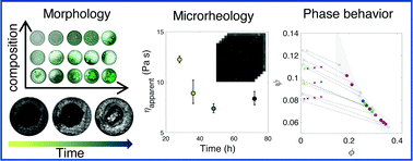
Evolution of composition, rheology, and morphology during phase separation in complex fluids is highly coupled to rheological and mass transport processes within the emerging phases, and understanding this coupling is critical for materials design of multiphase complex fluids. Characterizing these dependencies typically requires careful measurement of a large number of equilibrium and transport properties that are difficult to measure in situ as phase separation proceeds. Here, we propose and demonstrate a high-throughput microscopy platform to achieve simultaneous, in situ mapping of time-evolving morphology and microrheology in phase separating complex fluids over a large compositional space. The method was applied to a canonical example of polyelectrolyte complex coacervation, whereby mixing of oppositely charged species leads to liquid–liquid phase separation into distinct solute-dense and dilute phases. Morphology and rheology were measured simultaneously and kinetically after mixing to track the progression of phase separation. Once equilibrated, the dense phase viscosity was determined to high compositional accuracy using passive probe microrheology, and the results were used to derive empirical relationships between the composition and viscosity. These relationships were inverted to reconstruct the dense phase boundary itself, and further extended to other mixture compositions. The resulting predictions were validated by independent equilibrium compositional measurements. This platform paves the way for rapid screening and formulation of complex fluids and (bio)macromolecular materials, and serves as a critical link between formulation and rheology for multi-phase material discovery.
- This article is part of the themed collection: Soft Matter Editorial Board Highlights of 2022
 Please wait while we load your content...

