
 Open Access Article
 This Open Access Article is licensed under a
 Creative Commons Attribution 3.0 Unported Licence
Correction: Synthesis of TiO2 nanotubes with ZnO nanoparticles to achieve antibacterial properties and stem cell compatibility
Wenwen
Liu
abc,
Penglei
Su
b,
Su
Chen
a,
Na
Wang
a,
Yuanping
Ma
a,
Yiran
Liu
a,
Jinshu
Wang
b,
Zhenting
Zhang
*a,
Hongyi
Li
*b and
Thomas J.
Webster
*cd
aLaboratory of Biomaterials and Biomechanics, Beijing Key Laboratory of Tooth Regeneration and Function Reconstruction, School of Stomatology, Capital Medical University, Tian Tan Xi Li No. 4, Beijing, 100050, China. E-mail: veromcawen@gmail.com; zztttxl@hotmal.com
bPhotoelectrochemical Research Group, Key Laboratory of Advanced Functional Materials, School of Materials Science and Engineering, Beijing University of Technology, Beijing, 100124, China. E-mail: lhy06@bjut.edu.cn
cChemical Engineering Department, Northeastern University, Boston, MA 02115, USA. E-mail: th.webster@neu.edu
dCenter of Excellence for Advanced Materials Research, King Abdulaziz University, Jeddah, Saudi Arabia
First published on 10th February 2022
Abstract
Correction for ‘Synthesis of TiO2 nanotubes with ZnO nanoparticles to achieve antibacterial properties and stem cell compatibility’ by Wenwen Liu et al., Nanoscale, 2014, 6, 9050–9062, DOI: 10.1039/C4NR01531B.
The authors regret that, in the original article, Fig. 5 and 12 contained errors and are therefore replaced in this notice. Fig. 5(b) contained an incorrect XPS depth scale which has been amended. Fig. 12, which displayed fluorescence images showing the viability of the P. gingivalis, was found to have errors in panels (b) and (c) as the same sample was imaged in each by mistake.
 | ||
| Fig. 5 (b) High-resolution XPS spectra of Zn2p at different depths in TNT–Zn0.015. | ||
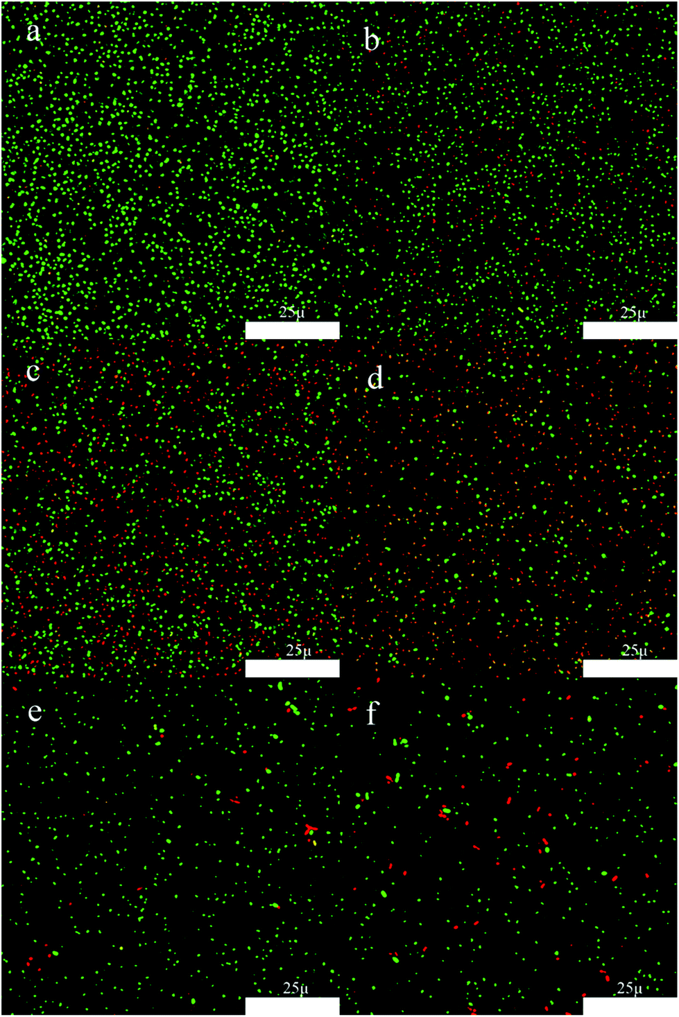 | ||
| Fig. 12 Fluorescence images showing the viability of the P. gingivalis on samples: (a) Ti, (b) TNT, (c) TNT–Zn0.005, (d) TNT–Zn0.015, (e) TNT–Zn0.030, and (f) TNT–Zn0.075. The live bacteria appear green while the dead ones appear orange. | ||
The procedure for imaging the P. gingivalis samples involves exposure to an aerobic environment, which in time compromises the samples. Given the time since original publication and the complicated procedure required to mitigate the effect of such aerobic conditions, the authors have re-imaged all panels of Fig. 12 to ensure that they are correct. The authors confirm that the discussion and conclusions of the original article are unaffected by the reproduction and replacement of Fig. 5(b) and 12.
The Royal Society of Chemistry apologises for these errors and any consequent inconvenience to authors and readers.
| This journal is © The Royal Society of Chemistry 2022 |