Distinct lipid membrane-mediated pathways of Tau assembly revealed by single-molecule analysis†

Abstract
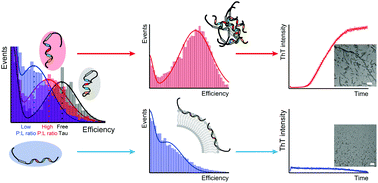
The conversion of intrinsically disordered Tau to highly ordered amyloid aggregates is associated with a wide range of neurodegenerative diseases termed tauopathies. The presence of lipid bilayer membranes is a critical factor that accelerates the abnormal aggregation of Tau protein. However, the lipid membrane-induced conformational changes of Tau and the mechanism for the accelerated fibrillation remain elusive. In this study, single-molecule Förster resonance energy transfer (smFRET) and fluorescence correlation spectroscopy (FCS) were applied to detect the conformational changes and intermolecular interactions of full-length Tau in the presence of different concentrations of 1,2-dimyristoyl-sn-glycero-3-phosphatidylserine (DMPS) vesicles. The results show that the conformation of Tau becomes expanded with opening of the N-terminal and C-terminal domains of Tau upon binding to DMPS. At low DMPS concentrations, Tau forms oligomers with a partially extended conformation which facilitates the amyloid fibrillization process. At high DMPS concentrations, Tau monomer binds to lipid membranes in a fully expanded conformation at low density thus inhibiting intermolecular aggregation. Our study reveals the underlying mechanisms by which lipid membranes influence amyloid formation of Tau, providing a foundation for further understanding of the pathogenesis and physiology of the interplay between Tau protein and lipid membranes.
 Please wait while we load your content...

