
The Art in Science of MicroTAS 2020
Gregory A.
Cooksey†
 *
*
National Institute of Standards and Technology (NIST), 100 Bureau Drive, Gaithersburg, MD 20899, USA. E-mail: gregory.cooksey@nist.gov
With respect to the Art in Science contest, the COVID-19 pandemic certainly limited time in the laboratory when great images could have been created. Yet, this year's submissions were no less inspiring despite the 50% decrease in submissions compared to pre-pandemic years.
Fundamentally, scientific imagery informs us about structure or function, generally within the context of experimental and imaging conditions. But an appealing image goes further, capturing natural phenomena or creating a novel microcosm that both distils information and inspires deeper inspection of a compelling visual story. At the microscale, the world beyond unaided human vision begs for a juxtaposition of both familiarity and novelty. Translation and transformation of these visions can involve use of color, texture, and contextual cues that relate the subject to the viewer and focus the viewer on points of emphasis. Getting the “right” image can go very far to communicate the structure, orientation, or scale of novel things, but also it can draw in the curiosity of a broader community, whose interest may lead to additional support for an important achievement.
The entries for this year's contest appropriately explore themes of familiarity and regularity, drawing us into the material with our curiosity about the small world. The images provided a wonderful means to connect through our work and to communicate more broadly to the general public. We appreciate all who contributed their work for consideration of the Art in Science award.
This year's winning image is “A microvascular ring” by Qinyu Li, from Shanghai Jiao Tong University, China (Fig. 1). The artist notes that the image is a “fluorescence photograph of a 3D vasculogenic network from Human Umbilical Vein Endothelial Cells inside the ring-shaped polymethylmethacrylate microfluidic chamber”. It is a wonderfully balanced composition that captures an impressive accomplishment in our field: recreation of both the form and function of the body's great microfluidic system.
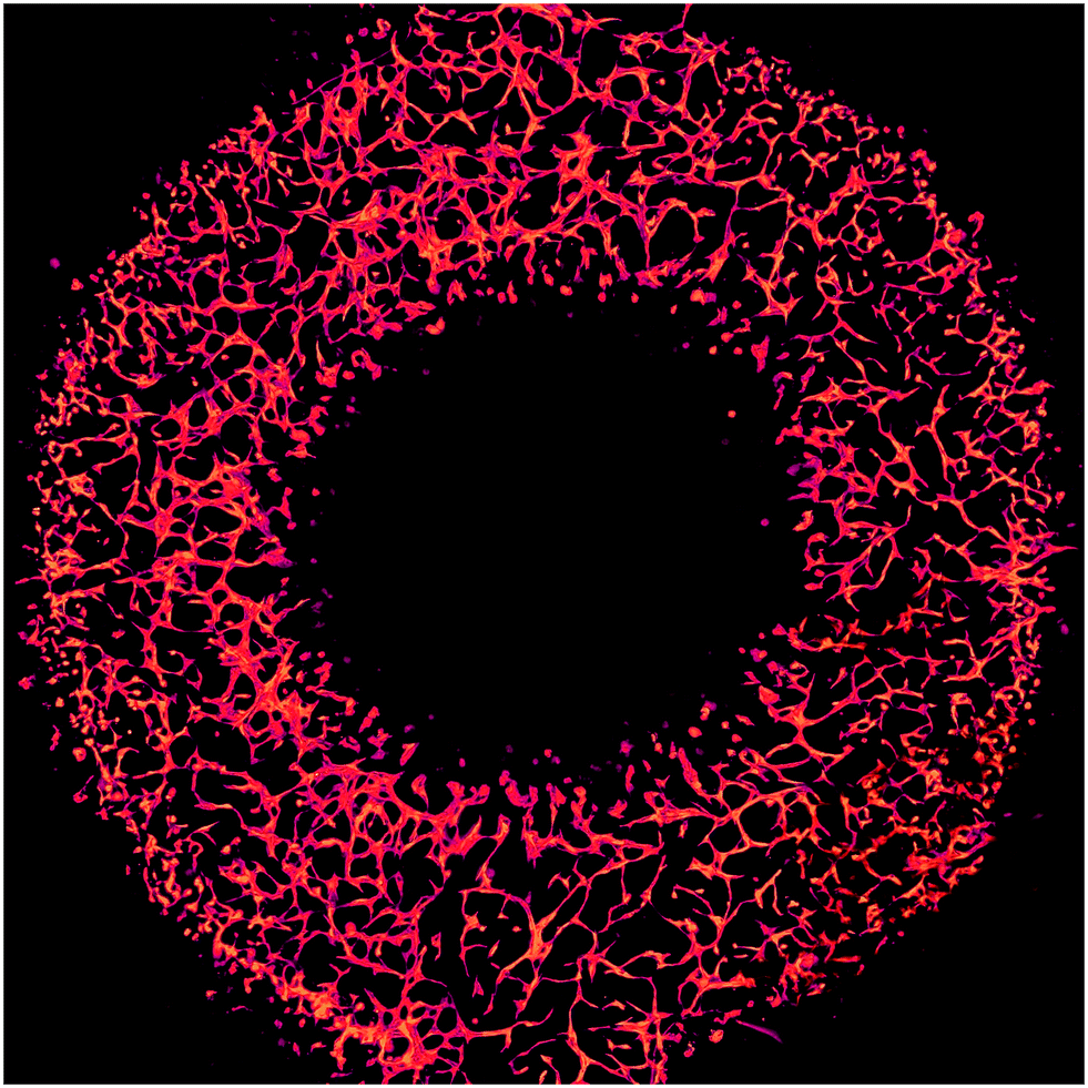 | ||
| Fig. 1 “A microvascular ring” by Qinyu Li, from Shanghai Jiao Tong University in China was selected as the winning image from the 2020 Art in Science competition. The image details the organization of human umbilical vein endothelial cells grown inside a plastic microstructure. | ||
The selection committee for the award consisted of Millie Newman, Lab on a Chip Deputy Editor; Greg Cooksey, project leader in the Microsystems and Nanotechnology Division at NIST; Dino DiCarlo, professor of Bioengineering and Mechanical and Aerospace Engineering at the University of California, Los Angeles; and Jianhua Qin, a chair professor at the Dalian Institute of Chemical Physics.
The award was presented during the awards ceremony at the closing of MicroTAS 2020. The committee also recognizes runner-up honors for Pierre-Alexandre Goyette, Polytechnique Montréal (Fig. 2), Minji Kim, Washington University in St Louis (Fig. 3), and Yage Zhang, University of Hong Kong (Fig. 4).
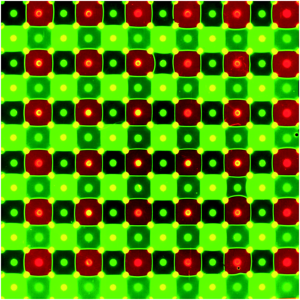 | ||
| Fig. 2 Pierre-Alexandre Goyette from Polytechnique Montréal, Canada, was honored with first runner up for his submission “The Pixelated Chemical Display”. The displays are open-space microfluidic devices that use an array of apertures to move fluids on the surface. | ||
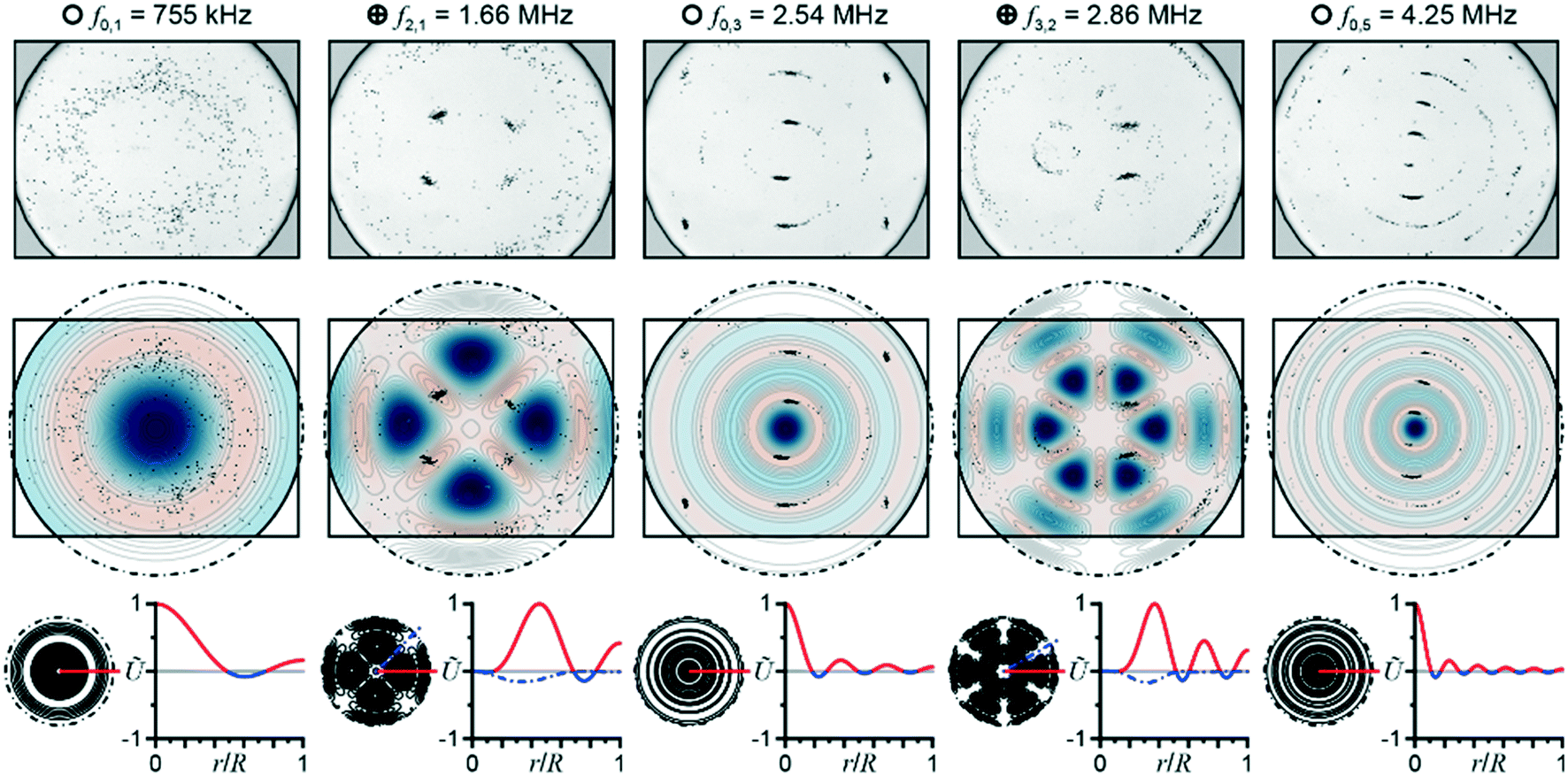 | ||
| Fig. 3 “Swimming in Sound” earned second runner up for Minji Kim from Washington University in St Louis, USA. The image shows cell confinement (top), field patterns (middle) and line plots (bottom) resultant from standing waves formed inside microfluidic device at various drive frequencies. | ||
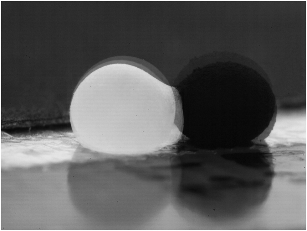 | ||
| Fig. 4 “Black or White – A Fantasy” by Yage Zhang, University of Hong Kong, Hong Kong SAR. The high-speed acquisition shows electrostatically driven coalescence of liquid marbles filled with 6 μm silicone resin particles (white) or 5 μm silicone particles (black). | ||
Acknowledgements
The Art in Science competition is sponsored and supported by MicroTAS, the Chemical and Biological Microsystems Society (CBMS), the Lab on a Chip journal, and NIST. The 2020 award consisted of a monetary prize ($500), an award certificate, and a coveted front cover of the Lab on a Chip journal. Please check the MicroTAS 2021 conference website for further details regarding changes to the competition format and image submission deadline for the next MicroTAS conference.Footnote |
| † Any opinions or views expressed in this article are entirely those of the author and do not represent the views of the National Institute of Standards and Technology, the journal (Lab on a Chip), or the Royal Society of Chemistry. |
| This journal is © The Royal Society of Chemistry 2022 |