
Effects of synbiotic supplementation on regulatory T cells’ response in patients with axial spondyloarthritis: a randomized double-masked placebo-controlled trial†
Alireza
Khabbazi
a,
Masoud
Ahangari Maleki
a,
Mohammad Sadegh
Soltani-Zangbar
b,
Mehdi
Yousefi
b and
Aida
Malek Mahdavi
 *acd
*acd
aConnective Tissue Diseases Research Center, Tabriz University of Medical Sciences, Tabriz, Iran. E-mail: aidamalek@gmail.com
bStem Cell Research Center, Tabriz University of Medical Sciences, Tabriz, Iran
cTuberculosis and Lung Disease Research Center, Tabriz University of Medical Sciences, Tabriz, Iran
dRahat Breathe and Sleep Research Center, Tabriz University of Medical Sciences, Tabriz, Iran
First published on 31st October 2022
Abstract
This study was conducted on samples from patients enrolled in a randomized double-masked placebo-controlled trial on the effect of synbiotic supplementation on the IL-17/IL-23 pathway and disease activity in patients with axial spondyloarthritis (axSpA) to investigate the effects of synbiotic supplementation on regulatory T (Treg) cells’ response in these patients. Forty-eight axSpA patients were randomized to take one synbiotic capsule or placebo daily for 12 weeks. Treg cell proportion, gene expression of forkhead box protein P3 (Foxp3), microRNA (miRNA)-25, miRNA-106b, miRNA-146a, interleukin (IL)-10, and transforming growth factor (TGF)-β as well as serum IL-10 and TGF-β levels were assessed before and after the trial. Thirty-eight patients (19 in each group) completed the trial. The proportion of Treg cells (P < 0.001), the gene expression of FoxP3 (P < 0.001), IL-10 (P = 0.001), TGF-β (P < 0.001), and miRNA-146a (P < 0.001) and serum IL-10 (P = 0.003) and TGF-β (P = 0.002) levels significantly increased compared to the baseline in the synbiotic group. Additionally, a significant reduction in the gene expression of miRNA-25 (P < 0.001) and miRNA-106b (P < 0.001) was observed in the synbiotic group. Significant between-group differences were observed in the proportion of Treg cells (P = 0.024) and the gene expression of FoxP3 (P = 0.010), IL-10 (P = 0.002), TGF-β (P = 0.016), miRNA-25 (P = 0.008), miRNA-106b (P = 0.001), and miRNA-146a (P = 0.010). Differences in the serum levels of IL-10 and TGF-β between the groups were not significant. As a conclusion, synbiotic supplementation could modulate Treg cells’ response in axSpA patients and thus can be promising as an adjunctive therapy. Additional investigations would help in further clarifying the subject.
1. Introduction
Axial spondyloarthritis (axSpA) is an inflammatory ailment that influences the spine and sacroiliac joints and leads to back pain, incapacitation and poor quality of life.1 In addition to musculoskeletal involvement, extra-articular involvement like uveitis, enthesitis and inflammatory bowel disease (IBD) can be noticed in axSpA.1 Commonly, the disease initiates in the third decade of life and affects males more than females.2 The global prevalence of axSpA ranges from 0.1% to 1.4%.3 Although the cause of axSpA is not well understood, genetic susceptibility, environmental factors (e.g., microbial infections) and immune reaction are related to disease pathogenesis.4 It is also suggested that intestinal microbiota and dysbiosis may play a role in axSpA.5 Variation in intestinal permeability and induction of immune responses are mechanisms that have been suggested to explicate the plausible function of the microbiome in axSpA progression.5T lymphocytes are the main cells involved in axSpA pathogenesis. Alteration in the proportion of peripheral blood CD4+ T cells including higher T-helper 17 (Th17) cells and lower regulatory T (Treg) cells has been noticed in axSpA.6–8 Treg cells are a subpopulation of CD4+ T cells that are involved in controlling immune tolerance and sustaining immune homeostasis.9,10 These cells are responsible for expressing forkhead box protein P3 (Foxp3), a transcription factor that suppresses the proliferation and activity of a variety of immune cells.11,12 Moreover, Treg cells generate interleukin (IL)-10 and transforming growth factor (TGF)-β, which are immunosuppressive agents.13 MicroRNAs (miRNAs) are non-coding ribonucleic acids that have a significant role in several biological processes via acting at the post-transcriptional level.14 It has been proposed that the microbiota–miRNA interplay influences host immunity.15 Dysbiosis can alter the expression of miRNAs.15 An altered expression of miRNAs occurs in autoimmune inflammatory conditions including rheumatic diseases.16 It is indicated that miRNA-25 and miRNA-106b have a role in regulating the TGF-β pathway and enhanced expression of these miRNAs disrupts the TGF-β signaling pathway and affects Tregs.17,18 It has also been reported that miRNA-146a down-regulation in monocytes and its level have a negative correlation with the axSpA disease activity score.19 Thus, these miRNAs modulate the biology and function of Treg cells and can be considered as potential therapeutic targets.
Non-steroidal anti-inflammatory drugs (NSAIDs), conventional synthetic disease-modifying anti-rheumatic drugs (csDMARDs) and biological DMARDs (bDMARDs) are common medications used for treating axSpA.20 Because of undesirable effects, limited efficacy and sometimes high expenses of these medications, research on safer and inexpensive alternative treatments appears to be indispensable. Interest in complementary treatments including nutritional supplements has been lately increasing to identify safer alternatives for disease management.
Synbiotics are nutritional supplements containing both probiotics and prebiotics.21 It has been indicated that synbiotic supplementation has a notably more synergistic impact on the intestinal and fecal microflora and the immune system than either prebiotic or probiotic supplementation alone.22,23 Limited studies are available regarding the syn/probiotics supplementation in autoimmune inflammatory diseases including rheumatic disorders, which indicate conflicting results.24–32 To the authors’ knowledge, no study evaluates the effect of synbiotic supplementation on Treg cells in axSpA patients; therefore, the current study was conducted to assess the effect of synbiotic supplementation on Treg cells’ response in patients with axSpA.
2. Methods
2.1. Participants
This study was conducted on samples collected in our previous study about the effect of synbiotic supplementation on the IL-17/IL-23 pathway and disease activity in patients with axSpA.33 The protocol of the current study was approved by the Ethics Committee of Tabriz University of Medical Sciences (ethics code: IR.TBZMED.REC.1399.152) and the Iranian Registry of Clinical Trials (code: IRCT20190917044794N2). Inclusion criteria were determined as: (i) diagnosis of axSpA based on the international criteria34 and (ii) age range between 20–60 years. Exclusion criteria were determined as: taking nutritional supplements or antioxidants one month before and during the trial, use of antibiotics, pregnancy and breast-feeding, having other autoimmune diseases, diabetes mellitus, IBD, gastrointestinal infection and other chronic diseases. Sample size determination was performed considering the information from the previous research.30 Assuming 95% confidence level and 80% power, the sample was minimum 19 subjects per group, which was increased to 24 subjects in each group for a potential 25% dropout. Patients were aware of the research and signed written informed consents.2.2. Design and measurements
Forty-eight axSpA patients were designated from the rheumatology outpatient clinic of Tabriz University of Medical Sciences and divided into intervention and placebo groups randomly via a block randomization method (Random Allocation Software), which matched subjects according to the gender and age per block. The intervention group (n = 24) received one synbiotic capsule (Zist-Takhmir Company, Tehran, Iran) per day for 12 weeks. This capsule contains 109 colony-forming units (CFUs) of 7 beneficial bacteria including L. casei, L. acidophilus, L. rhamnosus, L. bulgaricus, B. longum, B. breve, and S. thermophiles together with fructooligosaccharide and other constituents (lactose-magnesium acetate-talc). The control group (n = 24) received one placebo capsule (Zist-Takhmir Company, Tehran, Iran) per day for 12 weeks. The placebo capsules were identical in shape and color and contained a non-therapeutic substance, cornstarch. Patients took the supplements with lunch and they were asked not to modify diet and physical activity. Patients were monitored every two weeks. The study flowchart was presented in our previous report.338 mL of fasting blood samples were gathered at the baseline and at the end of the trial. In order to separate peripheral blood mononuclear cells (PBMCs), the Ficoll separation method (Biosera, East Sussex, UK) and centrifugation (25 minutes, 450 g) were used. Then, 5 × 10⁶ of cells were cultured in a 5 mL medium (fetal bovine serum, penicillin, and L-glutamine) in the presence of 10 ng mL−1 of phorbol myristate acetate (eBioscience, San Diego, CA) and incubated (37 °C, 5% CO2) for 48 hours. In order to assess Treg cells, the proportion of CD4+CD25+CD127− T cells was determined by flow cytometry. Overall, incubation of 1 × 10⁶ of PBMCs was performed at 4 °C for 15 minutes using anti-human CD4, anti-human CD25 and anti-human CD127 (eBioscience). Stained cells were assessed using a FACSCalibur flow cytometer (BD Biosciences) and Flowing Software 2.4.1. Then, gating of CD4-positive lymphocytes was performed for analyzing the expression of CD25 and CD127. In order to analyze the mRNA expression of FoxP3, TGF-β, and IL-10, the real-time polymerase chain reaction (PCR) was performed. Homogenization was performed on cultured PBMCs and extraction of total RNA was conducted using RNX-PLUS Solution (SinaClon, Tehran, Iran). The complementary DNA (cDNA) was produced using the RevertAid Reverse Transcriptase kit (Thermo Fisher, Waltham, MA). miRNA-25, miRNA-106b and miRNA-146a were extracted by TaqMan RT-PCR. The reverse transcription of RNA was conducted using the TaqMan MicroRNA Reverse Transcription Kit and Master Mix. Quantification of transcripts of β-actin and RNA, U6 small nuclear (RNU6) was performed as the endogenous RNA control. β-Actin was assumed as a housekeeping control gene for FoxP3, TGF-β, and IL-10 genes and RNU6 was assumed as an endogenous control for miRNAs. In order to analyze the results, the 2−ΔΔCt procedure was used. Furthermore, enzyme-linked immunosorbent assay (ELISA) kits (Biosource, Nivelles, Belgium) and a Medgenix ELISA reader (BP-800; Biohit, Helsinki, Finland) were used for measuring IL-10 and TGF-β in the supernatant of cultured PBMCs.
2.3. Statistical analysis
For analyzing data, SPSS 16.0 software (SPSS Inc., Chicago, IL) was used. The Kolmogorov–Smirnov test was performed to evaluate the normality of variable distribution. Categorical and continuous variables were displayed as frequency (percentage) and mean ± standard deviation (SD), respectively. Differences in categorical variables between two groups were compared by the Chi-square test. Intragroup differences were assessed by a paired t-test. At the baseline, intergroup comparisons were made with an independent sample t-test. After research, analysis of covariance (ANCOVA) was performed for intergroup comparisons and considering adjustment for baseline measures and axSpA duration. P < 0.05 was assumed to be statistically significant.3. Results
A total of 48 patients were randomized into the synbiotic and placebo groups. Five patients were excluded from each study group. Therefore, thirty-eight patients completed the study. Baseline characteristics of the studied patients were presented in our previous report.33As illustrated in Fig. 1, the baseline proportion of Treg cells did not differ between the two groups (P > 0.05). The proportion of Treg cells increased considerably in the synbiotic group compared with the baseline (4.68 ± 2.28 vs. 2.57 ± 1.44, P < 0.001), whilst no considerable changes occurred in the placebo group (3.06 ± 1.80 vs. 2.62 ± 1.40, P > 0.05). After the study, significant between-group differences in the proportion of Treg cells, adjusted baseline measures and the disease duration were observed (P = 0.024).
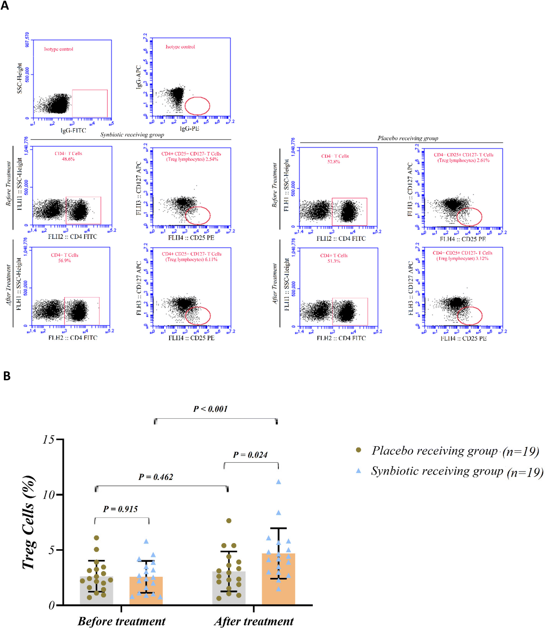 | ||
| Fig. 1 (A) Flow cytometry plots for identifying regulatory T cells in whole blood. (B) Percentage of circulating regulatory T cells in study groups. Values are mean ± SD. Intragroup differences were assessed by a paired t-test. Intergroup differences were assessed by an independent sample t-test at the baseline or the ANCOVA test, adjusted baseline values and the disease duration after 12 weeks. P < 0.05 is considered significant. | ||
As depicted in Fig. 2, the baseline mRNA expression of FoxP3, IL-10 and TGF-β did not vary notably between the two groups (P > 0.05). The mRNA expression of FoxP3, IL-10, and TGF-β increased remarkably in the synbiotic group compared with the baseline (P < 0.001, P = 0.001, and P < 0.001, respectively); whereas in the placebo group, no changes occurred in the mRNA expression of FoxP3, IL-10, and TGF-β (P > 0.05). After the research, the results of the ANCOVA test indicated significant between-group differences in the mRNA expression of FoxP3, IL-10 and TGF-β, adjusted baseline values and the disease duration (P = 0.010, P = 0.002, and P = 0.016, respectively).
 | ||
| Fig. 2 The mRNA expression level of regulatory T cells related to transcription factor FoxP3 and cytokines IL-10 and TGF-β in study groups. Values are mean ± SD. Intragroup differences were assessed by a paired t-test. Intergroup differences were assessed by an independent sample t-test at the baseline or the ANCOVA test, adjusted baseline values and the disease duration after 12 weeks. P < 0.05 is considered significant. | ||
According to Fig. 3, there were no significant differences in the mRNA expression of miRNA-25, miRNA-106b and miRNA-146a between the two groups at the baseline (P > 0.05). Compared to the baseline, significant reduction in the mRNA expression of miRNA-106b and a significant increase in the mRNA expression of miRNA-146a were observed in both synbiotic (P < 0.001 and P < 0.001) and placebo (P = 0.027 and P = 0.007) groups. Furthermore, a significant reduction in the mRNA expression of miRNA-25 was observed in the synbiotic group compared to the baseline (P < 0.001); however, no remarkable change was noted (P > 0.05) in the placebo group. After the research, the results of the ANCOVA test showed significant between-group differences in the mRNA expression of miRNA-25, miRNA-106b and miRNA-146a adjusted baseline measures and the disease duration (P = 0.008, P = 0.001, and P = 0.010, respectively).
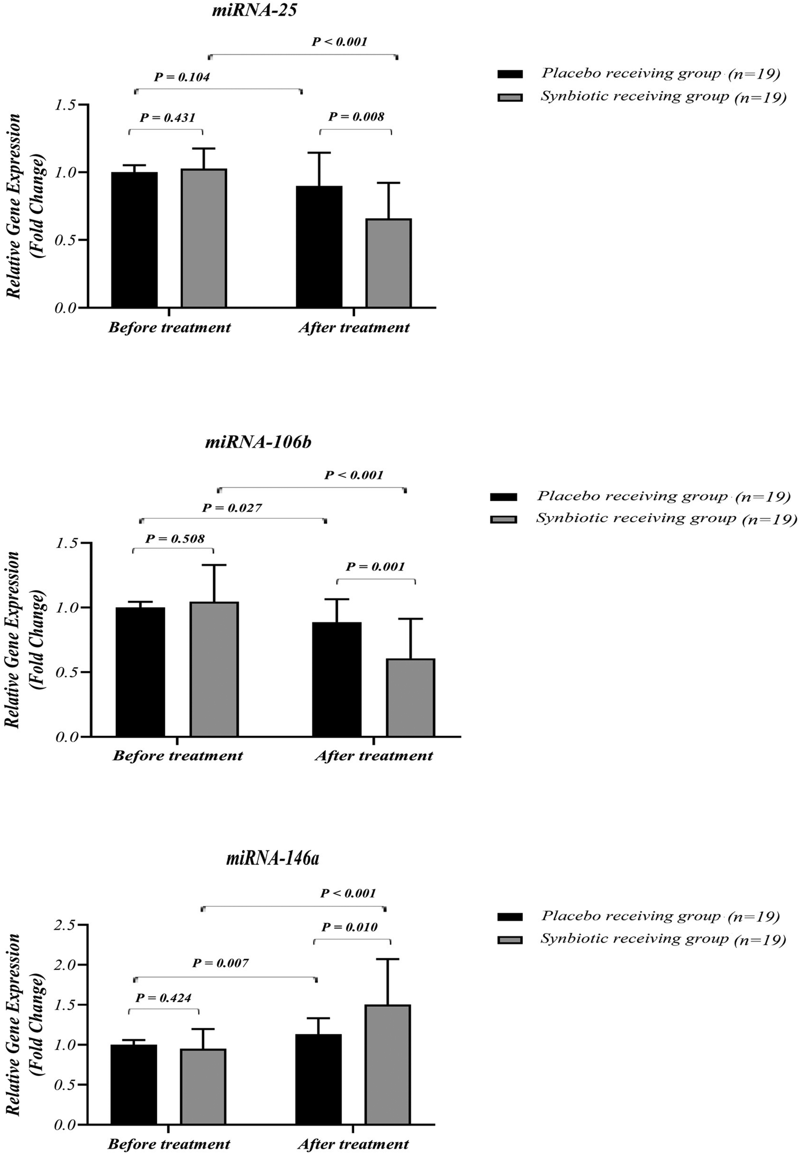 | ||
| Fig. 3 The mRNA expression level of miRNA-25, miRNA-106b, and miRNA-146a in study groups. Values are mean ± SD. Intragroup differences were assessed by a paired t-test. Intergroup differences were assessed by an independent sample t-test at the baseline or the ANCOVA test, adjusted baseline values and the disease duration after 12 weeks. P < 0.05 is considered significant. | ||
In addition, we assessed the serum levels of IL-10 and TGF-β and disease activity in the studied groups (Table 1). No significant differences existed in the baseline serum IL-10 and TGF-β between groups (P > 0.05). Compared with the baseline, serum IL-10 and TGF-β levels enhanced notably in the synbiotic group (P = 0.003 and P = 0.002, respectively), whilst in the placebo group, no significant changes occurred in serum IL-10 and TGF-β levels (P > 0.05). After the research, no significant differences were observed between the two groups in serum IL-10 and TGF-β, adjusted baseline measures and the disease duration (P > 0.05).
| Variable | Synbiotic group (n = 19) | Placebo group (n = 19) | P-value* | |
|---|---|---|---|---|
| IL, interleukin; TGF, transforming growth factor. Variables are reported as mean ± SD. P < 0.05 is considered significant. *P values indicate a comparison between groups (an independent sample t-test at the baseline or the ANCOVA test, adjusted baseline values and disease duration after 12 weeks). **A paired t-test. | ||||
| IL-10 (pg mL−1) | Baseline | 693.20 ± 291.09 | 819.61 ± 346.33 | 0.223 |
| After 12 weeks | 1139.30 ± 401.28 | 922.22 ± 349.19 | 0.074 | |
| P-value** | 0.003 | 0.369 | ||
| TGF-β (pg mL−1) | Baseline | 346.11 ± 158.99 | 337.28 ± 142.56 | 0.862 |
| After 12 weeks | 513.28 ± 194.62 | 402.50 ± 166.34 | 0.105 | |
| P-value** | 0.002 | 0.174 | ||
4. Discussion
Considering the importance of Tregs in modulating immune responses as well as the abnormalities occurred in the proportion and activity of these cells in autoimmune inflammatory diseases,35 it is supposed that Treg cell-inducing therapies are possibly an innovative modality to treat axSpA. Synbiotics are nutritional supplements with immunomodulatory properties36 that have been shown to be useful in the management of autoimmune inflammatory disorders.37 The present randomized double-masked placebo-controlled trial was the first investigation assessing the effects of synbiotic supplementation on Tregs in patients with axSpA. Our research demonstrated that synbiotic supplementation led to a significant elevation in Treg cells’ proportion in comparison with the baseline and placebo. In agreement with our results, Lorea Baroja et al.38 indicated that consumption of probiotic yogurt increases the CD4+ CD25+ Tregs proportion in IBD patients. López et al.39 and Chen et al.40 reported that treatment with probiotics could stimulate Treg CD4+ CD25+ FoxP3+ differentiation. However, differences were not significant. Inconsistent with our research, van der Aa et al.41 did not show a significant difference in Treg cell percentage between synbiotic and placebo receiving infants with atopic dermatitis. In addition, Shukla et al.42 declared no significant variation in Treg cell percentage between probiotic and placebo receiving children with active enthesitis-related arthritis. Although the mechanism of increasing Treg cells by syn/probiotics is not well understood, several mechanisms have been proposed, including (i) activation of tolerogenic dendritic cells (DCs) via binding to the lectin dendritic cell (DC)-specific intercellular adhesion molecule 3-grabbing nonintegrin (DC-SIGN), which in turn promotes Foxp3+ Treg cells’ production,43,44 (ii) stimulation of Toll like receptors (TLR) like TLR9 which led to release of immunosuppressive cytokines such as TGF-β and IL-10,43 and (iii) direct activation of Tregs by producing short chain fatty acids (SVFAs).43 Short-chain fatty acids (SCFAs) bind to G protein-coupled receptors (GPCRs) on Tregs and activate them to suppress CD8 and Th17 cells,43 (iv) modulating the host miRNA expression15 and (v) interfering with nuclear factor-κB degradation.44This study demonstrated a significant increase in the expression of FoxP3, IL-10 and TGF-β with synbiotic supplementation. Inconsistent with our research, Dehnavi et al.45 did not report a significant change in the gene expression of FoxP3, IL-10, and TGF-β after synbiotic supplementation in patients with allergic rhinitis. In addition, studies in patients with IBD46 and recurrent aphthous stomatitis47 indicated no significant differences in the serum IL-10 level between synbiotic and placebo groups. Pineda et al.27 did not report a significant change in serum IL-10 with probiotic consumption in RA patients. Similarly, Inoue et al.,48 Wang et al.49 and Smecuol et al.50 demonstrated that oral administration of probiotics did not lead to a significant change in serum IL-10 concentrations compared with placebo in subjects with atopic dermatitis, peritoneal dialysis and celiac disease, respectively. Additionally, our study was different from Shukla et al.42 and Vaghef-Mehrabany et al.51 studies that reported a substantial difference in serum IL-10 levels between probiotic and placebo receiving patients with active enthesitis-related arthritis and rheumatoid arthritis (RA). Furthermore, Inoue et al.48 reported that oral administration of probiotics significantly increases the serum TGF-β level compared with placebo in subjects with atopic dermatitis. Discrepancy between the findings of this study with previous studies on other diseases may be related to variations in pathophysiological conditions (e.g., various ailments), the baseline Treg cell proportion, the baseline FoxP3, IL-10 and TGF β status, the dosage and type of syn/probiotic supplements and the duration of supplementation.
This study demonstrated a significant increase in the expression of miRNA-146a genes with synbiotic supplementation. Moreover, synbiotic supplementation led to a significant decrease in the expression of miRNA-25 and miRNA-106b genes in comparison with the baseline and placebo. MiR-146a is usually expressed in Treg cells and has a critical function in Treg-mediated inhibition. The lack of miR-146a in Treg cells causes an enhanced expression of STAT1, a target gene of miR-146a that stimulates the proinflammatory phenotype of Tregs. Up-regulation of miR-146a can inhibit Th1 responses derived from Tregs function, thereby maintaining the optimal range of STAT1 activity and decreasing severe failure of immunologic tolerance.52,53 Furthermore, miRNA-25 and miRNA-106b have a role in regulating the TGF-β pathway and enhanced expression of these miRNAs disrupts the TGF-β signaling pathway and affects Tregs.17,18 Thus, miR-25 and miR-106b dysregulation can alter Treg cells’ action principally via changing the TGF-β actions.17 Down-regulation of miR-25 and miR-106b can induce the TGF-β pathway as well as Treg cell differentiation and maturation.
The weak points of the study include a relatively small number of patients. The strengths of the present study were regular monitoring of patients by a phone call and a relatively favorable acceptance of interventions.
5. Conclusion
As a conclusion, synbiotic supplementation could modulate Treg cells’ response in axSpA patients and thus can be promising as an adjunctive therapy. Additional investigations would help in further clarifying the subject.Conflicts of interest
The authors have no conflicts to declare.Acknowledgements
We would like to appreciate the cooperation of the Clinical Research Development Unit of Imam Reza General Hospital, Tabriz, Iran in conducting this research. We also thank all the patients for participating in this study. This work was financially supported by a grant from Research Vice-Chancellor of Tabriz University of Medical Sciences, Tabriz, Iran (Grant No. 65554).References
- J. Walsh, T. Hunter, K. Schroeder, D. Sandoval and R. Bolce, Trends in diagnostic prevalence and treatment patterns of male and female ankylosing spondylitis patients in the United States, 2006–2016, BMC Rheumatol., 2019, 3, 39 CrossRef PubMed.
- J. Sieper and D. Poddubnyy, Axial spondyloarthritis, Lancet, 2017, 390, 73–84 CrossRef.
- J. Braun and J. Sieper, Ankylosing spondylitis, Lancet, 2007, 369, 1379–1390 CrossRef.
- W. Zhu, X. He, K. Cheng, L. Zhang, D. Chen, X. Wang, G. Qiu, X. Cao and X. Weng, Ankylosing spondylitis: etiology, pathogenesis, and treatments, Bone Res., 2019, 7, 22 CrossRef PubMed.
- L. Yang, L. Wang, X. Wang, C. J. Xian and H. Lu, A Possible Role of Intestinal Microbiota in the Pathogenesis of Ankylosing Spondylitis, Int. J. Mol. Sci., 2016, 17, 2126 CrossRef PubMed.
- B. Szalay, G. Mészáros, Á. Cseh, L. Ács, M. Deák, L. Kovács, B. Vásárhelyi and A. Balog, Adaptive immunity in ankylosing spondylitis: phenotype and functional alterations of T-cells before and during infliximab therapy, Clin. Dev. Immunol., 2012, 808724 Search PubMed.
- H. Guo, M. Zheng, K. Zhang, F. Yang, X. Zhang, Q. Han, Z. N. Chen and P. Zhu, Functional defects in CD4+ CD25high FoxP3+ regulatory cells in ankylosing spondylitis, Sci. Rep., 2016, 6, 37559 CrossRef CAS PubMed.
- L. Xueyi, C. Lina, W. Zhenbiao, H. Qing, L. Qiang and P. Zhu, Levels of circulating Th17 cells and regulatory T cells in ankylosing spondylitis patients with an inadequate response to anti-TNF-α therapy, J. Clin. Immunol., 2013, 33, 151–161 CrossRef PubMed.
- E. Aktas Cetin, F. Cosan, A. Cefle and G. Deniz, IL-22-secreting Th22 and IFN-γ-secreting Th17 cells in Behçet's disease, Mod. Rheumatol., 2014, 24, 802–807 CrossRef CAS.
- D. V. Sawant and D. A. Vignali, Once a Treg, always a Treg?, Immunol. Rev., 2014, 259, 173–191 CrossRef CAS.
- J. Guo and X. Zhou, Regulatory T cells turn pathogenic, Cell. Mol. Immunol., 2015, 12, 525–532 CrossRef CAS.
- D. Rostamzadeh, M. Yousefi, M. R. Haghshenas, M. Ahmadi, S. Dolati and Z. Babaloo, mTOR Signaling pathway as a master regulator of memory CD8+ T-cells, Th17, and NK cells development and their functional properties, J. Cell. Physiol., 2019, 234, 12353–12368 CrossRef CAS.
- Z. Li, D. Li, A. Tsun and B. Li, FOXP3+ regulatory T cells and their functional regulation, Cell. Mol. Immunol., 2015, 12, 558–565 CrossRef PubMed.
- A. Mehta and D. Baltimore, MicroRNAs as regulatory elements in immune system logic, Nat. Rev. Immunol., 2016, 16, 279–294 CrossRef.
- M. Li, W. D. Chen and Y. D. Wang, The roles of the gut microbiota–miRNA interaction in the host pathophysiology, Mol. Med., 2020, 26, 101 Search PubMed.
- Z. Qu, W. Li and B. Fu, MicroRNAs in autoimmune diseases, BioMed. Res. Int., 2014, 2014, 527895 Search PubMed.
- S. Yamagiwa, J. D. Gray, S. Hashimoto and D. A. Horwitz, A role for TGF-beta in the generation and expansion of CD4+ CD25+ regulatory T cells from human peripheral blood, J. Immunol., 2001, 166, 7282–7289 CrossRef CAS.
- G. De Santis, M. Ferracin, A. Biondani, L. Caniatti, M. Rosaria Tola, M. Castellazzi, B. Zagatti, L. Battistini, G. Borsellino, E. Fainardi, R. Gavioli, M. Negrini, R. Furlan and E. Granieri, Altered miRNA expression in T regulatory cells in course of multiple sclerosis, J. Neuroimmunol., 2010, 226, 165–171 CrossRef CAS.
- O. Fogel, A. Bugge Tinggaard, M. Fagny, N. Sigrist, E. Roche, L. Leclere, J. F. Deleuze, F. Batteux, M. Dougados, C. Miceli-Richard and J. Tost, Deregulation of microRNA expression in monocytes and CD4+ T lymphocytes from patients with axial spondyloarthritis, Arthritis Res. Ther., 2019, 21, 51 CrossRef.
- R. S. Y. Wong, Disease-Modifying Effects of long-term and continuous use of nonsteroidal anti-inflammatory drugs (NSAIDs) in Spondyloarthritis, Adv. Pharmacol. Pharm. Sci., 2019, 2019, 1–6 Search PubMed.
- M. de Vrese and J. Schrezenmeir, Probiotics, prebiotics, and synbiotics, Adv. Biochem. Eng./Biotechnol., 2008, 111, 1–66 CrossRef.
- J. Frece, B. Kos, I. K. Svetec, Z. Zgaga, J. Beganović, A. Lebos and J. Susković, Synbiotic effect of Lactobacillus helveticus M92 and prebiotics on the intestinal microflora and immune system of mice, J. Dairy Res., 2009, 76, 98–104 CrossRef CAS.
- D. L. Worthley, R. K. Le Leu, V. L. Whitehall, M. Conlon, C. Christophersen, D. Belobrajdic, K. A. Mallitt, Y. Hu, N. Irahara, S. Ogino, B. A. Leggett and G. P. Young, A human, double-blind, placebo-controlled, crossover trial of prebiotic, probiotic, and synbiotic supplementation: effects on luminal, inflammatory, epigenetic, and epithelial biomarkers of colorectal cancer, Am. J. Clin. Nutr., 2009, 90, 578–586 CrossRef CAS.
- B. Alipour, A. Homayouni-Rad, E. Vaghef-Mehrabany, S. K. Sharif, L. Vaghef-Mehrabany, M. Asghari-Jafarabadi, M. R. Nakhjavani and J. Mohtadi-Nia, Effects of Lactobacillus casei supplementation on disease activity and inflammatory cytokines in rheumatoid arthritis patients: a randomized double-blind clinical trial, Int. J. Rheum. Dis., 2014, 17, 519–527 CAS.
- B. Zamani, H. R. Golkar, S. Farshbaf, M. Emadi-Baygi, M. Tajabadi-Ebrahimi, P. Jafari, R. Akhavan, M. Taghizadeh, M. R. Memarzadeh and Z. Asemi, Clinical and metabolic response to probiotic supplementation in patients with rheumatoid arthritis: a randomized, double-blind, placebo-controlled trial, Int. J. Rheum. Dis., 2016, 19, 869–879 CrossRef CAS PubMed.
- B. Zamani, S. Farshbaf, H. R. Golkar, F. Bahmani and Z. Asemi, Synbiotic supplementation and the effects on clinical and metabolic responses in patients with rheumatoid arthritis: a randomised, double-blind, placebo-controlled trial, Br. J. Nutr., 2017, 117, 1095–1102 CrossRef PubMed.
- L. M. Pineda, S. F. Thompson, K. Summers, F. de Leon, J. Pope and G. Reid, A randomized, double-blinded, placebo-controlled pilot study of probiotics in active rheumatoid arthritis, Med. Sci. Monit., 2011, 17, CR347–CR354 Search PubMed.
- K. Hatakka, J. Martio, M. Korpela, M. Herranen, T. Poussa, T. Laasanen, M. Saxelin, H. Vapaatalo, E. Moilanen and R. Korpela, Effects of probiotic therapy on the activity and activation of mild rheumatoid arthritis – a pilot study, Scand. J. Rheumatol., 2003, 32, 211–215 CrossRef PubMed.
- F. Esmaeili, M. Salesi, G. Askari, A. Esmaeilisharif, M. Maracy, H. Karimzadeh and B. Shojaie, Efficacy of synbiotic supplementation in improving rheumatoid arthritis, Res. Pharm. Sci., 2020, 15, 263–272 CrossRef.
- K. Jenks, S. Stebbings, J. Burton, M. Schultz, P. Herbison and J. Highton, Probiotic therapy for the treatment of spondyloarthritis: a randomized controlled trial, J. Rheumatol., 2010, 37, 2118–2125 CrossRef PubMed.
- S. Brophy, C. L. Burrows, C. Brooks, M. B. Gravenor, S. Siebert and S. J. Allen, Internet-based randomised controlled trials for the evaluation of complementary and alternative medicines: probiotics in spondyloarthropathy, BMC Musculoskeletal Disord., 2008, 9, 4 CrossRef PubMed.
- G. Askari and A. R. Moravejolahkami, Synbiotic supplementation may relieve anterior uveitis, an ocular manifestation in Behcet's syndrome, Am. J. Case Rep., 2019, 20, 548–550 CrossRef PubMed.
- M. Ahangari Maleki, A. Malek Mahdavi, M. S. Soltani-Zangbar, M. Yousefi and A. Khabbazi, Randomized double-blinded controlled trial on the effect of synbiotic supplementation on IL-17/IL-23 pathway and disease activity in patients with axial spondyloarthritis, Immunopharmacol. Immunotoxicol., 2022, 1–9 CrossRef.
- B. Amor, M. Dougados and M. Mijiyawa, Criteria of the classification of spondylarthropathies, Rev. Rhum. Mal. Osteoartic., 1990, 57, 85–89 CAS.
- S. Sakaguchi, M. Ono, R. Setoguchi, H. Yagi, S. Hori, Z. Fehervari, J. Shimizu, T. Takahashi and T. Nomura, Foxp3+ CD25+ CD4+ natural regulatory T cells in dominant self-tolerance and autoimmune disease, Immunol. Rev., 2006, 212, 8–27 CrossRef CAS PubMed.
- P. Markowiak and K. Śliżewska, Effects of Probiotics, Prebiotics, and Synbiotics on Human Health, Nutrients, 2017, 9, 1021 CrossRef.
- G. Askari, A. Ghavami, F. Shahdadian and A. R. Moravejolahkami, Effect of synbiotics and probiotics supplementation on autoimmune diseases: A systematic review and meta-analysis of clinical trials, Clin. Nutr., 2021, 40, 3221–3234 CrossRef CAS PubMed.
- M. Lorea Baroja, P. V. Kirjavainen, S. Hekmat and G. Reid, Anti-inflammatory effects of probiotic yogurt in inflammatory bowel disease patients, Clin. Exp. Immunol., 2007, 149, 470–479 CrossRef.
- P. López, I. González-Rodríguez, B. Sánchez, M. Gueimonde, A. Margolles and A. Suárez, Treg-inducing membrane vesicles from Bifidobacterium bifidum LMG13195 as potential adjuvants in immunotherapy, Vaccine, 2012, 30, 825–829 CrossRef.
- Y. S. Chen, R. L. Jan, Y. L. Lin, H. H. Chen and J. Y. Wang, Randomized placebo-controlled trial of lactobacillus on asthmatic children with allergic rhinitis, Pediatr. Pulmonol., 2010, 45, 1111–1120 CrossRef.
- L. B. van der Aa, R. Lutter, H. S. Heymans, B. S. Smids, T. Dekker, W. M. van Aalderen, J. H. Sillevis Smitt, L. M. Knippels, J. Garssen, A. J. Nauta, A. B. Sprikkelman and Synbad Study Group, No detectable beneficial systemic immunomodulatory effects of a specific synbiotic mixture in infants with atopic dermatitis, Clin. Exp. Allergy, 2012, 42, 531–539 CrossRef.
- A. Shukla, P. Gaur and A. Aggarwal, Effect of probiotics on clinical and immune parameters in enthesitis-related arthritis category of juvenile idiopathic arthritis, Clin. Exp. Immunol., 2016, 185, 301–308 CrossRef CAS PubMed.
- M. Dwivedi, P. Kumar, N. C. Laddha and E. H. Kemp, Induction of regulatory T cells: A role for probiotics and prebiotics to suppress autoimmunity, Autoimmun. Rev., 2016, 15, 379–392 CrossRef CAS.
- S. Issazadeh-Navikas, R. Teimer and R. Bockermann, Influence of dietary components on regulatory T cells, Mol. Med., 2012, 18, 95–110 CAS.
- S. Dehnavi, F. J. Azad, R. F. Hoseini, N. Moazzen, J. Tavakkol-Afshari, A. R. Nikpoor, A. A. Salmani, H. Ahanchian and M. Mohammadi, A significant decrease in the gene expression of interleukin-17 following the administration of synbiotic in patients with allergic rhinitis who underwent immunotherapy: A placebo-controlled clinical trial, J. Res. Med. Sci., 2019, 24, 51 CrossRef CAS.
- A. Federico, C. Tuccillo, E. Grossi, R. Abbiati, N. Garbagna, M. Romano, A. Tiso, V. Blanco Cdel and C. Loguercio, The effect of a new symbiotic formulation on plasma levels and peripheral blood mononuclear cell expression of some pro-inflammatory cytokines in patients with ulcerative colitis: a pilot study, Eur. Rev. Med. Pharmacol. Sci., 2009, 13, 285–293 CAS.
- M. A. M. Mimura, R. C. Borra, C. H. W. Hirata and N. de Oliveira Penido, Immune response of patients with recurrent aphthous stomatitis challenged with a synbiotic, J. Oral Pathol. Med., 2017, 46, 821–828 CrossRef CAS PubMed.
- Y. Inoue, T. Kambara, N. Murata, J. Komori-Yamaguchi, S. Matsukura, Y. Takahashi, Z. Ikezawa and M. Aihara, Effects of oral administration of Lactobacillus acidophilus L-92 on the symptoms and serum cytokines of atopic dermatitis in Japanese adults: a double-blind, randomized, clinical trial, Int. Arch. Allergy Immunol., 2014, 165, 247–254 CrossRef CAS.
- I. K. Wang, Y. Y. Wu, Y. F. Yang, I. W. Ting, C. C. Lin, T. H. Yen, J. H. Chen, C. H. Wang, C. C. Huang and H. C. Lin, The effect of probiotics on serum levels of cytokine and endotoxin in peritoneal dialysis patients: a randomized, double-blind, placebo-controlled trial, Benefic. Microbes, 2015, 6, 423–430 CrossRef.
- E. Smecuol, H. J. Hwang, E. Sugai, L. Corso, A. C. Cherñavsky, F. P. Bellavite, A. González, F. Vodánovich, M. L. Moreno, H. Vázquez, G. Lozano, S. Niveloni, R. Mazure, J. Meddings, E. Mauriño and J. C. Bai, Exploratory, randomized, double-blind, placebo-controlled study on the effects of Bifidobacterium infantis natren life start strain super strain in active celiac disease, J. Clin. Gastroenterol., 2013, 47, 139–147 CrossRef PubMed.
- E. Vaghef-Mehrabany, B. Alipour, A. Homayouni-Rad, S. K. Sharif, M. Asghari-Jafarabadi and S. Zavvari, Probiotic supplementation improves inflammatory status in patients with rheumatoid arthritis, Nutrition, 2014, 30, 430–435 CrossRef.
- L. F. Lu, M. P. Boldin, A. Chaudhry, L. L. Lin, K. D. Taganov, T. Hanada, A. Yoshimura, D. Baltimore and A. Y. Rudensky, Function of miR-146a in controlling Treg cell-mediated regulation of Th1 responses, Cell, 2010, 142, 914–929 CrossRef.
- Q. Zhou, S. Haupt, J. T. Kreuzer, A. Hammitzsch, F. Proft, C. Neumann, J. Leipe, M. Witt, H. Schulze-Koops and A. Skapenko, Decreased expression of miR-146a and miR-155 contributes to an abnormal Treg phenotype in patients with rheumatoid arthritis, Ann. Rheum. Dis., 2015, 74, 1265–1274 CrossRef.
Footnote |
| † Electronic supplementary information (ESI) available. See DOI: https://doi.org/10.1039/d2fo01377k |
| This journal is © The Royal Society of Chemistry 2022 |