
 Open Access Article
 This Open Access Article is licensed under a
 Creative Commons Attribution 3.0 Unported Licence
Correction: Glycyrrhizic acid promotes neural repair by directly driving functional remyelination
Jing
Tian
a,
Xing
Li
a,
Li
Zhao
a,
Peixin
Shen
a,
Zhezhi
Wang
a,
Lin
Zhu
b,
Cuiqin
Li
a,
Chun
Su
a and
Yuan
Zhang
*a
aNational Engineering Laboratory for Resource Development of Endangered Crude Drugs in Northwest China, The Key Laboratory of Medicinal Resources and Natural Pharmaceutical Chemistry, The Ministry of Education, College of Life Sciences, Shaanxi Normal University, Xi'an, Shaanxi 710119, China. E-mail: yuanzhang_bio@126.com
bDepartment of Pharmacy, The First Affiliated Hospital of Zhengzhou University, Zhengzhou, Henan 450052, China
First published on 24th May 2022
Abstract
Correction for ‘Glycyrrhizic acid promotes neural repair by directly driving functional remyelination’ by Jing Tian et al., Food Funct., 2020, 11, 992–1005, https://doi.org/10.1039/C9FO01459D.
The authors regret that Fig. 2 and Fig. 3 are incorrect in the original article. In Fig. 2E the images for both Naïve and EAE were incorrect. In Fig. 3D the images for Cup-4wk + GA were incorrect. This does not change the scientific conclusions of the article in any way and the correct versions of Fig. 2 and Fig. 3 are presented below.
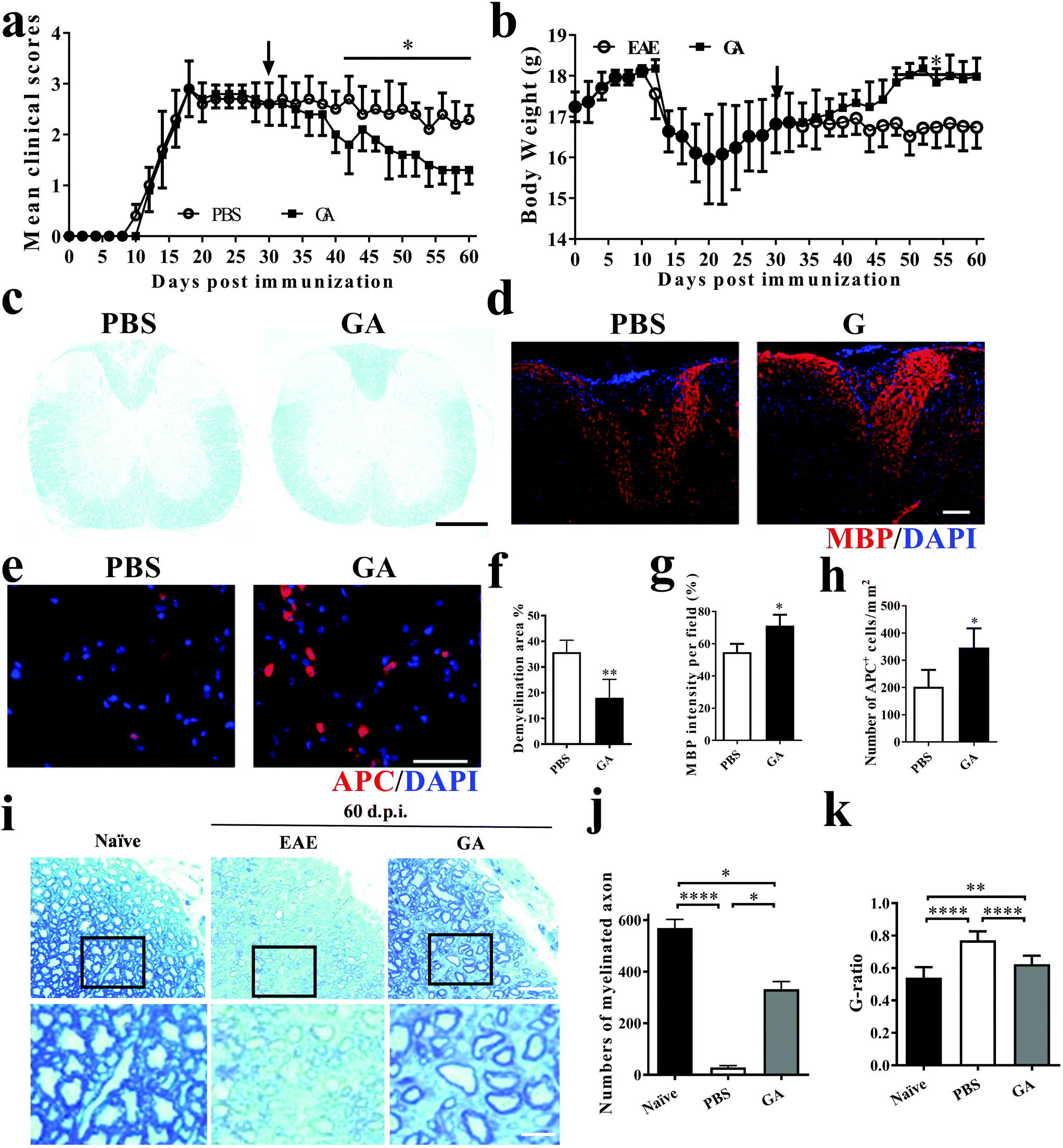 | ||
| Fig. 1 GA improved the clinical signs in chronic EAE and promoted remyelination of myelin at the chronic stage of EAE. Clinical scores (a) and body weight (b) of EAE mice that were administered daily with GA (50 mg kg−1 day−1) or vehicle control from day 30 p.i. (n = 5, therapeutic treatment). (c) Mice were sacrificed on day 60 p.i. (n = 5 each group), and spinal cords were harvested from control EAE mice, and GA-treated mice were stained with LFB. (d) MBP staining of spinal cord sections for the detection of MBP expression. (e) The effect of GA on the number of mature oligodendrocytes in the spinal cord compared with PBS-treated mice. APC and DAPI co-immunolabeling was evaluated to determine the number of mature oligodendrocytes. (f) Demyelinated area was measured by manually outlining the total white matter area with Image-Pro Plus software. The demyelinated area is expressed as a percentage of the total white matter area. (g) MBP intensity was measured and quantified in the lesion areas in the white matter of lumbar spinal cord. (h) Quantification of total APC + cells. (i) Histological sections stained with toluidine blue show the extent of remyelination and occurrence of remyelination in the lesions of naïve, PBS-treated and GA-treated animals sacrificed at day 60 p.i. (j) Quantification of the percentage of myelinated axons among total axons as shown. (k) Mean G ratio (axon diameter divided by the entire myelinated fiber diameter) was determined using Image-Pro Plus software. Data are mean ± SD (n = 5 each group). Scale bar = 1 mm in (c). Scale bar = 10 μm in (d, e). Scale bar = 25 μm and 5 μm in (i). *p < 0.05, **p < 0.01, and ****p < 0.0001 as determined by two-way ANOVA. One representative of three experiments is shown. | ||
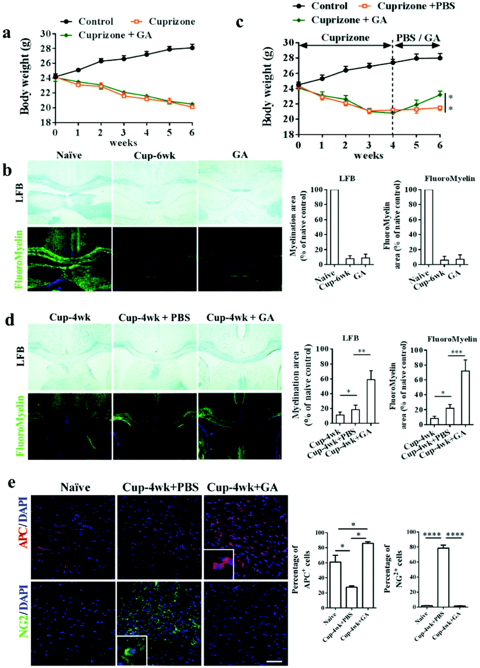 | ||
| Fig. 2 GA enhanced remyelination in the cuprizone-induced demyelinated model. For the demyelination model, 8-week old naïve male C57BL/6 mice were fed daily with standard rodent diets, and cuprizone model mice were treated with PBS and GA (50 mg kg−1) for 6 weeks, respectively. Body weights (a) of mice were recorded every week. (b) LFB and FluoroMyelin stains of the corpus callosum. For the remyelination model, 8-week old naïve male C57BL/6 mice were fed daily with cuprizone for 4 weeks; PBS or GA (50 mg kg−1) was administered daily between weeks 4 and 6. Quantitative analysis of myelinated and fluoromyelinated areas measured at random areas in the white matter of spinal cords using Image Pro software. (c) Body weights of mice were recorded every week. (d) LFB and myelin stains of the corpus callosum. Quantitative analysis of myelinated and fluoromyelinated areas measured at random areas in the white matter of spinal cords using Image Pro. (e) Immunofluorescence stains of the corpus callosum for APC+ OLGs and NG2+ OPCs. Quantification analysis of the percentage of APC+ cells and NG2+ cells measured at random areas using Image Pro. Data are mean ± SD (n = 5 each group). Scale bar = 80 μm in (b, d). Scale bar = 200 μm in (e). *p < 0.05, **p < 0.01, and ***p < 0.001 as determined by two-way ANOVA. One representative of three experiments is shown. | ||
The Royal Society of Chemistry apologises for these errors and any consequent inconvenience to authors and readers.
| This journal is © The Royal Society of Chemistry 2022 |