
 Open Access Article
 This Open Access Article is licensed under a
 Creative Commons Attribution 3.0 Unported Licence
Multi-mass velocity map imaging study of the 805 nm strong field ionization of CF3I†
Stuart W.
Crane‡
 a,
Jason W. L.
Lee
bc and
Michael N. R.
Ashfold
*a
a,
Jason W. L.
Lee
bc and
Michael N. R.
Ashfold
*a
aSchool of Chemistry, University of Bristol, Bristol, BS8 1TS, UK. E-mail: mike.ashfold@bristol.ac.uk
bDepartment of Chemistry, University of Oxford, Oxford, OX1 3TA, UK
cDeutsches Elektronen-Synchrotron DESY, D-22607 Hamburg, Germany
First published on 22nd July 2022
Abstract
Multi-mass velocity map imaging studies of charged fragments formed by near infrared strong field ionization together with covariance map image analysis offer a new window through which to explore the dissociation dynamics of several different highly charged parent cations, simultaneously – as demonstrated here for the case of CF3IZ+ cations with charges Z ranging from 1 to at least 5. Previous reports that dissociative ionization of CF3I+ cations yields CF3+, I+ and CF2I+ fragment ions are confirmed, and some of the CF3+ fragments are deduced to undergo secondary loss of one or more neutral F atoms. Covariance map imaging confirms the dominance of CF3+ + I+ products in the photodissociation of CF3I2+ cations and, again, that some of the primary CF3+ photofragments can shed one or more F atoms. Rival charge symmetric dissociation pathways to CF2I+ + F+ and to IF+ + CF2+ products and charge asymmetric dissociations to CF3 + I2+ and CF2I2+ + F products are all also identified. The findings for parent cations with Z ≥ 3 are wholly new. In all cases, the fragment recoil velocity distributions imply dissociation dynamics in which coulombic repulsive forces play a dominant role. The major photoproducts following dissociation of CF3I3+ ions are CF3+ and I2+, with lesser contributions from the rival CF2I2+ + F+ and CF32+ + I+ channels. The CF32+ fragment ion images measured at higher incident intensities show a faster velocity sub-group consistent with their formation in tandem with I2+ fragments, from photodissociation of CF3I4+ parent ions. The measured velocity distributions of the I3+ fragment ions contain features attributable to CF3I5+ photodissociation to CF32+ + I3+ and the images of fragments with mass to charge (m/z) ratio ∼31 show formation of I4+ products that must originate from parent ions with yet higher Z.
1. Introduction
Most studies of gas phase molecular photodissociation processes employ ultraviolet or visible excitation wavelengths, and neutral systems. The dominant process following excitation with higher energy photons in the range 10–25 eV is single ionization. Excitation with yet higher energy photons leads to double ionization (formation of dications) and higher (multiple) ionization. Energy permitting, the multiple ionization will usually be accompanied by fragmentation. Some, at least, of the resulting fragments will be charged and these charged fragments can be detected with unit efficiency. Since all charged fragments arising from the same dissociation event are formed at the same time, it is possible to detect and correlate these various charged particles. Eland was in the vanguard of those developing and using coincidence methods to explore the two- and three-body fragmentation dynamics of small multiply charged cations.1–3 Early studies of this type generally used atomic resonance lamps, then synchrotrons, as the source of the extreme ultraviolet (XUV) photons required for preparing the multiply charged parent cations,4,5 but many such studies are now performed using ultrashort (femtosecond (fs)) laser pulses.Removing multiple electrons from an isolated molecule causes it to undergo a Coulomb explosion (CE)6–9 and, as noted above, many CEs are now initiated using fs laser pulses at near infrared (NIR, e.g. λ ∼ 805 nm) or short XUV wavelengths. The route to forming multiply charged cations is different in the two cases: strong field ionization (SFI) when using NIR photons, Auger cascade following removal of a core electron when using photons from an X-ray free electron laser.10 In both cases, the multiply charged cation ‘explodes’ yielding two or more proximal positively charged atomic and/or molecular fragments. Determining the fragment ion velocities and momenta using, for example, coincidence velocity map imaging (VMI) methods or VMI in conjunction with covariance analysis methods offers a route to determine static molecular structures,10–12 to distinguish isomers13–18 and enantiomers,19 to explore the fragmentation dynamics of multiply charged cations20 and, in appropriately-designed ultrashort laser pulse pump–probe experiments, to follow the time-evolving structure and/or the fragmentation of photoexcited small molecules21–23 and ions.24–26
The present study explores the fragmentation dynamics of multiply charged CF3IZ+ ions formed by non-resonant NIR SFI. Each excitation pulse creates a distribution of multiply-charged parent cations, which display Z-dependent explosion dynamics. The hydrogen analogue, methyl iodide (CH3I), has been a very popular molecule for testing dynamical understanding of CEs induced both by NIR27–36 and X-ray37,38 excitation. Our recent studies of the NIR SFI of CH3I at three different incident intensities (I)39 revealed a clear transition from ‘diatomic-like’ explosion to CH3+ and Iq+ (q = 1, 2) fragments from parent ions in low Z states (Z = 2, 3), through a complex pattern of fragmentation behaviours involving loss of one or more H atoms or protons from parent ions with intermediate Z, to limiting Coulomb-driven dissociation to the constituent (charged) atoms once Z ≥ 6. Aspects of this behaviour could be rationalised by reference to complementary on-the-fly ab initio trajectory simulations,40 but the finding that the observed velocities of the Iq+ (q > 2) fragments were consistently lower than those predicted by such calculations hinted at non-adiabatic coupling effects in the evolution from the photo-prepared parent cation to asymptotic fragments. Thorough investigation of the transition from ‘low’ to ‘high’ Z fragmentation behaviour in the case of CH3I was hampered by the unfavourable mass ratios: the experimental mass resolution precluded mass-selective imaging of heavier fragments differing by just one mass unit, while the recoil velocities of many of the light H+ fragments were too high to permit detection with the available time and position sensitive detector.
Hence the motivation of the present multi-mass imaging study, wherein we present and analyse velocity map images of all major fragment ions arising in the NIR SFI of jet-cooled CF3I molecules following excitation with ultrashort (40 fs), non-resonant NIR (805 nm) laser pulses at the same three peak intensities as used in the CH3I studies (I ≈ 2.6 × 10¹⁴–1.3 × 10¹⁵ W cm−2 (260–1300 TW cm−2)). As in the case of CH3I, many aspects of the CE dynamics of CF3IZ+ cations with low Z can be understood using a 1-D picture that recognises the competition between (attractive) bonding and (repulsive) coulombic forces along the C–I bond (RC–I). In distinction to CH3I, however, where the loss of one (or more) light H atoms causes only a small (and generally unresolvable) change to the momentum of the CHnp+ (and Iq+) fragments formed in any CE and the H+ ions can only ever carry a q = 1 charge, F atom (or F+ ion) loss during the CE of CF3I has a discernible effect on the momenta of any CFnp+ partner fragments and the F atoms deriving from CE of CF3I ions in high Z states can carry multiple charges, offering the prospect of richer, further from ‘diatomic-like’ explosion dynamics.
Unravelling these dynamics can be challenging, given that there will generally be more than one route to forming any given fragment ion in a NIR SFI-induced CE experiment. The velocity (and momentum) information encoded in the fragment ion images helps in unravelling this complexity, but the present multi-mass imaging measurements carry time and position of arrival (at the detector) information for all fragments which allows identification of correlated product-pairs (or, at least in principle, even triplets) via analysis of 2-D (and 3-D) covariance-map images.41,42 Covariance-map imaging methods have already been applied in analysing the CE of CF3I molecules following electron impact at collision energies in the range 20–100 eV43,44 and are here used to provide further insights into dominant two-body and some of the more probable three-body SFI-induced fragmentations of CF3IZ+ cations.
Analyses of the velocity-map and covariance-map images of the various fragment ions observed at the three NIR field strengths offers a new way of exploring the fragmentation dynamics of multiply-charged CF3IZ+ cations. The data reported here complements and extends previous discussions of the dissociative ionization of CF3I+ cations,45–47 provides additional insights into the more important fragmentation pathways available to the CF3I2+ cations48 and provides completely new information about rival fragmentation pathways for CF3IZ+ cations with Z ≥ 3. More generally, the present work also illustrates the multiplex advantage afforded by multi-mass imaging coupled with covariance analysis methods; just in the case of CF3I, this single family of experiments informs on more than 15 fragmentation channels from five different CF3IZ+ cations.
2. Experimental
The ultrafast Ti-sapphire laser, the optical set-up, the VMI spectrometer, data collection and sample preparation methods have been described previously.39 CF3I (Aldrich, 99%) samples, exhaustively degassed by prior freeze–thaw cycling, diluted (1%) in He (Air Liquide, >99.999%), were expanded as a pulsed molecular beam and skimmed en route to the differentially-pumped interaction region, where they were intercepted mid-way between the repeller and extractor plates of a cryo-shielded ion optics assembly by the focussed (f = 30 cm), fundamental output of the Ti-sapphire laser. The resulting ions were accelerated along the time-of-flight (TOF, z) axis and passed through a field-free flight region towards a 40 mm diameter microchannel plate (MCP)/phosphor screen detector. This allowed measurement of the ion TOF spectrum and, using a second-generation Pixel Imaging Mass Spectrometry (PImMS2) sensor,49,50 2-D (x, y) images of the ions incident at the detector within individual 25 ns time bins. A recognized limitation of the method is that multiple ions hitting the same detector region (most notably the central spot) in a narrow time interval will be under-detected. The data were analysed to provide covariance-map images between different pairs of product ions.51 Characterisation of the 805 nm laser output returned a pulse duration of 40 fs (full width half maximum) and maximum pulse intensities of ∼1300 TW cm−2, ∼650 TW cm−2 and ∼260 TW cm−2 for the studies reported here.3. Results and discussion
Fig. 1 shows time-of-flight (TOF) mass spectra obtained following 805 nm SFI of CF3I at I = 260, 650 and 1300 TW cm−2, with the detector gain gated to preclude detection of the CF3I+ parent ion with mass to charge ratio, m/z 196. The spectrum recorded at lowest I is dominated by peaks with m/z 127 (I+) and 69 (CF3+) but other weaker features at m/z 177 (CF2I+), 146 (IF+), 139 (CI+), 88.5 (CF2I2+), 63.5 (I2+), 50 (CF2+), 42 (I3+), 34.5 (CF32+), 31.5 (CF+, with varying amounts of contamination from I4+), 25 (CF22+), 19 (F+), 15.5 (CF2+) and 12 (C+) are observed in this spectrum and/or in spectra recorded at higher I. The relative yields of the Iq+ (q = 2–4) and F+ peaks increase with increasing I, and weak peaks attributable to both F2+ and C2+ fragment ions also become apparent at the highest I. Clearly, the data allows the opportunity to monitor and image the different CFnp+ (n = 0–3, p = 1, 2) fragment ions and, by determining covariance map images, to identify correlations between the various product ions (i.e. products arising via a common fragmentation process).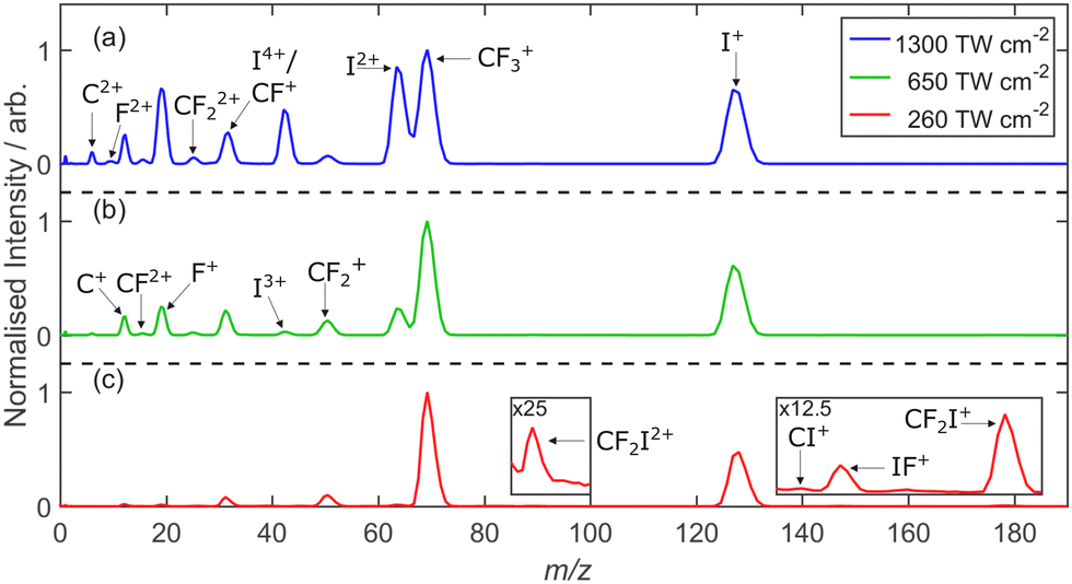 | ||
| Fig. 1 TOF mass spectra of the fragment ions observed following SFI of CF3I with λ = 805 nm photons at I = (a) 1300, (b) 650 and (c) 260 TW cm−2, displayed with a mass/charge (m/z) scale, with the regions appropriate for CF2I+, IF+, CI+ and CF2I2+ ions (i.e. m/z 135–185 and 85–100) shown on expanded vertical scales in the insets within panel (c). For ease of comparison, the three spectra are displayed so that the m/z 69 (CF3+) peak appears with the same intensity. | ||
3.1 Fragment velocity map and covariance map images
Fig. 2 shows images of the Iq+ (q = 1–4) fragment ions measured at the three different peak intensities. These and all other images presented in this work have been symmetrized prior to display. All were recorded using the highest extraction voltages that satisfied VMI conditions and are displayed on a common radial dispersion. The I+ images reveal two distinct product channels. One is ‘slow’ (with a small radius, r), with an anisotropic recoil velocity distribution that is preferentially aligned parallel to the ε vector of the SFI laser radiation. Throughout, an italic θ will be used to define recoil velocities relative to this ε vector. The second is associated with ‘fast’ I+ fragments (appearing at larger r). The recoil velocity distribution of this latter component evolves from preferentially parallel (i.e. θ ∼ 0°) to preferentially perpendicular (i.e. θ ∼ 90°) with increasing I. The relative importance of the slow and fast channels also varies with I. The I2+ images recorded at lower I also show a slow component but the Iq+ (q > 1) images recorded at higher I are dominated by fast products with an increasing propensity for recoil parallel to ε. Such aligned fragment recoil is typical of SFI processes wherein the interaction is strongest when ε is parallel to a particular molecular axis (here the polar C–I bond) and dissociation then occurs on a timescale much shorter than the parent rotational period.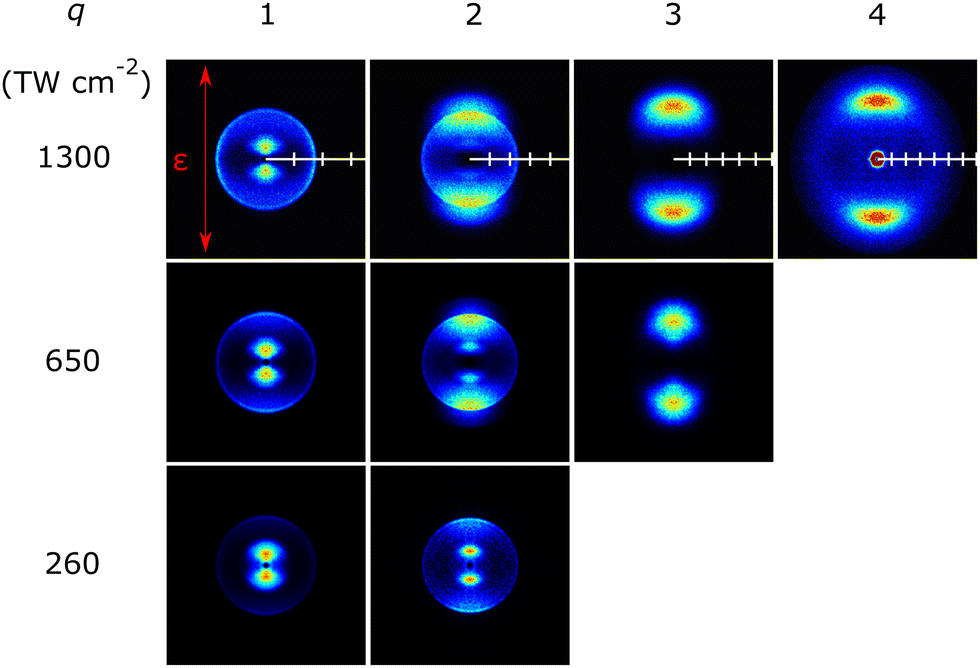 | ||
| Fig. 2 Symmetrized images of the different Iq+ fragments observed following SFI of CF3I with linearly polarized λ = 805 nm photons at I = 1300, 650 and 260 TW cm−2. The ticks on the white horizontal scale overlaid on each of the 1300 TW cm−2 images indicate 1000 m s−1 intervals to provide a relative comparison of achievable velocities. The orientation of the ε vector of the SFI laser radiation is shown by the double headed red arrow included in the top left panel. The central feature in the I4+ image is attributable to O2+ ions from SFI of trace air impurity, and the more isotropic component at large radius in this same image is attributable to CF+ fragments. | ||
These data were converted to the corresponding product velocity (v) and kinetic energy (KE) distributions using the r to v scaling determined via two-colour pump (267 nm)–SFI probe (805 nm) measurements of the CH3 and I fragments (measured via the CH3+ and I+ cations) from the well-documented photolysis of CH3I52 at long pump–probe delays using the same ion optics conditions. Fig. 3 shows the velocity distributions P(v) determined for each Iq+ fragment at each incident I with, in each case, the CE-induced feature of most interest scaled to unit peak intensity. The KEs of these Iq+ fragments (KEI) are shown on the upper abscissa. As before,39 the reported P(v) distributions have been derived by summing signal in three adjacent time bins (i.e., a δt = 75 ns time slice) that span the centre of the TOF peak of interest rather than by Abel inverting the ‘crushed’ image. The three-time-bin analysis approach allows better m/z resolution, but the returned P(v) distributions are somewhat less resolved (though show better signal to noise ratio) – as illustrated in Fig. S1 in ESI.† Key in the present context, the P(v) distributions returned by the two approaches peak at reassuringly similar velocities.
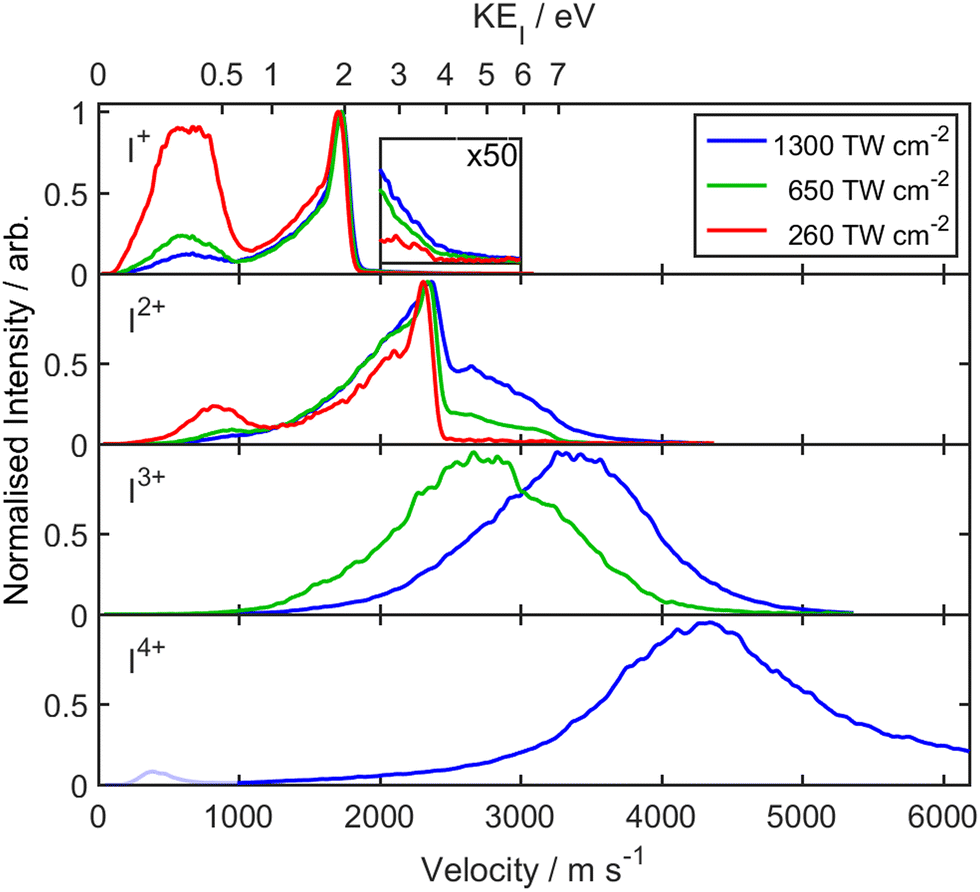 | ||
| Fig. 3 P(v) distributions for the different Iq+ (q = 1–4) fragments derived from analysis of images obtained from SFI of CF3I with λ = 805 nm photons at I = 1300, 650 and 260 TW cm−2 with, in each case, the strongest feature normalised to unit intensity. The corresponding KEI scale is shown on the top x-axis. The inset in the top panel shows the weak I-dependent yield of fast I+ fragments, while the low velocity peak in the I4+ spectrum (shown in a fainter hue) is attributable to O2+ ions from air impurity. | ||
Fig. 4 shows symmetrized images of the various CFnp+ (n = 1–3, p = 1, 2) fragment ions measured at these same peak intensities, recorded with the same extraction voltages. The CFn+ (n = 1–3) images also show both ‘slow’ and ‘fast’ components with different recoil anisotropies and I-dependent relative intensities, whereas the CFn2+ (n = 1–3) images all show at least two ‘fast’ components with parallel recoil anisotropy. Note that Fig. 4 shows no image attributed to CF+ ions following SFI at I = 1300 TW cm−2. Given the available TOF mass resolution, CF+ and I4+ (and O2+) ions all appear at the same m/z. The CF+ and I4+ fragment ions appear at similar radius but are deduced to have clearly different angular distributions. Both recoil preferentially along the axis defined by the laser polarization vector but the I4+ fragments, which only contribute significantly at the highest incident intensity, are much more tightly aligned with ε, as shown in Fig. 5. Hence the decision to display the image of the fragment ions with m/z ∼ 31.5 recorded at I = 1300 TW cm−2 in Fig. 2 rather than Fig. 4. [Note, the small O2+ contribution (from SFI of O2 in any trace air impurity) is also readily recognizable as a central dot in the relevant image in Fig. 2.] The P(v) distributions determined from the various CFnp+ images are shown in Fig. 6. The images of the F+, F2+, IF+, CF2I+ and CF2I2+ fragment ions all imply that the formation of at least a fraction of each species is also accompanied by some dynamical energy release. These images and the associated P(v) distributions are collected in, respectively, Fig. S2–S4 in the ESI.†
 | ||
| Fig. 4 Symmetrized images of the CFnp+ (n = 1–3, p = 1, 2) fragments observed following SFI of CF3I with λ = 805 nm photons at I = 1300, 650 and 260 TW cm−2. The ticks on the white horizontal scale overlaid on each of the 650 TW cm−2 images indicate 1000 m s−1 intervals to provide a relative comparison of achievable velocities. The orientation of the ε vector of the SFI laser radiation is shown by the double headed red arrow in the top left panel. The m/z ∼ 31.5 (CF+) image recorded at the highest I is omitted as I4+ fragment ions are adjudged to be the dominant contributor (see Fig. 2). | ||
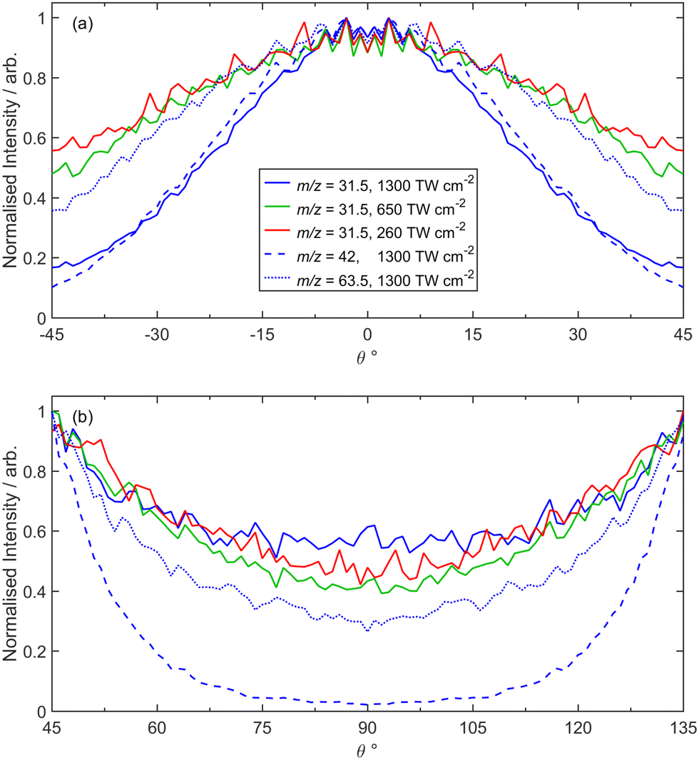 | ||
| Fig. 5 Comparisons of P(θ) distributions showing that I4+ ions make a dominant contribution to the m/z ∼ 31.5 images recorded following SFI of CF3I with λ = 805 nm photons at the highest I. Panel (a) compares P(θ) distributions for fragments with m/z ∼ 31.5 recorded at I = 260, 650 and 1300 TW cm−2 and with m/z ∼ 63.5 (I2+) and m/z ∼ 42 (I3+) recorded at I = 1300 TW cm−2, plotted over the range −45° ≤ θ ≤ 45°, which highlight the similar P(θ) distributions for the I3+ and (assumed) I4+ fragments at high I. The corresponding P(θ) distributions over the range 45° ≤ θ ≤ 135° shown in panel (b) highlight the relatively much greater ‘perpendicular’ contribution in the m/z ∼ 31.5 (cf. m/z ∼ 42 (I3+)) trace, indicating that CF+ fragment ions also contribute to the measured recoil velocity distribution in the former case. All P(θ) distributions have been normalised to a peak intensity of unity at the θ value associated with maximum signal intensity. | ||
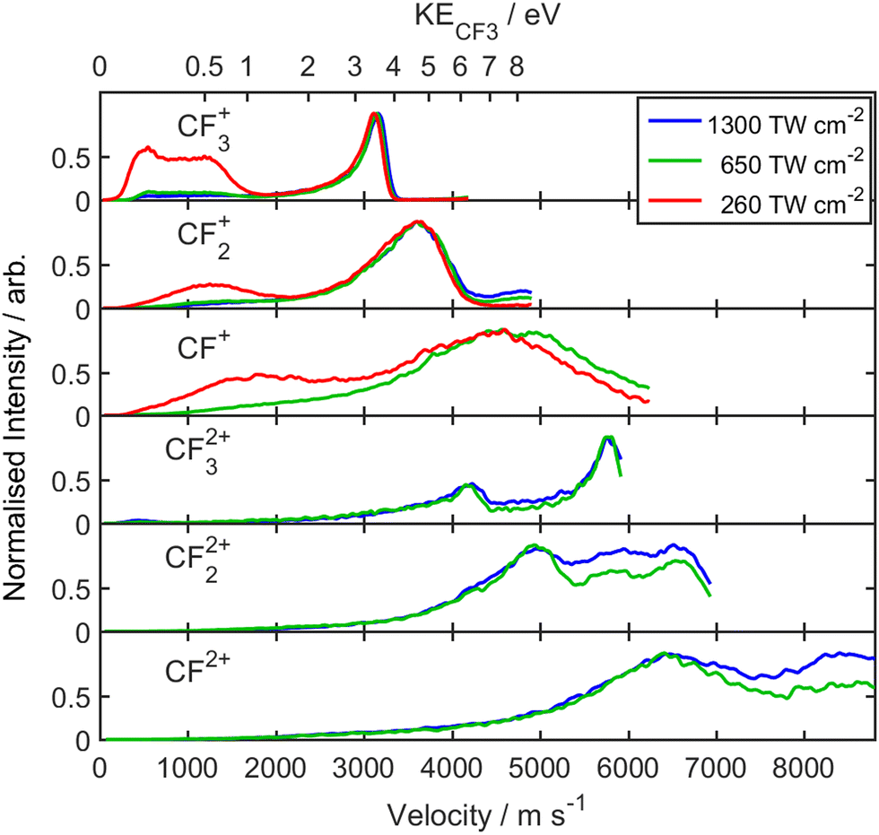 | ||
| Fig. 6 P(v) distributions for the CFnp+ (n = 1–3, p = 1, 2) fragments derived from analysis of images obtained from SFI of CF3I with λ = 805 nm photons at I = 1300, 650 and 260 TW cm−2 with, in each case, the strongest feature normalised to unit intensity. The corresponding KECF3 scale is shown on the top x-axis. | ||
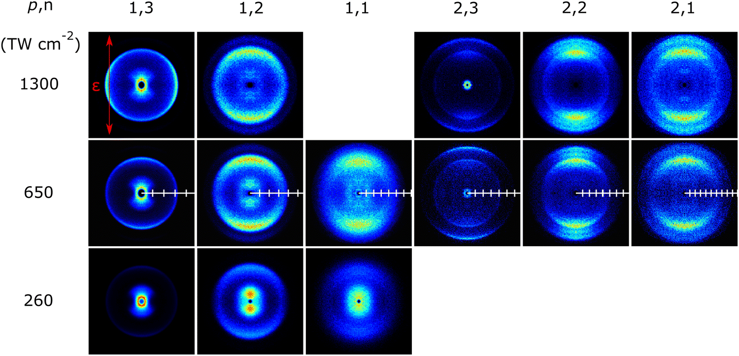
Fig. 7 presents covariance map images recorded at I = 260 TW cm−2 that show clear correlations between the I+ and I2+ fragments with both the CF3+ and CF2+ fragment ions. These are displayed as (CFn+, Iq+) covariance map images (i.e. where the Iq+ species has been selected as the reference ion and its velocity fixed to be vertically upwards (defined as θ = 0°, with a non-italic θ, by the red arrow in the top left hand panel, and displaying the correlated 2-D velocity distribution of the CFn+ partner in the frame defined by the reference ion). Fig. S5 in the ESI† confirms that the same correlations are revealed in the corresponding (Iq+, CFn+ (n = 1–3)) covariance map images. The correlations between the Iq+ (q = 1, 2) and CF3+ products are tightly focussed along θ = 180°, as required by momentum conservation in the case of a two-body fragmentation, and as demonstrated in Fig. 7(c). The correlations between the CF2+ (and CF+) and Iq+ (q = 1, 2) products are also centred along θ = 180°, but the covariance map images are more diffuse, consistent with these ion pairs arising via three- (or more-) body fragmentation processes. The tight correlation between CF3+ and both I+ and I2+ products is also evident in covariance map images derived from data recorded at the higher laser intensities (Fig. S6 in the ESI†). Fig. S7 and S8 in the ESI† illustrate further detectable covariances between, respectively, CF32+ and I2+ fragment ions in images recorded at I = 650 TW cm−2, and between IF+ and CF2+ and between CF2I+ and F+ ions fragment ions in images recorded at I = 260 TW cm−2.
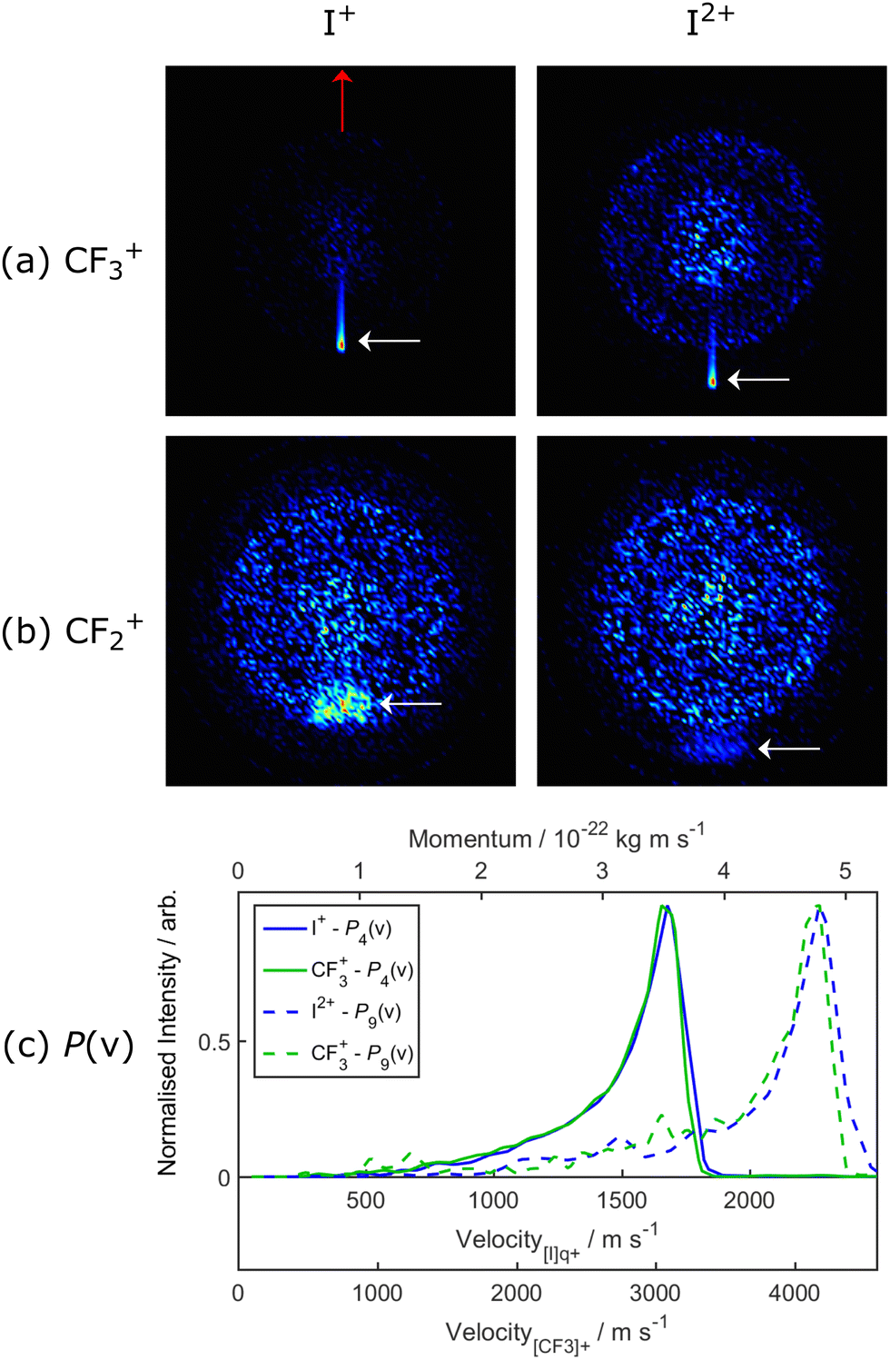 | ||
| Fig. 7 Covariance map images from the I = 260 TW cm−2 data from SFI of CF3I, selecting I+ (left column) and I2+ (right hand column) as the reference ion, and fixing the respective velocity to be vertically upwards (as indicated by the red arrow in the top left hand plot), and displaying the correlated 2-D velocity distributions of the (a) CF3+ and (b) CF2+ counter-fragments in the frame of the reference ion. The covariance signal of interest in each case is bounded by dashed white lines. Panel (c) illustrates the momentum matching between the product pairs from dissociation channels (4) and (11), with each P(v) distribution plotted so that the peak signal is displayed with unit intensity. | ||
3.2 Fragmentation channels
These data are now discussed from the perspective of the CE dynamics of CF3IZ+ ions, in order of increasing Z.| CF3I + hν → CF3+ + I, E = 11.384 eV | (1) |
| CF3I + hν → CF3 + I+, E = 12.80 eV | (2) |
| CF3I + hν → CF2I+ + F, E = 13.40 eV. | (3) |
The present data show signatures of all three of these DI processes, most clearly in the images recorded at low intensity (I = 260 TW cm−2). The I+ image (Fig. 2) shows a ‘slow’ feature with parallel recoil anisotropy. The breadth of the P(v) distribution of these I+ fragments (which spans the range ∼400 < v < 900 m s−1 though, given the previously noted under-detection of on-axis fragments, this distribution may well extend down to zero velocity) may indicate DI from SFI-produced parent ions in a range of vibronic states, and/or a spread of internal energies in the neutral CF3 partner fragments, and/or that the SFI process yields I+ products in all three 3PJ (J = 0–2) spin–orbit states.
The P(v) distribution for the CF3+ fragments (Fig. 6) shows a double humped feature spanning the range ∼300 < v < 1500 m s−1 that is plausibly attributable to branching into ground (2P3/2) and spin–orbit excited (2P1/2) I atoms in DI process (1) – as proposed in previous studies of the DI of CF3I+ cations in, respectively, the X and A electronic states.46,47 Again, we caution that under-detection issues on axis mean that we cannot be certain that this distribution does not extend down to zero velocity. Higher excited states of CF3I+ have been identified in one photon excitation studies46 and will likely be populated in the present SFI environment. The fragmentation behaviour of these higher excited states has not been characterised in any detail, but the similarities between the ‘slow’ (small r) features in the CF3+, CF2+ and CF+ images suggests that some of the primary CF3+ fragments are formed with sufficient excitation that they decay further, by sequential loss of one (or two) neutral F atoms.
CF2I+ fragments are also observed, with a small relative yield (cf. CF3+, Fig. 1) and a P(v) distribution that peaks at low velocity but also shows a second, more clearly anisotropic component at larger v that grows in relative importance with increasing I (Fig. S3, ESI†). We first focus on the low velocity component, again noting that the ‘hole’ in the centre of the image may reflect known under-detection issues and lead to a somewhat exaggerated mean recoil velocity for this yield of slow CF2I+ fragments. Following Powis et al.,46 it is likely that these ions arise via near-threshold autoionization (process (3)). The form of the kinetic energy distribution is also reminiscent of that found for the CF2I+ fragments arising in the dissociative electron ionization of CF3I using incident electron energies throughout the range 20–80 eV.43 We recognise that secondary decay of internally excited CF2I+ fragments could also contribute to the observed yield of ‘slow’ CF2+ fragments and, conceivably, to the observed yield of IF+ products, but note that covariance map image analysis also demonstrates that Z = 2 parent ions contribute to the yields of these fragment ions (see below).
| CF3I2+ → CF3+ + I+, | (4) |
Both fragments display very similar (I-dependent) recoil anisotropies, which evolve from preferential parallel to preferential perpendicular recoil with increasing I (Fig. 4). Such observations serve as reminders that the fragmentation of CF3I2+ cations is still far from the pure Coulomb limit. The valence bonding forces are sufficient to sustain several diabatically bound states of the CF3I2+ cation,48 with symmetries that can be accessed by parallel or perpendicular transitions from the ground state. The present data suggest that the latter gain in relative importance at higher I. As also noted in our recent SFI studies of CH3I,39 the P(v) distributions of the fragments arising from the two-body dissociation process (4) are narrow. Expressed in terms of total kinetic energy release (TKER) derived from the ‘sliced’ image data following Abel inversion (see Fig. S1, ESI†), the CF3+ + I+ products display a TKER of ∼5.7 eV with a full width half maximum of only ∼0.6 eV. The earlier double photoionization study at 40.8 eV photon energy identified a similarly narrow TKER distribution for the CF3+ + I+ products from dissociation of the CF3I2+ cation.48 These energy widths are comparable to the spin–orbit splitting in the I+ cation, and less than half that of an 805 nm photon as used in the present study. Both experiments imply that a near-constant amount of the available energy is released as product translation, i.e. that more internally excited CF3I2+ parent cations must dissociate to correspondingly more internally excited CF3+ (and/or I+) products.
Weaker CF2+, CF+ and even C+ fragment ion signals are evident in the TOF mass spectrum (Fig. 1). As Fig. 4 and 6 show, the associated images show an annular ring reminiscent of that seen for the CF3+ fragments but, with decreasing F-content, the peak in the associated P(v) distribution broadens and its centre of gravity shifts to higher velocity. The (CF2+, I+) covariance map image shown in Fig. 7 confirms a correlation between the I+ and CF2+ ions and, as in the case of the CF3+ products from DI process (1), it is logical to attribute these CF2+ ions to the dissociation of Z = 2 parent ions formed with sufficient internal energy that the resulting CF3+ fragments (formed via process (4)) themselves have sufficient internal energy to dissociate further, by losing one (or more) F atoms to yield eventual CF2+, CF+ and even C+ fragments, i.e.
| CF3+ → CF2+ + F → CF+ + 2F → C+ + 3F, | (5) |
Eland et al.48 noted the operation of several rival fragmentation pathways for Z = 2 parent ions. One is process (6):
| CF3I2+ → CF2I+ + F+, | (6) |
| CF3I2+ → IF+ + CF2+. | (7) |
| CF3I2+ → CF2I2+ + F | (8) |
| CF3I2+ → CF3 + I2+. | (9) |
The observation of CF32+ cations with m/z ∼ 34.5 in the TOF mass spectrum (Fig. 1) and the associated ion images (Fig. 4) confirms the intrinsic stability of this dication,57,58 but the absence of any slow feature in the CF32+ (or other CFn2+) images tends to rule out any role in the present experiments for the third possible charge asymmetric dissociation process
| CF3I2+ → CF32+ + I, | (10) |
| CF3I3+ → CF3+ + I2+ | (11) |
Notwithstanding the weakness of the m/z ∼ 34.5 (CF32+) peak in the TOF mass spectrum (Fig. 1), well-resolved velocity map images of the CF32+ fragment ions were readily obtainable at I = 650 and 1300 TW cm−2 (Fig. 4). These show at least two velocity sub-groups, exhibiting parallel recoil anisotropy. The P(v) distribution associated with the weak, slowest sub-group peaks at v ∼ 4200 m s−1 (Fig. 6). As noted above, momentum matching requires that the corresponding Iq+ fragments must have v ∼ 2300 m s−1 and the P(v) distribution of the I+ cations shows a weak pedestal (shown in the inset to Fig. 3) stretching out to such velocities. We conclude that CE of Z = 3 parent ions occurs predominantly via process (11), with only a minor contribution from the rival fragmentation
| CF3I3+ → CF32+ + I+. | (12) |
Fig. S4 (ESI†) shows the m/z 88.5 (CF2I2+) image recorded at all three incident intensities. The image recorded at I = 1300 TW cm−2 and its analysis must be viewed with some caution. Scrutiny of the image data suggests that the small peak at v ∼ 1050 m s−1 is real, but we cannot exclude the possibility that the more obvious peak centred at v ∼ 2400 m s−1 is associated with fragment ions from SFI of trace bromothiophene contaminant (the immediate prior experiments in the apparatus involved this molecule). Notwithstanding, it is tempting to associate the v ∼ 1050 m s−1 signal (at least) with some contribution from another rival fragmentation channel
| CF3I3+ → CF2I2+ + F+. | (13) |
| CF3I4+ → CF32+ + I2+, | (14) |
| CF32+ → CF22+ + F → CF2+ + 2F | (15) |
| CF32+ → CF2+ + F+ → CF+ + F+ + F → C+ + F+ + 2F | (16) |
The I3+ fragment ion images recorded at I = 650 and 1300 TW cm−2 both show peaked recoil velocity distributions aligned parallel to the ε-vector, but the peaks of the P(v) distributions derived from these images clearly shift to higher velocity with increasing I. The distribution measured at I = 650 TW cm−2 peaks at v ∼ 2700 m s−1 and is logically assigned to a rival, charge-asymmetric, decay process for Z = 4 parent ions
| CF3I4+ → CF3+ + I3+. | (17) |
The I3+ fragment ion image measured at I = 1300 TW cm−2 peaks at a larger radius, corresponding to v ∼ 3400 m s−1. Increasing the incident intensity will increase the relative proportion of higher Z parent ions in the interaction region, and the logical source of these ions is the process
| CF3I5+ → CF32+ + I3+, | (18) |
Given that parent cations with Z = 5 are present in the interaction region at the higher incident intensities, we note that the rival process
| CF3I5+ → {CF33+} + I2+ | (19) |
![[thin space (1/6-em)]](https://www.rsc.org/images/entities/char_2009.gif) } signifies that this highly charged 4-atom species is likely to have a very fleeting nature) may contribute to the fast tail in the P(v) distribution of the I2+ fragments formed at I = 1300 TW cm−2 (Fig. 3). We also note that parent cations with Z = 5 are the first that could, in principle, atomize to singly charged atomic products – the threshold energy for which is ∼90.9 eV.
} signifies that this highly charged 4-atom species is likely to have a very fleeting nature) may contribute to the fast tail in the P(v) distribution of the I2+ fragments formed at I = 1300 TW cm−2 (Fig. 3). We also note that parent cations with Z = 5 are the first that could, in principle, atomize to singly charged atomic products – the threshold energy for which is ∼90.9 eV.
Evidence for CE of CF3IZ+ cations with Z > 5 in the present experiments is limited to observations of (i) I3+ fragment ions with velocities v > 4500 m s−1 in the data recorded at I = 1300 TW cm−2 (i.e. much faster than can be accommodated by dissociation process (18)) and (ii) I4+ fragments. Interpretation of the image of the m/z ∼ 31.5 ions measured at high I (Fig. 2) required rather more thought. The central feature is attributable to O2+ ions from SFI of trace air impurity. The feature centred at v ∼ 4200 m s−1 is plausibly consistent with that expected by extrapolating the CF+ data measured at lower I (Fig. 4 and 6) but, as Fig. 5 convincingly shows, I4+ fragments also contribute to the image recorded at I = 1300 TW cm−2. No I5+ fragments are discernible in the TOF mass spectrum measured at this highest incident intensity.
4. Conclusions
This study demonstrates how multi-mass VMI and covariance map imaging studies of the charged fragments formed by near infrared strong field ionization of a neutral gas phase molecule (here CF3I) can provide many new insights into the dissociation dynamics of the resulting parent cations formed in a range of charge states. The distribution of Z states necessarily varies with the incident intensity and, within any one focussed laser pulse, is both time- and position-dependent. This multiplex aspect is both an asset and a limitation of the approach. The obvious advantage is that one experiment allows the study of multiple fragmentation channels from different parent cations, simultaneously. The most obvious limitation is that a given fragment ion image may contain overlapping contributions from the dissociation of several CF3IZ+ ions, but these overlaps can be revealed by probing the image sensitivities to I and, in favourable cases, deconvolved by covariance map imaging methods.Previous reports that DI of CF3I+ cations can result in CF3+, I+ and CF2I+ fragment ions (together with, respectively, I atoms in both spin–orbit states, CF3 neutrals, and an F atom) are confirmed and some of the CF3+ fragments are deduced to be formed with sufficient internal energy to undergo secondary loss of one or more neutral F atoms.46,47
The covariance map images confirm the dominance of CF3+ and I+ products in the photodissociation of CF3I2+ ions.48 The I-dependent recoil anisotropy of these fragments shows that the dissociation of Z = 2 cations is far from Coulomb dominated. Valence bonding forces have an obvious influence on the fragmentation dynamics, and the I-dependent recoil anisotropy is most readily explained by assuming that different incident laser intensities sample different distributions of dissociative parent electronic states of CF3I2+. The measured images reveal that the CF3+ + I+ products are formed with a narrow spread of kinetic energies. The data imply a strong propensity for parent internal excitation to map into internal excitation of the products and show that some of the primary CF3+ fragments are formed with sufficient internal energy to decay further by secondary loss of one or more F atoms. Rival dissociations to CF2I+ + F+ and to CF2+ + IF+ products are revealed by covariance map image analysis. Charge asymmetric dissociations to CF3 + I2+ and CF2I2+ and F are also both identified, but the present study finds no evidence for the alternative (higher energy) CF32+ + I product channel.
The findings reported here for CF3IZ+ ions with Z ≥ 3 are entirely new. In all cases, the coulombic repulsive forces are sufficient to ensure that the photofragments recoil along an axis parallel to ε. The primary products formed in the photodissociation of CF3I3+ ions are mainly CF3+ and I2+, with only minor contributions from the rival, more endoergic, CF2I2+ + F+ and CF32+ + I+ channels. Again, the covariance map images indicate that the internal energies of some of the CF3+ products are sufficient to allow the subsequent loss of one or more F atoms. The CF32+ fragment ion images measured at higher I show another faster velocity sub-group consistent with their formation in tandem with I2+ fragments from the photodissociation of CF3I4+ parent ions. Again, the measured fragment ion yields and ion images suggest that some of the nascent CF32+ fragments decay further by losing F and/or F+ atoms. The P(v) distributions derived from I3+ fragment ion images recorded at the highest I show a component attributable to CF3I5+ photodissociation to CF32+ + I3+ and the m/z ∼ 31.5 images clearly show I4+ products from parent ions with Z > 5.
Dissociative pathways from parent ions with CF3IZ+ ions where Z ≥ 3 could, in principle, be investigated by application of three-fold covariance methods. Such studies should complement the conclusions in this manuscript, by confirming the kinetic energies of the fragment ions directly. To date, however, successful examples of three-fold covariance analysis generally require the collection of significantly more data (better statistics) than achieved in the present work and/or have been limited to relatively simple molecules with a modest number of dissociative pathways (e.g. the covariance between the D+, D+, O+ fragments arising in the dissociation of D2O3+ parent ions 59). Any such experiment on highly charged CF3IZ+ ions would surely benefit from pre-alignment of the parent molecules to simplify the data analysis and interpretation, as demonstrated in earlier ground-breaking CEI studies of pre-aligned 3,5-dibromo-3′,5′-difluoro-4′-cyanobiphenyl molecules.60
Finally, comparisons with CH3I are revealing: analogous SFI experiments on CH3I, at the same incident intensities, yielded measurable images of I5+ and even I6+ fragment ions.39 These differences (cf. CF3I) might be explicable if the cross-sections for SFI of CF3I at λ ∼ 805 nm are relatively smaller than for CH3I, or if the SFI-induced charges are less heavily concentrated on the I atom in CF3I than in CH3I, i.e. that F atoms (and a CF3 group) are a better sink for positive charge than H atoms (and a CH3 group).
Data availability
All underpinning data are available at the University of Bristol data repository, data.bris, at https://doi.org/10.5523/bris.1jmu8pj7rzm6p2td4lwhl569ej.Conflicts of interest
The authors declare no conflicts of interest.Acknowledgements
The authors are grateful to EPSRC for funding via Programme Grant EP/L005913/1. JWLL acknowledges financial support via the Helmholtz-ERC Recognition Award (ERC-RA-0043) of the Helmholtz Association (HGF).References
- D. M. Curtis and J. H. D. Eland, Int. J. Mass Spectrom. Ion Phys., 1985, 63, 241–264 CrossRef CAS.
- J. H. D. Eland, Mol. Phys., 1989, 61, 725–745 CrossRef.
- H. Hsieh and J. H. D. Eland, J. Phys. B: At. Mol. Phys., 1997, 30, 4515–4534 CrossRef.
- D. Mathur, Phys. Rep., 2004, 391, 1–118 CrossRef CAS.
- K. Ueda and J. H. D. Eland, J. Phys. B: At. Mol. Phys., 2005, 38, S839–S859 CrossRef CAS.
- Z. Vager, R. Naaman and E. P. Kanter, Science, 1989, 244, 426–431 CrossRef CAS PubMed.
- L. J. Frasinski, K. Codling and P. A. Hatherly, Science, 1989, 246, 1029–1031 CrossRef CAS PubMed.
- K. W. D. Ledingham, R. P. Singhal, D. J. Smith, T. McCanny, P. Graham, H. S. Kilic, W. X. Peng, S. L. Wang, A. J. Langley and P. F. Taday, et al. , J. Phys. Chem. A, 1998, 102, 3002–3005 CrossRef CAS.
- F. Légaré, K. F. Lee, I. V. Litvinyuk, P. W. Doley, A. D. Bandrauk, D. M. Villeneuve and P. B. Corkum, Phys. Rev. A: At., Mol., Opt. Phys., 2005, 72, 052717 CrossRef.
- T. Yatsuhashi and N. Nakashima, J. Photochem. Photobiol., C, 2018, 34, 52–84 CrossRef CAS.
- X. Li, A. Rudenko, M. S. Schöffler, N. Anders, Th. M. Baumann, S. Eckart, B. Erk, A. De Fanis, K. Fehre and R. Dörner, et al. , Phys. Rev. Res., 2022, 4, 013029 CrossRef CAS.
- R. Boll, J. M. Schäfer, B. Richard, K. Fehre, G. Kastirke, Z. Jurek, M. S. Schöffler, M. M. Abdullah, N. Anders and T. M. Baumann, et al. , Nat. Phys., 2022, 18, 423–428 Search PubMed.
- M. Pitzer, M. Kunitski, A. S. Johnson, T. Jahnke, H. Sann, F. Sturm, L. P. H. Schmidt, H. Schmidt-Bocking, R. Dorner and J. Stohner, et al. , Science, 2013, 341, 1096–1100 CrossRef CAS PubMed.
- M. Pitzer, G. Kastirke, M. Kunitski, T. Jahnke, T. Bauer, C. Goihl, F. Trinter, C. Schober, K. Henrichs and J. Becht, et al. , ChemPhysChem, 2016, 17, 2465–2472 CrossRef CAS PubMed.
- U. Ablikim, C. Bomme, H. Xiong, E. Savelyev, R. Obaid, B. Kaderiya, S. Augustin, K. Schnorr, I. Dumitriu and T. Osipov, et al. , Sci. Rep., 2016, 6, 38202 CrossRef PubMed.
- M. Burt, K. Amini, J. W. L. Lee, L. Christiansen, R. R. Johansen, Y. Kobayashi, J. D. Pickering, C. Vallance, M. Brouard and H. Stapelfeldt, J. Chem. Phys., 2018, 148, 091102 CrossRef.
- U. Ablikim, C. Bomme, T. Osipov, H. Xiong, R. Obaid, R. C. Bilodeau, N. G. Kling, I. Dumitriu, S. Augustin and S. Pathak, et al. , Rev. Sci. Instrum., 2019, 90, 055103 CrossRef PubMed.
- S. Pathak, R. Obaid, S. Bhattacharyya, J. Burger, X. Li, J. Tross, T. Severt, B. Davis, R. C. Bilodeau and C. A. Trallero-Herrero, et al. , J. Phys. Chem. Lett., 2020, 11, 10205–10211 CrossRef CAS PubMed.
- C. Saribal, A. Owens, A. Yachmenev and J. Küpper, J. Chem. Phys., 2021, 154, 071101 CrossRef CAS PubMed.
- J. Yadav, C. P. Safvan, P. Bhatt, P. Kamari, A. Kumar and J. Rajput, J. Chem. Phys., 2022, 156, 141101 CrossRef CAS PubMed.
- M. Burt, R. Boll, J. W. L. Lee, K. Amini, H. Köckert, C. Vallance, A. S. Gentleman, S. R. Mackenzie, S. Bari and C. Bomme, et al. , Phys. Rev. A, 2017, 96, 043415 CrossRef.
- M. E. Corrales, J. González-Vázquez, R. de Nalda and L. Bañares, J. Phys. Chem. Lett., 2019, 10, 138–143 CrossRef CAS PubMed.
- R. Forbes, F. Allum, S. Bari, R. Boll, K. Borne, M. Brouard, P. H. Bucksbaum, A. J. Howard, P. Johnsson and J. W. L. Lee, et al. , J. Phys. B: At. Mol. Phys., 2020, 53, 224011 Search PubMed.
- M. Li, M. Zhang, O. Vendrell, Z. N. Guo, Q. R. Zhu, X. Gao, L. S. Cao, K. Y. Guo, Q. Q. Su and W. Cao, et al. , Nat. Commun., 2021, 12, 4233 CrossRef CAS PubMed.
- S. T. Alavi, G. A. Cooper and A. G. Suits, Mol. Phys., 2022, 120, e1988170 CrossRef.
- D. Mishra, J. Reino-González, R. Obaid, A. C. LaForge, S. Diáz-Tendero, F. Martin and N. Berrah, Phys. Chem. Chem. Phys., 2022, 24, 433–443 RSC.
- P. Graham, K. Ledingham, R. Singhai, S. Hankin, T. McCanny, X. Fang, C. Kosmidis, P. Tzallas, P. Taday and A. J. Langley, J. Phys. B: At. Mol. Opt. Phys., 2021, 34, 4015–4026 CrossRef.
- H. T. Liu, Z. Yang, Z. Gao and Z. C. Tang, J. Chem. Phys., 2017, 126, 044316 CrossRef PubMed.
- Y. M. Wang, S. Zhang, Z. R. Wei and B. Zhang, J. Phys. Chem. A, 2008, 112, 3846–3851 CrossRef CAS PubMed.
- M. E. Corrales, G. Gitzinger, J. Gonzalez-Vazquez, V. Loriot, R. de Nalda and L. Bañares, J. Phys. Chem. A, 2012, 116, 2669–2677 CrossRef CAS PubMed.
- S. G. Walt, N. B. Ram, A. von Conta, O. I. Tolstikhin, L. B. Madsen, F. Jensen and H. J. Wörner, J. Phys. Chem. A, 2015, 119, 11772–11782 CrossRef CAS PubMed.
- D. D. Zhang, S. Z. Luo, H. F. Xu, M. X. Jin, F. C. Liu, B. Yan, Z. G. Wang, H. Liu, D. W. Jiang and A. Eppink, et al. , Eur. Phys. J. D, 2017, 71, 148 CrossRef.
- Z. R. Wei, J. L. Li, S. T. See and Z.-H. Loh, J. Phys. Chem. Lett., 2017, 8, 6067–6072 CrossRef CAS PubMed.
- Y. Malakar, W. L. Pearson, M. Zohrabi, B. Kaderiya, P. K. Raju, F. Ziaee, S. Xue, A. T. Le, I. Ben-Itzhak and D. Rolles, et al. , Phys. Chem. Chem. Phys., 2019, 21, 14090–14102 RSC.
- A. Sen, S. Mandal, S. Sen, B. Bapat, R. Gopal and V. Sharma, Phys. Rev. A, 2021, 103, 043107 CrossRef CAS.
- F. Allum, N. Anders, M. Brouard, P. Bucksbaum, M. Burt, B. Downes-Ward, S. Grundmann, J. Harries, Y. Ishimura and H. Iwayama, et al. , Faraday Discuss., 2021, 228, 571–596 RSC.
- R. Boll, B. Erk, R. Coffee, S. Trippel, T. Kierspel, C. Bomme, J. D. Bosek, M. Burkett, S. Carron and K. R. Ferguson, et al. , Struct. Dyn., 2016, 3, 043207 CrossRef PubMed.
- M. Wallner, J. H. D. Eland, R. J. Squibb, J. Andersson, A. Hult Roos, R. Singh, O. Talaee, D. Koulentianos, M. N. Piancastelli and M. Simon, et al. , Sci. Rep., 2020, 10, 1246 CrossRef CAS PubMed.
- S. W. Crane, L. F. Ge, G. A. Cooper, B. P. Carwithen, M. Bain, J. A. Smith, C. S. Hansen and M. N. R. Ashfold, J. Phys. Chem. A, 2021, 125, 9594–9608 CrossRef CAS PubMed.
- W. W. Zhou, L. F. Ge, G. A. Cooper, S. W. Crane, M. H. Evans, M. N. R. Ashfold and C. Vallance, J. Chem. Phys., 2020, 153, 184201 CrossRef CAS PubMed.
- J. W. L. Lee, H. Köckert, D. Heathcote, D. Popat, R. T. Chapman, G. Karras, P. Majchrzak, E. Springate and C. Vallance, Commun. Chem., 2020, 3, 72 CrossRef CAS.
- L. Minion, J. W. L. Lee and M. Burt, Phys. Chem. Chem. Phys., 2022, 24, 11636–11645 RSC.
- H. Köckert, D. Heathcote, J. W. L. Lee, W. W. Zhou, V. Richardson and C. Vallance, Phys. Chem. Chem. Phys., 2019, 21, 14296–14305 RSC.
- H. Köckert, D. Heathcote, J. W. L. Lee and C. Vallance, Mol. Phys., 2021, 119, e1811909 CrossRef.
- R. L. Asher and B. Ruscic, J. Chem. Phys., 1997, 106, 210–221 CrossRef CAS.
- I. Powis, O. Dutuit, M. Richard-Viard and P. M. Guyon, J. Chem. Phys., 1990, 92, 1643–1652 CrossRef CAS.
- F. Aguirre and S. T. Pratt, J. Chem. Phys., 2003, 119, 9476–9485 CrossRef CAS.
- J. H. D. Eland, R. Feifel and M. Hochlaf, J. Chem. Phys., 2008, 128, 234303 CrossRef CAS PubMed.
- A. Nomerotski, M. Brouard, E. Campbell, A. Clark, J. Crooks, J. Fopma, J. J. John, A. J. Johnsen, C. Slater and R. Turchetta, et al. , J. Instrum., 2010, 5, C07007 Search PubMed.
- C. Vallance, M. Brouard, A. Lauer, C. Slater, E. Halford, B. Winter, S. J. King, J. W. L. Lee, D. Pooley and I. Sedgwick, et al. , Phys. Chem. Chem. Phys., 2014, 16, 383–395 RSC.
- C. Vallance, D. Heathcote and J. W. L. Lee, J. Phys. Chem. A, 2021, 125, 1117–1133 CrossRef CAS PubMed.
- A. T. J. B. Eppink and D. H. Parker, J. Chem. Phys., 1999, 110, 832–844 CrossRef CAS.
- N. A. Macleod, S. Wang, J. Hennessy, T. Ridley, K. P. Lawley and R. J. Donovan, J. Chem. Soc., Faraday Trans., 1998, 94, 2689–2694 RSC.
- A. Vredenborg, W. G. Roeterdink, C. A. de Lange and M. H. M. Janssen, Chem. Phys. Lett., 2009, 478, 20–27 CrossRef CAS.
- B. Ruscic and D. H. Bross, Active Thermochemical Tables (ATcT) Values based on ver. 1.122r of the Thermochemical Network, 2021, available at Active Thermochemical Tables – Thermochemical Data (anl.gov).
- A. Kramida, Yu Ralchenko, J. Reader and NIST ASD Team, NIST Atomic Spectra Database (ver. 5.9), (Online), 2018, available: https://physics.nist.gov/asd, [2022, January 6], National Institute of Standards and Technology, Gaithersburg, NM DOI:10.18434/T4W30F.
- C. J. Proctor, C. J. Porter, T. Ast and J. H. Beynon, Int. J. Mass Spectrom. Ion Phys., 1982, 41, 251–263 CrossRef CAS.
- J. Hrušák, N. Sändik and W. Koch, Int. J. Mass Spectrom., 1999, 185, 701–706 CrossRef.
- F. Allum, C. Cheng, A. J. Howard, P. H. Bucksbaum, M. Brouard, T. Weinacht and R. Forbes, J. Phys. Chem. Lett., 2021, 34, 8302–8308 CrossRef PubMed.
- J. D. Pickering, K. Amini, M. Brouard, M. Burt, I. J. Bush, L. Christensen, A. Lauer, J. H. Nielsen, C. S. Slater and H. Stapelfeldt, J. Chem. Phys., 2016, 144, 161105 CrossRef PubMed.
Footnotes |
| † Electronic supplementary information (ESI) available: A compilation of the thermochemical threshold energies for many of the fragmentation processes relevant to this study and a summary of all fragmentation channels identified; a comparison of the P(v) distributions of the I+ and CF3+ fragment ions derived from the symmetrized 3-time bin images shown in Fig. 2 and 4 and by Abel inverting the symmetrized crushed images; symmetrized images (and P(v) distributions derived therefrom) of F+, F2+, CF2I+, IF+ and CF2I2+ fragments obtained following SFI of CF3I at I = 1300, 650 and 260 TW cm−2; the following covariance map images: (Iq+, CF3+), (Iq+, CF2+) and (Iq+, CF+) (q = 1, 2) from the I = 260 TW cm−2 data; (CF3+, Iq+) and (Iq+, CF3+) (q = 1, 2) from the I = 650 and 1300 TW cm−2 data; (I2+, CF32+) and (CF32+, I2+) from the I = 650 TW cm−2 data; and (IF+, CF2+) and (CF2I+, F+) from the I = 260 TW cm−2 data. See DOI: https://doi.org/10.1039/d2cp02449g |
| ‡ Current address: Department of Chemistry, Brown University, Providence, RI 02912, USA. |
| This journal is © the Owner Societies 2022 |