
 Open Access Article
 This Open Access Article is licensed under a
 Creative Commons Attribution 3.0 Unported Licence
Wet-spinning multi-component low-molecular-weight gelators to print synergistic soft materials†
Emma N.
Drew
a,
Carmen C.
Piras
 a,
Juliette
Fitremann
b and
David K.
Smith
*a
a,
Juliette
Fitremann
b and
David K.
Smith
*a
aDepartment of Chemistry, University of York, Heslington, York, YO10 5DD, UK. E-mail: david.smith@york.ac.uk
bIMRCP, UMR 5623, CNRS, Université de Toulouse, 118 route de Narbonne, F-31062 Toulouse, France
First published on 14th September 2022
Abstract
Two different low-molecular-weight gelators (LMWGs) have been 3D-printed as filaments by wet-spininng. When the two LMWGs are simultaneously wet-spun, the co-assembled hybrid gel filaments combine the individual properties of the two gelators (dynamic pH response and in-situ metal nanoparticle formation) in synergistic ways, creating gel objects with new properties.
Supramolecular hydrogels based on LMWGs that assemble via non-covalent interactions have in vivo applications such as drug delivery and tissue engineering.1 They are responsive to stimuli, like solvent, pH, temperature etc., and their properties can be influenced by self-assembly conditions.2 The ability to shape and pattern such gels is increasingly valued, with a variety of techniques being explored.3 3D-printing has great potential, having been exploited with polymer-based gels.4 However, 3D-printing remains relatively unexplored for LMWGs.
Adams and co-workers optimised parameters for extrusion printing a peptide gelator.5 Others have printed peptide gels and in some cases, achieved 3D tissue culture.6 Different types of supramolecular gel have also been printed.7 Recently, Adams and co-workers 3D-printed LMWGs with layers of different concentrations.8 The groups of Bai and Adams have printed layers of different peptide LMWGs.9 Alternative to extrusion printing a pre-formed gel, a gel can rapidly form in situ after printing. Fitremann and co-workers pioneered LMWG wet-spinning.10 An LMWG is dissolved at relatively high concentration in a good solvent; on injection into a poor solvent it assembles into a gel filament. This approach can also 3D-print LMWGs layer-by-layer onto a wet substrate. The stability of the printed objects can be enhanced by tuning hydrophobicity.11
Recently, we demonstrated that our thermally-responsive gelator DBS-CONHNH2 based on 1,3:2,4-dibenzylidenesorbitol (DBS) was ideal for wet-spinning (Fig. 1), giving very stable 3D-printed objects.12 Here, we extend this to a second LMWG – pH-responsive DBS-COOH (Fig. 1). We also determine whether the two gelators can be wet-spun in combination, with the goal of creating multi-component, multi-functional gel filaments. Multi-component gels are of considerable interest.13 LMWGs may disrupt one another's assembly, self-sort, or co-assemble. Previously, multi-component gels of DBS-CONHNH2 and DBS-COOH were formed, with the former assembled by slow cooling, and the latter via a slow pH drop.14 The orthogonal approaches yielded self-sorted materials. Given printing multiple LMWGs is rare,8 we were interested to know if two LMWGs could be simultaneously wet-spun giving gel filaments with synergistic behaviour.
 | ||
| Fig. 1 Chemical structures of LMWGs DBS-CONHNH2 and DBS-COOH, schematics of the experimental set-up for wet spinning and random co-assembly mode, and photos of gel filaments formed by DBS-COOH and DBS-COOH/DBS-CONHNH2. | ||
The LMWGs were synthesised using our well-established methodology.15 Gel filaments of DBS-CONHNH2 were prepared by wet spinning as described previously.12 We then explored wet-spinning DBS-COOH. A solution of DBS-COOH in DMSO formed gel filaments on injection into water (Fig. 1 and Fig. S1, ESI†). This was optimised (Fig. S2–S5, ESI†) in terms of loading (1.5–4.5% wt/vol‡), needle gauge (15–26G, 1.37–0.26 mm internal diameter, i.d.) and extrusion rate (3.4–27 μL min−1). In general, at lower gelator loadings (1.5% wt/vol), with higher extrusion rates (>15 μL min−1) uncontrolled gelation was observed in which a gel formed as an amorphous mass and was not spatially-constrained as a filament. Uncontrolled gelation resolved at higher loadings (≥3.0% wt/vol). At low extrusion rates (3.4 μL min−1), needles with larger internal diameters (15G, 1.37 mm i.d. and 18G, 0.84 mm i.d.) sometimes gave issues with clogging. Our preferred conditions to extrude a continuous gel filament at all loadings were a flow rate of 3.4 or 6.7 μL min−1 through a 23G (0.34 mm i.d.) needle. Gel filament diameters ranged from 100–200 μm (Table S1, ESI†). Needle gauge had little effect, but concentration and flow rate had some impact, with higher concentrations and faster flow rates giving wider filaments. We estimated Tgel values by heating filaments in the absence of water. They had very good thermal stability with Tgel values of 88–98 °C, at loadings of 1.5–4.5% wt/vol (Table S2, ESI†). Unfortunately, the fragility of the filaments prevented rheology.
Scanning electron microscopy (SEM) showed filaments had a well-defined interior nanofibrillar network (Fig. 2(A) and Fig. S6, ESI†). Transmission electron microscopy (TEM) indicated nanofibers assembled with a relatively crystalline rigid, structure (Fig. 2(C) and Fig. S7, ESI†). This is different to DBS-COOH assembled via slow protonation, which has longer flexible gel fibres ca. 8–22 nm in diameter.14a We suggest the rapid assembly induced by wet-spinning leads to small crystal-like nanofibres rather than allowing for slow assembly of longer fibrillar objects.
 | ||
| Fig. 2 SEM images of nanoscale structures that underpin the gel filaments formed by wet-spinning (A) DBS-COOH (4.5% wt/vol), scale bar = 1 μm and (B) DBS-CONHNH2/DBS-COOH (4.5% wt/vol), scale bar = 1 μm. TEM images of gel filaments formed by wet-spinning (C) DBS-COOH (4.5% wt/vol), scale bar = 200 nm and (D) DBS-CONHNH2/DBS-COOH (4.5% wt/vol), scale bar = 50 nm. | ||
We quantified the LMWG in the filament by drying a sample, fully dissolving it in d6-DMSO and adding an internal standard (CH3CN). 1H NMR indicated >99.5% of LMWG was incorporated at loadings of 1.5–3.0% wt/vol, and >95% at 4.5% wt/vol. The residual DMSO in the filaments was calculated as <1% by suspending filaments in D2O with an internal standard. As in previous work,12 washing filaments removed even more DMSO.
We then determined filament stability/dynamics (Table S3, ESI†). The LMWG (100 μL) was wet-spun into D2O (6 ml), then transferred into an NMR tube with D2O (0.7 ml). On standing, the DBS-COOH filaments formed at the lowest loading (1.5% wt/vol) slowly disassembled. After 1 day, 13% of the LMWG had become mobile, rising to 45% after 4 days (Fig. 3). Disassembly was less pronounced at higher loadings, with <5% of the LMWG becoming mobile. This suggests that at low DBS-COOH loading, ambient pH sufficiently deprotonates the LMWG to induce disassembly. Deprotonation will also occur at higher loadings, but presumably the nanofibers do not become sufficiently deprotonated for disassembly to trigger. In support of this, the pH of the water after wet-spinning fell (Fig. S9, ESI†), but at lower loadings the pH (as expected) fell less, giving a final pH (ca. 5.7) above the pKa of DBS-COOH (5.4).16 This demonstrates the inherent instability of DBS-COOH to ambient pH.14 When gel filaments were exposed to NaOD/D2O, DBS-COOH disassembly was complete (Fig. 3) in <5 min.
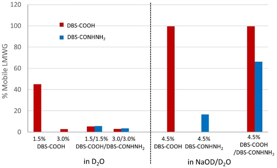 | ||
| Fig. 3 Disassembly of gel filaments as monitored by 1H NMR spectroscopy. (Left) % Mobile LMWG observed after 4 days standing in D2O. (Right) % Mobile LMWG observed after 12 h standing in NaOD/D2O. | ||
We then explored wet-spinning a combination of DBS-COOH and DBS-CONHNH2 (Fig. S10–S15, ESI†). Equal mass loadings of each gelator were employed. At the lowest loading (0.75% wt/vol of each gelator, total loading 1.5% wt/vol), uncontrolled gelation was obtained at most flow rates, except the very slowest. As LMWG loading increased, filament formation increased, but at the highest loading (total load 6.0% wt/vol), clogs became increasingly common at low flow rates in larger needles. Our preferred conditions were a 23G needle at a flow rate of 3.4–10.2 μL min−1, with total LMWG loading of 3.0% or 4.5% wt/vol. Filament diameters (130–230 μm) were somewhat dependent on loading; higher loadings giving wider filaments (Table S4, ESI†). Tgel values indicated very good thermal stability being 87–96 °C at LMWG loadings of 1.5–6.0% wt/vol (Table S5, ESI†).
SEM revealed a well-defined nanofibrillar interior network, similar to DBS-COOH alone, although there did appear to be some additional ‘film-like’ morphologies which could result from a drying artefact17 (Fig. 2(B), and Fig. S16, ESI†). TEM, however, indicated different, very well-defined nanostructures (Fig. 2(D) and Fig. S17, ESI†). These appeared to be longer and more flexible than the DBS-COOH objects, and with darker edges, there was evidence they were nanotubular, with consistent diameters of 12.3 ± 2.5 nm. They were better defined than the nanofibers previously observed by TEM imaging of DBS-CONHNH2-only filaments (with equivalent sample preparation).12 The presence of a single well-defined nanoscale morphology suggests the LMWGs co-assemble into a new architecture when wet-spun together. This is different to self-sorted networks of these gels, formed using stepwise cooling/pH triggering,14 which had 10–30 nm fibres. This indicates a different self-assembly pathway arises from rapid triggering achieved by the solvent switch mechanism used in wet spinning. We suggest this leads to random organisation of the gelators in co-assembled nanofibers. We were somewhat surprised by this as we had not previously observed co-assembly of these LMWGs, even though they have similar structures. It would be interesting in future to wet-spin LMWGs with very different structures to further understand this.
IR spectroscopy found that the N–H stretch shifted from a sharp band at 3295 cm−1 for filaments formed by DBS-CONHNH212 to a broader band from 3180–3290 cm−1 for the two-component filament (Fig. S18 and S19, ESI†). The C![[double bond, length as m-dash]](https://www.rsc.org/images/entities/char_e001.gif) O stretch of DBS-COOH shifts slightly from 1689 cm−1 for wet-spun DBS-COOH to 1692 cm−1 for the two-component filaments (Fig. S20, ESI†). These changes suggest hydrogen bond interactions between the components, meaning the molecules intimately interact with each other when wet-spun together.
O stretch of DBS-COOH shifts slightly from 1689 cm−1 for wet-spun DBS-COOH to 1692 cm−1 for the two-component filaments (Fig. S20, ESI†). These changes suggest hydrogen bond interactions between the components, meaning the molecules intimately interact with each other when wet-spun together.
1H NMR indicated >99% of each gelator assembled into filaments, with <1% residual DMSO present. On standing the filaments in D2O, there was very little disassembly (<10%) over 4 days – even at the lowest total loading of 1.5% wt/vol (Fig. 3 and Table S6, ESI†). This is very different to DBS-COOH, where ca. 50% of LMWG became mobile. Wet-spinning in combination with DBS-CONHNH2 stabilises the gel nanostructures at ambient pH. We suggest that once co-assembled with DBS-CONHNH2, the ambient pH of water has less impact. In support of this, although pH initially falls on printing the two-component gel filaments, it then rises again suggesting an active role of DBS-CONHNH2 in meditating pH (Fig. S21, ESI†) The stability of DBS-COOH in combination with DBS-CONHNH2 in the filaments is different to self-sorted gels, in which, DBS-COOH disassembled even in the presence of DBS-CONHNH2,14 because they were sorted into their own nanostructures rather than in an intimate co-assembly. Wet-spinning is therefore promising for applying this two-component gel in tissue engineering,18 as it should have the stability to survive in tissue growth medium.
We tested disruption of the two-component filaments with NaOD/D2O. The DBS-COOH network was fully disrupted (Fig. 4 and Table S7, ESI†). In addition the DBS-CONHNH2 network became >50% mobile (66% after 12 h). This suggests basic conditions fully deprotonate DBS-COOH, destabilising the nanostructures, breaking them down and releasing DBS-CONHNH2. Not all of the DBS-CONHNH2 becomes ‘mobile’, but its solubility is limited in water, which will limit the amount that can be observed. In contrast, DBS-CONHNH2-only gel filaments are stable in basic conditions, with <20% of the gelator being mobilised (Fig. 3 and Table S8, ESI†). This supports the hypothesis that in the two-component wet-spun filaments, the gelators affect one another's behaviour because they are integrated into the same nanofibres. As such, DBS-COOH endows DBS-CONHNH2 with base-responsiveness.
 | ||
| Fig. 4 Photographs of gel filaments based on (A) DBS-COOH and (B) DBS-CONHNH2/DBS-COOH on exposure to aqueous AuCl3 solutions. (C) TEM image of DBS-CONHNH2/DBS-COOH gel after exposure to AuCl3 and in situ formation of AuNPs (scale bar = 100 nm). | ||
Finally, we tested the ability of these gel filaments to extract precious metals (e.g. Au(III)) from solution and form metal nanoparticles (e.g. AuNPs) in situ (Fig. 4). This is a distinctive behaviour of DBS-CONHNH2-based gels not observed for other LMWGs and acts as a ‘signature’ of this molecule within a gel.19 AuNP-loaded gels have applications as a result of their conductivity and biocompatibility (see below). On exposure of DBS-CONHNH2/DBS-COOH gel filaments to AuCl3 solution, an almost immediate colour change of filaments from white to purple was observed, consistent with AuNP formation (Fig. 4(B)). Conversely, for DBS-COOH-only gel filaments there was no colour change (Fig. 4(A)).
Gold uptake was quantified by UV-Vis spectroscopy (Table S9, ESI†). The two-component system incorporated increasing amounts of gold as the loading of DBS-CONHNH2 increased. At total loadings of 1.5%, 3.0% and 4.5% wt/vol (0.75%, 1.5% and 2.25% wt/vol of DBS-CONHNH2), Au uptake was 115, 140 and 164 μmol mL−1, respectively. In our previous work on DBS-CONHNH2-only gels, an LMWG loading of 1.5% wt/vol led to Au uptake of 127 μmol mL−1.12 This is comparable to the 140 μmol mL−1 observed for the multicomponent system with equivalent DBS-CONHNH2 loading, indicating DBS-CONHNH2 retains its ability to reduce Au(III) to Au(0) in the presence of co-assembled DBS-COOH. Although DBS-COOH-only gels took up some Au(III), there was no evidence of reduction, and uptake was constant across all LMWG loadings, suggesting this is simple diffusion/partitioning, rather than specific interactions with the LMWG.
TEM images show AuNPs fairly uniformly dispersed within the two-component gel filaments (Fig. 4(C), and Fig. S23, ESI†). There was some evidence of AuNPs in the DBS-COOH-only gel filaments (Fig. S22, ESI†) – but significantly fewer, and there were also significant areas of larger aggregates suggesting no control over metal uptake.
In conclusion, DBS-COOH, like DBS-CONHNH2, can be wet-spun into gel filaments via DMSO/H2O solvent-exchange. The DBS-COOH filaments can be dynamically disassembled by the addition of base, and at low loadings are prone to disassemble in water. On wet-spinning DBS-COOH in combination with DBS-CONHNH2, a new co-assembled tubular nanostructure results. Importantly, the presence of DBS-CONHNH2 stabilises the gel filaments in water and prevents DBS-COOH disassembly. However, the presence of DBS-COOH makes the system base-sensitive and enables base-triggered DBS-CONHNH2 disassembly, which cannot be achieved for DBS-CONHNH2-only gel filaments. The presence of DBS-CONHNH2 allows the gel filaments to reduce precious metal salts to nanoparticles in situ, which cannot be achieved by DBS-COOH-only gels. Wet-spinning a combination of gelators therefore yields nanostructured gel filaments with a randomly co-assembled nanostructure, that could not previously be accessed. This harnesses the dynamic properties of DBS-COOH alongside the reducing ability of DBS-CONHNH2 in a synergistic way to form stable but responsive filaments with incorporated AuNPs. Future work will test the biocompatibility of these filaments, and use them in layer-by-layer 3D-printing. Loading gels with AuNPs can enhance cell growth, and endow conductivity,19 which could give such filaments impact in 3D scaffolds for electrical stimulus-responsive cells (e.g. stem cells, neurons, muscles).
The project was supervised by DKS who developed the concept. END performed experimental work and data interpretation. CCP provided training in wet-spinning. JF helped develop the experimental set-up in York. DKS helped with data analysis, and wrote the manuscript with input from all other authors.
We thank Karen Hodgkinson (Dept of Biology, Univ. York) for SEM and TEM imaging and Lamisse El-Qarra for image analysis. CCP thanks RSC (Mobility Grant, M19-9519) for funding.
Conflicts of interest
There are no conflicts to declare.Notes and references
- (a) X. Du, J. Zhou, J. Shi and B. Xu, Chem. Rev., 2015, 115, 13165–13307 CrossRef CAS PubMed; (b) J. Hoque, N. Sangaj and S. Varghese, Macromol. Biosci., 2019, 19, e1800259 CrossRef PubMed.
- (a) R. G. Weiss, J. Am. Chem. Soc., 2014, 136, 7519–7530 CrossRef CAS PubMed; (b) E. R. Draper and D. J. Adams, Chem, 2017, 3, 390–410 CrossRef CAS; (c) D. B. Amabilino, D. K. Smith and J. W. Steed, Chem. Soc. Rev., 2017, 46, 2404–2420 RSC; (d) S. Panja and D. J. Adams, Chem. Soc. Rev., 2021, 50, 5165–5200 RSC.
- (a) P. R. A. Chivers and D. K. Smith, Nat. Rev. Mater., 2019, 4, 463–478 CrossRef CAS; (b) G. A. Primo and A. Mata, Adv. Funct. Mater., 2021, 31, 2009574 CrossRef CAS.
- (a) J. Li, C. Wu, P. K. Chu and M. Gelinksy, Mater. Sci. Eng. Rep., 2020, 140, 100543 CrossRef; (b) A. Kjar, B. McFarland, K. Mecham, N. Harward and Y. Huang, Bioact. Mater., 2021, 6, 460–471 CrossRef CAS PubMed; (c) S. Bom, R. Ribeiro, H. M. Ribeiro, C. Santos and J. Marto, Int. J. Pharm., 2022, 615, 121506 CrossRef CAS PubMed.
- M. C. Nolan, A. M. Fuentes Caparrós, B. Dietrich, M. Barrow, E. R. Cross, M. Bleuel, S. M. King and D. J. Adams, Soft Matter, 2017, 13, 8426–8432 RSC.
- (a) Y. Xia, M. Qin, Y. Cao, Y. Li and W. Wang, Sci. Rep., 2017, 7, 9691 CrossRef PubMed; (b) B. Raphael, T. Khalil, V. L. Workman, A. Smith, C. P. Brown, C. Streuli, A. Saiaini and M. Domingos, Mater. Lett., 2017, 190, 103–106 CrossRef CAS; (c) H. H. Susapto, D. Alhattab, S. Abdelrahman, Z. Khan, S. Alshehri, K. Kahin, R. Ge, M. Moretti, A.-H. Emwas and C. A. E. Hauser, Nano Lett., 2021, 21, 2719–2729 CrossRef CAS; (d) S. Rauf, H. H. Susapto, K. Kahin, S. Alshehri, S. Abdelrahman, J. H. Lam, S. Asad, S. Jadhav, D. Sundaramurthi, X. Gao and C. A. E. Hauser, J. Mater. Chem. B, 2021, 9, 1069–1081 RSC; (e) N. A. Sather, H. Sai, I. R. Sasselli, K. Sato, W. Ji, C. V. Synatschke, R. T. Zambrotta, J. F. Edelbrock, R. R. Kohlmeyer, J. O. Hardin, J. D. Berrigan, M. F. Durstock, P. Mirau and S. I. Stupp, Small, 2021, 17, 2005743 CrossRef CAS PubMed; (f) J. Yang, M. Chen, H. Lee, Z. Xu, Z. Zhou, S.-P. Feng and J. T. Kim, ACS Appl. Mater. Interfaces, 2021, 13, 20573–20580 CrossRef CAS PubMed.
- (a) H. Yang, S. Zhang, K. Liu and Y. Fang, RSC Adv., 2016, 6, 109969 RSC; (b) K. Liu, S. Zang, R. Xue, J. Yang, L. Wang, J. Huang and Y. Yan, ACS Appl. Mater. Interfaces, 2018, 10, 4530–4539 CrossRef CAS PubMed; (c) A. Biswas, S. Malferran, D. M. Kalaskar and A. K. Das, Chem. Commun., 2018, 54, 1778–1781 RSC; (d) B. Dessane, R. Smirani, G. Bouguéon, T. Kauss, E. Ribot, R. Devillard, P. Barthélémy, A. Naveau and S. Crauste-Manciet, Sci. Rep., 2020, 10, 2850 CrossRef CAS PubMed; (e) Z. Zhou, M. Samperi, L. Santu, G. Dizon, S. Aboarkaba, D. Límon, C. Tuck, L. Pérez-García, D. J. Irvine, D. B. Amabilino and R. Wildman, Mater. Des., 2021, 206, 109792 CrossRef CAS; (f) G. Yang, R. Guan, H. Zhen, K. Ou, J. Fang, D. Li, Q. Fu and Y. Sun, ACS Appl. Mater. Interfaces, 2022, 14, 10998–11005 CrossRef CAS PubMed.
- A. M. Fuentes-Caparrós, Z. Canales-Galarza, M. Barrow, B. Dietrich, J. Läuger, M. Nemeth, E. R. Draper and D. J. Adams, Biomacromolecules, 2021, 22, 1625–1638 CrossRef PubMed.
- (a) H. Jian, M. Wang, Q. Dong, J. Li, A. Wang, X. Li, P. Ren and S. Bai, ACS Appl. Mater. Interfaces, 2019, 11, 46419–46426 CrossRef CAS; (b) M. J. S. Hill and D. J. Adams, Soft Matter, 2022, 18, 5960–5965 RSC.
- (a) A. Chalard, P. Joseph, S. Souleille, B. Lonetti, N. Saffon-Merceron, I. Loubinoux, L. Vaysse, L. Malaquin and J. Fitremann, Nanoscale, 2019, 11, 15043–15056 RSC; (b) A. Chalard, M. Mauduit, S. Souleille, P. Joseph, L. Malaquin and J. Fitremann, Addit. Manuf., 2020, 33, 101162 CAS; (c) D. Bordignon, B. Lonetti, C. Coudret, P. Roblin, P. Joseph, L. Malaquin, A. Chalard and J. Fitremann, J. Colloid Interface Sci., 2021, 603, 333–343 CrossRef CAS PubMed.
- F. Andriamiseza, D. Bordignon, B. Payré, L. Vaysse and J. Fitremann, J. Colloid Interface Sci., 2022, 617, 156–170 CrossRef CAS PubMed.
- C. C. Piras, A. G. Kay, P. G. Genever, J. Fitremann and D. K. Smith, Chem. Sci., 2022, 13, 1972–1981 RSC.
- E. R. Draper and D. J. Adams, Chem. Soc. Rev., 2018, 47, 3395–3405 RSC.
- (a) C. C. Piras and D. K. Smith, Chem. – Eur. J., 2019, 25, 11318–11326 CAS; (b) L. Schlichter, C. C. Piras and D. K. Smith, Chem. Sci., 2021, 12, 4162–4172 RSC; (c) H. S. Cooke, L. Schlichter, C. C. Piras and D. K. Smith, Chem. Sci., 2021, 12, 12156–12164 RSC.
- (a) D. J. Cornwell, B. O. Okesola and D. K. Smith, Soft Matter, 2013, 9, 8730–8736 RSC; (b) B. O. Okesola and D. K. Smith, Chem. Commun., 2013, 49, 11164–11166 RSC.
- D. J. Cornwell, O. J. Daubney and D. K. Smith, J. Am. Chem. Soc., 2015, 137, 15486–15492 CrossRef CAS PubMed.
- D. J. Adams, Gels, 2018, 4, 32 CrossRef.
- B. O. Okesola, Y. Wu, B. Derkus, S. Gani, D. Wu, D. Knani, D. K. Smith, D. J. Adams and A. Mata, Chem. Mater., 2019, 31, 7883–7897 CrossRef CAS PubMed.
- B. O. Okesola, S. K. Suravaram, A. Parkin and D. K. Smith, Angew. Chem., Int. Ed., 2016, 55, 183–187 CrossRef CAS PubMed.
Footnotes |
| † Electronic supplementary information (ESI) available: Materials and methods, full characterisation and additional data. See DOI: https://doi.org/10.1039/d2cc04003d |
| ‡ A loading of 1.5% wt/vol means 15 mg of LMWG are dissolved in 1.0 ml DMSO. Given the density of DMSO is 1.10 mg mL−1, this is equivalent to a loading of 1.36% wt/wt. |
| This journal is © The Royal Society of Chemistry 2022 |