 Open Access Article
Creative Commons Attribution 3.0 Unported Licence
Correction: Novel anilino quinazoline-based EGFR tyrosine kinase inhibitors for treatment of non-small cell lung cancer
Lili
Yang
ab,
Shanshan
Liu
ab,
Jingjing
Chu
a,
Shuang
Miao
ab,
Kai
Wang
ab,
Qingwei
Zhang
ab,
Yingyi
Wang
a,
Yadi
Xiao
a,
Lina
Wu
ab,
Yang
Liu
ab,
Longjian
Yu
ab,
Caihong
Yu
ab,
Xiang
Liu
ab,
Mingxing
Ke
ab,
Zhen
Cheng
*c and
Xilin
Sun
*ab
aNHC and CAMS Key Laboratory of Molecular Probe and Targeted Theranostics, Molecular Imaging Research Center (MIRC), Harbin Medical University, Harbin, Heilongjiang 150028, China
bTOF-PET/CT/MR Center, The Fourth Hospital of Harbin Medical University, Harbin, Heilongjiang 150028, China. E-mail: sunxl@ems.hrbmu.edu.cn; Tel: +86-451-88118600
cMolecular Imaging Program at Stanford (MIPS), Department of Radiology, Stanford University School of Medicine, Stanford, California 94305, USA. E-mail: zcheng@stanford.edu; Fax: +1-650-736-7925
First published on 11th April 2022
Abstract
Correction for ‘Novel anilino quinazoline-based EGFR tyrosine kinase inhibitors for treatment of non-small cell lung cancer’ by Lili Yang et al., Biomater. Sci., 2021, 9, 443–455. DOI: 10.1039/D0BM00293C.
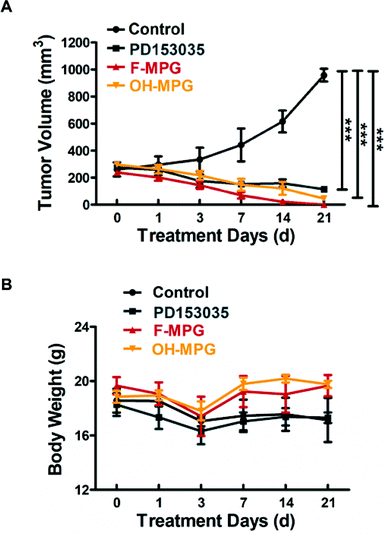
The authors regret that an incorrect image and scale bar were used in Fig. 4, as well as the incorrect scale in the figure caption. The authors also regret a spelling mistake in the Y axis of Fig. 6.
The corrected Fig. 4 and 6 are as shown here. The results and the conclusions of the manuscript remain unaffected.
The Royal Society of Chemistry apologises for these errors and any consequent inconvenience to authors and readers.
| This journal is © The Royal Society of Chemistry 2022 |


