EDC/NHS activation mechanism of polymethacrylic acid: anhydride versus NHS-ester

Qin Yan, Hong-Ning Zheng, Chuan Jiang, Kun Li and Shou-Jun Xiao*
State Key Laboratory of Coordination Chemistry, School of Chemistry and Chemical Engineering, Nanjing University, Nanjing 210093, Jiangsu, China. E-mail: sjxiao@nju.edu.cn

Received 14th July 2015 , Accepted 3rd August 2015

First published on 3rd August 2015


Abstract

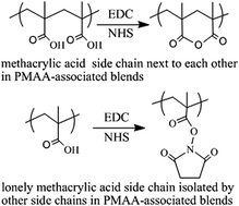
Polymer brushes of polymethacrylic acid (PMAA) and PMAA-associated polymer blends of PMAA/PNIPAM (poly-N-isopropylacrylamide) were prepared on porous silicon for further investigation of the EDC/NHS (1-ethyl-3-(3-dimethylaminopropyl)carbodiimide/N-hydroxysuccinimide) activation mechanisms by infrared spectroscopy. When the fragmentation degree of PMAA blocks in PMAA-associated polymer blends is increased, the production of anhydride wanes from dominant to recessive, whereas a complementary product of the NHS-ester waxes from recessive to dominant. The Thorpe–Ingold effect was applied to explain the formation of anhydride: the gem-dialkyl groups of PMAA next to the carboxylic acids compress the acid side chains close to each other; thus, once the intermediate of O-acylisourea forms, it will be attacked by the intramolecular neighboring acid much faster than any other nucleophiles such as NHS and water, and therefore the six-membered ring of the anhydride will be formed. All acid side chains in PMAA standing next to each other will form an anhydride, primarily due to the Thorpe–Ingold effect, unless they are sterically hindered, whereas only isolated acid side chains form the NHS-ester. The EDC/NHS activation results for four small molecules of dicarboxylic acids in aqueous media, namely, glutaric acid and 2,2-dimethyl glutaric acid, which generate disuccinimidyl ester with high yield, and succinic acid and 2,2-dimethyl succinic acid, which remain intact, can also be explained by the Thorpe–Ingold effect. A clear understanding of the EDC/NHS activation mechanisms of PMAA will take us a step closer for resolving the mechanistic ambiguity of the carbodiimide/additive coupling reactions for amide bond formation.


Introduction

Amide bond formation (or amidation) from carboxylic acid by carbodiimide activation with the assistance of additives such as NHS, followed by the addition of amine sequentially in one-pot or in stepwise reactions, is so broadly used in peptide synthesis, bio-conjugation, immunochemistry, and biosensing, that it would be difficult to overstate its importance.1 However, the reaction mechanisms still pose a challenge,2 especially due to uncertainty regarding intermediates and the large deviation in product yields for very similar carboxylic acids under the same reaction conditions. The initial activation intermediate is believed to be the unstable O-acylisourea,3 which goes through several transition routes and mechanisms to form stable intermediates or byproducts; this is the focal point of debates regarding these reactions. Taking one of the most common activation recipes as an example, namely EDC and an additive of NHS or sulfo-NHS with carboxylic acid in an aqueous media; the initial intermediate O-acylisourea converts to stable intermediates or byproducts through the following 4 pathways: 1st, nucleophilic addition of NHS to generate the NHS-ester, which efficiently forms an amide with an amine in a weak alkaline solution at room temperature; 2nd, nucleophilic addition of carboxylic acid to form a symmetrical anhydride (a stable anhydride intermediate in aqueous media was only captured and confirmed recently in the PMAA system by us4), which can react with the amine directly to form an amide, or undergo a nucleophilic addition with NHS to form the NHS-ester, or undergo hydrolysis to form the carboxylic acid; 3rd, rearrangement to a stable side product of N-acyl urea; and 4th, hydrolysis to urea and carboxylic acid by consuming carbodiimide only. All the abovementioned pathways complicate the production of an amide from the individual acid, due to the need to deal with the thermodynamics and kinetics of different pathways.5

In our previous study,4b two different intermediates were observed to be formed from two similar acid polymers after EDC/NHS activation, namely, NHS-ester from polyacrylic acid, as expected, while an anhydride was formed from PMAA, which was not predicted. We believed that the capture of polymethacrylic anhydride occurred for two reasons: the intra-molecular nucleophilic attack on O-acylisourea from a neighboring side chain of carboxylic acid, which occurred faster than any other pathways and the stable thermodynamic structure of polymethacrylic anhydride, which prevented further nucleophilic attack from NHS and water. Similar to the NHS-ester, polymethacrylic anhydride, which is a good bioconjugate reagent, possesses a longer shelf life and bears metastability towards hydrolysis for hours in weak alkaline solutions. Obviously, both the kinetic and thermodynamic properties of PMAA during EDC/NHS activation relate to the side chains of methyl groups, a phenomenon that is specifically called the Thorpe–Ingold effect (or gem-dialkyl effect), which is one of the neighboring group effects.6 The captured stable intermediate anhydride structure renders PMAA so special that further investigation of the EDC/NHS activation of PMAA and polyacrylic acid (PAA) will be not only beneficial for practical applications but also for achieving a better understanding of the carbodiimide-triggered amidation mechanisms.

In this study, we have investigated the EDC/NHS activation of two PMAA-based polymer brushes grafted on porous silicon:4b,7 (1) reiterative EDC/NHS activation and amidation of PMAA brushes and (2) EDC/NHS activation of the polymer blend brushes of PMAA/PNIPAM (poly-N-isopropylacrylamide) grown from the mixed monomers of methacrylic acid (MAA) and N-isopropylacrylamide (NIPAM) with blending ratios of 10%, 20%, 30%, 40%, 50%, 60%, 70%, 80%, 90%, and 100% MAA. We adapted our previous method:4b infrared measurements of stepwise surface reactions, which possess the merit of ignoring the laborious separation and purification procedure, especially for multiple reaction steps in aqueous media. 3-Amino-1-azidopropane (AAP),8 an infrared probe of the azido tag with a strong stretching band at 2105 cm−1, was used as the amidation reagent. We observed the complementary waxing of the NHS-ester from recessive to dominant and waning of the anhydride from dominant to recessive as the degree of fragmentation of the PMAA blocks in the above-mentioned two polymer systems was increased after EDC/NHS activation. Further amidation kinetics for both PMAA anhydride and NHS-ester with AAP was investigated using infrared absorption evolution against the reaction time. Finally, the Thorpe–Ingold effect was applied to interpret the EDC/NHS activation of 4 small dicarboxylic acids: glutaric acid, 2,2-dimethyl glutaric acid, 2,2-dimethyl succinic acid, and succinic acid. All the experimental results helped us to speculate that the faster nucleophilic attack on O-acylisourea for PMAA, 2,2-dimethyl succinic acid, and succinic acid is from the intramolecular neighboring acid due to the Thorpe–Ingold effect, thus generating the anhydride, whereas the faster nucleophilic attack on O-acylisourea for PAA, glutaric acid, and 2,2-dimethyl glutaric acid is from the intermolecular NHS, therefore producing the NHS-ester.

Experimental section

Materials

Single-sided polished silicon wafers (〈100〉, p-type, boron-doped, 5.0–8.0 Ω cm, 500 μm thick) were purchased from Hefei Kejing Materials Technology Co. Ltd. N-Ethyl-N′-(3-(dimethylamino)propyl)carbodiimide hydrochloride (EDC) (98%), N-hydroxysuccinimide (NHS) (98%), and sodium azide (NaN3, 98%) were obtained from Aladdin. Sodium methacrylate (NaMA, 99%), ω-undecylenyl alcohol (98%), 2-bromoisobutyryl bromide (98%), 3-bromopropylamine hydrobromide (98%), and 4-morpholineethanesulfonic acid hydrate (MES) were obtained from Alfa Aesar. N-Isopropylacrylamide (NIPAM, 99%), copper(I) bromide (CuBr, 98%), and pentamethyldiethylenetriamine (PMDETA, 98%) were obtained from Aldrich. Glutaric acid, 2,2-dimethyl glutaric acid, 2,2-dimethyl succinic acid, and succinic acid were obtained from TCI. PMAA (Mn 1100) and PAA (Mn 1300) were obtained from Polymer Source Inc. Water (18 MΩ cm) was obtained from a Milli-Q Ultrapure water purification system.

Fourier-transform infrared spectrometry

A Bruker V80 spectrometer equipped with a deuterated triglycine sulfate (DTGS) detector was used to record spectra for the stepwise reactions. In all the cases, the samples were mounted in a vacuum chamber and thus the interference of CO2 and water vapor from air was greatly attenuated. Spectra were recorded with 100 scans at a frequency of 7.5 kHz and a resolution of 4 cm−1, using a cleaned planar Si(100) chip as the reference. All the spectra were disposed with the OPUS software and spectral windows were magnified to highlight the significant changes.

Preparation of porous silicon

The wafer was cut into 15 × 15 × 0.5 mm3 pieces. Silicon chips were sonicated in ethanol, acetone and deionized water, each for 15 min. Then, they were immersed in piranha solution (H2SO4/H2O2 (3/1 in v/v)) at the boiling temperature for 2 h to remove any organic residues. The cleaned chips were rinsed extensively with water and ethanol and subsequently dried in a stream of nitrogen (unless stated otherwise, these steps were adopted in all the following chip cleaning and drying procedures). We used metal-assisted stain etching to produce porous silicon.7c Briefly, the chip was immersed in 1% HF aqueous solution for 15 min to remove the surface silicon oxide film. After cleaning, the hydrided chip was deposited with a thin metallic film of Pt (10 nm thick) using a sputter-coater (SCD 500) at 15 mA for 180 s. The Pt-coated chip was etched in a mixture of 40% HF, 30% H2O2 and anhydrous ethanol at a 2[thin space (1/6-em)]:[thin space (1/6-em)]2[thin space (1/6-em)]:[thin space (1/6-em)]1 (v/v/v) ratio for 4 min in the dark and sequentially immersed in 1% HF aqueous solution for 1 min. It was then rinsed with copious amounts of water and ethanol and dried with N2; hydrogen-terminated porous silicon chips were thus produced.

Surface hydrosilylation and introduction of surface initiator4b,7c

The freshly etched silicon chips were transferred into a glass bottle containing 10 mL neat ω-undecylenyl alcohol. The bottle was purged with N2 for 15 min to vent the air. The reaction was performed in a CEM Discovery microwave reactor, which was controlled with a dynamic mode to reach 120 °C in 10 min and was held at this temperature for 20 min. After the reaction, the chip was washed sonically with anhydrous alcohol for 3 min and with water for 3 min. The undecylenyl alcohol-modified chip with hydroxy-termini was placed into a glass bottle containing 10 mL CH2Cl2 and 2 mL Et3N. The reaction vessel was cooled in an ice bath for 15 min, then 2 mL 2-bromoisobutyryl bromide was dropped in slowly, and finally the reaction solution was allowed to stand at room temperature for 12 h. After the reaction, the chip with a surface initiator containing 2-bromoisobutyryl groups was rinsed with copious amounts of CH2Cl2 and ethanol and dried in a stream of nitrogen.

Preparation of PMAA and PMAA/PNIPAM brushes4b,7c,9

Surface-initiated polymerization of NaMA was performed in a N2-filled vial with 1.69 g NaMA (15 mmol) and 75 μL PMDETA dissolved in a 5 mL solution of H2O/CH3OH (1/1 v/v). The solution was adjusted to pH 9.0. The bottle was purged with N2 for 15 min to vent the air, followed by the addition of 20 mg CuBr and held at 40 °C for 5 h. Then, the chips were removed from the vial, washed with THF, H2O and ethanol and dried under a stream of N2. PMAA brushes were obtained by the immersion of the chip into 0.1 M phosphate–citric acid buffer (pH = 3.0) for 3 h to change carboxylate anions (COO) into carboxylic acid (COOH) species (the acidification procedure in the reiterative reactions followed the same protocol). Random polymer blend brushes of PMAA/PNIPAM were prepared using the same procedure. The total amount of monomers was 15 mmol. Ten samples of random PMAA/PNIPAM blend brushes with the following NIPAM[thin space (1/6-em)]:[thin space (1/6-em)]NaMA molar ratios were prepared: 9[thin space (1/6-em)]:[thin space (1/6-em)]1, 8[thin space (1/6-em)]:[thin space (1/6-em)]2, 7[thin space (1/6-em)]:[thin space (1/6-em)]3, 6[thin space (1/6-em)]:[thin space (1/6-em)]4, 5[thin space (1/6-em)]:[thin space (1/6-em)]5, 4[thin space (1/6-em)]:[thin space (1/6-em)]6, 3[thin space (1/6-em)]:[thin space (1/6-em)]7, 2[thin space (1/6-em)]:[thin space (1/6-em)]8, and 1[thin space (1/6-em)]:[thin space (1/6-em)]9.

Surface EDC/NHS activation of PMAA and PMAA/PNIPAM brushes

EDC/NHS activation of the chip was performed under the optimum reaction conditions reported previously by us:4b 0.1 mol L−1 EDC and 0.2 mol L−1 NHS in 0.1 mol L−1 MES buffer at pH 6.5 at room temperature for 1 h. After the reaction, the chip was rinsed with water and dried under a stream of nitrogen.

EDC/NHS activation of dicarboxylic acids

The EDC/NHS activation of dicarboxylic acids of glutaric acid, 2,2-dimethyl glutaric acid, 2,2-dimethyl succinic acid, and succinic acid was performed with 0.1 mol L−1 dicarboxylic acid, 0.25 mol L−1 EDC, and 0.25 mol L−1 NHS in 0.1 mol L−1 MES buffer (pH 6.5) at room temperature for 1 h. For example, 15 mL freshly prepared MES buffer containing 2.4 g EDC (0.0125 mol) was added immediately to 35 mL stirred MES buffer containing glutaric acid (0.66 g, 0.005 mol) and NHS (1.44 g, 0.0125 mol) at 0 °C. Then, the mixture was stirred at room temperature for 1 h. The white suspension was filtered, washed with water, then dissolved in dichloromethane, dried over anhydrous Na2SO4, and concentrated under vacuum. A white solid of glutaric acid di-succinimidyl ester (GA-NHS) was obtained after recrystallization from isopropyl alcohol (1.2 g, 73% yield). Following the same protocol, another white solid of 2,2-dimethyl glutaric acid di-succinimidyl ester (DMGA-NHS) was obtained after recrystallization with a yield of 85%. No products were obtained from either 2,2-dimethyl succinic acid or succinic acid solutions, following the same EDC/NHS activation protocol.

Preparation of 3-amino-1-azide propane (AAP)

AAP was prepared according to the literature.8 3-Bromopropylamine hydrobromide (15 mmol, 3.2 g) was suspended in water (10 mL), followed by the addition of NaN3 (50 mmol, 3.2 g) in 15 mL of water. The mixture was heated to 80 °C for 12 h, followed by the removal of 2/3rd of water under vacuum. The resulting mixture was cooled in an ice bath, and diethyl ether (50 mL) and KOH pellets (4 g) were added while keeping the temperature °C. The organic layer was separated and the aqueous phase was extracted with diethyl ether (2 × 30 mL), dried over K2CO3, and concentrated. The pure product was obtained by silica column chromatography with CH3OH/CH2Cl2 (v[thin space (1/6-em)]:[thin space (1/6-em)]v = 1[thin space (1/6-em)]:[thin space (1/6-em)]3) to obtain a clear, yellow oil (1.1 g, 73%).

Amidation

Generally, the PMAA anhydride or NHS-ester chips were incubated in 5 mL 0.1 M NaHCO3/Na2CO3 buffer (v/v = 1/1 with DMSO, pH = 8.5) containing 0.1 mM AAP at room temperature for 1 h, whereas for kinetic studies, the chips were withdrawn at 2, 20, 40, 80, and 120 min, rinsed with water, dried with nitrogen, and studied by infrared spectrometry.

Results and discussion

Iterative EDC/NHS activation and amidation of PMAA

We have previously reported that the predominant product of EDC/NHS activation of PMAA is the anhydride.4 However, the anhydride has its own limitations for amidation; theoretically, only half the carboxylic acids can be converted to amide, while the rest return to carboxylic acids. To overcome this limitation, we reiterated the EDC/NHS activation for the rest of the carboxylic acids before inducing amidation again three times. We define the products of the 1st, 2nd and 3rd EDC/NHS activations as 0.5G, 1.5G, and 2.5G, respectively, while the amidation products produced from 0.5G, 1.5G, and 2.5G are denoted as 1.0G, 2.0G and 3.0G, respectively. Their respective infrared traces are shown in Fig. 1 and the infrared peak assignments in the carbonyl stretching region from 1500 to 2200 cm−1 are listed in Table S1 (see Table S1 in ESI).
image file: c5ra13844b-f1.tif
Fig. 1 Infrared spectra of PMAA, 0.5G, 1.0G, 1.5G, 2.0G, 2.5G and 3.0G are displayed in the left frame from top to bottom. The region of 1650–1850 cm−1 is magnified and fitted to have a clear view of the deconvoluted anhydride (blue doublet bands), the NHS-ester (pink triplex bands) and the acid (green curved empty single band) in 0.5G, 1.5G and 2.5G, respectively in the right-hand frame.

The molar concentrations of surface species can be quantitatively estimated from their corresponding absorption bands using the Beer–Lambert law: A = εbc (where A is the absorbance, ε is the molar extinction coefficient, b is the thickness of the polymer brushes, and c is the concentration). To execute quantitative analyses using the infrared spectra in Fig. 1, we classified the reiterative reactions into two groups: 0.5G, 1.5G, 2.5G and 1.0G, 2.0G, 3.0G. In the first group, all three products in the infrared region of 1650–1850 cm−1 can be fitted as shown in the right-hand column of Fig. 1: NHS-ester (associated pink triplex infrared bands at 1738, 1780, and 1811 cm−1), anhydride (associated blue doublet bands at 1760 and 1802 cm−1), and acid residue (green curved empty single band at 1714 cm−1). Byproducts of N-acylurea will be ignored in the calculations because in the second group no n-trace in 1.0G and a neglectable trace in 2.0G and 3.0G of N-acylurea can be found in the carbonyl stretching region (1700–2000 cm−1).4b The Beer–Lambert law for NHS-ester, anhydride, and acid in the first group can be written as follows:

ANHS-ester = εNHS-esterbcNHS-ester

Aanhydride = εanhydridebcanhydride

Aacid = εacidbcacid
where b has the same polymer thickness for all the 3 species.

We adapted the linear relationship between the 3 individual molar extinction coefficients from our previous report: εNHS-ester/εanhydride/εacid = 2[thin space (1/6-em)]:[thin space (1/6-em)]1.5[thin space (1/6-em)]:[thin space (1/6-em)]1.4b The largest peak heights of each species in Fig. 1 were used as the absorbance (A) for calculations: 1760 cm−1 for anhydride, 1738 cm−1 for NHS-ester, and 1714 cm−1 for carboxylic acid.

Assuming that all the reactants of the side chain acid groups are converted to NHS-ester, anhydride, and acid residue and not to any other compound, the molar percentages (represented with β) with an acid group as the unit converting to NHS-ester (βNHS-ester), anhydride (βanhydride) and acid (βacid) can be calculated individually using eqn (1)–(3).

 
image file: c5ra13844b-t1.tif(1)
 
image file: c5ra13844b-t2.tif(2)
 
image file: c5ra13844b-t3.tif(3)

We obtained βanhydride, βNHS-ester, and βacid as 83, 10, 7; 52, 28, 20; and 17, 46, 37 for 0.5G, 1.5G, and 2.5G, respectively (see Table S2 in ESI).

For the second group, 1.0G, 2.0G, and 3.0G, there are only two products of the amide and the carboxylate; this can be proven from their respective infrared traces in Fig. 1. The as-obtained amide was confirmed with amides I and II at 1650 and 1529 cm−1, respectively. Because the weak amide II overlaps with the carboxylate (1570 cm−1) and amide I is always complicated by factors such as water vapor and its orientation in the supporting substrate,10 we used the independent and strong vibration band of the azide at 2105 cm−1, as well as that of the carboxylate (1570 cm−1) to calculate the amide yield (αazide) as follows:

 
image file: c5ra13844b-t4.tif(4)
 
image file: c5ra13844b-t5.tif(5)
where Aazide and ACOO can be obtained from the peak heights of 2105 and 1570 cm−1 in Fig. 1, respectively. We adapted the molar extinction coefficients of εazide = 1000 LM−1 cm−1 (ref. 11) and εCOO = 380 LM−1 cm−1 (ref. 12) from previous studies, and the amide yields were obtained as 32%, 48%, and 62% for 1.0G, 2.0G, and 3.0G, respectively. Combining the calculated results of βanhydride, βNHS-ester, βacid and αazide, we can easily derive the component compositions (see Table S3 in ESI), which are shown in Scheme 1 under their corresponding structures.


image file: c5ra13844b-s1.tif
Scheme 1 Schematic drawing of molecular structures with three reiterative EDC/NHS activations and amidations of PMAA (molar percentages are listed under the subunit structures). The structures in parentheses are subunits, while the units for the polymer blend are in brackets. Curved lines represent connections, but not only in this order; subunits in parentheses with the subscripts m, n, p, and k (≥1) are variable integers, meaning that they can be isolated or next to each other, while subunits in parentheses without subscripts indicate that they are isolated and cannot be situated next to each other. 0.5G, 1.5G, 2.5G represent the products after the 1st, 2nd and 3rd EDC/NHS activations, respectively; 1.0G, 2.0G, and 3.0G represent the products after successive amidations of 0.5G, 1.5G, 2.5G, respectively.

The molecular structures and their stoichiometric molar ratios shown in Scheme 1 are derived from the quantitative analyses of infrared spectra. Details are illustrated as follows: (i) during the first EDC/NHS activation (0.5G), the anhydride is the overwhelming product (83%) due to the faster kinetic ring-closing mechanism and the thermodynamically stable anhydride; then, the isolated acid side chains are partly esterified to NHS-esters (10%) and partly left as unreacted acid residues (7%) due to steric hindrance such as the one-end fixed backbone on silicon having less freedom of translation and rotation. (ii) Successive AAP amidation (1.0G) generates amides (32%) and carboxylates (68%), which can be assigned to three subunit structures. The green subunit can be inferred statistically if amidation occurs in an even space when half of the anhydrides react with AAP. Supposing that the NHS-ester converts completely to the amide and the other chemical conversions rationally follow the dashed arrows from 0.5G to 1.0G, an algebraic approach for balancing the chemical equation gives perfectly matching compositions for the subunit structures depicted in 1.0G, among which the green subunit is the key structure to consistently interpret the reaction mechanisms. (iii) For the 2nd EDC/NHS activation (1.5G), the independently measured molar percentages of the anhydride (35%), the NHS-ester (19%), and the acid (14%) perfectly follow the chemical reaction logistics from 1.0G. (vi) Successive amidation (2.0G) produces amides (48%) and carboxylates (52%). (v) The 3rd EDC/NHS activation (2.5G) of the remaining 48% of the acids provides anhydride (9%), NHS-ester (24%), and acid (19%). (vi) Successive AAP amidation generates amides (62%) and carboxylate residues (38%). Repeating the EDC/NHS activation further will overwhelmingly generate NHS-ester intermediates and the AAP amidation will further achieve over 80% amides. All the subunits depicted as monomers are suggested rationally and are inferred logistically from the green subunits.

From the abovementioned self-consistent quantitative analyses, we can deduce the key idea about the structure–reactivity relationship as follows: all the acid side chains standing next to each other will form the anhydride primarily due to the Thorpe–Ingold effect, unless they are sterically hindered, while the isolated acid side chains and acid side chains that act as isolated ones, such as in the trimer after anhydride formation at either side or in the tetramer after anhydride formation at the middle, form the NHS-ester.

EDC/NHS activation of random polymer blend brushes of PMAA/PNIPAM

To prove the abovementioned structure–reactivity statement further, we applied the EDC/NHS activations on another PMAA-based acid system, i.e., the random polymer blends of PMAA/PNIPAM with a series of molar blending ratios of 10, 20, 30, 40, 50, 60, 70, 80, 90, and 100% PMAA in increasing concentration steps of 10% PMAA. In the polymer blends, methacrylic acids are interrupted by N-isopropylacrylamide monomers or oligomers to form monomers, dimers, trimers, and multimers sequentially and statistically from low to high PMAA concentrations in the backbone. After EDC/NHS activation, we observed similar evolution phenomena for both products of NHS-ester and anhydride. These are illustrated in Fig. 2: as the molar concentration of PMAA is decreased from 100% to 10%, the anhydride evolves from dominant to recessive, while the NHS-ester changes from recessive to dominant.
image file: c5ra13844b-f2.tif
Fig. 2 Infrared spectra of the EDC/NHS activated polymer blends of PMAA/PNIPAM with molar blending ratios of 10%, 20%, 30%, 40%, 50%, 60%, 70%, 80%, 90%, and 100% PMAA. The two intermediates of the NHS-ester (associated triplet bands at 1738, 1780, and 1807 cm−1), the anhydride (associated doublets at 1760 and 1804 cm−1) and the acid residue (1717 cm−1) are indicated. Amide I at 1652 cm−1 and amide II at 1535 cm−1 are obtained from PNIPAM. A few wavenumber shifts of the same species from Fig. 1 are due to different neighboring group effects.

Quantitative analysis for Fig. 2 with eqn (1)–(3) after deconvolution (ignored) of the 10 traces in the infrared region of 1700–1900 cm−1 provide the product composition against the molar percentage of PMAA in Fig. 3, without counting PNIPAM. Obviously, 10–20% of the acid residues remain un-reacted, which can be deduced from the knowledge of chemical reactions; the anhydride increases with PMAA concentration roughly in a linear relationship, from 6% in the 10% PMAA/PNIPAM blend up to 83% in pure PMAA; while the NHS-ester, in contrast to the anhydride, decreases linearly, from 76% in the 10% PMAA/PNIPAM blend to 9% in pure PMAA. In the medium PMAA concentration range (30–70%), both the anhydride and the NHS-ester are generated in considerable quantities. The observed phenomenon can be explained in the same way as that for the reiterative EDC/NHS activations on PMAA brushes. Isolated acid side chains are dominant at lower PMAA concentrations; oligomers of MAA, such as dimers, trimers, and tetramers are the major structures of PMAA in the medium concentration range, while multimers (≥4) become the dominant structures at higher PMAA concentrations. Such structural distributions result in the complementary waxing of the NHS-ester and waning of the anhydride with the fragmentation of PMAA in the polymer blend.


image file: c5ra13844b-f3.tif
Fig. 3 Molar percentage of anhydride, NHS-ester, and acid residue against the molar percentage of PMAA for polymer blends of PMAA/PNIPAM after EDC/NHS activation, without counting PNIPAM.

From the abovementioned qualitative and quantitative analysis of the infrared traces, we suggest the following mechanism for EDC/NHS activation of PMAA: one acid side chain next to another in a multimer or even in a dimer of a polymer chain will primarily form the anhydride, and an isolated acid side chain will convert to the NHS-ester.

Amidation kinetics

We have demonstrated the EDC/NHS activation product details of PMAA with different contents in its random polymer blends. While the main goal of EDC/NHS activation of carboxylic acids is to couple free amine-containing biomolecules, what is the kinetic behavior of PMAA anhydride or NHS-ester during the amidation process? To compare their kinetics, we used an anhydride, directly derivatised from PMAA brushes with a dominant content of ∼80% in all acid derivatives, and an NHS-ester, derivatised from a 30% PMAA-containing a PMAA/PNIPAM blend at a content of ∼60% in all acid derivatives, as examples to run kinetic experiments with AAP. Although a higher NHS-ester content of over 70% in all acid derivatives can be derivatised from the 20% or less PMAA-containing PMAA/PNIPAM blend, the absolute amount of NHS-ester is too low to precisely assay the amidation kinetics; therefore, we preferred to use the NHS-ester from a 30% PMAA-containing PMAA/PNIPAM blend. Fig. 4 illustrates the amidation kinetics of PMAA anhydride; Fig. 4a demonstrates the spectral evolution over time, whereas Fig. 4b shows the absorbance curve of the height at 1760 cm−1 and 2100 cm−1 versus time, indicating the rate of anhydride consumption and the rate of amide formation, respectively. Similarly, Fig. 5 presents the amidation kinetics of the NHS-ester in the PMAA/PNIPAM blend containing 70% PNIPAM with strong and broad amide bands at 1652 cm−1 and 1535 cm−1; Fig. 5a demonstrates the spectral evolution over time, whereas Fig. 5b shows the absorbance curve of the height at 1738 cm−1 and 2100 cm−1 versus time, indicating the rate of NHS-ester consumption and the rate of amide formation, respectively. From both Fig. 4 and 5, we conclude that their amidation kinetics are similar. More details are summarized as follows: (1) in the first 2 min, the amidation reactions were very fast; 50% and 64% amidation products were obtained for the anhydride and the NHS-ester, whereas 30% anhydride and 50% NHS-ester were consumed, respectively. (2) At 40 min, both amidation reactions were close to completion, therefore a reaction time of 1 h was adequate for the termination of the amidation reactions. (3) Trace amounts of residues of anhydrides or NHS-esters at even 80 min should be due to the buried species within the polymer brushes, which would be mainly hydrolysed late because they were more likely to be surrounded by water molecules. (4) Hydrolysis as a competitive reaction is much slower than amidation at pH 8.5 for both PMAA anhydride and NHS-ester because the water molecule is a weaker nucleophile than the primary amine during the nucleophilic substitution reaction. Generally, most of the anhydrides are considerably less stable than the NHS-ester and will therefore be hydrolysed immediately in aqueous media, whereas PMAA anhydride possesses a stable structure due to the gem-dialkyl group effect; therefore, it proceeds through a slow hydrolysis over hours in weak alkaline solutions, by a process comparable to the NHS-ester.
image file: c5ra13844b-f4.tif
Fig. 4 Amidation kinetics of PMAA anhydride with AAP: (a) infrared trace against reaction time and (b) absorbance height at 1760 cm−1 and 2100 cm−1 against reaction time.

image file: c5ra13844b-f5.tif
Fig. 5 Amidation kinetics of PMAA NHS-ester with AAP: (a) infrared trace against reaction time and (b) absorbance height at 1738 cm−1 and 2100 cm−1 against reaction time.

EDC/NHS activation of glutaric acids and succinic acids

Because anhydride formation in the EDC/NHS activation of PMAA is due to the Thorpe–Ingold effect: the side methyl groups compress the acid side chains close to each other; therefore, the formation of the six-membered anhydride ring by nucleophilic attack of the intramolecular neighboring acid is considerably faster than any other pathways for the derivatisation of O-acylisourea; however, for PAA, the neighboring acid distance should be slightly longer than that in PMAA; its dominant product is NHS-ester, not the anhydride, due to the faster nucleophilic attack on O-acylisourea from NHS rather than from a neighboring acid.4 Does the Thorpe–Ingold effect also play a role for small molecules in the EDC/NHS activation reaction with the structures analogous to the MAA dimer in PMAA? We ran the EDC/NHS reactions for 4 available dicarboxylic acids: glutaric acid, 2,2-dimethyl glutaric acid, succinic acid, and 2,2-dimethyl succinic acid. The di-NHS-ester products (GA-NHS13 and DMGA-NHS, their IR, 1H-NMR and MS are presented in Figs. S1-5 of ESI) were easily obtained for glutaric acid and 2,2-dimethyl glutaric acid as white precipitates (shown in Fig. S1), whereas the di-NHS-ester products were not observed for succinic acid and 2,2-dimethyl succinic acid; the reaction solutions were clear and the acids remained unreacted. Why do similar dicarboxylic acids exhibit such different products? From our understanding of the EDC/NHS activation of PMAA and PAA, we give our interpretation here to coordinate the observed phenomena together: the distance between the two end carboxylic acids in glutaric acid and 2,2-dimethyl glutaric acid is a little bit longer than in succinic acid and 2,2-dimethyl succinic acid; once an O-acylisourea is formed, the nucleophilic attack competition between NHS and the neighboring acid on O-acylisourea determines the final product. For glutaric acid and 2,2-dimethyl glutaric acid, the nucleophilic attack from NHS is faster than from its neighboring acid; therefore, the di-NHS-ester products are formed easily with high yields. For succinic acid and 2,2-dimethyl succinic acid, the nucleophilic attack from their neighboring acid is faster than from NHS due to the Thorpe–Ingold effect; therefore, the five-membered ring anhydride, succinic anhydride or 2,2-dimethyl succinic anhydride, is formed primarily. However, both succinic anhydride and 2,2-dimethyl succinic anhydride are so vulnerable in aqueous media, that they will be hydrolysed to acids immediately. Both O-acylisourea and succinic anhydrides can react with NHS thermodynamically; however, the kinetic probability of attack from NHS is considerably lower than that of attack from its competitors. Therefore, the formation and hydrolysis of succinic anhydrides during the EDC/NHS activation forms a cycle. By the end, the only consumed reactants are EDC and water; the reaction follows the 4th pathway as described in the Introduction: O-acylisourea was hydrolysed to urea and carboxylic acid by consuming carbodiimide only.

By the way, ideal small dicarboxylic acid molecules, for investigation of the gem-dialkyl effect in EDC/NHS activation, will be gem-dialkyl glutaric acids, i.e. 2,2,4,4-tetramethyl glutaric acid and 2,4-dimethyl-2,4-diethyl glutaric acid, because they resemble the MAA dimer structure in PMAA better. However, these small molecules have not yet been synthesized, maybe due to the crowded side groups.

Discussion

From the abovementioned experimental results, we infer that, in the analogous structures of glutaric acids including PMAA and PAA, the initial EDC activation of the carboxylic acid to O-acylisourea is a slow step, then the conversion of O-acylisourea to NHS-ester (the 1st pathway) or to anhydride (the 2nd pathway) is a fast step. Formation of either the NHS-ester or the anhydride depends on the competition of nucleophilic attack from a neighboring acid and from NHS. The gem-dialkyl groups in PMAA demonstrated the typical Thorpe–Ingold effect in the EDC/NHS activation, in which a slightly shorter distance between neighboring acid groups brought by the gem-dialkyl groups in PMAA resulted in a faster nucleophilic attack on O-acylisourea from its neighboring acid and therefore generated the anhydride. However, for PAA, the nucleophilic attack on O-acylisourea from NHS was faster than from its neighboring acid; thus, the dominant EDC/NHS activation product was the NHS-ester.4b It is also reasonable to deduce that the 3rd conversion pathway of O-acylisourea to a stable side product of N-acyl urea by intramolecular rearrangement and the 4th pathway of hydrolysis of O-acylisourea by water attack would be slower than the 1st and 2nd pathways. To precisely measure and distinguish the in situ kinetics of the formation of the NHS-ester or the anhydride at a molecular level is beyond the scope of our experimental skills. However, we adapted a very rough analysis approach to demonstrate their kinetic difference at the macroscale by following the precipitation rate of PMAA anhydride and that of PAA NHS-ester with the measurement of the optical density (OD) at 600 nm, as shown in Fig. S6 and S7. We observed that PMAA anhydride precipitated immediately after the addition of EDC to the reaction solution of PMAA and NHS and the optical density of the solution achieved a saturation of 0.8 OD at 240 s (or 4 min). It was very likely that the OD saturation meant the completion of the reaction, while the precipitation of PAA NHS-ester started to occur at 120 s (or 2 min) after the addition of EDC, and the precipitation became slower after 360 s (or 6 min) and still continued very slowly until our measurement time window of 1000 s (or ∼17 min). All the abovementioned experimental results, including the precipitation rate measurements, lead us to suggest that the formation of either anhydride or NHS-ester depends on the nucleophilic attack kinetics on O-acylisourea from the intramolecular neighboring acid or from the intermolecular NHS at the molecular level. Therefore, except for the succinic acid and gem-dialkyl glutaric acid structures, which can obviously be affected by the Thorpe–Ingold effect, all other alkyl acids with a distance between two adjacent acid molecules longer than the length of glutaric acid will form the dominant intermediate product of NHS-esters.

Conclusion

In conclusion, for the irregular EDC/NHS activation products of PMAA, we observed the waxing of NHS-ester and the waning of anhydride as the fragmentation degree of PMAA blocks in PMAA-associated polymer blends was increased. It is the delicate kinetics and thermodynamics at a molecular level that bring complicated production results at a macroscale level such as variable products and yields for different types of acids. Our discovery of the different EDC/NHS activation products between PMAA and PAA and the following understanding of the Thorpe–Ingold effect on the EDC/NHS activation of PMAA allowed us to explain the phenomenon of the waxing of the NHS-ester versus the waning of the anhydride clearly. Although the Thorpe–Ingold effect is key to interpreting the observed phenomena, steric hindrance should also be included, especially for the small part of NHS-ester in pure PMAA and for nontrivial NHS-esters in PMAA oligomers such as dimers, trimers, and tetramers. From our abovementioned speculations about the EDC/NHS activation kinetics, we would suggest some modifications to the well-known EDC/NHS reaction procedures that are currently widely used. For example, the molar ratios of EDC/NHS/acid = 1.2[thin space (1/6-em)]:[thin space (1/6-em)]1.2[thin space (1/6-em)]:[thin space (1/6-em)]1, in general reaction conditions, are commonly accepted, whereas we suggest adding more NHS to the acid, i.e. in a ratio of 2 to 5 or even higher, in the reaction to obtain the NHS-ester with a higher yield because more NHS molecules will increase the probability of their nucleophilic attack on O-acylisourea. Because the EDC/NHS activation of PMAA is so popularly used for biomolecular immobilization and conjugation in biomaterials, pharmaceuticals, immunology, and food science, such a clear understanding of the reaction mechanism could lead to more economical ways of loading more biomolecules. Only EDC and not any other additives, such as NHS or HOBt, is needed in the first reaction to activate PMAA and synthesize amide linkages; reiterating the EDC/NHS activation and amidation on the product from the first round will then increase the amidation efficiency dramatically. We believe that more precise experimental measurements and theoretical studies of the EDC/NHS activation mechanisms, both in kinetics and thermodynamics, will help to resolve the ambiguity of the carbodiimide/additive coupling reaction for better amide bond formation.

Acknowledgements

This study was financially supported by the National Basic Research Program of China, No. 2013CB922101, the Natural Science Foundation of Jiangsu Province, No. BK20130054 and the State Key Laboratory of Bioelectronics of Southeast University.

Notes and references

  1. (a) A. Williams and T. I. Ibrahim, Chem. Rev., 1981, 81, 589 CrossRef CAS; (b) V. R. Pattabiraman and J. W. Bode, Nature, 2011, 480, 471 CrossRef CAS PubMed; (c) L. H. H. Olde Damink, P. J. Dijkstra, M. J. A. van Luyn, P. B. van Wachem, P. Nieuwenhuis and J. Feijen, Biomaterials, 1996, 17, 765 CrossRef CAS; (d) C. A. G. N. Montalbetti and V. Falque, Tetrahedron, 2005, 61, 10827 CrossRef CAS PubMed; (e) M. M. Joullié and K. M. Lassen, ARKIVOC, 2010, 189 Search PubMed; (f) Z. Grabarek and J. Gergely, Anal. Biochem., 1990, 185, 131 CrossRef CAS; (g) M. J. B. Wissink, R. Beernink, J. S. Pieper, A. A. Poot, G. H. M. Engbers, T. Beugeling, W. G. van Aken and J. Feijen, Biomaterials, 2001, 22, 151 CrossRef CAS; (h) J. V. Staros, R. W. Wright and D. M. Swingle, Anal. Biochem., 1986, 156, 220 CrossRef CAS.
  2. (a) T. Iwasawa, P. Wash, C. Gibson and J. Rebek Jr, Tetrahedron, 2007, 63, 6506 CrossRef CAS PubMed; (b) N. Nakajima and Y. Ikada, Bioconjugate Chem., 1995, 6, 123 CrossRef CAS; (c) B. M. Prentice, J. D. Gilbert, J. R. Stutzman, W. P. Forrest and S. A. McLuckey, J. Am. Soc. Mass Spectrom., 2013, 24, 30 CrossRef CAS PubMed.
  3. (a) D. F. de Tar and R. Silverstein, J. Am. Chem. Soc., 1966, 88, 1013 CrossRef CAS; (b) D. F. de Tar and R. Silverstein, J. Am. Chem. Soc., 1966, 88, 1020 CrossRef CAS.
  4. (a) H. B. Liu, Q. Yan, C. E. Wang, X. Liu, C. Wang, X. H. Zhou and S. J. Xiao, Colloids Surf., A, 2011, 386, 131 CrossRef CAS PubMed; (b) C. Wang, Q. Yan, H. B. Liu, X. H. Zhou and S. J. Xiao, Langmuir, 2011, 27, 12058 CrossRef CAS PubMed.
  5. (a) M. R. Lockett, M. F. Phillips, J. L. Jarecki, D. Peelen and L. M. Smith, Langmuir, 2008, 24, 69 CrossRef CAS PubMed; (b) R. L. Touahir, J.-N. Chazalviel, S. Sam, A. Moraillon, C. Henry de Villeneuve, P. Allongue, F. Ozanam and A. C. Gouget-Laemmel, J. Phys. Chem. C, 2011, 115, 6782 CrossRef; (c) S. Sam, L. Touahir, J. S. Andresa, P. Allongue, J. N. Chazalviel, A. C. Gouget-Laemmel, C. Henry de Villeneuve, A. Moraillon, F. Ozanam, N. Gabouze and S. Djebba, Langmuir, 2010, 26, 809 CrossRef CAS PubMed; (d) C. Y. Lim, N. A. Owens, R. D. Wampler, Y. Ying, J. H. Granger and M. D. Porter, Langmuir, 2014, 30, 12868 CrossRef CAS PubMed; (e) F. Palazon, C. M. Benavides, D. Leonard, E. Souteyrand, Y. Chevolot and J. P. Cloarec, Langmuir, 2014, 30, 4545 CrossRef CAS PubMed.
  6. (a) R. M. Beesley, C. K. Ingold and J. F. Thorpe, J. Chem. Soc., Trans., 1915, 107, 1080 RSC; (b) M. E. Jung and G. Piizzi, Chem. Rev., 2005, 105, 1735 CrossRef CAS PubMed; (c) C. Galli and L. Mandolini, Eur. J. Org. Chem., 2000, 3117 CrossRef CAS; (d) C. Galli and L. Mandolini, Acc. Chem. Res., 1981, 14, 94 Search PubMed; (e) R. Salvio, L. Mandolini and C. Savelli, J. Org. Chem., 2013, 78, 7259 CrossRef CAS PubMed.
  7. (a) H. F. Li, H. M. Han, Y. G. Wu and S. J. Xiao, Appl. Surf. Sci., 2010, 256, 4048 CrossRef CAS PubMed; (b) C. Wang, X. M. Jia, C. Jiang, G. N. Zhuang, Q. Yan and S. J. Xiao, Analyst, 2012, 137, 4539 RSC; (c) M. Xiao, H. M. Han and S. J. Xiao, Sci. China: Chem., 2013, 56, 1152 CrossRef CAS.
  8. N. S. Hatzakis, H. Engelkamp, K. Velonia, J. Hofkens, P. C. Christianen, A. Svendsen, S. A. Patkar, J. Vind, J. C. Maan, A. E. Rowan and R. J. Nolte, Chem. Commun., 2006, 2012 RSC.
  9. S. Tugulu, R. Barbey, M. Harms, M. Fricke, D. Volkmer, A. Rossi and H. A. Klok, Macromolecules, 2007, 40, 168 CrossRef CAS.
  10. (a) S. J. Xiao, S. Brunner and M. Wieland, J. Phys. Chem. B, 2004, 108, 16508 CrossRef CAS; (b) S. J. Xiao, M. Wieland and S. Brunner, J. Colloid Interface Sci., 2005, 290, 172 CrossRef CAS PubMed.
  11. I. T. Suydam and S. G. Boxer, Biochemistry, 2003, 42, 12050 CrossRef CAS PubMed.
  12. A. Barth, Prog. Biophys. Mol. Biol., 2000, 74, 141 CrossRef CAS.
  13. T. A. van den Berg, B. L. Feringa and G. Roelfes, Chem. Commun., 2007, 180 RSC.

Footnote

Electronic supplementary information (ESI) available: Supplementary tables and figures. See DOI: 10.1039/c5ra13844b

This journal is © The Royal Society of Chemistry 2015
Click here to see how this site uses Cookies. View our privacy policy here.