
 Open Access Article
 This Open Access Article is licensed under a
 Creative Commons Attribution 3.0 Unported Licence
Correction: Engineering Cu2−xS-conjugated upconverting nanocomposites for NIR-II light-induced enhanced chemodynamic/photothermal therapy of cancer
Kaimin
Du
ab,
Shuang
Zhao
a,
Jing
Feng
 *ab,
Xuan
Gao
ab,
Kai
Liu
ac,
Xiaozhen
Wang
*d,
Manli
Zhang
ab,
Yao
Li
ab,
Yu
Lu
a and
Hongjie
Zhang
*abc
*ab,
Xuan
Gao
ab,
Kai
Liu
ac,
Xiaozhen
Wang
*d,
Manli
Zhang
ab,
Yao
Li
ab,
Yu
Lu
a and
Hongjie
Zhang
*abc
aState Key Laboratory of Rare Earth Resource Utilization, Changchun Institute of Applied Chemistry, Chinese Academy of Sciences, 5625 Renmin Street, Changchun 130022, China. E-mail: fengj@ciac.ac.cn; hongjie@ciac.ac.cn; Fax: +86 431 85698041; Tel: +86 431 85262127
bUniversity of Science and Technology of China, Hefei 230026, China
cDepartment of Chemistry, Tsinghua University, Beijing 100084, China
dThe First Hospital of Jilin University, Changchun 130021, China. E-mail: xzwang@jiu.edu.cn
First published on 11th May 2021
Abstract
Correction for ‘Engineering Cu2−xS-conjugated upconverting nanocomposites for NIR-II light-induced enhanced chemodynamic/photothermal therapy of cancer’ by Kaimin Du et al., J. Mater. Chem. B, 2021, DOI: 10.1039/d1tb00337b.
The authors apologise for omitting scale bars from Fig. 3c and d and for including an incorrect version of the H&E-stained slices of tumor tissues collected from the “UCNPs–Cu2−xS + laser” group in Fig. 5g.
The corrected versions of Fig. 3 and 5 are provided below. The authors confirm that these do not influence any of the experimental results and conclusions of the study.
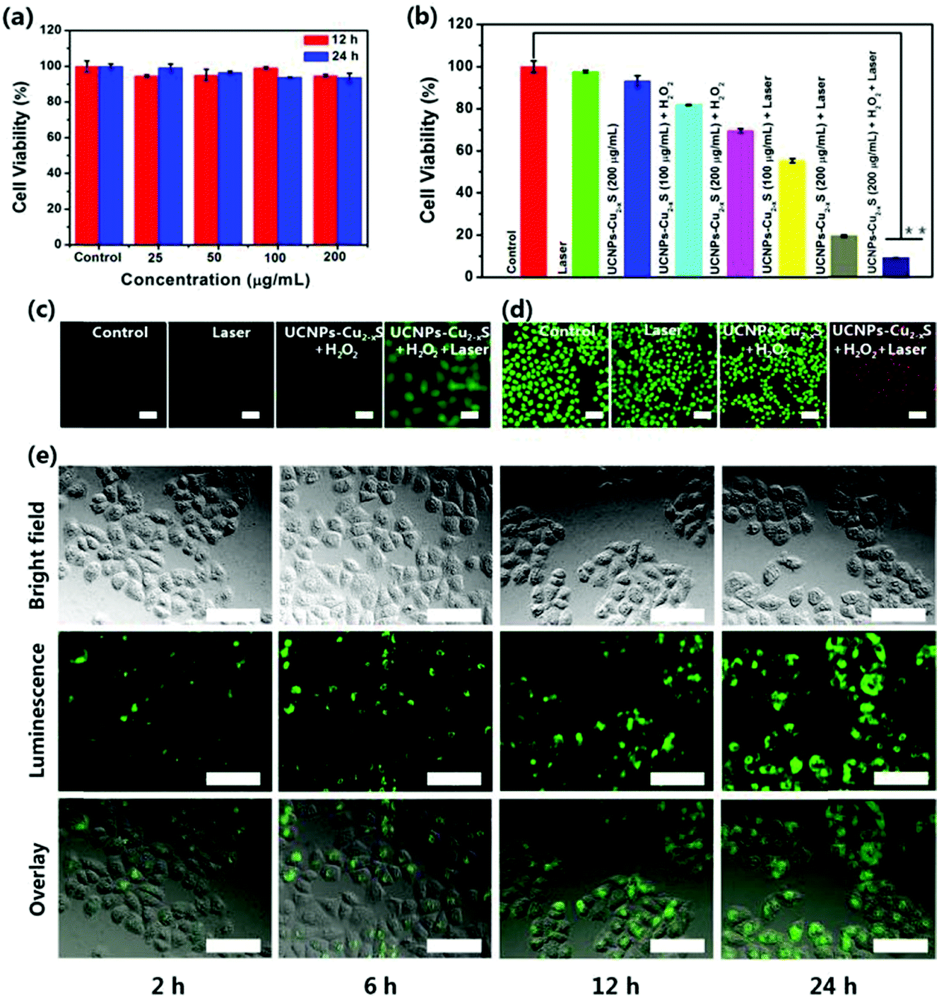 | ||
| Fig. 3 (a) Viability of HeLa cells after incubation with UCNPs–Cu2−xS at varying concentrations (0–200 μg mL−1) for 12 h and 24 h. (b) CCK-8 assay of HeLa cells treated in different groups, **p < 0.01 (two-tailed t test). (c) Fluorescence microscopy images of HeLa cells stained by DCFH-DA in different groups: control, laser, UCNPs–Cu2−xS + H2O2 and UCNPs–Cu2−xS + H2O2+ laser; scale bars are 20 μm. (d) Fluorescence microscopy images of HeLa cells co-stained with calcein AM (live cells, green) and PI (dead cells, red) after different treatments: control, laser, UCNPs–Cu2−xS + H2O2 and UCNPs–Cu2−xS + H2O2 + laser; scale bars are 50 μm. (e) Inverted fluorescence microscope images of HeLa cells incubated with UCNPs–Cu2−xS for 2 h, 6 h, 12 h and 24 h at 37 °C. Each series can be classified into the bright field image, luminescence image and overlay of the above two. The scale bar in each image is 50 μm. | ||
 | ||
| Fig. 5 (a) Infrared thermal images of the tumor site of tumor-bearing mice intravenously injected with 5% glucose solution (control) and UCNPs–Cu2−xS nanocomposites followed by 1064 nm laser irradiation for 6 min. (b) Corresponding temperature change curves at the tumor sites based on thermal images. (c) Body weight of mice under different treatments. (d) Relative tumor growth curves of different groups after various treatments, **p < 0.01 (two-tailed t-test). (e) The digital photographs of excised tumors from representative euthanized mice and (f) mean tumor weight of each group after various treatments from the last day of the experiment (day 14), **p < 0.01 (two-tailed t-test). (g) H&E-stained slices of tumor tissues collected from different groups. All scale bars are 100 μm. | ||
The Royal Society of Chemistry apologises for these errors and any consequent inconvenience to authors and readers.
| This journal is © The Royal Society of Chemistry 2021 |