 Open Access Article
Creative Commons Attribution 3.0 Unported Licence
New Tb3+–simvastatin optical biosensor for sensitive determination of folic acid, progesterone, testosterone and vitamin D3 in biological fluids
Mohamed S. Attia *a,
Amal M. Ahmeda,
Tarek A. Amina,
Ahmed. O. Youssefa,
Mohammed A. Amin
 b,
Ekram H. Mohamed
 c,
Safwat A. Mahmoud*d and
Mona N. Abou-Omare
aChemistry Department, Faculty of Science, Ain Shams University, Cairo 11566, Egypt. E-mail: Mohd_mostafa@sci.asu.edu.eg; Mohamed_sam@yahoo.com; Tel: +202 1229867311 Tel: +202 1060819022
bDepartment of Chemistry, Collage of Science, Taif University, P. O. BOX 11099, Taif 21944, Saudi Arabia
cPharmaceutical Analytical, Chemistry Department, Faculty of Pharmacy, The British University in Egypt, 11837, El Sherouk City, Egypt
dPhysics Department, Faculty of Science, Northern Border University, Arar, Saudi Arabia. E-mail: samahmoud2002@yahoo.com
eDepartment of Chemistry, Faculty of Women for Arts, Science and Education, Ain Shams University, Cairo, Egypt
First published on 6th October 2021
Abstract
An innovative, simple and cost effective Tb3+–simvastatin photo probe was designed and used as a core for a spectrofluorometric approach to sensitively determine four vital biological compounds in different matrices. A Tb3+–simvastatin complex displays a characteristic electrical band with λem at 545 nm with significant luminescence intensity, which is quenched in the presence of folic acid, progesterone, testosterone and vitamin D3 at four variant sets of pH: 5.0, 6.2, 7.5 and 9.0, respectively. The conditions were optimized and the best solvent for operation was found to be acetonitrile at λex at 340 nm. Folic acid was successfully estimated in tablet dosage form, urine and serum in the concentration range of 2.49 × 10⁻⁹ to 1.28 × 10⁻⁶ mol L−1. Progesterone, testosterone and vitamin D3 were also assessed in serum samples using the same optimal conditions within concentration ranges of 5 × 10⁻⁹ to 1.9 × 10⁻⁶, 5 × 10⁻⁹ to 2.8 × 10⁻⁶ and 5 × 10⁻⁹ to 4.2 × 10⁻⁶ mol L−1, respectively. The proposed luminescence method was validated in accordance with ICH guidelines and found to be accurate, precise, and specific and free from any interference at the working pH for each analyte. The cost effectiveness and applicability of the method make it a good choice for routine analysis of the four compounds and early diagnosis of chronic diseases associated with abnormalities in their physiological levels.
1. Introduction
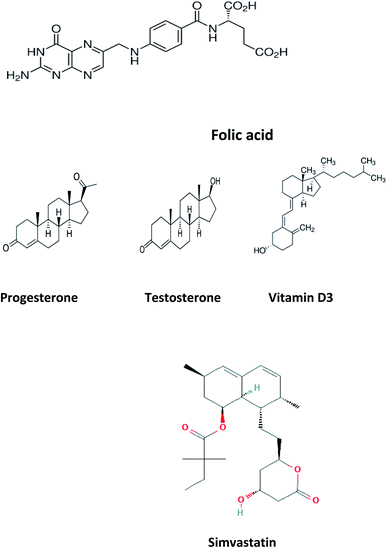
Folic acid (FCA), vitamin B9, is a water-soluble vitamin1 found naturally in various types of foods as legumes, leafy green vegetables, wheat germs, beets, broccoli, citrus fruits, fermented products, beef liver and eggs. FCA is an essential supplement for pregnant women in the first trimester to avoid birth abnormalities including congenital heart diseases and neural tube defects and autism.2 FCA is essential for DNA and RNA production and amino acid metabolism.3 Untreated deficiency of FCA is linked with different health problems, including neurological and psychological manifestations like psychosis, depression, insomnia, and Alzheimer's disease, increase risk of cancer and osteoporosis.4 Elevated levels of homocysteine, a biomarker for arteriosclerosis, is also associated with FCA deficiency. Other symptoms include poor cognitive performance, hearing loss and other symptoms including fatigue, heart palpitations, shortness of breath, hair and skin discoloration, mouth sores, and swollen tongue.5 Different analytical techniques were reported in the literature for FCA determination in dosage form, dietary supplements, beverages and biological samples including spectroscopy6 and chromatography7 and electrochemistry.8 The chemical structure of folic acid is presented in Fig. 1.Progesterone (PGS) is a member of progestogen steroid hormones group, secreted mainly during the menstrual cycle by the corpus luteum preparing the body for conception in case of ova fertilization.9 PGS is used in oral contraception either in single form or combined with estrogen and as hormonal replacement therapy to alleviate menopause symptoms. Low levels of progesterone may lead to abnormal bleeding during menstruation, premature labor and miscarriage during pregnancy and considered as sign for poly-cystic ovarian syndrome. While PGS elevated level may increase the risk of breast cancer development and marker for adrenal hyperplasia. PGS concentrations was recently estimated using different analytical approaches; spectroscopic,10 chromatographic,11 electrochemical methods12 and immunological assay.13 The chemical structure of PGS is presented in Fig. 1.
Testosterone (TST), an anabolic steroid, is the primary male sex hormone where it regulates RBCs production, libido, fertility, spermatogenesis, fat distribution, bone and muscle mass.14 Imbalance in levels TST may can cause serious body dysfunctions where diminished levels have an inverse impact sexual drive, erection, sperm count and muscle strength. Abnormal high TST levels may trigger early puberty in males and menstrual irregularities and baldness in females.15 TST could also be as a medication to replenish its insufficiency, manage breast cancer in women and enhance physique and performance, for instance in athletes.16 Its concentration in different matrices as plasma, serum, saliva was recently measured through spectroscopic17 chromatographic18 electrochemical methods19 and capillary electrophoresis.20 Fig. 1 shows the structure of TST.
Vitamin D3, one of fat-soluble vitamins, is naturally found in different types of foods as oily or fatty fish, dairy products, beefy liver and egg yolk and synthetized endogenously in human body upon exposure to sun. Vit. D3 is converted to its active form through two successive hydroxylation steps forming calcidiol (25-hydroxyvitamin D) in liver followed by calcitriol (1,25-dihydroxyvitamin D) in kidney. It has a major role in regulation concentration of phosphate and calcium in serum and essential in bone remodeling and growth.21 It is also used to improve the cognitive functions and in treatment of specific type of psoriasis. In addition, it contributed to the management of Covid-19 by reducing the cytokine storms and thrombotic episodes associated with the infection.22 The deficiency of Vit. D may lead to serious conditions as rickets and osteomalacia in young and adults, respectively.23 Low levels of Vit. D is also associated with increased risk of colon and pancreatic cancer respiratory acute infections.24 On the other hand, the excessive intake of Vit. D may increase the levels of calcium both in soft tissues (calcinosis) and blood (hypercalcemia). To evaluate the status of Vit. D in human body, calcidiol level in blood is used as best indicator. The chemical structure of Vit. D is displayed in Fig. 1. In the last decade, quantification of Vit. D and/or its metabolites was established through chemiluminescent assay,25 chromatography.26 The reported methods showed relatively high limits of detection which restricts their practical applications. Moreover, the measurement of low concentrations of folic acid, progesterone, testosterone and vitamin D3 in biological samples along with interference from some biomolecules such as uric acid (UA), ascorbic acid (AA), and different hormones requires to efficiently improve the sensitivity of chromatographic methods and the electrochemical sensors for practical applications. Therefore, developing a simple method for accurate determination of folic acid, progesterone, testosterone and vitamin D3 in the presence of each other in the same sample is still of great significance. Today, the research field in which the lanthanide complexes were used as biosensors has a great interest.27–42 Luminescent optical biosensor Tb(simvastatin)3 (Tb–SIM) complex embedded in PEG matrix have many advantages over the mentioned traditional methods. Terbium ion has sharp and precise emission bands in green light region. The terbium ion is used as photo probe for many analytes with a high selectivity depends on the excitation wavelength of terbium–analyte complex, pH and the type of solvent of the test solution. Doping of the optical sensors in the polymer matrix increases its stability and durability.30–36 The sensor can provide a constant signal response for two years, which makes it 24-fold better balance compared to the lifetime warranted for the chromatographic and electrochemical methods. The source of error of the present work eliminated as it more stables for a long time; it gives a low standard deviation value. The higher stability of the current sensor can be attributed to the doping of the optical sensor in the polymer matrix.
2. Experimental
2.1. Instrumentation
A double beam UV-Visible spectrophotometer (PerkinElmer Lambda 25), fluorescence Spectrometer (Thermo Scientific Lumina, Meslo-PN; 222-263000). pH meter (Jenway; 33300)2.2. Materials and reagents
Pure folic acid standard was kindly supplied by the National Organization for Drug control and Research (Giza, Egypt). Pharmaceutical preparation of folic acid tablets dosage form labelled to contain 500 μg manufactured by Mepaco-Medifood (Arab Company for Pharmaceutical and Medicinal plants, Egypt) was purchased from community pharmacy in the Egyptian market. Progesterone, testosterone, vitamin D3, solvents including ethanol, acetonitrile, dimethylformamide (DMF), chloroform and dimethyl sulfoxide (DMSO) were purchased from Sigma Aldrich. Analytical grade ammonium hydroxide (NH4OH), hydrochloric acid (HCl), Tb (NO3)3·5H2O, simvastatin and polyethylene glycol (PEG) were purchased from Sigma Aldrich.The Human real samples were gathered from both Ain Shams Specialized and Teaching New Al-Kasr-El-Aini Hospitals, Cairo, Egypt in accordance with the approved protocol of World Health Organization (WHO) for the collection of human specimens and the use of the clinically related information and data for the purpose of research. The patients approved and were all consented before using their samples.
2.3. Preparation of standard solutions
Stock solutions of Tb (NO3)3·5H2O and simvastatin; were prepared separately by accurately weighing and transferring 0.11 g and 0.039 g, respectively of their authentic pure forms into separate 25 mL volumetric flasks by the aid of the least amount of ethanol till dissolution and completing the volume with the same solvent to obtain final concentration of (10−2 mol L−1) for each of them.Tb3+–simvastatin complex solution; was prepared by mixing 0.1 mL of Tb(NO3)3 stock solution with 0.3 mL of simvastatin (Fig. 1) stock solution in 10 mL volumetric flask and completing the volume to the mark with acetonitrile.
For the four compounds under study, all stock solutions were separately prepared in 10 mL volumetric flasks in concentration of solution (10−2 mol L−1). This was achieved by dissolving 0.044 g of FCA in least amount of DMF and then completing the volume to the mark using acetonitrile. For PGS, TST and Vit. D 0.031 g, 0.0288 g, 0.033 g, were dissolved, respectively in small amount of ethanol and then volume was diluted to the mark with acetonitrile. Further dilutions for the stock solutions using acetonitrile were performed to obtain working solutions with concentrations of 1.0 × 10⁻⁴ to 1.0 × 10⁻⁹ mol L−1 of FCA, PGS, TST and Vit. D.
0.1 mol L−1 of NH4OH and HCl were used to adjust the pH to 9.0, 5.0, 6.2, 7.5 for FCA, PGS, TST and Vit. D, respectively. All the prepared solutions should be kept at low temperature (2–8 °C) to remain stable.
2.4. Preparation of FCA pharmaceutical dosage form solution
Ten tablets of Folic acid® 500 μg were weighed and grinded into fine homogenous powder. The average weight of one tablet was calculated and dissolved in few mL of DMF and sonicated for 20 minutes. The solution was then filtered using whattman filter papers (12 mm) into 10 mL volumetric flask to obtain final concentration of FCA equivalent to 1.1 × 10⁻³ mol L−1. Further dilution was performed to obtain different solutions with concentration range of (1.0 × 10⁻⁴ to 1.0 × 10⁻⁷ mol L−1) was prepared by appropriate dilution with acetonitrile.2.5. Preparation of urine sample spiked with FCA
The urine sample was collected from a healthy volunteer who didn't administer any previous medications, it was then manipulated in the lab as follows; 10 mL of the collected urine sample were centrifuged at 4000 rpm for 15 min to remove all interferants including crystals, salts, pus and red blood cells. 1.0 mL of urine was spiked with 1.0 mL of previously prepared drug solution with concentration of 1.0 × 10⁻⁶ mol L−1 and completed by acetonitrile to the mark in 10 mL measuring flask.2.6. Preparation serum samples spiked with FCA, PGS, TST and Vit. D3
A 1.0 mL of samples of blood collected from healthy volunteers was centrifuged for 15 min at 4000 rpm to remove proteins. 0.1 mL of the serum sample was added to 1.0 mL of each drug working solution of concentration 1.0 × 10⁻⁶ mol L−1 and the volume was complete to 10 mL by acetonitrile to obtain 1.0 × 10⁻⁷ mol L−1 for each drug in four separate 10 mL measuring flasks.2.7. Preparation of Tb–SIM biosensor embedded in PEG
Tb–SIM complex was prepared in the solid state by mixing an equal volume of 1.0 × 10⁻⁴ mol L−1 Tb ion and 3.0 × 10⁻⁴ mol L−1 simvastatin in ethanol, then evaporation near the dryness of the solution, a pale pink solid was obtained after cooling in air. The thin film was prepared by dissolving 0.1 g of the solidified and seamless complex in 3 mL ethanol and then adding 10 mL of viscose freshly prepared PEG with stirring for about one hour until a homogenous solution was obtained. A thin film was fabricated by spin-coating on a small quartz slide (width 8.5 mm, height 25 mm) to quick fit in the cuvette of the spectrofluorometer.2.8. Recommended procedure
An appropriate volume (100 μL) of various standard concentrations of folic acid, progesterone, testosterone and vitamin D3 should be diluted to 3 mL with acetonitrile. The dilute solution was mixed with a thin film of biosensing Tb–SIM doped in PEG matrix in the quartz cell of a spectrofluorometer. The luminescence spectra were recorded at the excitation wavelength λex = 340 nm. After each measurement, the optical sensor was washed with acetonitrile, and the calibration curve was built by applying the Stern's Volmer equation by plotting (F/Fo) the at λem = 545 nm on the y-axis versus the folic acid, progesterone, testosterone and vitamin D3 concentration in mol L−1 on the x-axis.2.9. Determination of FCA in tablet dosage form
The tablet dosage form solutions previously prepared under (2.4) were analyzed using the following procedures: in the spectrofluorometer cell, 1.0 mL of the tablet solutions was separately added followed by the 1.5 mL of acetonitrile in presence of the biosensor film. After mixing, the obtained solutions were scanned, and luminescence spectra were recorded at λex/λem = 340/545 nm. The concentrations of the real samples were calculated using corresponding regression equation.2.10. Determination of FCA in spiked urine samples
The luminescence spectra of the previously prepared spiked urine samples as detailed under (2.5) were scanned at λex/λem = 340/545 nm and the concentration of spiked FCA was determined using the corresponding regression equation adopting the standard addition technique.2.11. Determination of FCA, progesterone, testosterone and vitamin D3 in serum samples
The luminescence spectra of the serum samples previously prepared as described under (2.6) were measured adopting the same procedures followed under (3.2). The concentrations of each real sample were calculated using corresponding regression equation.3. Result and discussion
3.1. General features of absorption and emission spectra of Tb–SIM complex
Owing to the f–f transition forbiddance of trivalent ion (Tb3+), there is a restriction to directly absorb light which could be overcome through the antenna effect via the coupling between Tb3+ and a prominently absorbing organic ligand leading to efficient energy transfer and light absorption processes. Regarding the proposed photo probe, Tb3+ is surrounded covalently by 3 molecules of simvastatin ligand responsible for efficient absorption of light and transfer of energy to populate 5D4 state of Tb3+.43The emission of the formed complex Tb–SIM exhibited four specific and intense bands because of the 5D4–7FJ transitions (J = 6, 5, 4 and 3).44
3.2. Absorption and emission spectra
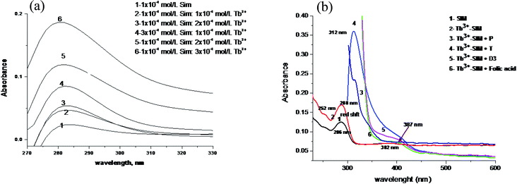
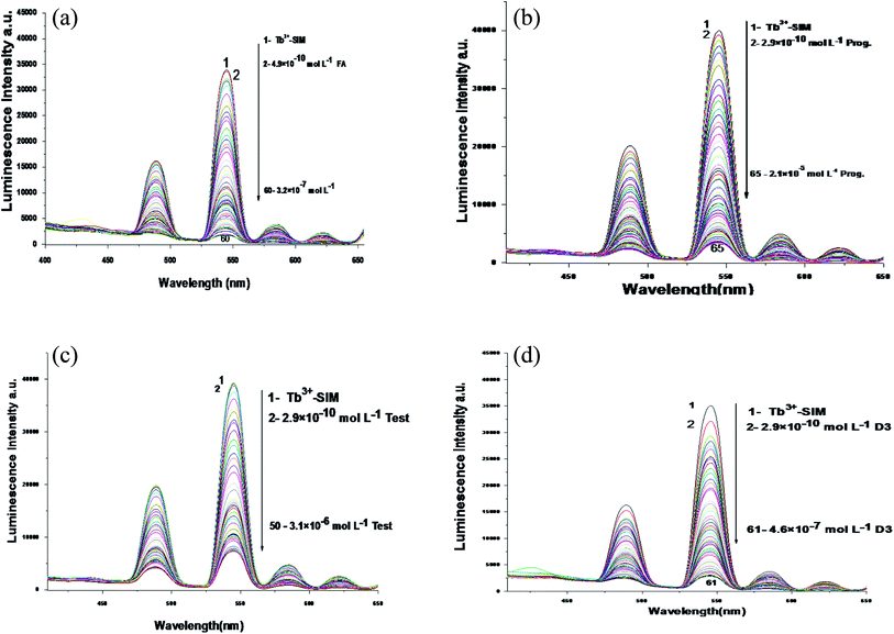
The absorption spectra of Tb (NO3)3, simvastatin and Tb3+–simvastatin complex are shown in Fig. 2a. A red shift by 7 nm and the absorbance value is enhanced denoting that simvastatin could form a stable complex with Tb3+. The absorption spectra of FCA, PGS, TST and Vit. D3 were scanned alone and in the presence of the optical sensor are shown in Fig. 2b.The emission spectra of Tb3+–SIM complex after adding different concentrations of FCA, PGS, TST and Vit. D3 using acetonitrile as solvent are shown in Fig. 3a–d, respectively. The characteristic electrical emission band of Tb3+ exhibited at λem 545 nm was quenched due to energy transfer from the optical sensor to FCA, PGS, TST and Vit. D3.
3.3. Experimental variables
3.4. Mechanism of emission quenching
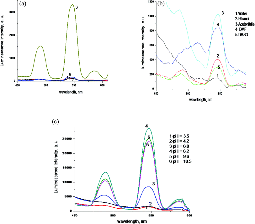
Upon adding different concentrations of FCA, PGS, TST and Vit. D3 to the Tb–SIM photo probe a notified quenching in its luminescent intensity occurs owing to the approach of the analytes under study and formation of H-bond between the hydroxyl group in both of TST and Vit. D3, carboxylic group in FCA and enol group in PGS with the SIM. The formation of H-bonding lead to the depression or decrease in the transfer of energy to the Tb3+ ion and consequently the luminescence intensity is significantly quenched.The pH effect on the luminescence intensity after the addition of the studied analytes to the proposed photoprobe was studied and the luminescence quenching was observed at pH 5.0, 6.2, 7.5 and 9.0 for FCA, PGS, TST and Vit. D3 respectively.
4. Analytical performance61
4.1. Linearity
Correlations between the luminescence of emission intensity of optical sensor at λem 545 nm and FCA, PGS, TST and Vit. D3 within concentration ranges of (2.4 × 10⁻⁹ to 1.28 × 10⁻⁶), (5 × 10⁻⁹ to 1.9 × 10⁻⁶), (5 × 10⁻⁹ to 2.8 × 10⁻⁶) and (5 × 10⁻⁹ to 4.2 × 10⁻⁶) mol L−1 respectively were found to be linear as presented in respective calibration graphs, Fig. 5a and b obtained by applying the Stern–Völmer plot.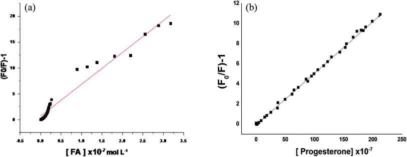 | ||
| Fig. 5 (a) Stern–Volmer plot (Fo/F) − 1 against corresponding concentrations of folic acid. (b) Stern–Volmer plot (Fo/F) − 1 against corresponding concentrations of progesterone. | ||
The critical concentration of FCA, PGS, TST and Vit. D3 values are (3.31, 3.1, 2.2, 1.3) and (0.005 × 10⁻⁷ to 3.18 × 10⁻⁷, 2.49 × 10⁻¹⁰ to 2.12 × 10⁻⁵, 2.4 × 10⁻¹⁰ to 3.18 × 10⁻⁶, 4.9 × 10⁻¹⁰ to 4.8 × 10⁻⁷) mol L−1 respectively. The distance between the cited compounds and the ionophore is 3.36 Å indicating the electron transfer mechanism of quenching.
The regression equations were computed and the regression parameters in addition the LOD and LOQ were calculated, and results were presented in Table 1.
| Parameter | Folic acid | Progesterone | Testosterone | Vitamin D3 |
|---|---|---|---|---|
| a Where Y: intensity of luminescence, X: analyte concentration (mol L−1), a: intercept and b: slope. | ||||
| λem (nm) | 545 | |||
| Linearity (mol L−1) | 1.28 × 10⁻⁶ to 2.49 × 10⁻⁹ | 1.9 × 10⁻⁶ to 5 × 10⁻⁹ | 2.8 × 10⁻⁶ to 5 × 10⁻⁹ | 4.2 × 10⁻⁶ to 5 × 10⁻⁹ |
| LOD (mol L−1) | 1.99 × 10⁻⁹ | 1.5 × 10⁻⁹ | 3.02 × 10⁻⁹ | 1.59 × 10⁻⁹ |
| LOQ (mol L−1) | 5.94 × 10⁻⁹ | 4.5 × 10⁻⁹ | 9.0 × 10⁻⁹ | 2.8 × 10⁻⁹ |
| Regression equation | (Y = a + bX)a | |||
| Intercept (a) | 0.32 | 47.2 | 105 | 84.5 |
| Slope (b) | 3.31 | 3.1 | 2.2 | 1.30 |
| Standard deviation | 0.04 | 15.5 | 20.5 | 6.40 |
| Variance (S2) | 0.0016 | 240.25 | 420.25 | 4.90 |
| Regression coefficient (r) | 0.99 | 0.99 | 0.99 | 0.99 |
4.2. Accuracy and precision
The accuracy of the developed method was further investigated via applying the standard addition technique and calculating the recovery%. Assessing the obtained recovery was performed through determination of agreement extent between the measured and actual added standard concentration of analyte. All assays were repeated 3 times within the same day and different days to assess the repeatability and intermediate precision, respectively. Three different levels of the analyte concentrations were used in the assays and the results were summarized and presented in (Table 2).| Sample | Concentration taken (×10−7 mol L−1) | Repeatability | Intermediate precision | ||||
|---|---|---|---|---|---|---|---|
| Average found ± CLb | % REc | % RSDd | Drug average found ± CL | % RE | % RSD | ||
| a n = 3.b CL: confidence limits (supplementary material).c % RE: percent relative error.d RSD: relative standard deviation. | |||||||
| Progesterone in serum | 1.0 | 1.03 ± 0.13 | 3.0 | 3.39 | 1.06 ± 0.11 | 6.0 | 2.13 |
| 2.0 | 1.95 ± 0.18 | 2.5 | 2.33 | 2.05 ± 0.17 | 2.5 | 3.12 | |
| 4.0 | 4.19 ± 0.24 | 4.75 | 2.99 | 4.20 ± 0.23 | 5.0 | 2.11 | |
| Testosterone in serum | 1.0 | 1.11 ± 0.13 | 11.0 | 3.46 | 0.99 ± 0.11 | 1.00 | 3.11 |
| 2.0 | 2.02 ± 0.18 | 1.00 | 2.41 | 2.04 ± 0.16 | 2.00 | 2.06 | |
| 4.0 | 3.89 ± 0.26 | 2.75 | 2.95 | 4.13 ± 0.21 | 3.25 | 3.02 | |
| Vitamin D3 in serum | 1.0 | 1.06 ± 0.23 | 6.00 | 2.22 | 1.09 ± 0.21 | 9.00 | 2.01 |
| 2.0 | 2.05 ± 0.28 | 2.50 | 2.26 | 2.14 ± 0.36 | 7.00 | 4.35 | |
| 4.0 | 4.19 ± 0.48 | 4.75 | 2.25 | 4.23 ± 0.31 | 5.75 | 2.51 | |
| Tablet, 500 μg of folic acid MEPACO | 3.0 | 3.04 ± 0.024 | 1.33 | 0.33 | 3.07 ± 0.052 | 2.33 | 0.68 |
| 6.0 | 5.99 ± 0.050 | 0.16 | 0.35 | 6.08 ± 0.070 | 1.33 | 0.47 | |
| 9.0 | 8.96 ± 0.025 | 0.33 | 0.11 | 9.09 ± 0.062 | 1.00 | 0.28 | |
| Folic acid in serum | 4.0 | 3.98 ± 0.20 | 0.50 | 0.38 | 4.08 ± 0.038 | 2.00 | 0.37 |
| 6.0 | 5.98 ± 0.15 | 0.33 | 0.61 | 6.09 ± 0.080 | 1.50 | 0.53 | |
| 9.0 | 9.01 ± 0.22 | 0.22 | 0.33 | 9.06 ± 0.062 | 0.67 | 0.28 | |
| Folic acid in urine | 4.0 | 3.99 ± 0.20 | 0.50 | 0.10 | 4.06 ± 0.043 | 2.03 | 0.32 |
| 6.0 | 5.99 ± 0.15 | 0.33 | 0.66 | 6.07 ± 0.070 | 1.54 | 0.51 | |
| 9.0 | 8.99 ± 0.22 | 0.22 | 0.44 | 9.04 ± 0.066 | 0.74 | 0.38 | |
4.3. Selectivity
The selectivity of the proposed method was investigated through analyzing placebo blank and synthetically prepared mixtures. All possible interfering inactive compounds were used to prepare a placebo containing; 50 mg calcium carbonate, 20 mg calcium dihydrogen orthophosphate, 30 mg lactose, 100 mg magnesium stearate, 40 methyl cellulose, 70 mg sodium alginate, 300 mg starch and 250 mg Talc. Extraction was performed using water and the solution was manipulated as detailed under 2.4. A suitable aliquot of the obtained solution was analyzed after the addition of the optical sensor Tb3+–simvastatin, and the luminescence spectra were recorded at λex/λem = 340/545 nm following the optimized conditions.The validity and selectivity were further assessed in presence of some proteins and hormones that may interfere as cortisol, Thyroid stimulating hormone, norepinephrine, dopamine and albumin within concentration range of 0.08 g L−1. The interference of 0.0.06 g L−1 urea, 0.08 g L−1 glucose, uric acid and folic acid was also studied, and the resulting data revealed that there was no significant effect on the observed luminescence activity of the proposed photo probe under optimized conditions.
In addition, the proposed optical probe was successfully applied for selective determination of FCA, PGS, TST and Vit. D3 either as single or in combination in synthetically prepared mixtures. Four synthetic mixtures were prepared by adding different concentrations of FCA, PGS, TST and Vit. D3 within their linearity range in 4 similar sets of 10 mL volumetric flasks containing 1.0 mL of the serum sample as mentioned under 2.6.
The pH of the first set was adjusted to 5.0 for selective determination of FCA in presence of PGS, TST and Vit. D3, the pH of the second set was adjusted to 6.2 for the determination of PGS in presence of FCA, TST and Vit. D3, the pH of the third set was adjusted to 7.5 for determination of TST in presence of FCA, PGS, and Vit D3, finally the pH of the fourth set was adjusted to 9 for determination of Vit D3 in presence of FCA, PGS, TST and the volume was completed with acetonitrile for the four sets. Thus, each mixture was prepared 4 times but at different pH (5.0, 6.2, 7.5 and 9.0) for selective estimation of FCA, PGS, TST and Vit. D3, respectively. Each solution was in triplicates and yielded recovery% of 99.60 ± 0.47,100.8 ± 2.10, 99.4 ± 2.60 and 101.9 ± 2.20 for FCA, PGS, TST and Vit. D3, respectively.
Results in Fig. 6 show that the luminescence of Tb3+–SIM complex in its second coordination sphere in which the quaternary mixture of FCA, PGS, TST and Vit. D3 is quite sensitive to four variant sets of pHs. For Tb3+–SIM–FCA, λex = 340 and pH 5.0, give the more quenching of luminescence intensity of Tb3+–SIM while that for Tb3+–SIM–PGS was of λex = 340 and pH 6.2 and that for Tb3+–SIM–TST was of λex = 340 and pH 7.5, and that for Tb3+–SIM–Vit-D3 was of λex = 340 and pH 9.0. Thus, a dual-controlled luminescence of smoothly dynamic reversibility is achieved and a reversible on/off switchable Tb3+ emission of one system was observed by tuning its optimal values of pH to the optimal ones of the second and so on for the third and fourth. By this dual controlled luminescence, the quaternary mixture of FCA, PGS, TST and Vit. D3 was simultaneously resolved with average error <3.5%.
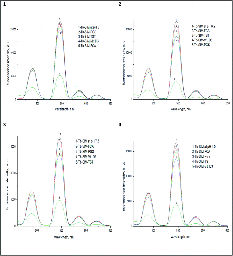 | ||
| Fig. 6 The luminescence spectra of the complexes Tb–SIM, Tb–SIM–FCA, Tb–SIM–PGS, Tb–SIM–TST and Tb–SIM–Vit-D3 at λex = 340 nm and different pHs; (1) 5.0, (2) 6.2, (3)7.5 and (4) 9.0. | ||
Also, the data obtained upon assaying single PGS, TST and Vit. D3 separately in serum sample and FCA in serum, urine and dosage form, without any interference from inactive excipients, were processed and results were tabulated as shown in Table 3. The results of the proposed method were comparable to that obtained from the reference chromatographic methods mentioned in the British pharmacopeia.62 The limitations of the proposed method in real samples in which a hormones and proteins are existed. These biological molecules contain OH, NH and SH groups may make an interference with the analytes at different pHs.
| Sample | Added (× 10−7 mol L−1) | Found (× 10−7 mol L−1) | Averagea (× 10−7 mol L−1) | Average recovery ± % R.S.D | B·P. (LC) |
|---|---|---|---|---|---|
| a Average of nine measurements. | |||||
| Progesterone serum sample | 3.5 | 3.52, 3.48, 357 | 3.52 | 100.2 ± 2.1 | 98.6 ± 0.5 |
| 7.0 | 6.97, 7.05, 7.03 | 7.01 | |||
| 9.5 | 9.55, 9.46, 9.46 | 9.49 | |||
| Testosterone serum sample | 3.5 | 3.49, 3.53, 3.52 | 3.51 | 99.6 ± 2.5 | 99.2 ± 0.6 |
| 7.0 | 6.95, 6.99, 6.98 | 6.97 | |||
| 9.5 | 9.51, 9.45, 9.48 | 9.48 | |||
| Vitamin D3 serum sample | 3.5 | 3.39, 3.43, 3.62 | 3.48 | 103.1 ± 2.9 | 99.4 ± 0.5 |
| 7.0 | 6.85, 6.89, 6.88 | 6.87 | |||
| 9.5 | 9.41, 9.55, 9.58 | 9.51 | |||
| Tablet, 500 μg of folic acid MEPACO-MEDIFOOD | 3.0 | 3.04, 3.05, 3.03 | 3.04 | 101.33 ± 0.33 | 99.8 ± 0.055 |
| 6.0 | 6.02, 5.98, 5.99 | 5.99 | 99.83 ± 0.35 | ||
| 9.0 | 8.97, 8.96, 8.95 | 8.96 | 99.66 ± 0.11 | ||
| Folic acid serum sample | 4.0 | 3.98, 3.97, 4.00 | 3.98 | 99.5 ± 0.38 | 99.6 ± 0.050 |
| 6.0 | 6.01, 5.97, 5.95 | 5.98 | 99.66 ± 0.61 | ||
| 9.0 | 8.98, 8.95, 9.01 | 9.01 | 99.77 ± 0.33 | ||
| Folic acid urine sample | 4.0 | 3.99, 3.97, 4.01 | 3.99 | 99.75 ± 0.10 | 99.5 ± 0.050 |
| 6.0 | 6.02, 5.98, 5.99 | 5.99 | 99.83 ± 0.66 | ||
| 9.0 | 8.99, 8.97, 9.00 | 8.99 | 99.88 ± 0.44 | ||
4.4. Comparison with previously reported methods
The results obtained from the proposed spectrofluorometric technique was compared with obtained from other previously reported methods assuring the applicability, accuracy, and precision of the proposed method as presented in Table 4,8,9,14,15,17,20,21,29–33,63,64| Analyte | Methods | Linearity | Limit of detection | References |
|---|---|---|---|---|
| Progesterone | HPLC-MS-MS | 0.2–50 ng mL−1 | 0.2 ng mL−1 | 18 |
| Microfluidic immunosensor system | 0.5–12.5 ng mL−1 | 0.2 ng mL−1 | 21 | |
| Enzyme-linked fluorescence assay | 3–40.0 ng mL−1 | — | 22 | |
| Spectrofluorometric using Tb3+–SIM | 1.9 × 10⁻⁶ to 5 × 10⁻⁹ mol L−1 | 1.49 × 10⁻⁹ mol L−1 | ||
| Testosterone | HPLC in plasma | 1.6–400 ng mL−1 | 1.6 ng mL−1 | 32 |
| HPLC in serum | 1–20 ng mL−1 | 0.4 | 33 | |
| HPLC in urine | 10–500 ng mL−1 | 1 ng mL−1 | 30 | |
| HPLC in dosage form | 50–200 μg mL−1 | 5 μg mL−1 | 31 | |
| HPLC in urine | 2–300 ng mL−1 | 2 ng mL−1 | 29 | |
| Spectrofluorometric using Tb3+–SIM | 2.8 × 10⁻⁶ to 5 × 10⁻⁹ mol L−1 | 3.1 × 10⁻⁹ mol L−1 | ||
| Vitamin D3 | HPLC | 15–200 nmol L−1 | 3 nmol L−1 | 62 |
| LC-MS/MS | 3.5 to 75 ng mL−1 | 14 ng mL−1 | 63 | |
| HPLC-APCI-MS | 5–400 nmol L−1 | 1–4 nmol L−1 | 64 | |
| Spectrofluorometric using Tb3+–SIM | 4.2 × 10⁻⁶ to 5 × 10⁻⁹ mol L−1 | 1.6 × 10⁻⁹ mol L−1 | ||
| Folic acid | LC-MS/MS | 4.5 × 10⁻⁸ to 5 × 10⁻¹⁰ mol L−1 | 5 × 10⁻¹⁰ mol L−1 | 15 |
| Chemiluminometric and fluorimetric determination | 114–6.0 μg mL−1, 1.10–0.022 μg mL−1 | 2.0 μg mL−1, 0.002 μg mL−1 | 8 | |
| Chemiluminescence | 8 × 10⁻⁷ to 6 × 10⁻⁹ mol L−1 | 6 × 10−103 mol L−1 | 9 | |
| HPLC method | 2500 to 50 μg mL−1 | 1.3 ng mL−1 | 14 | |
| Spectrofluorometric method: using Tb3+–SIM | 1.28 × 10⁻⁶ to 2.49 × 10⁻⁹ mol L−1 | 1.99 × 10⁻⁹ mol L−1 |
5. Conclusion
The proposed analytical method based on the use of Tb3+–simvastatin complex is simple and economic and can be successfully applied for sensitive and accurate determination of folic acid, progesterone, testosterone and vitamin D3 in different matrices including dosage forms, urine and serum. The analysis of the FCA, PGS, TST and Vit. D in biological samples can contribute to early diagnosis of some chronic diseases associated with their abnormal levels.Conflicts of interest
The authors declare that they have no known competing financial interests or personal relationships that could have appeared to influence the work reported in this paper.Acknowledgements
Authors thank Taif University Researchers Supporting Project (TURSP-2020/03), Taif University, Taif, Saudi Arabia, for supporting this work.References
- U. Binesh, Y.-L. Yang and S.-M. Chen, Amperometric Determination of Folic Acid at Multi-Walled Carbon Nanotube-Polyvinyl Sulfonic Acid Composite Film Modified Glassy Carbon Electrode, Int. J. Electrochem. Sci., 2011, 6, 3224–3227 Search PubMed.
- D. B. Kirsten, C. G. David, J. C. Susan, W. D. Karina, W. E. John, A. R. G. Francisco, R. K. Alex, H. K. Alex, E. K. Ann, C. Landefeld, M. M. Carol, R. P. William, G. P. Maureen, P. P. Michael, S. Michael and T. Chien-Wen, Folic Acid Supplementation for the Prevention of Neural Tube Defects: US Preventive Services Task Force Recommendation Statement, JAMA, J. Am. Med. Assoc., 2017, 317, 183–189 CrossRef PubMed.
- Y. Feng, S. Wang, R. Chen, X. Tong, Z. Wu and X. Mo, Maternal folic acid supplementation and the risk of congenital heart defects in offspring: a meta-analysis of epidemiological observational studies, Sci. Rep., 2015, 5, 8506 CrossRef CAS PubMed.
- I. M. Ebisch, C. M. Thomas, W. H. Peters, D. D. Braat and R. P. Steegers-Theunissen, The importance of folate, zinc and antioxidants in the pathogenesis and prevention of subfertility, Hum. Reprod. Update, 2007, 13, 163–174 CrossRef CAS PubMed.
- A. M. Treon, M. Shea-Budgell, B. Shukitt-Hale, D. E. Smith, J. Selhub and I. H. Rosenberg, B-vitamin deficiency causes hyperhomocysteinemia and vascular cognitive impairment in mice, Proc. Natl. Acad. Sci. U. S. A., 2008, 105, 12474–12479 CrossRef PubMed.
- M. Jägerstad, Folic acid fortification prevents neural tube defects and may also reduce cancer risks, Acta Paediatr., 2012, 101, 1007–1012 CrossRef PubMed.
- M. V. Ribeiro, I. S. Melo, F. C. Lopes and G. C. Moita, Development and validation of a method for the determination of folic acid in different pharmaceutical formulations using derivative spectrophotometry, Braz. J. Pharm. Sci., 2016, 52, 741–750, DOI:10.1590/S1984-82502016000400019.
- T. Meropi, A. Panagiotis, M. Triantafillos, S. Magdalini and T. Thalia, Chemiluminometric and Fluorimetric Determination of Folic Acid, Anal. Lett., 2007, 40, 2203–2216 CrossRef.
- B. T. Zhang, L. Zhao and J. M. Lin, Determination of folic acid by chemiluminescence based on peroxomonosulfate-cobalt (II) system, Talanta, 2008, 74, 1154–1159 CrossRef CAS PubMed; E. P. Azevedo, E. M. Alves, S. Khan, L. Silva, J. R. de Souza and B. S. Santos, Folic acid retention evaluation in preparations with wheat flour and corn submitted to different cooking methods by HPLC/DAD, PLoS One, 2020, 15, e0230583, DOI:10.1371/journal.pone.0230583.
- A. Mahato, S. Vyas and N. S. Chatterjee, HPLC-UV Estimation of Folic Acid in Fortified Rice and Wheat Flour using Enzymatic Extraction and Immunoaffinity Chromatography Enrichment: An Interlaboratory Validation Study, J. AOAC Int., 2020, 103, 73–77 CrossRef PubMed.
- E. Mine Öncü-Kaya, Determination of Folic Acid by Ultra-High Performance Liquid Chromatography in Certain Malt-based Beverages after Solid-Phase Extraction, CBU J. Sci., 2017, 13, 623–630 CrossRef.
- K. Sasaki, H. Hatate and R. Tanaka, Determination of 13 Vitamin B and the Related Compounds Using HPLC with UV Detection and Application to Food Supplements, Chromatographia, 2020, 83, 839–851 CrossRef CAS.
- R. M. Kok, D. E. C. Smith, J. R. Dainty, J. T. V. Akker, P. M. Finglas, Y. M. Smulders, C. Jakobs and K. D. Meera, 5-Methyltetrahydrofolic acid and folic acid measured in plasma with liquid chromatography tandem mass spectrometry: applications to folate absorption and metabolism, Anal. Biochem., 2004, 326, 129–138 CrossRef CAS PubMed.
- E. Dokur, Ö. Gördük and Y. Şahin, Differential Pulse Voltammetric Determination of Folic Acid Using a Poly(Cystine) Modified Pencil Graphite Electrode, Anal. Lett., 2020, 53, 2060–2078, DOI:10.1080/00032719.2020.1728540.
- M. C. Brucker and T. L. King, Pharmacology for Women's Health, 2nd edn, Jones & Bartlett Publishers. 2010, p. 372 Search PubMed.
- D. Maliwal, P. Jain, A. Jain and V. Patidar, Determination of progesterone in capsules by high-performance liquid chromatography and UV- spectrophotometry, J. Young Pharm., 2009, 1, 371–374 CrossRef CAS.
- P. Naderi and F. Jalali, Poly-L-serine/AuNPs/MWCNTs as a Platform for Sensitive Voltammetric Determination of Progesterone, J. Electrochem. Soc., 2020, 167, 027524, DOI:10.1149/1945-7111/ab6a7f.
- A. F. Javier, M. G. Alejandro, M. P. Gabriela, Z. M. Alicia, R. Julio and F. Héctor, Determination of progesterone (P4) from bovine serum samples using a microfluidic immunosensor system, Talanta, 2010, 80, 1986–1992 CrossRef PubMed.
- N. Brugger, C. Otzdorff, B. Walter, B. Hoffmann and J. Braun, Quantitative Determination of Progesterone (P4) in Canine Blood Serum Using an Enzyme-linked Fluorescence Assay, Reprod. Domest. Anim., 2011, 46, 870–873 CrossRef CAS PubMed.
- C. Yu, Y. Mehrdad, R. H. Barry, P. D. Eleftherios and W. Pui-Yuen, Rapid determination of serum testosterone by liquid chromatography-isotope dilution tandem mass spectrometry and a split sample comparison with three automated immunoassays, Clin. Biochem., 2009, 42, 484–490 CrossRef PubMed.
- J. Bain, The many faces of testosterone, Clin. Interventions Aging, 2007, 2, 567–576 CrossRef CAS PubMed.
- D. French, Development and validation of a serum total testosterone liquid chromatography-tandem mass spectrometry (LC-MS/MS) assay calibrated to NIST SRM 971, Clin. Chim. Acta, 2013, 415, 109–117 CrossRef CAS PubMed.
- R. S. Tan and S. J. Pu, A pilot study on the effects of testosterone in hypogonadal aging male patients with Alzheimer's disease, Aging Male, 2003, 6, 13–17 CrossRef CAS PubMed.
- B. A. Andre, M. D. Julia, A. S. Elizabeth, M. M. Hassan, T. G. Lin and A. W. Gary, Endogenous Testosterone and Mortality in Men: A Systematic Review and Meta-Analysis, J. Clin. Endocrinol. Metab., 2011, 96, 3007–3019 CrossRef PubMed.
- S. P. Tuck and R. M. Francis, Testosterone, bone and osteoporosis, Front. Horm. Res., 2009, 37, 123–132 CAS.
- A. J. Kadhem, S. Xiang, S. Nagel, C.-H. Lin and M. Fidalgo de Cortalezzi, Photonic Molecularly Imprinted Polymer Film for the Detection of Testosterone in Aqueous Samples, Polymers, 2018, 10, 349–352 CrossRef PubMed.
- M. M. Abd-Elzaher, M. A. Ahmed, A. B. Farag, M. S. Attia, A. O. Youssef and S. M. Sheta, A Fast and Simple Method for Determination of Testosterone Hormone in Biological Fluids Based on a New Eu(III) Complex Optical Sensor, Sens. Lett., 2017, 15, 977–981 CrossRef.
- L. Konieczna, A. Plenis, I. Olędzka, P. Kowalski and T. Bączek, Optimization of LC method for the determination of testosterone and epitestosterone in urine samples in view of biomedical studies and anti-doping research studies, Talanta, 2011, 83, 804–814 CrossRef CAS PubMed.
- C. He, S. Li, H. Liu, K. Li and F. Liu, Extraction of testosterone and epitestosterone in human urine using aqueous two-phase systems of ionic liquid and salt, J. Chromatogr. A, 2005, 1082, 143–149 CrossRef CAS PubMed.
- R. K. Gupta, R. K. Roy, Anurag and A. N. Jha, HPLC Method Development for Testosterone Cipionate in Bulk Drug and Oil-Based Injectables, Biosci., Biotechnol. Res. Asia, 2010, 7, 505–511 CAS.
- K. H. Yuen and B. H. Ng, Determination of plasma testosterone using a simple liquid chromatographic method, J. Chromatogr. B: Anal. Technol. Biomed. Life Sci., 2003, 793, 421–426 CrossRef.
- V. Loi, M. Vertzoni, A. Vryonidou and C. Phenekos, Development and validation of a simple reversed-phase high-performance liquid chromatography method for the determination of testosterone in serum of males, J. Pharm. Biomed. Anal., 2006, 41, 527–532 CrossRef CAS PubMed.
- J. Albrethsen, M. L. Ljubicic and A. Juul, Longitudinal Increases in Serum Insulin-like Factor 3 and Testosterone Determined by LC-MS/MS in Pubertal Danish Boys, J. Clin. Endocrinol. Metab., 2020, 105, 3173–3178, DOI:10.1210/clinem/dgaa496.
- S. Alvi and M. Hammami, An improved method for measurement of testosterone in human plasma and saliva by ultra-performance liquid chromatography-tandem mass spectrometry, J. Adv. Pharm. Technol. Res., 2020, 11, 64, DOI:10.4103/japtr.JAPTR_162_19.
- W. C. Chang, D. A. Cowan, C. J. Walker, N. Wojek and A. D. Brailsford, Determination of anabolic steroids in dried blood using microsampling and gas chromatography-tandem mass spectrometry: application to a testosterone gel administration study, J. Chromatogr. A, 2020, 1628, 461445, DOI:10.1016/j.chroma.2020.461445.
- X. Li, T. Yuan, T. Zhao, X. Wu and Y. Yang, An Effective Acid–Base-Induced Liquid–Liquid Microextraction Based on Deep Eutectic Solvents for Determination of Testosterone and Methyltestosterone in Milk, J. Chromatogr. Sci., 2020, bmaa051, DOI:10.1093/chromsci/bmaa051.
- W. Xu, H. Li, Q. Guan, Y. Shen and L. Cheng, A rapid and simple liquid chromatography-tandem mass spectrometry method for the measurement of testosterone, androstenedione, and dehydroepiandrosterone in human serum, J. Clin. Lab. Anal., 2017, 31, e22102, DOI:10.1002/jcla.22102.
- R. Heidarimoghadam, O. Akhavan, E. Ghaderibi, E. Hashemi, S. S. Mortazavi and A. Farmany, Graphene oxide for rapid determination of testosterone in the presence of cetyltrimethylammonium bromide in urine and blood plasma of athletes, Mater. Sci. Eng., C, 2016, 61, 246–250 CrossRef CAS PubMed.
- K. H. Liu, D. O'Hare, J. L. Thomas, H. Z. Guo, C. H. Yang and M. H. Lee, Self-assembly Synthesis of Molecularly Imprinted Polymers for the Ultrasensitive Electrochemical Determination of Testosterone, Biosensors, 2020, 10, 16, DOI:10.3390/bios10030016.
- B. Du, J. Zhang, Y. Dong, J. Wang, L. Lei and R. Shi, Determination of testosterone/epitestosterone concentration ratio in human urine by capillary electrophoresis, Steroids, 2020, 161, 108691, DOI:10.1016/j.steroids.2020.108691.
- A. W. Norman, From vitamin D to hormone D: fundamentals of the vitamin D endocrine system essential for good health, Am. J. Clin. Nutr., 2008, 88, 491–499 CrossRef PubMed.
- M. S. Attia, W. H. Mahmoud, A. O. Youssef and M. S. Mostafa, Cilostazol Determination by the Enhancement of the Green Emission of Tb3+ Optical Sensor, J. Fluoresc., 2011, 21, 2229–2235, DOI:10.1007/s10895-011-0927-y.
- M. S. Attia, M. H. Khalil, M. S. A. Abdel-Mottaleb, M. B. Lukyanova, Yu. A. Alekseenko and B. Lukyanov, Effect of Complexation with Lanthanide Metal Ions on the Photochromism of (1,3,3-Trimethyl-5-Hydroxy-6-Formyl-Indoline-Spiro2,2-[2H] chromene) in Different Media, Int. J. Photoenergy, 2006, 1–9, DOI:10.1155/IJP/2006/42846.
- M. S. Attia, A. A. Essawy, A. O. Youssef and M. S. Mostafa, Determination of Ofloxacin using a Highly Selective Photo Probe Based on the Enhancement of the Luminescence Intensity of Eu3+ - Ofloxacin Complex in Pharmaceutical and Serum Samples, J. Fluoresc., 2012, 2, 557–564, DOI:10.1007/s10895-011-0989-x.
- M. S. Attia, A. M. Othman, A. O. Youssef and E. El-Raghi, Excited state interaction between Hydrochlorothiazide and Europium ion in PMMA polyme, and its application as optical sensor for Hydrochlorothiazide in tablet and serum samples, J. Lumin., 2012, 132, 2049–2053, DOI:10.1016/j.jlumin.2012.03.012.
- M. S. Attia, A. O. Youssef and A. A. Essawy, A novel method for tyrosine assessment in vitro by using fluorescence enhancement of the ion-pair tyrosine- neutral red dye photo probe, Anal. Methods, 2012, 4, 2323–2328, 10.1039/C2AY25089F.
- M. S. Attia, A. O. Youssef and R. H. El-Sherif, Durable diagnosis of seminal vesicle and sexual gland diseases using the nano optical sensor thin film sm-doxycycline complex, Anal. Chim. Acta, 2014, 835, 56–64, DOI:10.1016/j.aca.2014.05.016.
- M. S. Attia, M. Diab and M. F. El-Shahat, Diagnosis of some diseases related to the histidine level in human serum by using the nano optical sensor Eu–Norfloxacine complex, Sens. Actuators, B, 2015, 207, 756–763, DOI:10.1016/j.snb.2014.10.132.
- M. S. Attia, A. O. Youssef, Z. A. Khan and M. N. Abou-Omar, Alpha fetoprotein assessment by using a nano optical sensor thin film binuclear Pt-2-aminobenzimidazole-Bipyridine for early diagnosis of liver cancer, Talanta, 2018, 186, 36–43, DOI:10.1016/j.talanta.2018.04.043.
- M. S. Attia and N. S. Al-Radadi, Progress of pancreatitis disease biomarker alpha amylase enzyme by new nano optical sensor, Biosens. Bioelectron., 2016, 86, 413–419, DOI:10.1016/j.bios.2016.06.079.
- M. S. Attia and N. S. Al-Radadi, Nano optical sensor binuclear Pt-2-pyrazinecarboxylic acid-bipyridine for enhancement of the efficiency of 3-nitrotyrosine biomarker for early diagnosis of liver cirrhosis with minimal hepatic encephalopathy, Biosens. Bioelectron., 2016, 86, 406–412, DOI:10.1016/j.bios.2016.06.074.
- M. S. Attia, Nano optical probe samarium tetracycline complex for early diagnosis of histidinemia in newborn children, Biosens. Bioelectron., 2017, 94, 81–86, DOI:10.1016/j.bios.2017.02.018.
- M. S. Attia, K. Ali, M. El-Kemary and W. M. Darwish, Phthalocyanine-doped polystyrene fluorescent nanocomposite as a highly selective biosensor for quantitative determination of cancer antigen 125, Talanta, 2019, 201, 185–193, DOI:10.1016/j.talanta.2019.03.119.
- M. S. Attia, M. N. Ramsis, L. H. Khalil and S. G. Hashem, Spectrofluorimetric assessment of chlorzoxazone and Ibuprofen in pharmaceutical formulations by using Eu-tetracycline HCl optical sensor doped in sol–gel matrix, J. Fluoresc., 2012, 22, 779–788, DOI:10.1007/s10895-011-1013-1.
- A. A. Elabd and M. S. Attia, A new thin film optical sensor for assessment of UO22+ based on the fluorescence quenching of Trimetazidine doped in sol gel matrix, J. Lumin., 2015, 165, 179–184, DOI:10.1016/j.jlumin.2015.04.024.
- A. A. Elabd and M. S. Attia, Spectroflourimetric assessment of UO22+ by the quenching of the fluorescence intensity of Clopidogrel embedded in PMMA matrix, J. Lumin., 2016, 169, 313–318, DOI:10.1016/j.jlumin.2015.08.007.
- A. A. Essawy and M. S. Attia, Novel application of pyronin Y fluorophore as high sensitive optical sensor of glucose in human serum, Talanta, 2013, 107, 18–24, DOI:10.1016/j.talanta.2012.12.033.
- E. Hamed, M. S. Attia and K. Bassiony, Synthesis, spectroscopic and thermal characterization of Copper (II) complexes of folic acid and their absorption efficiency in the blood, Bioinorg. Chem. Appl., 2009, 1–7, DOI:10.1155/2009/979680.
- M. S. Attia, K. Ali, M. El-Kemary and W. M. Darwish, Phthalocyanine-doped polystyrene fluorescent nanocomposite as a highly selective biosensor for quantitative determination of cancer antigen 125, Talanta, 2019, 201, 185–193 CrossRef CAS PubMed.
- IHT Guideline, Validation of analytical procedures: text and methodology, Q2 (R1), 2005, vol. 1, pp. 1–15 Search PubMed.
- Stationery Office (Great Britain), British pharmacopoeia 2009, Stationery Office, London, 2008 Search PubMed.
- G. Snellman, H. Melhus, R. Gedeborg, L. Byberg, L. Berglund, L. Wernroth and K. Michaëlsson, Determining vitamin D status: a comparison between commercially available assays, PLoS One, 2010, 5, e11555 CrossRef PubMed.
- U. Turpeinen, S. Linko, O. Itkonen and E. Hämäläinen, Determination of testosterone in serum by liquid chromatography-tandem mass spectrometry, Scand. J. Clin. Lab. Invest., 2008, 68, 50–57 CrossRef CAS PubMed.
- M. S. Newman, T. R. Brandon, M. N. Groves, W. L. Gregory, S. Kapur and D. T. Zava, A liquid chromatography/tandem mass spectrometry method for determination of 25-hydroxy vitamin D2 and 25-hydroxy vitamin D3 in dried blood spots: a potential adjunct to diabetes and cardiometabolic risk screening, J. Diabetes Sci. Technol., 2009, 3, 156–162 CrossRef PubMed.
| This journal is © The Royal Society of Chemistry 2021 |

![[thin space (1/6-em)]](https://www.rsc.org/images/entities/char_2009.gif) :
: