 Open Access Article
An in vitro study of lipase inhibitory peptides obtained from de-oiled rice bran
Titima Ketprayoona,
Sajee Noitangb,
Papassara Sangtanoob,
Piroonporn Srimongkolb,
Tanatorn Saisavoeyb,
Onrapak Reamtongc,
Kiattawee Choowongkomon d and
Aphichart Karnchanatat
 *b
*b
aProgram in Biotechnology, Faculty of Science, Chulalongkorn University, 254 Phayathai Road, Pathumwan, Bangkok 10330, Thailand
bResearch Unit in Bioconversion/Bioseparation for Value-Added Chemical Production, Institute of Biotechnology and Genetic Engineering, Chulalongkorn University, 254 Phayathai Road, Pathumwan, Bangkok 10330, Thailand. E-mail: Aphichart.K@chula.ac.th
cDepartment of Molecular Tropical Medicine and Genetics, Faculty of Tropical Medicine, Mahidol University, 420/6 Ratchawithi Road, Ratchathewi, Bangkok 10400, Thailand
dDepartment of Biochemistry, Faculty of Science, Kasetsart University, 50 Ngamwongwan Road, Chatuchak, Bangkok 10900, Thailand
First published on 25th May 2021
Abstract
De-oiled rice bran (DORB) is a potentially useful by-product of the rice bran oil industry. DORB may prove to be an important protein source, and also contains many other micronutrients. This study has the principal aim of optimizing the process of DORB protein hydrolysate preparation, and then testing the hydrolysate to determine its lipase inhibitory activity. DORB underwent hydrolysis using Alcalase® and response surface methodology (RSM). The resulting degree of hydrolysis (DH) was then monitored along with the extent of any lipase inhibitory activity. The optimum levels of lipase inhibition were obtained at a temperature of 49.88 °C, a duration of 150.43 minutes, and 1.53% Alcalase® used for the sample 5% (w/v) solution. In these conditions, the DH value was 35.65%, and the IC50 value for lipase inhibitory activity was 2.84 μg mL−1. Five ranges of different molecular weights were obtained via fractionation, whereupon it was determined that the highest level of inhibitory activity was achieved by the <0.65 kDa fraction. This fraction was then further purified via RP-HPLC, and the resulting peak had a retention time of 21.75 minutes (F2 sub-fraction) and exhibited high lipase inhibitory activity. Mass spectrometry was used to determine the amino acid sequence for this peak, identified as FYLGYCDY. This particular peptide is categorized as bitter, with a non-toxic profile, and having poor water solubility. The synthesized form of this peptide showed lipase inhibitory activity measured by an IC50 value of 0.47 ± 0.02 μM. The Lineweaver–Burk plot revealed that FYLGYCDY is a non-competitive inhibitor, while analysis of the docking results provided details of the FYLGYCDY peptide binding site with the porcine pancreatic lipase (PPL) complex, which is a competitive type. It can be inferred from these findings that DORB may prove a useful raw material source for the production of anti-obesity peptides which might enhance the therapeutic and commercial performance of functional foods and healthcare products.
1. Introduction
Obesity is becoming increasingly prevalent as a consequence of modern lifestyles which couple high intake levels of unhealthy food with a lack of physical activity.1,2 It is widely understood to be linked to a range of chronic diseases including hypertension, hyperlipidemia, type II diabetes, coronary heart disease, and various forms of cancer.3–5 One technique for treating obesity is to block lipid digestion and absorption in the gastrointestinal tract, accompanied by obstruction of new fat tissue development. Lipid metabolism is a process in which pancreatic lipase is the vital enzyme required to digest triglycerides. Triglycerides in the diet undergo hydrolysis by pancreatic lipase to form monoglycerides and free fatty acids, along with other small molecules which can be absorbed by the intestines where they resynthesize triglycerides, eventually resulting in obesity. For this reason, one key strategy to eliminate obesity is to inhibit this lipase activity, which can be tested in vitro. A number of different techniques to address and reduce obesity are understood, including the use of natural and synthetic pancreatic lipase inhibitors, which are able to block intestinal absorption of lipids.6–8 Studies conducted in animals have shown that an important tea catechin, (−)-epigallocatechin-3-gallate,9 along with polyphenol extracts from black tea,10 can stop obesity resulting from a diet containing high levels of fat through pancreatic lipase inhibition. One drug, orlistat, is a pancreatic inhibitor which is widely used as a means of limiting obesity.11 Nevertheless, they may cause side effects when used long-term.According to recent developments in the functional food market, proteins are a significant driving force, providing energy and essential amino acids that are helpful to human health. The enzymatic hydrolysis mechanism generates different sizes of peptides, and also reducing unwanted side reactions. The nutritional and functional qualities of enzymatic protein hydrolysates allow them to play a role in both clinical practice and as protein supplements.12–15 It has been reported that enzymatic protein hydrolysates obtained from plants including rice,16 corn gluten,17 soy,18 potatoes,19 lemon basil seeds,20 and rice bran21 present bioactive properties which can act against oxidation and hypertension, and provide antithrombotic and hypocholesterolemic activity. Besides, plant protein hydrolysates are also rich sources of both proteins and other bioactive compounds, and can therefore be used as functional ingredients in nutritional food products.22
Rice is the most important of Thailand's agricultural crops, but paddy rice also yields a by-product, rice bran, at a rate of around 10%. The subject of this study, de-oiled rice bran (DORB), is a by-product obtained when rice bran oil is extracted from whole rice bran. Following the extraction of the oil, up to 20% of the protein will still be present in the DORB, which now serves as the main source of protein. As a result of its high protein content, rice bran offers has good potential as a source of beneficial hydrolysates. Useful bioactive properties have already been reported in rice bran proteins by a number of researchers, indicating the high value available from this particular by-product.23–26 There are a number of useful properties attributed to rice bran protein hydrolysates, including anti-oxidant, anti-diabetic, and anti-inflammatory activity.26 Due to the extensive disulfide bonds and compact agglomeration, rice bran proteins are insoluble in all common solvents.27 Most of the protein from rice bran is extracted using alkali. This processing method also results in protein denaturation and hydrolysis, as well as an increase in Maillard reaction, and causes increasing precipitation of non-protein material. Therefore, enzymatic methods have been applied as a method of enhancing rice bran protein solubility, yielding a wide variety of peptide hydrolysates.28
In this study, hydrolysis of DORB was performed using Alcalase® so as to derive a set of lipase inhibitory fractions. Response surface methodology (RSM) was used to determine optimal hydrolysis conditions, taking into consideration hydrolysis temperature, pH, and E/S ratio. The purification of lipase inhibitory peptides was performed using ultrafiltration along with RP-HPLC (reversed-phase high-performance liquid chromatography). Peptide sequences were established using an LC-MS/MS quadrupole time-of-flight (Q-TOF) mass spectrometer LC-MS/MS, while inhibition mode was evaluated with Lineweaver–Burk plots. In the last step, DORB peptides which showed effective activity against obesity had their molecular docking examined to assess the relationship between lipase and peptides, and the nature of the molecular inhibitory mechanism. This would provide useful information to guide the preparation of suitable peptides to counter obesity.
2. Materials and methods
2.1 Biomaterials and chemicals
Thai Edible Oil Co., Ltd (Bangkok, Thailand) supplied the DORB (Oryza sativa), while Novo (Novo Industries, Bagsvaerde, Denmark) provided the Alcalase®, stored at 4 °C until required and offering activity of 3.018 U mL−1 from Bacillus licheniformis. Pancreatic lipase (EC 3.1.1.3) from porcine pancreas type II, gum arabic (Acacia gum), p-nitrophenyl palmitate, t-octylphenoxypoly-ethoxyethanol (Triton X 100), 99% o-phthalaldehyde (OPA), bovine serum albumin (BSA), dimethyl sulfoxide (DMSO), and 99% trifluoroacetic acid were obtained from Sigma-Aldrich (St. Louis, MO, USA). Ajax Finechem (Tauren Point NSW, Australia) provided potassium dihydrogen orthophosphate (KH2PO4), di-potassium hydrogen orthophosphate (K2HPO4), sodium hydroxide (NaOH), sodium chloride (NaCl), and sodium tetraborate (Na2[B4O5(OH)4]·8H2O), while 8% phosphoric acid was obtained from Avantor™ Performance Materials (St. Broad, Phillipsburg, USA). March KGaA (St. Concord, Billerica MA, USA) supplied tris-base, Molecular Biology Grade, 37% hydrochloric acid (HCl), L-serine, dithiothreitol (DTT), sodium dodecyl sulfate (SDS), 2-propanol, and 95% ethanol. Coomassie Brilliant Blue G was supplied by USB Corporation (Cleveland, OH, USA). HPLC grade methanol (MeOH) was purchased from Merck KGaA (Merck KGaA, Darmstadt, Germany) while HPLC grade Acetonitrile was obtained from Fisher Scientific Korea Ltd (St. Gwangpyeong-ro, Gangnam-Gun, Seoul). All chemicals used in the course of the research were of analytical grade.2.2 Preparation of DORB powder
The preparation of DORB followed the approach recommended by Saisavoey et al.21 albeit with minor adjustments. A fine powder of DORB was first produced using a grinder. This powder was then sieved using a mesh of 90 μm to ensure that the surface of the powder would be ready to carry out protein hydrolysis. Once prepared, the sieved DORB powder was held until required in a desiccator at room temperature, which was itself stored in a vacuum-sealed polypropylene bag.2.3 Proximate analysis
The standardized approach of the Association of Official Analytical Chemists (AOAC) was employed to establish nutritional composition. In the first step, the DORB powder was placed in a crucible in an oven for 6 hours at a temperature of 105 °C, before being allowed to cool in a desiccator. The moisture content of the samples was then assessed by weighing (925.09 and 926.08).29 Petroleum ether extraction was then conducted in Soxhlet apparatus for 1 hour in order to measure the fat content (AOAC 2003.06).30 The ash content of the sample was determined by kindling the sample overnight using a muffle furnace at 550 °C (AOAC 923.03).31 Protein content was then determined using the Kjeldahl method and the application of a 6.25 nitrogen conversion factor (AOAC 968.06 and 992.15).32 Crude fiber content was assessed in line with AOAC 962.09.33 In each case, analysis was performed in triplicate. Using the data obtained, carbohydrate content was then determined by subtracting the amounts of fat, crude protein, moisture, ash, and fiber from total sample weight. Energy values of different date varieties were determined by considering the fat, carbohydrate, and crude protein content through the application of a formula devised by Crisan and Sands: energy value (kcal per 100 g) = (2.62 × % protein) + (8.37 × % fat) + (4.2 × % carbohydrate).342.4 Initial experimentation for DORB hydrolysates via enzymatic hydrolysis
The initial investigation of the DORB protein hydrolysate via single-factor analysis used a range of temperatures from 30 to 60 °C, a range of processing times from 1 to 4 hours, and concentrations of Alcalase® from 0.5 to 2.5%. The hydrolysis process involved the use of 2.5 g of sieved DORB powder, which then underwent suspension in 50 mL of 20 mM phosphate buffer at a pH of 8.0 prior to incubation in a shaker operating at 150 rpm. The process was ended by heating in a water bath up to a temperature of 90 °C for 20 minutes. Centrifugation was then carried out for 30 minutes at 15![[thin space (1/6-em)]](https://www.rsc.org/images/entities/char_2009.gif) 900×g. Protein hydrolysate was the resulting supernatant, which was recovered and stored under refrigerated conditions at −20 °C until required. Analysis was then performed to determine degree of hydrolysis and level of pancreatic lipase inhibition. For each factor, an optimal value was then selected for response surface methodology.
900×g. Protein hydrolysate was the resulting supernatant, which was recovered and stored under refrigerated conditions at −20 °C until required. Analysis was then performed to determine degree of hydrolysis and level of pancreatic lipase inhibition. For each factor, an optimal value was then selected for response surface methodology.
2.5 DORB hydrolysate optimization via RSM
To optimize the preparation of the DORB hydrolysate, CCD (central composite design) was used to design the experiments and carry out statistical analysis. RSM testing design permitted analysis of a number of different independent variables: temperature, enzyme, and duration. Five levels were coded accordingly as −1.68, −1, 0, +1, and +1.68 using Design Expert software (Version 11, Stat-Ease, Inc., USA). Response variables to be optimized were the degree of hydrolysis and lipase inhibitory activity. RSM was capable of determining the effects of independent variables (temperature, X1; enzyme, X2; and time, X3) upon DH and lipase inhibitory activity (Y1 and Y2). A five-level and three-factor CCD allowed assessment during preliminary experiments of temperature effects on hydrolysis in the range of 41.95 °C to 48.4 °C, concentration of Alcalase® effects in the range of 0.5% to 2.5%, and incubation time effects in the range of 79.09 to 280.91 minutes. Overall, the design allowed the testing of 20 different combinations in optimizing response values during protein hydrolysis. Stat-Ease software (Design Expert version 11.0.5 Trial) was employed to design the experiment and conduct statistical analysis.Hydrolysis was conducted through the addition of different Alcalase® concentrations to a suspension containing 2.5 g of DORB powder in 50 mL of 20 mM phosphate buffer at a pH of 8.0, and shaken in an incubator at 150 rpm for varying durations and at varying temperatures. Following hydrolysis under the specific conditions for variables set according to experimental design, the resulting protein hydrolysate solutions were heated to 90 °C for 20 minutes using boiling water in order to terminate enzymatic hydrolysis reactions via the deactivation of protease. Centrifugation was then performed at 15![[thin space (1/6-em)]](https://www.rsc.org/images/entities/char_2009.gif) 900×g for 30 minutes. The supernatant was then placed in storage until required, at a temperature of 4 °C.
900×g for 30 minutes. The supernatant was then placed in storage until required, at a temperature of 4 °C.
2.6 DH determination
An approach explained by Nielsen et al.35 was slightly adjusted and used to find the degree of hydrolysis of the DORB protein. Following hydrolysis of the DORB protein produced under each experimental condition, 400 mL of the supernatant was introduced to 3 mL of OPA reagent and mixed. The resulting mixture underwent precisely 2 minutes of room temperature incubation before detection using a spectrophotometer at 340 nm. This process was carried out in triplicate. Measurement of hydrolyzed proteins was based on the reaction occurring between OPA and amino acid groups when β-mercaptoethanol was present to form a colored compound which could be detected at 340 nm. The standard employed was serine, and the definition for DH held that it represented the percentage of cleaved peptide bonds, calculated using eqn (1):| DH (%) = [((B × Nb)/MP) × (1/α) × (1/htot)] × 100 | (1) |
2.7 Inhibition of pancreatic lipase
To assess pancreatic lipase inhibition, assay followed the approach set out by Adisakwattana et al.36 The test made use of 0.5 mg mL−1 of PPL (porcine pancreatic lipase) in 0.061 M Tris–HCl buffer at a pH of 8.0. For the substrate, p-nitrophenyl palmitate (p-NPP) was dissolved in 10 mL isopropanol in the form of a 1.66 mM substrate, which was then added to the Tris–HCl buffer containing gum arabic as an emulsifier and Triton X-100 as a surfactant. In the experimental phase, a 96-well plate was used to hold 50 μL as the sample solution, 25 μL of PPL solution, and 75 μL of p-NPP solution, while the blank sample in the absence of PPL instead contained Tris–HCl buffer. The negative control involved altering the sample to 50 μL of Tris–HCl buffer, and orlistat was used as a positive control. After 30 minutes of incubation, the reaction solution was then tested via spectrophotometer to evaluate the absorbance of released p-nitrophenol at 410 nm. The eqn (2) given below was then used to calculate lipase inhibitory percentage:
 | (2) |
2.8 Soluble protein evaluation
The soluble protein concentration was determined using the previously described method (Bradford 1976).37 To perform the test, 200 μL of Bradford reagent working solution was added to 20 μL of DORB hydrolysate in a 96-well plate, prior to a room temperature incubation period of not less than 2 minutes, and not more than one hour. A spectrophotometer was then used to measure absorbance at 595 nm, while the concentration of proteins in the DORB hydrolysate can be specified through comparisons with a BSA of protein standard curve. This process was carried out in triplicate.2.9 Fractionation and purification of lipase inhibitory peptides
2.10 Amino acid sequence identification via LC-ESI-Q-TOF-MS/MS
Characterization of the amino acid sequences of the lipase inhibitory peptide isolated using RP-HPLC was performed using a Q-TOF mass spectrometer along with an electrospray ionization source mass spectrometer (Model Amazon SL, Bruker, Germany). When calibrated for the initial step, ESI-Q-TOF aimed to determine those peptide chains with a mass ranging from 50 to 1200 m/z, whereupon the collected data were assessed via de novo sequencing, which involves the analysis of mass differences arising between fragment ion pairs, allowing the mass of amino acid residues to be subsequently calculated for the peptide chain. The determination of that residue thus permits identification.Having identified the peptide sequence, the next step was to examine the profiles further to determine the extent of lipase inhibitory activity which would be linked to the synthetic peptide which could potentially serve as a biological agent. This information is available from the BIOPEP database (http://www.uwm.edu.pl/biochemia/index.php/en/biopep). The toxicity level of lipase inhibitory peptide can also be discovered using ToxinPred (http://crdd.osdd.net/raghava/toxinpred/) to determine the suitability of the peptide for use as an ingredient in functional food products. SVM (support vector machine) analysis was used for predictions in order to separate the peptides into those which were toxic and those which were considered safe, by setting a threshold of 0.00. In addition, the BIOPEP database was used to make predictions of the taste qualities of the selected lipase inhibitory peptide, since amino acids and peptides can have a significant influence on the taste of functional foods.
2.11 Synthesis of peptides
The DORB hydrolysate-derived lipase inhibitory peptide which was found to offer the greatest potential can by synthesized chemically using Fmoc solid-phase synthesis. This is performed by Biotech Bioscience & Technology Co., Ltd, in Shanghai, China, making use of an Applied Biosystems Model 433A Synergy peptide synthesizer. The purity of this lipase inhibitory peptide underwent analysis for verification using the MS system (Thermo Mod. Finnigan™ LXQ™) linked to a Surveyor HPLC. For this synthetic peptide, the lipase inhibitory peptide sequence was shown to achieve at least 98% purity.2.12 Kinetics evaluation
Lineweaver–Burk plots allowed the identification of the inhibitory mechanisms used by the synthesized peptide FYLGYCDY. The Michaelis equation was used to determine kinetic parameters, including Km, Vmax, and Ki, while the measurement of lipase inhibitory activity was performed using differing substrate concentrations (0.07, 0.1, 0.13, and 0.16 mM) as well as varying synthesized peptide concentrations (0, 0.2, 0.5, and 0.8 mM). The substrate concentration reciprocal was then used as Km on the x-axis for linear regression analysis of Lineweaver–Burk plots, while the reciprocal of the absorbed substrate product at 410 nm served as Vmax on the y-axis. From these data it was then possible to establish the value of Ki, the inhibitor constant.2.13 FYLGYCDY molecular docking at the lipase binding site
Discovery Studio 2019 Software was employed to produce a 3D FYLGYCDY peptide model. The molecular docking study made use of a 3D crystal structure of a PPL complex (PDB: 1ETH) to serve as the receptor, obtained online from the RCSB PDB Protein Data Bank (http://www.rcsb.org/pdb/home/home.do). Orlistat, which was used as the positive control, was obtained online from the PubChem substance and compound databases (https://pubchem.ncbi.nlm.nih.gov). The study of molecular docking involving FYLGYCDY, orlistat, and PPL was performed using GOLD 5.7.1 software. Before docking takes place, it is necessary to first eliminate all heteroatoms from the PPL model, such as water molecules which are not needed, and ligands of the inhibitor. Polar hydrogens, de-oiled rice bran peptides, and orlistat were then added to the PPL model. A number of different scoring functions were then employed to carry out evaluations, including ChemPLP, ChemScore, GoldScore, ASP, and User Defined Score. Docking scores and measurements of binding energy were employed to determine the ideal pose for each residue. This helped in achieving a better understanding of de-oiled rice bran peptide binding to PPL and molecular docking of ligands lipase inhibitory peptide, as well as orlistat binding to the PPL receptor. Discovery Studio 19 software was employed to investigate the different interactions which arose between FYLGYCDY, PPL and orlistat. These interactions involved various forces, including electrostatic, van der Waals, hydrophilic, hydrophobic, and coordination forces. The optimal inhibitor peptide pose was also identified involving access to the active PPL site, while catalytic PPL sites included Ser153, Asp177, and His264, whereas substrate-binding sites included Phe78, His152, and Phe216.382.14 Statistical analysis
Design Expert was used to analyze the dependent and independent variables applied in the experimental, aiming to optimize preparation conditions for the DORB hydrolysate. Variables were measured in triplicate for all experimental results and presented in the form of mean ± standard deviation before analysis using the SPSS 11.5 statistical package. Data were analyzed with one-way analysis of variance (ANOVA) and least significant difference (LSD) test, with a significance level of 0.05. The best result obtained in terms of cost and convenience was then analyzed further for validation and to carry out further optimization.3. Results and discussion
3.1 Proximate composition
By establishing the basic composition of a product in terms of its fat, fiber, moisture, ash, carbohydrates, and protein, it is possible to determine the nutritional value of that product, albeit on the understanding that the values may be altered when the proportions of materials used in creating the product are changed. Table 1 presents information concerning the chemical composition of DORB. DORB is typically the second by-product obtained from rice bran when it is used for oil extraction purposes. As a consequence of the oil removal, the relative quantities of nutrients in DORB may change, usually resulting in greater proportions of ash, carbohydrates, crude proteins, and fiber. DORB has been shown to have moisture content of just 6.72%, presenting a storage challenge given that the safe level of moisture content to store processed rice is 14%, while 12% is considered reasonable for storage over the longer term if some nutrients are to be maintained.39 Among the nutrients will be dietary fiber, which is found in cereal by-products, one of which is rice bran. As understanding of its health benefits grows, foods which are rich in fiber are increasingly promoted to consumers. There is very strong and growing demand for foods which offer added dietary fiber,40 so DORB makes an excellent choice, reaching as much as 13.94% dietary fiber.| Parameter | Proximate in dry weight (% w/w) |
|---|---|
| a Values are expressed as mean ± standard deviation and tests were performed in triplicate. | |
| Moisture | 6.72 ± 0.11 |
| Fat | 1.44 ± 0.05 |
| Fiber | 6.11 ± 0.06 |
| Ash | 13.57 ± 0.12 |
| Carbohydrate | 59.20 ± 0.24 |
| Protein | 19.68 ± 0.30 |
Ash levels can provide critical information about the presence of inorganic minerals which play key roles in nutrition. These can be seen in Table 1. When a food sample is tested for ash content, this gives useful insights into the extent of any mineral elements in that sample.41 In DORB, the ash content was reported to be 13.57%, while in rice bran, levels up to 13% have been recorded. In rice husks, ash content is affected by climate and location, as well as rice type. The quality of milled rice is partly determined by ash content, since the extent of milling which will be carried out on the rice grain will depend on ash and crude fat content, whiteness, and polished rice yield.42 DORB can have carbohydrate content reaching as high as 59.20%, possibly as a result of the starchy endosperm ridges which are present, along with broken pieces which are produced during the final milling stage, which eliminates the last traces of the bran layer along with the kernel endosperm.43 Extracted lipid content will also be lower. Rice bran carbohydrates take various forms and typically contain celluloses, hemicelluloses, starch, polysaccharides, oligosaccharides, and various sugars. Rice bran contains a number of water-soluble polysaccharides such as hemicelluloses, oligosaccharides as well as non-starchy polysaccharides and some phytochemicals which have water-soluble properties.44 The conformational structure of plant polysaccharides is highly specific, and leads to the generation of anti-inflammatory cytokines while preventing pro-inflammatory cytokines. Plant polysaccharides also have the ability serve as immunomodulators via both innate and cell-mediated immunity, and via the interactions they have with monocytes, macrophages, T-cells, and polymorphonuclear lymphocytes.45
DORB has a protein content of approximately 19.68%. RBP are mostly high nutrient conservation protein, such as globulin, glutein, albumin, and prolamin, and their general properties tend to match those of other cereal proteins. The greatest nutritional value is found in proteins derived from rice bran, which are often hypoallergenic, and for this reason proteins which might be obtained from rice bran may be very valuable.46 Furthermore, the RBP amino acid profile is excellent due to the abundance of essential amino acids, one of which is lysine, which in cereal grains acts as a limiting amino acid. The easy digestibility of RBP allows its use as a food product ingredient. Since rice bran is rich in protein, it offers great potential as a source of protein hydrolysates. In addition, proteins and peptides obtained from rice bran are already well known to be effective against certain health conditions and can also work well as nutraceuticals.47–49 The preparation of these bio-functional peptides from rice bran typically involves enzymatic hydrolysis. RBP are known to serve as antioxidants and also offer immunomodulatory and antihypertensive properties. They can also inhibit cancer and ACE (angiotensin-converting enzyme) as well as providing inflammatory cytokine signaling. RBP can also improve food matrix physicochemical stability as well as exhibiting viscoelastic properties, cake properties, and enzymatic browning inhibitory activity.24 The impact of food products on health can be assessed partly through energy value, which also provides important information to consumers who are particularly sensitive to the health qualities of the product. Food energy values indicate the extent to which the body can use the food as a fuel, and provide information concerning the chemical energy potential of the organic compound bonds in food as well as the extent of the fat, protein, and carbohydrate along with minor content including organic acids.50 This study sought to arrive at a figure for DORB energy value, which amounted to 328.48 kcal per 100 g.
3.2 Optimization of the DORB protein hydrolysis process
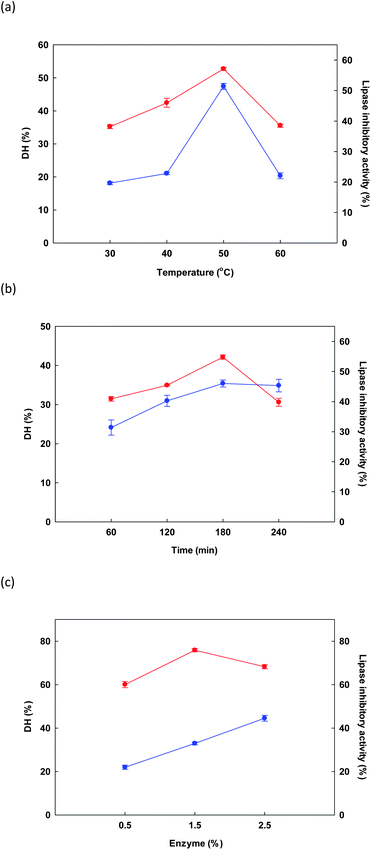 | ||
Fig. 1 Initial examination of the influence of changes in different factors: temperature (a), time (b) and enzyme (c) for the reaction upon DH ( ) and lipase inhibitory activity ( ) and lipase inhibitory activity ( ). ). | ||
In Fig. 1b, the influence on DH and lipase inhibition of the various hydrolysis times is shown, including 60, 120, 180, and 240 minutes. For hydrolysis experiments, temperature was 50 °C while Alcalase® concentration was held at 1.5%. As time increased from 50 to 180 minutes, response variables of DH and lipase inhibition also rose, reaching a maximum at 180 minutes. A decline was then observed at longer time periods. Liu et al.52 reported similar findings when evaluating the DH of fish protein during enzymatic hydrolysis, where DH climbed notably as the time period increased from zero to 8 hours, whereupon it reached an equilibrium, or underwent a very small decrease. Lipase inhibition results showed a similar pattern. Our findings confirmed that the influence of time was significant in the context of DORB protein hydrolysis (p < 0.05). On the basis of these experiments, the selected RSM central point was 180 minutes.
Finally, in Fig. 1c the influence on DH and lipase inhibition is shown for different enzyme concentrations in the range of 0.5% to 2.5%. Temperature was fixed at 50 °C while duration was 180 minutes. As enzyme concentration increased, DH also increased, finally reaching 44.55 ± 1.34% at the maximum 2.5% enzyme concentration. However, for lipase inhibition, the best outcome occurred at a concentration of 1.5%, while increasing the concentration further caused the hydrolyzed peptides to be shorter, and thus less effective for the inhibition of lipase.52 On the basis of cost, as well as the importance of lipase inhibition, the concentration of 1.5% was therefore selected to serve as the central point for RSM, as shown in Table 2.
| Independent variables | Code levels | ||||
|---|---|---|---|---|---|
| −1.68 | −1 | 0 | 1 | 1.68 | |
| X1: temperature (°C) | 41.59 | 45.00 | 50.00 | 55.00 | 58.41 |
| X2: E/S (% w/v) | 0.50 | 0.90 | 1.50 | 2.10 | 2.50 |
| X3: hydrolysis time (min) | 79.09 | 120.00 | 180.00 | 240.00 | 280.91 |
| Run | Space type | Coded levels | Independent variables | Response | |||||
|---|---|---|---|---|---|---|---|---|---|
| X1 | X2 | X3 | X1 | X2 | X3 | Y1 | Y2 | ||
| a All tests were performed in triplicate. | |||||||||
| 1 | Factorial | −1 | −1 | −1 | 45.00 | 0.90 | 120.00 | 25.03 | 9.77 |
| 2 | Factorial | +1 | −1 | −1 | 55.00 | 0.90 | 120.00 | 30.10 | 14.33 |
| 3 | Factorial | −1 | +1 | −1 | 45.00 | 2.10 | 120.00 | 31.23 | 11.00 |
| 4 | Factorial | +1 | +1 | −1 | 55.00 | 2.10 | 120.00 | 33.91 | 6.30 |
| 5 | Factorial | −1 | −1 | +1 | 45.00 | 0.90 | 240.00 | 30.56 | 7.06 |
| 6 | Factorial | +1 | −1 | +1 | 55.00 | 0.90 | 240.00 | 31.74 | 14.69 |
| 7 | Factorial | −1 | +1 | +1 | 45.00 | 2.10 | 240.00 | 36.13 | 19.25 |
| 8 | Factorial | +1 | +1 | +1 | 55.00 | 2.10 | 240.00 | 36.47 | 19.68 |
| 9 | Axial | −1.68 | 0 | 0 | 41.59 | 1.50 | 180.00 | 29.34 | 15.92 |
| 10 | Axial | +1.68 | 0 | 0 | 58.41 | 1.50 | 180.00 | 34.64 | 18.01 |
| 11 | Axial | 0 | −1.68 | 0 | 50.00 | 0.50 | 180.00 | 27.83 | 9.19 |
| 12 | Axial | 0 | +1.68 | 0 | 50.00 | 2.50 | 180.00 | 39.79 | 12.50 |
| 13 | Axial | 0 | 0 | −1.68 | 50.00 | 1.50 | 79.09 | 30.15 | 7.31 |
| 14 | Axial | 0 | 0 | +1.68 | 50.00 | 1.50 | 280.91 | 34.93 | 12.98 |
| 15 | Center | 0 | 0 | 0 | 50.00 | 1.50 | 180.00 | 34.54 | 3.02 |
| 16 | Center | 0 | 0 | 0 | 50.00 | 1.50 | 180.00 | 35.24 | 3.83 |
| 17 | Center | 0 | 0 | 0 | 50.00 | 1.50 | 180.00 | 34.36 | 4.12 |
| 18 | Center | 0 | 0 | 0 | 50.00 | 1.50 | 180.00 | 34.51 | 3.46 |
| 19 | Center | 0 | 0 | 0 | 50.00 | 1.50 | 180.00 | 33.07 | 3.65 |
| 20 | Center | 0 | 0 | 0 | 50.00 | 1.50 | 180.00 | 34.89 | 3.89 |
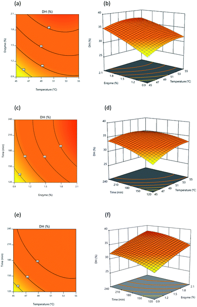
The 2D response graph presented in Fig. 2 shows the actual and predicted values for responses, with actual values illustrated on a straight line of the predicted value (Fig. 2a and b). Results confirmed the suitability of the independent variables for both of the response surface models. A summary of ANOVA (analysis of variance) results investigating regression models for the experiments testing DH and IC50 was provided in Design Expert 11 software and can be observed in Table 4. ANOVA results were shown to be significant (p < 0.05) for DH, taking into consideration the linear, interaction, and quadratic terms represented respectively by (X1, X2, and X3), (X1X3), and (X12, X32), as indicted by regression coefficients, while there was no significant influence exerted upon DH by interaction terms (X1X2 and X2X3) or quadratic term (X22). For an IC50 value of lipase inhibition, ANOVA results showed significance (p < 0.05) for linear, interaction, and quadratic terms represented respectively by (X1, X2, and X3), (X1X2, X1X3, and X2X3), and (X12, X22, and X32).
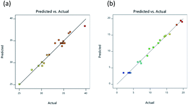 | ||
| Fig. 2 Relationship between the experimental and predicted values for DH (a), and lipase inhibitory activity (IC50; μg mL−1) (b). | ||
| Source | df | Y1 | Y2 | ||||||
|---|---|---|---|---|---|---|---|---|---|
| Sum of square | Mean square | F-value | P-value | Sum of square | Mean square | F-value | P-value | ||
| a Indicates those variables which significantly influence the response variables (p < 0.05). | |||||||||
| Model | 9 | 211.46 | 23.50 | 24.59 | <0.0001a | 619.70 | 68.86 | 107.72 | <0.0001a |
| X1 | 1 | 24.21 | 24.21 | 25.34 | 0.0005a | 9.58 | 9.58 | 14.99 | 0.0031a |
| X2 | 1 | 119.66 | 119.66 | 125.23 | <0.0001a | 18.60 | 18.60 | 29.10 | 0.0003a |
| X3 | 1 | 37.63 | 37.63 | 39.38 | <0.0001a | 60.83 | 60.83 | 95.17 | <0.0001a |
| X1X2 | 1 | 1.30 | 1.30 | 1.36 | 0.2698 | 33.88 | 33.88 | 53.01 | <0.0001a |
| X1X3 | 1 | 4.85 | 4.85 | 5.08 | 0.0479a | 8.39 | 8.39 | 13.13 | 0.0047a |
| X2X3 | 1 | 0.01 | 0.01 | 0.01 | 0.9185 | 71.90 | 71.90 | 112.48 | <0.0001a |
| X12 | 1 | 15.28 | 15.28 | 15.99 | 0.0025a | 316.33 | 316.33 | 494.90 | <0.0001a |
| X22 | 1 | 2.15 | 2.15 | 2.25 | 0.1645 | 91.64 | 91.64 | 143.37 | <0.0001a |
| X32 | 1 | 10.05 | 10.05 | 10.52 | 0.0088a | 74.49 | 74.49 | 116.54 | <0.0001a |
| Residual | 10 | 9.55 | 9.56 | 6.39 | 0.6392 | ||||
| Lack of fit | 5 | 6.81 | 1.36 | 2.49 | 0.1701 | 3.69 | 0.7388 | 1.39 | 0.3694 |
| Pure error | 5 | 2.74 | 0.55 | 2.70 | 0.5396 | ||||
| Cor total | 19 | 221.01 | 626.09 | ||||||
| Std dev. | 0.9775 | 0.7995 | |||||||
| R2 | 0.9568 | 0.9898 | |||||||
| Adj R2 | 0.9179 | 0.9806 | |||||||
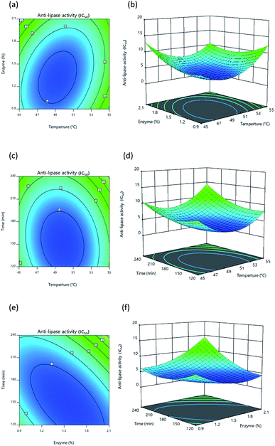
The p-values for both regression models were lower than 0.01, so the models can be considered significant. The p-values for lack of fit for the two models, however, exceeded 0.05 and hence were not significant. It can therefore be concluded that the models were suitably adjusted to responses. Model suitability can also be evaluated by coefficient of determination (adjusted R2) which was 0.9179, suggesting that 91.79% of response variability for DH could be explained by the model, while for lipase inhibition, the adjusted R2 value was 0.9806, thus demonstrating that 98.06% of response variability was explained by the model. In the case of our own R2 value, the models for both DH and lipase inhibition had values approaching 1.00, at 0.9568 and 0.9898 respectively, from which it can be inferred that the models fit well with experimental data. The response surface models which were developed had appropriate relationships involving independent variables constructed through the use of the regression coefficients for linear, interaction, and quadratic terms, along with a second-order polynomial model. Eqn (3) was used to calculate response for DH while lipase inhibition response was calculated with eqn (4):
| Y1 = −137.32471 + 5.0544X1 + 14.70023X2 + 0.239475X3 − 0.134583X1X2 − 0.002596X1X3 + 0.001007X2X3 − 0.04119X12 − 1.07299X22 − 0.000232X32 | (3) |
| Y2 = 492.32703 − 18.15846X1 + 0.240212X2 − 0.487773X3 − 0.685979X1X2 + 0.003414X1X3 + 0.083274X2X3 + 0.187405X12 + 7.0048X22 + 0.000632X32 | (4) |
In these equations, Y1 is DH prediction, Y2 is IC50 prediction, and X1, X2, and X3 represent temperature, enzyme, and time, respectively, as independent variables. Optimal levels for independent variables are those which generate the maximum DH value combined with minimum value for IC50.
Once protein hydrolysates had been obtained under the 20 different conditions, they were tested to determine their lipase inhibitory activity in order to develop response surface plots, which matched those defined for optimal conditions, showing convex shapes as presented in Fig. 4a–4c. Increasing the independent variables during hydrolysis results in an initial increase of lipase inhibition followed later by a decline. It is apparent, however, that the lipase inhibition from protein hydrolysates will be affected in part by the way the three independent variables interacting with each other. In general, it is typically found that response surfaces that maximize yield provide the greatest level of biological activity; for instance, according to Ahmad et al.55 high yields of Artocarpus altilis extract also exhibit heightened DPPH scavenging activity. However, in this research, maximized protein yield did not result in the highest level of lipase inhibition, thus matching the findings of Baharuddin et al.56 It is advisable, therefore, to focus primarily upon regression models for the IC50 value indicating lipase inhibition, since this will show hydrolyzed protein size, which is a more important factor in lipase inhibition.
3.3 Purification of the lipase inhibitory peptides
| Molecular weight (kDa) | Lipase inhibitory activitya (IC50; μg mL−1) |
|---|---|
| a All results are presented in the form of mean ± SD and tests are performed in triplicate. | |
| Crude protein | 2.8360 ± 0.2022b |
| >10 | 35.4567 ± 0.3009e |
| 5–10 | 19.2533 ± 0.3523c |
| 3–5 | 6.2967 ± 0.2571b |
| 0.65–3 | 20.1750 ± 0.4171d |
| <0.65 | 3.5097 ± 0.1729a |
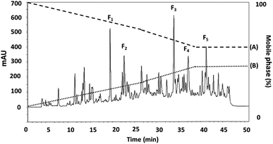 | ||
| Fig. 5 The RP-HPLC profile for the DORB protein hydrolysate active fraction (<0.65 kDa) separated using eluent A: 0.1% trifluoroacetic acid (TFA), and eluent B: 70% acetonitrile. | ||
3.4 Identification of peptides and their properties in F2 sub-fractions using quadrupole time-of-flight (Q-TOF) liquid chromatography-tandem mass spectrometry (LC-MS/MS)
The amino acid sequences of the lipase inhibitory peptide in the RP-HPLC fraction (F2) were identified using LC-ESI-Q-TOF-MS/MS de novo sequencing as Phe–Tyr–Leu–Gly–Tyr–Cys–Asp–Tyr (FYLGYCDY), as shown in Fig. 6. Molecular weight was determined as 1043.15 Da. The homologous region for FYLGYCDY was sought through the NCBI GenBank on the basis of the Oryzeae genus, which offered a similar sequence to the hypothesized protein EE612_044279 of O. sativa (accession number KAB8108513.1), U-box domain-containing protein 19 of O. sativa Japonica (accession number XP_015649021.1), and hypothetical protein OsI_29238 of O. sativa Indica (accession number EAZ06993.1). Database sequences had an E-value of 6.6, sequence score of 23.10%, with 63% equal identities (7/11). The FYLGYCDY peptide underwent synthesis as the database specified, resulting in production of the purified peptide. This synthesized peptide had an IC50 value of 0.47 ± 0.02 μM. The Innovagen server provides data indicating that the FYLGYCDY peptide offered solubility lower than 0.10 mg mL−1, attaining the categorization of ‘undissolved’, so the solution for the peptide will be less than 10 mg mL−1 of gross peptide concentration in dimethyl sulfoxide (DMSO). Data can be seen in Table 7. The key properties of lipase inhibitory peptides include chain length and hydrophobicity; after examination of a large number of peptide sequences, it is clear that the best inhibitors of lipase offer short chains comprising just 2–16 amino acids. It is easier for these short peptides to enter the enzyme active site.61 It is also easier for such short peptides to enter the bloodstream without any alteration to their general properties.62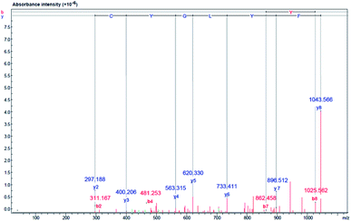 | ||
| Fig. 6 Identification of amino acid sequence and lipase inhibitor peptide molecular mass following purification of DORB hydrolysate (F2 from RP-HPLC). Mass fragmentation spectrum of FYLGYCDY peptide. | ||
| Synthesized peptides | Solubility in watera | Hydrophobicityb (%) | Potential biological activityd | Toxicity prfile (SVM score)c | Sensory characteristics |
|---|---|---|---|---|---|
| a Solubility data for the peptide comes from the Innovagen server (http://www.innovagen.com/proteomics-tools).b Peptide properties were calculated using the Peptide 2.0 server (http://www.peptide2.com).c Analysis of the peptide toxicity was performed using the ToxinPred server (http://crdd.osdd.net/raghava/toxinpred/).d Sensory attributes and potential biological properties of the peptide are obtained from the BIOPEP database (http://www.uwm.edu.pl/biochemia/index.php/en/biopep). | |||||
| FYLGYCDY | Poor | 25.0 | GY (ACE inhibitor and DPP IV inhibitor), FY (ACE inhibitor), LG (ACE inhibitor), DY (ACE inhibitor), YL (neuropeptide, DPP IV inhibitor and DPP-III inhibitor), LGY (immune-stimulating), LGY (regulating), YLGY (ACE inhibitor and antioxidative) | Non-toxic (−0.44) | GY (bitter), LG (bitter), FY (bitter), DY (bitter), YL (bitter) |
The BIOPEP database (http://www.uwm.edu.pl/biochemia/index.php/en/biopep) contains data on the properties of many of the peptides which are understood to offer potentially useful bioactive properties. To determine the likely properties of the FYLGYCDY peptide it is possible to examine similar sequences in the database, which are shown in Table 7. The investigated peptide sequences which showed bioactivity were GY (location 4–5), FY (location 1–2), LG (location 3–4), DY (location 7–8), and YLGY (location 2–5), all demonstrating inhibitory qualities. The peptide which serves to regulate the flow of ions was DY (location 7–8), while the peptide controlling the phosphoinositol metabolism was YLGY (location 2–5). Meanwhile, YL (location 2–3) is a neuropeptide of anxiolytic, a dipeptidyl peptidase IV inhibitor (DPP IV inhibitor), and a DPP-III inhibitor, while GY (location 4–5) is also a dipeptidyl peptidase IV inhibitor. LGY (location 3–5) demonstrated immunostimulatory activity while YLGY (location 2–5) showed antioxidative activity. There are certain concerns which remain in the context of using peptides to develop therapeutic products or functional foods, and one is the issue of toxicity.63 FYLGYCDY peptide, however, was obtained from plant-based proteins, and it is possible to evaluate its toxicity using the ToxinPred server (http://crdd.osdd.net/raghava/toxinpred/), which allows for predictions to be made which suggest that FYLGYCDY is not toxic and has SVM scores below zero, which can be seen in Table 7. It is therefore feasible for this lipase inhibitory peptide to undergo further development, but evaluations must be carried out both in vitro and in vivo prior to human use, in order to ensure that toxicity problems are avoided. Another important consideration for peptides and amino acids used as food ingredients is that of sensory properties, and in particular taste, which is the consequence of certain chemical structures. The sensory attributes of peptides can be predicted using data from the BIOPEP database by applying the procedures outlined by Iwaniak et al.64 It can be concluded that the taste of the FYLGYCDY peptide would be bitter, as Table 7 indicates.
3.5 Inhibition kinetic of the lipase inhibitory peptide
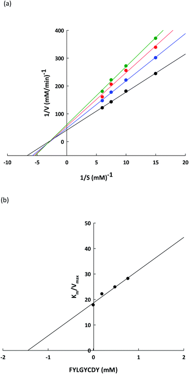
To verify the lipase inhibitory mechanism, a Lineweaver–Burk plot of the FYLGYCDY peptide derived from the DORB protein was analyzed. Analysis of the inhibition kinetics employed varying p-NPP concentrations as a substrate along with a fixed amount and concentration of the peptide. Fig. 7a shows that this peptide produces generally non-competitive inhibition. The four straight lines obtained had an intersection at one point on the 1/S axis and the value for Km was 0.6229 ± 0.1170 mM, while maximum velocity (Vmax) for the differing peptide concentrations along with the slopes of the straight lines were different. This occurred because an increase in peptide concentration resulted in a decline in the enzymatic reaction efficiency of the substrate. This is apparent in Table 8. Meanwhile, Ki represents the inhibition constant, which can be seen in Fig. 7b, revealing the bond strength between lipase and peptide, whereby a lower value is associated with higher bond strength. For FYLGYCDY, the value for Ki was 1.4493 ± 0.0961 mM, which is notable in peptides exhibiting non-competitive inhibition. The FYLGYCDY peptide therefore works by lowering the number of functional enzymes capable of reacting with the substrate. These findings are similar to those of Liu et al.,52 whose work on fish protein hydrolysates showed that the PPL inhibition mechanism was of a non-competitive inhibitor type, thus limiting the ability of the enzyme to generate efficient catalysis. It has also been reported that another non-competitive inhibitor for PPL is astaxanthin, which is able to block the channel to the enzyme catalytic site, thus delaying entrance to the substrate.| Kinetic parameter | Control | FYLGYCDY (mM) | ||
|---|---|---|---|---|
| 0.2 | 0.5 | 0.8 | ||
| a All results are presented in the form of mean ± SD and tests are performed in triplicate. | ||||
| KM (mM) | 0.6229 ± 0.1170 | 0.6229 ± 0.1170 | ||
| Vmax (mM min−1) | 0.0352 ± 0.0039 | 0.0282 ± 0.0023 | 0.0252 ± 0.0030 | 0.0222 ± 0.0027 |
| Ki (mM) | 1.4493 ± 0.0961 | |||
3.6 FYLGYCDY molecular docking at the lipase binding site
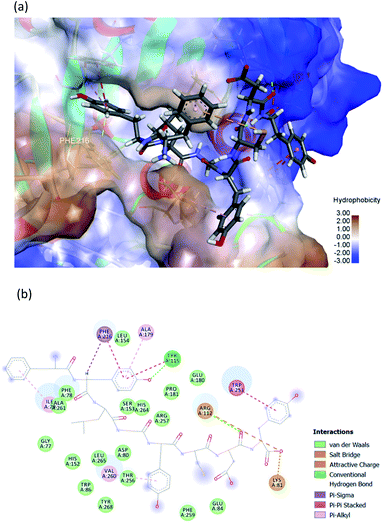
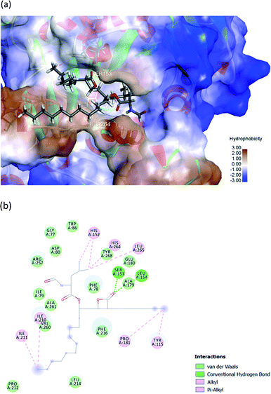
The hydrophobic peptide FYLGYCDY underwent docking with the crystal PPL structure (PDB: 1ETH) using GOLD 5.7.1 software to examine the interactions of the peptide (Fig. 8) binding with lipase and draw comparisons with orlistat which served as a positive control (Fig. 9). This process is shown in Fig. 8, which reveals that the Tyr amino acid, categorized as hydrophobic and located in position two of the inhibitor peptide, is capable of binding to the Phe216 of PPL.65 The hydrogen which forms part of Tyr2 serves as the donor atom, and Phe216 of PPL is the acceptor atom in the context of Pi–Sigma interaction. Phe216 acted as the substrate-binding site of the lipase, while Ser153, Asp177, and His264 acted as the PPL catalytic sites, whereby the inhibitor peptide did not bind to those catalytic sites but instead was under the influence of van der Waals forces. Table 9 shows this particular competitive type of molecular docking inhibition in the case of this peptide. It has been shown that hydrophobic peptides derived from the enzymatic hydrolysis of C. pryenoidose,58 with a greater molecular weight than the peptide which is the focus of this study, may bind to the PPL catalytic sites as a result of Pi-cation and conventional hydrogen bond interaction. Fig. 9 shows the properties of orlistat, which is designed to inhibit PPL as a mainly competitive inhibitor, when it binds to PPL at the principal catalytic sites, whereby Ser153 and His264 of the PPL complex by H-donor of Ser153 bind to the β-lactone ring of orlistat, while His264 binds to the side-chain hydrogen bond of the amino ester of orlistat, respectively binding through a conventional hydrogen bond and Pi–alkyl interaction.64 The evaluation was based on the different scoring functions for docking, the best score for which came from ChemPLP. The inhibitor peptide had a docking score of 122.54 for binding on PPL while orlistat achieved a docking score of 101.44. It therefore appears that the peptide would be able to inhibit the PPL function more effectively than orlistat.| Amino acid | Category | Donor atom | Acceptor atom | Interaction | Distance (Å) |
|---|---|---|---|---|---|
| Phe1 | Hydrophobic | Phe1 (Pi-orbitals) | Ile79 (alkyl) | Pi–alkyl | 3.9113 |
| Tyr2 | Hydrophobic | Tyr2 (Pi-orbitals) | Ala179 (alkyl) | Pi–alkyl | 5.0570 |
| Hydrophobic | Tyr2 (Pi-orbitals) | Phe216 (Pi-orbitals) | Pi–Pi stacked | 4.5563 | |
| Hydrophobic | Tyr2 (Pi-orbitals) | Try115 (Pi-orbitals) | Pi–Pi stacked | 4.4097 | |
| Hydrophobic | Tyr2 (C–H) | Phe216 (Pi-orbitals) | Pi–sigma | 2.7882 | |
| Hydrogen bond | Tyr115 (H-donor) | Tyr2 (H-acceptor) | Conventional hydrogen bond | 2.0815 | |
| Try5 | Hydrophobic | Try5 (Pi-orbitals) | Val260 (alkyl) | Pi–alkyl | 5.2900 |
| Asp7 | Hydrogen bond | Arg112 (H-donor) | Asp7 (H-acceptor) | Conventional hydrogen bond | 2.0172 |
| Try8 | Hydrogen bond | Try8 (Pi-orbitals) | Trp253 (Pi-orbitals) | Pi–Pi stacked | 3.9038 |
| Electrostatic | Arg112 (positive) | Try8 (negative) | Attractive charge | 3.8041 | |
| Hydrogen bond | Lys81 (H-donor; positive) | Try8 (H-acceptor; negative) | Salt bridge; attractive charge | 1.7087 | |
| Hydrophobic | Lrp253 (Pi-orbitals) | Try8 (Pi-orbitals) | Pi–Pi stacked | 4.1226 |
4. Conclusions
This study successfully identified a peptide which offered lipase inhibitory activity and which could be obtained from DORB proteins through the use of Alcalase®. In order to optimize the influence of temperature, time, and enzyme upon the degree of hydrolysis and lipase inhibitory activity as indicated by IC50 values, RSM was employed with CCD. To achieve the greatest level of lipase inhibitory activity from the hydrolysate, the necessary conditions were 49.88 °C, 150.43 minutes, and 1.53% enzyme for sample solutions of 5% w/v. Under these conditions, the DH value was 35.65%, and the IC50 value was 2.84 μg mL−1. The hydrolysate which offered the best performance in terms of lipase inhibition then underwent fractionation to obtain fractions bound by different molecular weight cut-offs, with the MW < 0.65 kDa proving to be the most effective inhibitor of lipase, having achieved an IC50 value of 3.51 μg mL−1. Following fractionation using ultrafiltration techniques, as well as purification using RP-HPLC, and peptide identification via LC-MS/MS, the lipase inhibitory peptide Phe–Tyr–Leu–Gly–Tyr–Cys–Asp–Tyr (FYLGYCDY) was identified, with an IC50 value of 0.47 μM and a molecular weight of 1043.15 Da. The peptide FYLGYCDY was shown to have the capacity to inhibit pancreatic lipase, and according to Lineweaver–Burk plots is a non-competitive inhibitor. However, molecular docking examination which aimed to predict the binding site for the peptide FYLGYCDY with the PPL complex showed a competitive binding type. From the results obtained, the peptide demonstrated the capacity to block the channel to the enzyme catalytic site, thus delaying the entrance of the substrate. The lipase inhibitory capacity of the DORB protein hydrolysate can function as a bioactive peptide, especially to serve as anti-obesity agents. This peptide, however, should also undergo further study in terms of its cytotoxicity and allergenicity in order that it might in future be used in the food or pharmaceutical sectors.Funding
Sincere thanks are expressed to we acknowledge the financial support from the grant for research: the Research Assistantship Fund, Faculty of Science, Chulalongkorn University (RAF_2562_003), the Thailand Science Research and Innovation (TSRI) Fund (CU_FRB640001_01_61_2), and the Center of Excellence on Medical Biotechnology (CEMB), S&T Postgraduate Education and Research Development Office (PERDO), Office of Higher Education Commission (OHEC), Thailand (SN-63-009-01) for providing the financial support for this research in recognition of the financial assistance offered which enabled this research study to be completed.Conflicts of interest
The authors have no conflict of interest. This publication has not been submitted earlier in any journal and is not being considered for publication elsewhere. All of the authors, including the corresponding authors, have read and approved the final submitted manuscript.Acknowledgements
The researchers are also grateful to the Institute of Biotechnology and Genetic Engineering at Chulalongkorn University for the facilities which they made available during the course of the study. The authors were grateful to Dr Benjamin Richard Poole, Department of English, Faculty of Art, Chulalongkorn University for reviewing this manuscript.References
- R. T. Hurt, C. Kulisek, L. A. Buchanan and S. A. McClave, Gastroenterol. Hepatol., 2010, 6, 780–792 Search PubMed.
- N. S. Mitchell, V. A. Catenacci, H. R. Wyatt and J. O. Hill, Psychiatr. Clin., 2011, 34, 717–732 Search PubMed.
- X. Pi-Sunyer, Postgrad. Med., 2009, 121, 21–33 CrossRef PubMed.
- C. M. Diaz-Melean, V. K. Somers, J. P. Rodriguez-Escudero, P. Singh, O. Sochor, E. M. Llano and F. Lopez-Jimenez, Curr. Atheroscler. Rep., 2013, 15, 364 CrossRef PubMed.
- K. B. Smith and M. S. Smith, Prim. Care., 2016, 43, 121–135 CrossRef PubMed.
- R. B. Birari and K. K. Bhutani, Drug Discov. Today, 2007, 12, 879–889 CrossRef CAS PubMed.
- J. W. Yun, Phytochemistry, 2010, 71, 1625–1641 CrossRef CAS PubMed.
- A. L. de la Garza, F. I. Milagro, N. Boque, J. Campión and J. A. Martínez, Planta Med., 2011, 77, 773–785 CrossRef CAS PubMed.
- K. A. Grove, S. Sae-tan, M. J. Kennett and J. D. Lambert, Obesity, 2012, 20, 2311–2313 CrossRef CAS PubMed.
- S. Uchiyama, Y. Taniguchi, A. Saka, A. Yoshida and H. Yajima, Nutrition, 2011, 27, 287–292 CrossRef CAS PubMed.
- A. Ballinger and S. R. Peikin, Eur. J. Pharmacol., 2002, 440, 109–117 CrossRef CAS PubMed.
- L. López-Barrios, J. A. Gutiérrez-Uribe and S. O. Serna-Saldívar, J. Food Sci., 2014, 79, R273–R283 CrossRef PubMed.
- S. Saadi, N. Saari, F. Anwar, A. Abdul Hamid and H. M. Ghazali, Biotechnol. Adv., 2015, 33, 80–116 CrossRef CAS PubMed.
- M. Nasri, Adv. Food Nutr. Res., 2017, 81, 109–159 CAS.
- Y. Wang, Q. Huang, D. Kon and P. Xu, Mini Rev. Med. Chem., 2018, 18, 1524–1535 CrossRef CAS PubMed.
- G. H. Li, M. R. Qu, J. Z. Wan and J. M. You, Asia Pac. J. Clin. Nutr., 2007, 16, 275–280 CAS.
- H. Zhuang, N. Tang and Y. Yuan, J. Funct. Foods, 2013, 5, 1810–1821 CrossRef CAS.
- F. Zhong, X. Zhang, J. Ma and C. F. Shoemaker, Food Res. Int., 2007, 40, 756–762 CrossRef CAS.
- M. Zhang, T. H. Mu and M. J. Sun, J. Funct. Foods, 2014, 7, 191–200 CrossRef CAS.
- N. Kheeree, P. Sangtanoo, P. Srimongkol, T. Saisavoey, O. Reamtong, K. Choowongkomon and A. Karnchanatat, Food Funct., 2020, 11, 8161–8178 RSC.
- T. Saisavoey, P. Sangtanoo, O. Reamtong and A. Karnchanatat, J. Food Biochem., 2016, 40, 731–740 CrossRef CAS.
- A. G. B. Wouters, I. Rombouts, E. Fierens, K. Brijs and J. A. Delcour, Compr. Rev. Food Sci. Food Saf., 2016, 15, 786–800 CrossRef CAS PubMed.
- K. Gul, B. Yousuf, A. K. Singh, P. Singh and A. A. Wani, Bioact. Carbohydr. Diet. Fibre, 2015, 6, 24–30 CrossRef CAS.
- H. Y. Park, K. W. Lee and H. D. Choi, Food Funct., 2017, 22, 935–943 RSC.
- M. Sohail, A. Rakha, M. S. Butt, M. J. Iqbal and S. Rashid, Crit. Rev. Food Sci. Nutr., 2017, 22, 3771–3780 CrossRef PubMed.
- Y. Q. Liu, P. Strappe, W. T. Shang and Z. K. Zhou, Crit. Rev. Food Sci. Nutr., 2019, 59, 349–356 CrossRef CAS PubMed.
- S. Tang, N. S. Hettiarachchy and T. H. Shellhammer, J. Agric. Food Chem., 2002, 50, 7444–7448 CrossRef CAS PubMed.
- C. Fabian and Y. H. Ju, Crit. Rev. Food Sci. Nutr., 2011, 51, 816–827 CrossRef CAS PubMed.
- Official Methods of Analysis of AOAC International. 18th ed. Methods 925.09 and 926.08, AOAC International, Gaithersburg, MD, USA, 2005 Search PubMed.
- Official Methods of Analysis of AOAC International. 18th ed. Methods 2003.06, AOAC International, Gaithersburg, MD, USA, 2005 Search PubMed.
- Official Methods of Analysis of AOAC International. 18th ed. Methods 923.03, AOAC International, Gaithersburg, MD, USA, 2005 Search PubMed.
- Official Methods of Analysis of AOAC International. 18th ed. Methods 968.06 and 992.15, AOAC International, Gaithersburg, MD, USA, 2005 Search PubMed.
- Official Methods of Analysis of AOAC International. 18th ed. Methods 962.09, AOAC International, Gaithersburg, MD, USA, 2005 Search PubMed.
- E. V. Crisan and A. Sands, Nutritional Value. Academic Press, New York, 1978, pp. 137–168 Search PubMed.
- P. Nielsen, D. Petersen and C. Dambmann, J. Food Sci., 2001, 66, 642–646 CrossRef CAS.
- S. Adisakwattana, J. Moonrat, S. Srichairat, C. Chanasit, H. Tirapongporn, B. Chanathong, S. Ngamukote, K. Mauml and S. Sapwarobol, J. Med. Plants Res., 2010, 4, 2113–2120 Search PubMed.
- M. M. Bradford, Anal. Biochem., 1976, 72, 248–254 CrossRef CAS PubMed.
- J. Hermoso, D. Pignol, B. Kerfelec, I. Crenon, C. Chapus and J. C. Fontecilla-Camps, J. Biol. Chem., 1996, 271, 18007–18016 CrossRef CAS PubMed.
- T. Genkawa, T. Uchino, A. Inoue, F. Tanaka and D. Hamanaka, Biosyst. Eng., 2008, 99, 515–522 CrossRef.
- S. Patel, J. Funct. Foods, 2015, 14, 255–269 CrossRef CAS.
- C. O. Edeogu, F. C. Ezeonu, A. N. C. Okaka, C. E. Ekuma and S. O. Elom, Int. J. Biotechnol. Biochem., 2007, 3, 1–8 CrossRef.
- C. Kalpanadevi, V. Singh and R. Subramanian, J. Food Sci. Technol., 2018, 55, 2259–2269 CrossRef CAS PubMed.
- B. J. Lloyd, T. J. Siebenmorgen and K. W. Beers, Cereal Chem., 2000, 77, 551–555 CrossRef CAS.
- T. Yamagishi, T. Tsuboi and K. Kikuchi, Cereal Chem., 2003, 80, 5–8 CrossRef CAS.
- L. Shi, Int. J. Biol. Macromol., 2016, 92, 37–48 CrossRef CAS PubMed.
- L. Amagliani, J. O'Regan, A. L. Kelly and J. A. O'Mahony, Trends Food Sci. Technol., 2017, 64, 1–12 CrossRef CAS.
- I. Sereewatthanawut, S. Prapintip, K. Watchiraruji, M. Goto, M. Sasaki and A. Shotipruk, Bioresour. Technol., 2008, 99, 555–561 CrossRef CAS PubMed.
- C. Fabian and Y. H. Ju, Crit. Rev. Food Sci. Nutr., 2011, 51, 816–827 CrossRef CAS PubMed.
- I. B. B. Piotrowicz, M. Garcés-Rimón, S. Moreno-Fernández, A. Aleixandre, M. Salas-Mellado and M. Miguel-Castro, Foods, 2020, 9, 812 CrossRef CAS PubMed.
- I. Popovic, B. A. G. Bossink and P. C. van der Sijde, Sustainability, 2019, 11, 7197 CrossRef.
- D. Gao, F. Zhang, Z. Ma, S. Chen, G. Ding, X. Tian and R. Feng, Int. J. Food Prop., 2019, 22, 1296–1309 CrossRef CAS.
- L. Liu, Y. Wang, C. Peng and J. Wang, Int. J. Mol. Sci., 2013, 14, 3124–3139 CrossRef CAS PubMed.
- P. Hanmoungjai, D. L. Pyle and K. Niranjan, J. Am. Oil Chem. Soc., 2001, 78, 817–821 CrossRef CAS.
- M. Montilha, M. Sbroggio, V. Figueiredo, E. Ida and L. Kurozawa, Int. Food Res. J., 2007, 24, 1067–1074 Search PubMed.
- M. N. Ahmad, N. U. Karim, E. Normaya, B. M. Piah, A. Iqbal and K. H. K. Bulat, Sci. Rep., 2020, 10, 1–14 CrossRef PubMed.
- N. A. Baharuddin, N. R. A. Halim and N. M. Sarbon, Int. Food Res. J., 2016, 23, 1424–1431 CAS.
- M. B. O'Keeffe, C. Conesa and R. J. FitzGerald, Int. J. Food Sci. Technol., 2017, 52, 1751–1759 CrossRef.
- R. Zhang, J. Chen, X. Mao, P. Qi and X. Zhang, Molecules, 2019, 24, 3527 CrossRef CAS PubMed.
- X. Wang, H. Chen, X. Fu, S. Li and J. Wei, LWT–Food Sci. Technol., 2017, 75, 93–99 CrossRef CAS.
- S. Jafar, H. Kamal, P. Mudgil, H. M. Hassan and S. Maqsood, LWT–Food Sci. Technol., 2018, 98, 212–218 CrossRef CAS.
- P. Mudgil, H. Kamal, G. C. Yuen and S. Maqsood, Food Chem., 2018, 259, 46–54 CrossRef CAS PubMed.
- R. E. Aluko, Annu. Rev. Food Sci. Technol., 2015, 6, 235–262 CrossRef CAS PubMed.
- S. Gupta, P. Kapoor, K. Chaudhary, A. Gautam, R. Kumar and G. P. S. Raghava, PLoS One, 2013, 8, e73957 CrossRef CAS PubMed.
- A. Iwaniak, P. Minkiewicz, M. Darewicz, K. Sieniawski and P. Starowicz, Food Res. Int., 2016, 85, 155–161 CrossRef CAS PubMed.
- J. Hermoso, D. Pignol, B. Kerfelec, I. Crenon, C. Chapus and J. C. Fontecilla-Camps, J. Biol. Chem., 1996, 271, 18007–18016 CrossRef CAS PubMed.
| This journal is © The Royal Society of Chemistry 2021 |

