 Open Access Article
Creative Commons Attribution 3.0 Unported Licence
Coexistence of metamagnetism and slow relaxation of magnetization in ammonium hexafluoridorhenate†
James Louis-Jean *a,
Samundeeswari M. Balasekaran‡
a,
Keith V. Lawler‡
 a,
Adrián Sanchis-Perucho‡b,
José Martínez-Lillo
 *b,
Dean Smith
 cd,
Paul M. Forstera,
Ashkan Salamatc and
Frederic Poineau
 *a
*a
aDepartment of Chemistry and Biochemistry, University of Nevada Las Vegas, 4505 South Maryland Parkway, Las Vegas, Nevada 89154-4003, USA. E-mail: louisjea@unlv.nevada.edu; poineauf@unlv.nevada.edu
bInstituto de Ciencia Molecular (ICMol), Departament de Química Inorgànica, Universitat de València, Catedrático José Beltrán 2, 46980 Paterna, València, Spain. E-mail: f.jose.martinez@uv.es
cDepartment of Physics and Astronomy, University of Nevada Las Vegas, 4505 South Maryland Parkway, Las Vegas, Nevada 89154-4003, USA
dHPCAT, X-ray Science Division, Argonne National Laboratory, Lemont IL 60439, USA
First published on 3rd February 2021
Abstract
The (NH4)2[ReF6] (1) salt was studied by X-ray diffraction, Raman spectroscopy, theoretical calculations, and magnetic measurements. 1 crystallizes in the trigonal space group P![[3 with combining macron]](https://www.rsc.org/images/entities/char_0033_0304.gif) m1 (Re–F = 1.958(5) Å). In the Raman spectrum of 1, splitting of the observed peaks was observed and correlated to the valence frequencies of vibration of the [ReF6]2− anion. The study of the magnetic properties of 1, through DC and AC magnetic susceptibility measurements, reveals the coexistence of metamagnetism and slow relaxation of magnetization at low temperature, which is unusual in the molecular systems based on the paramagnetic 5d metal ions reported so far.
m1 (Re–F = 1.958(5) Å). In the Raman spectrum of 1, splitting of the observed peaks was observed and correlated to the valence frequencies of vibration of the [ReF6]2− anion. The study of the magnetic properties of 1, through DC and AC magnetic susceptibility measurements, reveals the coexistence of metamagnetism and slow relaxation of magnetization at low temperature, which is unusual in the molecular systems based on the paramagnetic 5d metal ions reported so far.
Introduction
Salts of the [ReX6]2− (X = Cl, Br, I) anions have been known for decades and have been spectroscopically and structurally studied.1–3 However, the chemistry of the fluorido anion {i.e., [ReF6]2−} is not as well investigated; this matter of fact is probably due to the difficulty of preparing a [ReF6]2− precursor. Hexafluoridorhenate salts can be prepared from the solid-state melting reaction (SSMR) of A2[ReX6] (X = Cl, Br, I) salts with molten AHF2 (A = NH4+, K+). However, the isolation of the subsequent water-soluble A2[ReF6] salt has shown to be challenging.4,5 Recently, detailed procedures for the preparation of K2[ReF6]6 and (NH4)2[ReF6]7 have been reported. Currently, only eleven compounds containing the [ReF6]2− anion have been characterized by single crystal X-ray diffraction: K2[ReF6],6,8 A2[ReF6] (A = Rb, Cs),6 (AsPh4)2[ReF6]·2H2O, (PPh4)2[ReF6]·2H2O (Ph = C6H5: tetraphenylphosphonium),9,10 [M(viz)4(ReF6)]∞ (M = Zn, Ni; viz = C5H6N3: 1-vinylimidazole),9 [Co3(dpa)4][ReF6]·2DMF (DMF = HCON(CH3)2 and dpa = C10H8N3: 2,2′-dipyridylamine anion),11 (PPh4)2[ReF6]·CH3CN,11 (BEDO)4[ReF6]·6H2O (BEDO = C10H8O4S4: bis(ethylenedioxo)tetrathiafulvalene)12 and [Co(NH3)6]3[ReO4][ReF6]4·6H2O.13The (NH4)2[ReF6] salt was initially reported4 in 1956. While (NH4)2[ReF6] has been studied by X-ray absorption spectroscopy,7 thermal gravimetric analysis14,15 and was used as a precursor for preparing materials with remarkable magnetic properties,9,11,12 up to now there is no available single crystal X-ray data of the salt. In salts containing the [ReX6]2− (X = F, Cl, Br, I) anions, the Re(IV) ion (5d3) forms a hexakis-monodentate coordination with the halogen atoms resulting in either a regular or slightly distorted octahedral geometry. Studies on the magnetic behavior of [ReX6]2− species have shown the existence of strong antiferromagnetic interactions.16 The nature of such magnetic interaction occurred through space based on the Re–X⋯X–Re contacts and the size of the halogen. The magnetic properties of several species containing [ReX6]2− (X = Cl, Br, I) anions have been studied for more than 65 years.17–25 However, species containing the [ReF6]2− anion are underrepresented. To our knowledge, only six species of [ReF6]2− anion have been magnetically studied, namely K2[ReF6],21 (PPh4)2[ReF6]·2H2O (Ph = C6H5), [M(viz)4(ReF6)]∞ (M = Zn, Ni; viz = C5H6N3),9 [Co3(dpa)4][ReF6]·2DMF,11 and (BEDO)4[ReF6]·6H2O (BEDO = C10H8O4S4).12
Overall, there is a need to further develop and explore the structural and magnetic chemistry of hexafluoridorhenate(IV) compounds. In this context, the (NH4)2[ReF6] salt was prepared and characterized by X-ray diffraction (single crystal and powder), IR and Raman spectroscopies, DC and AC magnetic susceptibility measurements, and computational methods.
Results and discussion
Preparation of (NH4)2[ReF6] (1)
Bifluoride salts, AHF2 (A = NH4+, K+) are strong fluorinating agents and have been used for the preparation of [MF6]2− (M = Tc, Re) salts.4–6,9,26,27 Typically, those preparation involved the reactions of A2[MX6] salts (X = Cl, Br, I) in molten bifluorides. The isolation of the resulting A2[MF6] salts is followed by an aqueous workup. This method has been employed unsuccessfully by Peacock4 in an effort to prepare 1. Recently, an optimized method for the preparation of A2[ReX6] salts has been developed from the solid-state melting reaction of (NH4)2[ReX6] (X = Cl, Br) with excess NH4HF2.7,9,27 Using this method, 1 was prepared and separated after washing with a water–methanol mixture. After centrifugation and precipitation, the pink compound was obtained in 60% yield. Colorless single crystals of 1 could be obtained by recrystallization in water or in aqueous HF. The purity and homogeneity of the bulk sample of 1 was confirmed by EXAFS7 spectroscopy and powder XRD. The XRD pattern (Fig. 1) shows a single crystalline phase of 1.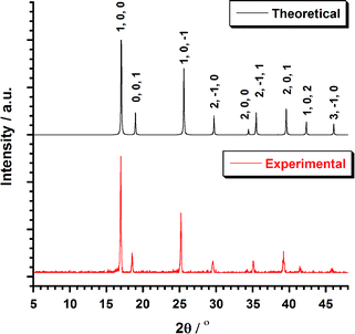 | ||
| Fig. 1 Plot of the theoretical and experimental XRD patterns profile (2θ/°) in the range 5–50° for compound 1. | ||
Structure description of 1
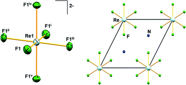
Similar to A2[MF6] (A = K, Rb, Cs; M = Tc, Re) and (NH4)2[TcF6] salts, 1 crystallizes in the trigonal space group P![[3 with combining macron]](https://www.rsc.org/images/entities/char_0033_0304.gif) m1.6,28 The structure of 1 consists of [NH4]+ cations and [ReF6]2− anions. The positions of the hydrogen atoms in 1 could not be determined reliably. Thus, the H atoms have not been considered during the refinement procedure. The overall solid-state structure is stabilized by a series of hydrogen bonding. In the [ReF6]2− anions, each Re(IV) ion forms a hexakis-monodentate coordination with fluorine atoms (Fig. 2). The Re(IV) ion is located at the origin of the trigonal unit cell occupying the site symmetry,
m1.6,28 The structure of 1 consists of [NH4]+ cations and [ReF6]2− anions. The positions of the hydrogen atoms in 1 could not be determined reliably. Thus, the H atoms have not been considered during the refinement procedure. The overall solid-state structure is stabilized by a series of hydrogen bonding. In the [ReF6]2− anions, each Re(IV) ion forms a hexakis-monodentate coordination with fluorine atoms (Fig. 2). The Re(IV) ion is located at the origin of the trigonal unit cell occupying the site symmetry, ![[3 with combining macron]](https://www.rsc.org/images/entities/char_0033_0304.gif) m (Wyckoff position 1a) whereas the F atom occupies the m site symmetry (Wyckoff position 6i). The six symmetry-related fluorine ligands in the anion result in a regular octahedral geometry (Fig. 2).
m (Wyckoff position 1a) whereas the F atom occupies the m site symmetry (Wyckoff position 6i). The six symmetry-related fluorine ligands in the anion result in a regular octahedral geometry (Fig. 2).
All Re–F bond lengths (1.958(5) Å) in 1 are of equal distances and are longer than the Tc–F bond length (1.922(6) Å) in (NH4)2[TcF6].28 The Re–F bond length in 1 is also in agreement (Table 1) with other species containing the [ReF6]2− anion. In [ReX6]2− (X = F, Cl, Br, I) salts, the Re–X bond length generally increases with increasing size of X, 1.958 Å, 2.353 Å, 2.502 Å and 2.721 Å respectively, as expected.6,16 DFT bonding analysis of the [ReF6]2− anion in its experimental geometry shows the presence of Re–F σ bonds. These σ bonds are nearly ionic with an 85.7% spin-up and 88.8% spin-down occupation on the F atom. The DFT optimized structure of a gas phase (in vacuo) [ReF6]2− anion is fully octahedral with the Re–F bond lengths extending to 2.004 Å. Bonding analysis provides a nearly identical description of the Re–F bonds in the optimized gas phase anion, indicating that the shorter observed bond lengths are due to packing forces.
In 1, the shortest inter-ionic Re–F⋯F–Re contact of 2.988(6) Å generates chains of [ReF6]2− anions (Fig. 3). This weak intermolecular interaction is expected to be responsible for the occurrence of magnetic interaction between the 5d3, paramagnetic Re(IV) atom centers; as seen in the series of [ReX6]2− (X = Cl, Br, I) salts.16,19 In the tetrahedral [NH4]+ cation, the short N⋯F contact of the cation and anion, 2.873 Å is responsible for the very weak hydrogen bonds and contributes (Fig. 4) to stabilizing the crystal structure of (NH4)2[ReF6] through a network of Re–F⋯N⋯F–Re contacts.
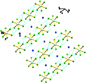 | ||
| Fig. 3 View of a fragment of the crystal packing of 1 showing intermolecular F⋯F contacts via Re–F⋯F–Re (dashed aqua line). Colour of atoms: Re in light-blue, N in dark-blue and F in green. | ||
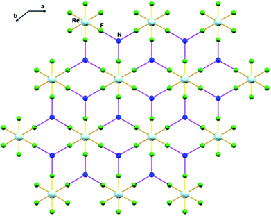 | ||
| Fig. 4 Intermolecular N⋯F interaction in 1 via Re–F⋯N⋯F–Re (dashed purple line). Colour of atoms: Re in light-blue, N in dark-blue and F in green. | ||
Further theoretical calculations indicate that there are 4 configurations of the hydrogen atoms of 1 within the P![[3 with combining macron]](https://www.rsc.org/images/entities/char_0033_0304.gif) m1 space group. They differ by whether the H atoms are staggered or eclipsed to the F atoms when viewed along [001] and if the apical H atoms of the [NH4]+ tetrahedra point towards (001) or (002). Plane-wave DFT optimizations of the atomic positions with the lattice fixed at the experimental value identifies the configuration with staggered H atoms and apical H atoms pointing towards (001) as the most favorable arrangement. The fractional coordinates of the [NH4]+ atoms in the DFT optimized asymmetric unit cell are N (2/3, 1/3, 0.6903), H1 (2/3, 1/3, 0.9102), and H2 (0.7594, 0.2406, 0.6132). The Re–F bonds are 1.967 Å in reasonable agreement with experiment, and the shortest H⋯F distance is 1.97 Å.
m1 space group. They differ by whether the H atoms are staggered or eclipsed to the F atoms when viewed along [001] and if the apical H atoms of the [NH4]+ tetrahedra point towards (001) or (002). Plane-wave DFT optimizations of the atomic positions with the lattice fixed at the experimental value identifies the configuration with staggered H atoms and apical H atoms pointing towards (001) as the most favorable arrangement. The fractional coordinates of the [NH4]+ atoms in the DFT optimized asymmetric unit cell are N (2/3, 1/3, 0.6903), H1 (2/3, 1/3, 0.9102), and H2 (0.7594, 0.2406, 0.6132). The Re–F bonds are 1.967 Å in reasonable agreement with experiment, and the shortest H⋯F distance is 1.97 Å.
Harmonic vibrational analysis indicates that each of the 4 P![[3 with combining macron]](https://www.rsc.org/images/entities/char_0033_0304.gif) m1 H configurations is dynamically unstable. The displacements of the unstable modes transform the configuration into the one described above or rotate the [NH4]+ tetrahedra lowering the symmetry to P
m1 H configurations is dynamically unstable. The displacements of the unstable modes transform the configuration into the one described above or rotate the [NH4]+ tetrahedra lowering the symmetry to P![[3 with combining macron]](https://www.rsc.org/images/entities/char_0033_0304.gif) . However, all of those structures are also dynamically unstable. Following the displacements of the P
. However, all of those structures are also dynamically unstable. Following the displacements of the P![[3 with combining macron]](https://www.rsc.org/images/entities/char_0033_0304.gif) instabilities or alternate initial placements of the H atoms result in lower energy triclinic structures with distorted [ReX6]2− octahedra. These distortions shorten some H⋯F distances to emphasize hydrogen bonding. Orientational disorder can occur in ammonium salts, and the disorder increases with temperature potentially changing the structure and Raman spectrum.29–31 While the energetic ordering of the disordered structures appears unphysical as they would have been more apparent by XRD, they do indicate that sufficient thermalization of 1 will lead to dynamic distortions that emphasize hydrogen bonding.
instabilities or alternate initial placements of the H atoms result in lower energy triclinic structures with distorted [ReX6]2− octahedra. These distortions shorten some H⋯F distances to emphasize hydrogen bonding. Orientational disorder can occur in ammonium salts, and the disorder increases with temperature potentially changing the structure and Raman spectrum.29–31 While the energetic ordering of the disordered structures appears unphysical as they would have been more apparent by XRD, they do indicate that sufficient thermalization of 1 will lead to dynamic distortions that emphasize hydrogen bonding.
Raman spectroscopy of 1
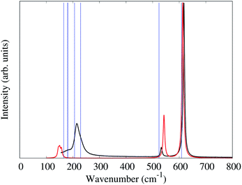
In the alkali salts of [MF6]2− (M = Tc, Re) where the anions are compressed along the crystallographic c-axis, the molecular symmetry of the [MF6]2− is lowered from Oh to D3d. The correlation between the effects of symmetry lowering and the vibrational spectra of the alkali salts of [MF6]2− (M = Tc, Re) are well studied.6,28,32,33 The slight increase of M–F bond lengths, moving from K2[MF6] to Cs2[MF6], as well as the different degrees of symmetry lowering lead the Raman bands to shift to lower frequencies.The unit-cell of 1 is shown in Fig. 2. Similar to the alkali salts of [MF6]2− (M = Tc, Re), parameters such as identical Re–F bond length (1.958(5) Å) and relative F–Re–F angles 90° and 180° are well represented in the Raman spectrum of 1. Here, the Raman spectrum of 1 (Fig. 5) exhibits three unique bands with central frequencies 617, 532, and 214 cm−1. The DFT predicted spectrum of a gas phase [ReF6]2− anion (in the crystalline geometry) provides a reasonable description of these bands, demonstrating that they primarily correspond to the 6 well-known Raman active vibrational modes of an octahedron.34 The 617 cm−1 band is the A1g symmetric stretch of the Re–F bonds. The 532 cm−1 band is the doubly degenerate Eg asymmetric stretch of the Re–F bonds. DFT predictions of these 3 modes for both gas phase [ReF6]2− and the P![[3 with combining macron]](https://www.rsc.org/images/entities/char_0033_0304.gif) m1 PW-DFT structure described previously are within 10 cm−1 of the measured values.
m1 PW-DFT structure described previously are within 10 cm−1 of the measured values.
The gas phase [ReF6]2− simulation assigns the 214 cm−1 band to the triply degenerate F2g octahedral Re–F bending modes which spilt by 7 cm−1 due to the slight deviation (>2°) from octahedral symmetry in the crystal structure. However, the gas phase frequencies are too low and cannot explain the lower frequency shoulder seen experimentally. PW-DFT predicts two of the F2g modes at 206 and the other at 229 cm−1. The cause of this splitting is a coupling with rocking/twisting motions of the [NH4]+ cations, but the inferred lineshape caused by the splitting seems in good agreement with the experimental spectrum. The PW-DFT simulations identify 5 additional modes within the span of the 214 cm−1 band: 2Eg [NH4]+ twisting modes at 181 cm−1, an A1g [NH4]+ translation mode at 180 cm−1, and 2Eg [NH4]+ translation modes at 165 cm−1. Although Raman intensities could not be calculated for the spin-polarized PW-DFT simulations, these center-of-mass motions are presumed to be weak and comprise the lower frequency shoulder of the 214 cm−1 band. The PW-DFT calculations also predict 5 modes between 1401 and 1678 cm−1, a range typically associated with ν4 and ν2 [NH4]+ bending/librational modes.35 There are also 4 Raman active modes between 3322–3445 cm−1 corresponding to stretching modes of the N–H bonds.35,36
Magnetic properties of 1
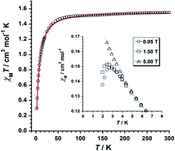
The χMT versus T plot (χM being the molar magnetic susceptibility per ReIV ion) is given in Fig. 6. The χMT value of 1.55 cm3 mol−1 K observed at T = 300 K is as expected for one Re(IV) ion with S = 3/2 and g ≈ 1.7–1.9.19Upon cooling, χMT decreases gradually with decreasing temperature, and much faster at approximately 50 K, reaching a minimum value of 0.29 cm3 mol−1 K at 2.0 K. The decrease of χMT observed for 1 is likely due to the presence of intermolecular antiferromagnetic interactions and zero-field splitting (ZFS) effects of the Re(IV) ion.19 The χM versus T plot obtained under small DC fields exhibits a maximum at ca. 2.9 K indicating the presence of antiferromagnetic exchanges between neighboring Re(IV) ions at very low temperature (see inset in Fig. 6), which would take place in the crystal lattice of 1 through short F⋯F interactions (Fig. 3). Nevertheless, this maximum wanes when higher magnetic fields are applied, thus suggesting the occurrence of a field-induced antiferromagnetic-to-paramagnetic ordering transition for 1 at TN ≈ 2.9 K (Fig. S2†), which is a magnetic behavior typical of metamagnetism phenomenon, as previously observed in systems based on Re(IV) ion.37
Taking the crystal structure of 1 into consideration, where short Re–F⋯F–Re interactions are observed (Fig. 3), we have used the Hamiltonian of eqn (1), and its derived theoretical expression for the magnetic susceptibility including a θ term to account for the intermolecular interactions,19,37–39 in order to analyze the magnetic behavior of 1.
| Ĥ = D[(ŜZ)2 − S(S + 1)/3] + gβHŜ | (1) |
Best least-squares fits of the experimental data in the 2–300 K temperature range afforded the magnetic parameters: D = +11.4(2) cm−1, g = 1.84(2) and θ = −3.1(1) K with R = 4.9 × 10⁻⁵ for 1 {R being the agreement factor defined as Σi[(χMT)obsi − (χMT)calcdi]2/[(χMT)obsi]2}.
The theoretical curve (red solid line in Fig. 6) matches the experimental data well in the whole temperature range. The g and D values calculated for 1 are in agreement with those previously reported for similar Re(IV) compounds.19 The negative θ value obtained from the fit implies the occurrence of significant antiferromagnetic exchange between Re(IV) ions through short Re–F⋯F–Re (Fig. 3) intermolecular interactions.
The field dependence of magnetization, or M versus H plot (M being the magnetization per Re(IV) ion and H the applied DC magnetic field), and its dM/dH curve measured at 2.0 K are shown in Fig. 7. M values increase with applied field first linearly and somewhat faster at high fields when a smooth inflexion is reached. A critical field (Hc) of ca. 3.0 T is detected through a maximum in the dM/dH versus H curve (Fig. 7), indicating that higher DC magnetic fields (H > Hc) overcome the antiferromagnetic interactions observed between metal ions in compound 1. Hence, these features support the occurrence of metamagnetic behavior in 1.37
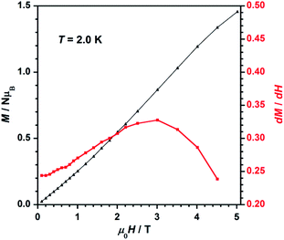 | ||
| Fig. 7 Dependence of the applied DC magnetic field on the magnetization (M, black-triangles) and on the derivative of M (dM/dH, red-squares) for 1. The solid lines are guides to the eye only. | ||
To study further the magnetic properties of the (NH4)2[ReF6] salt, AC magnetic susceptibility measurements were performed on a sample of this system in the temperature range of 2–5 K and under 0 and 1000 G DC fields oscillating at several frequencies (Fig. 8 and S3†). Interestingly, compound 1 exhibits incipient frequency-dependent out-of-phase AC signals  at very low temperatures (Fig. 8 and S3†), which is indicative of a system with slow relaxation of magnetization. A small dependence on the external DC field is observed in these AC signals for 1 (Fig. S4†). This phenomenon, that is typical of single-ion and single-molecule magnets, has been previously observed in similar Re(IV) complexes40–42 but this is the first time that it is observed in a Re(IV) system also showing metamagnetic behavior.
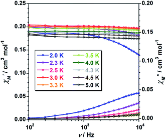 | ||
Fig. 8 Frequency dependence of the in-phase  and out-of-phase and out-of-phase  ac magnetic susceptibility signals for compound 1. Measurements performed at different temperatures and under a dc field of 1000 G. ac magnetic susceptibility signals for compound 1. Measurements performed at different temperatures and under a dc field of 1000 G. | ||
Unfortunately, the expected  maxima for 1 are not detected above 2.0 K (Fig. 8 and S3†), given that they would occur at lower temperatures or higher frequencies than those available in our device. So that, it is not possible to quantify magnetic parameters such as the anisotropy energy barrier to magnetization reorientation (Ea) and the preexponential factor (τo) for 1 through the Arrhenius expression. Nevertheless, these magnetic parameters could be roughly evaluated by means of the
  versus 1/T plot (Fig. 9), the
  expression, and by assuming only a single relaxation time,43 as previously done.42 In the case of compound 1, the thus estimated value of Ea parameter is 1.80 K (1.25 cm−1) and that of τo is 5.3 × 10⁻⁷ s, which were obtained in presence of an external DC field (HDC = 1000 G) (Fig. 9). These results are in good agreement with those found in the literature for some mono- and polynuclear Re(IV) systems showing slow relaxation of the magnetization.16,40,41 The presence of the NH4+ cation allows intermolecular halogen⋯halogen interactions that directly affect to the magnetic properties, leading to the magnetic behaviors observed in 1. However, the bulky PPh4+ cation in the previously reported (PPh4)2[ReF6] salt causes the magnetic dilution of the hexahalorhenate(IV) units (because of the great F⋯F separation that is generated) and magnetic interactions among neighboring paramagnetic [ReF6]2− anions are precluded.9
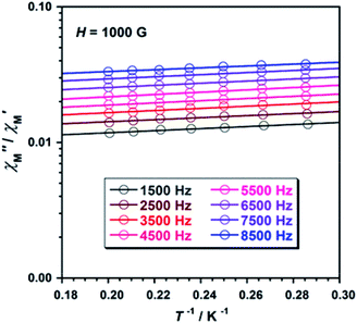 | ||
Fig. 9  versus 1/T plot for 1 at different frequencies (1500–8500 Hz range) and HDC = 1000 G. The solid lines are the best-fit curves (see text). versus 1/T plot for 1 at different frequencies (1500–8500 Hz range) and HDC = 1000 G. The solid lines are the best-fit curves (see text). | ||
Conclusions
For the first time, the (NH4)2[ReF6] (1) salt was characterized by X-ray diffraction, IR and Raman spectroscopies, DC and AC magnetic susceptibility measurements, and computational methods. 1 crystallizes in the trigonal space group P![[3 with combining macron]](https://www.rsc.org/images/entities/char_0033_0304.gif) m1 and is isostructural to its Tc homologue. The structure of 1 exhibits intermolecular F⋯F interactions (via Re–F⋯F–Re) and N⋯F interactions (via Re–F⋯N⋯F–Re). In 1, splitting of the Raman peaks (Re–F; F–Re–F) are correlated with the site symmetries of [ReF6]2− anion. The study of the magnetic properties, through DC and AC magnetic susceptibility measurements, reveals that metamagnetism and slow relaxation of magnetization phenomena coexist in 1, which is unusual in the molecular systems based on paramagnetic 5d metal ions reported so far. Hence, 1 exhibits a magnetic behaviour that is between that exhibited by the previously reported (PPh4)2[ReF6] single-ion magnet and the typical antiferromagnetic ordering of [ReX6]2− salts exhibiting short intermolecular X⋯X interactions. The magnetic chemistry of technetium is underdeveloped and magnetic measurements on [TcF6]2− species (4d3) have not been performed yet. Work on the preparation and magnetic characterisation of [TcF6]2− species is under progress and results will be reported in due course.
m1 and is isostructural to its Tc homologue. The structure of 1 exhibits intermolecular F⋯F interactions (via Re–F⋯F–Re) and N⋯F interactions (via Re–F⋯N⋯F–Re). In 1, splitting of the Raman peaks (Re–F; F–Re–F) are correlated with the site symmetries of [ReF6]2− anion. The study of the magnetic properties, through DC and AC magnetic susceptibility measurements, reveals that metamagnetism and slow relaxation of magnetization phenomena coexist in 1, which is unusual in the molecular systems based on paramagnetic 5d metal ions reported so far. Hence, 1 exhibits a magnetic behaviour that is between that exhibited by the previously reported (PPh4)2[ReF6] single-ion magnet and the typical antiferromagnetic ordering of [ReX6]2− salts exhibiting short intermolecular X⋯X interactions. The magnetic chemistry of technetium is underdeveloped and magnetic measurements on [TcF6]2− species (4d3) have not been performed yet. Work on the preparation and magnetic characterisation of [TcF6]2− species is under progress and results will be reported in due course.
Experimental section
Materials and reagents
All chemicals and reagents (≥99% purity) were obtained commercially from Sigma Aldrich® and used without any further purification. This work was performed in a well-ventilated fume hood due to the corrosive nature of molten ammonium bifluoride and evolution of gaseous hydrogen halides. The starting material, (NH4)2[ReBr6], was prepared from the reduction of NH4[ReO4] with H3PO2 in the presence of NH4Br and concentrated HBr as described in the literature.44Preparation and crystallization of (NH4)2[ReF6] (1)
The (NH4)2[ReF6] salt was prepared from the treatment of (NH4)2[ReBr6] (1.0 g, 1.42 mmol) with excess ammonium bifluoride (0.81 g, 14.2 mmol) at 300 °C under air. Upon cooling, the resulted solid cake was washed with H2O/MeOH and dissolved in warm H2O for crystallization. The resulting pink 1 was recrystallized in H2O and colorless crystals of 1 were obtained within a week. Yield: 300 mg, 60%. Anal. calcd for H8N2F6Re (1): H, 2.4; N, 8.3. Found: H, 2.5; N, 8.4. Raman (cm−1): Re–F: 617, 532 and 214. IR (cm−1): 472.63, 1000.87, 1078, 1123.94, 1403.89, 2848.8, 2921.8, 3234.59 and 3350.54.Physical measurements
Raman spectra were recorded on a HORIBA Jobin Yvon T64000 triple-grating spectrometer operating in subtractive mode. The spectra were collected from pure single crystals at room temperature using the 514.5 nm line of a Kr/Ar laser, with a total of 30 mW incident on the sample. Variable-temperature, solid-state (DC and AC) magnetic susceptibility data down to 2.0 K were collected on a Quantum Design MPMS-XL SQUID magnetometer equipped with a 5 T DC magnet.Single crystal XRD data collection and structure refinement
X-ray diffraction data were collected on a Bruker Apex II instrument with Mo-Kα radiation (λ = 0.71073 Å) and equipped with an Oxford nitrogen cryostream. The crystal was mounted under Paratone® on a glass fiber; data processing was performed using the Apex III suite software. Structural solution (direct methods) and refinements were completed using SHELXS and SHELX2014 software. Hydrogen atom positions were calculated using the “riding model” option of SHELXL software. The nitrogen atoms of the ammonium ions structure were calculated without the corresponding hydrogen. Crystal parameters and refinement results are summarized in Table 2. The graphical manipulations were performed using Mercury crystal programs.| (NH4)2[ReF6] | |
|---|---|
| Empirical formula | F6N2Re |
| Formula weight | 328.22 |
| Crystal system | Trigonal |
| Space group | P![[3 with combining macron]](https://www.rsc.org/images/entities/char_0033_0304.gif) m1 m1 |
| a (Å) | 6.015(1) |
| b (Å) | 6.015(1) |
| c (Å) | 4.680(1) |
| V (Å3) | 146.64(6) |
| Z | 1 |
| ρ calc (mg m−3) | 3.71665 |
| μ (mm−1) | 20.764 |
| Reflections collected | 1922 |
| Data/restraints/parameters | 182/0/12 |
| Goodness-of-fit | 1.139 |
| R1 indices [I > 2σ(I)] | 0.0282 |
| wR2 indices [I > 2σ(I)] | 0.0764 |
Simulations
Density functional theory (DFT) calculations45,46 were performed using the PBE47 generalized gradient approximation (GGA) functional. Molecular (in vacuo) simulations were performed with the Q-Chem 4.0 package48 using the def2-TZVP49,50 basis set with small core effective core potentials (ECPs), a 10−14 Eh integral cutoff, a 10−9 Eh (hartree) energy criterion, and a 10−4 Eh/Å force criterion. Plane-wave (PW-DFT) calculations on the periodic crystal structure of the solid were performed with CASTEP51 using the NCP17 pseudopotentials (Re: 5p66s25d5), a 4 × 4 × 4 Γ-centered k-point grid, a 600 eV plane-wave cutoff, a 10−12 eV energy criterion, and a 10−4 eV Å−1 force criterion. Initial structures were taken from the experimental single crystal structure with hydrogens added manually. For all PW-DFT structural optimizations, the atoms were allowed to move but the unit cell dimensions were fixed at the experimental values in Table 2. Localized orbital bonding analysis (LOBA)52 used the Pipek–Mezey53 localized orbitals. Vibrational analysis was performed by linear response for molecular frequencies and by finite difference for Γ-point plane-wave frequencies and molecular intensities.54 The PW-DFT phonon calculations were on a single unit cell that has only one S = 3/2 Re atom, therefore the Re magnetic moments were effectively aligned ferromagnetically.Funding sources
This material is based upon work supported by the Department of Energy National Nuclear Security Administration through the Nuclear Science and Security Consortium under Award Number(s) DE-NA0003180. Portions of this work were performed at HPCAT (Sector 16), Advanced Photon Source (APS), Argonne National Laboratory. HPCAT operations are supported by DOE-NNSA's Office of Experimental Sciences. The Advanced Photon Source is a U.S. Department of Energy (DOE) Office of Science User Facility operated for the DOE Office of Science by Argonne National Laboratory under Contract No. DE-AC02-06CH11357. The Spanish Ministry of Science, Innovation and Universities has also supported part of this work through the projects PID2019-109735GB-I00 and MDM-2015-0538 (Excellence Unit “María de Maeztu”).Conflicts of interest
The authors declare no conflict of interest.Acknowledgements
Thanks are due to Mrs Julie Bertoia, Mr Charles Bynum, Mr Kelly Seeley and Dr Hugues Badet for laboratory support. JLJ acknowledges Mr Kevin Louis-Jean and Ms. Ruthonce Stvil for their assistance. ASP thanks the “FPU fellowships” Programme and JML thanks the “Ramón y Cajal” Programme of the Spanish Ministry of Science, Innovation and Universities.References
- H. J. Berthold and G. Jakobson, Angew. Chem., 1964, 76, 497 CrossRef CAS.
- H. D. Grundy and I. D. Brown, Can. J. Chem., 1970, 48, 1151–1154 CrossRef CAS.
- C. K. Jørgensen and K. Schwochau, Z. Naturforsch., A: Astrophys., Phys. Phys. Chem., 1965, 20, 65–75 Search PubMed.
- R. D. Peacock, J. Chem. Soc., 1956, 1291–1293 RSC.
- E. Weise, Z. Anorg. Allg. Chem., 1956, 283, 377–389 CrossRef CAS.
- J. Louis-Jean, S. M. Balasekaran, D. Smith, A. Salamat, C. T. Pham and F. Poineau, Acta Crystallogr., Sect. E: Crystallogr. Commun., 2018, 74, 646–649 CrossRef CAS.
- F. Poineau, J. Louis-Jean, H. Jang, C. Higgins, S. M. Balasekaran, D. Hatchett and A. P. Sattelberger, SN Appl. Sci., 2019, 1, 1442 CrossRef CAS.
- G. R. Clark and D. R. Russell, Acta Crystallogr., Sect. B: Struct. Crystallogr. Cryst. Chem., 1978, 34, 894–895 CrossRef.
- K. S. Pedersen, M. Sigrist, M. A. Sørensen, A. L. Barra, T. Weyhermuller, S. Piligkos, C. A. Thuesen, M. G. Vinum, H. Mutka, H. Weihe, R. Clerac and J. Bendix, Angew. Chem., Int. Ed., 2014, 53, 1351–1354 CrossRef CAS.
- S. M. Balasekaran, A. Hagenbach and F. Poineau, Inorg. Chem. Commun., 2020, 119, 108064 CrossRef CAS.
- V. Bulicanu, K. S. Pedersen, M. Rouzières, J. Bendix, P. Dechambenoit, R. Clérac and E. A. Hillard, Chem. Commun., 2015, 51, 17748–17751 RSC.
- N. D. Kushch, L. I. Buravov, P. P. Kushch, G. V. Shilov, H. Yamochi, M. Ishikawa, A. Otsuka, A. A. Shakin, O. V. Maximova, O. S. Volkova, A. N. Vasiliev and E. B. Yagubskii, Inorg. Chem., 2018, 57, 2386–2389 CrossRef CAS.
- J. Louis-Jean, S. Mariappan Balasekaran, A. Hagenbach and F. Poineau, Acta Crystallogr., Sect. E: Crystallogr. Commun., 2019, 75, 1158–1161 CrossRef CAS.
- D. E. LaValle, R. M. Steele and W. T. Smith, J. Inorg. Nucl. Chem., 1966, 28, 260–263 CrossRef CAS.
- M. Cowie, C. J. L. Lock and J. Ozog, Can. J. Chem., 1970, 48, 3760–3763 CrossRef CAS.
- J. Martínez-lillo, J. Faus, F. Lloret and M. Julve, Coord. Chem. Rev., 2015, 289–290, 215–237 CrossRef.
- R. Chiozzone, A. Cuevas, R. González, C. Kremer, D. Armentano, G. De Munno and J. Faus, Inorg. Chim. Acta, 2006, 359, 2194–2200 CrossRef CAS.
- R. Gonza, C. Kremer, G. De Munno, F. Nicolo, F. Lloret, M. Julve and J. Faus, Inorg. Chem., 2003, 42, 2512–2518 CrossRef.
- J. Mrozinski, A. Tomkiewicz, H. Hartl, I. Brüdgam and F. Villain, Pol. J. Chem., 2002, 76, 285–293 CAS.
- C. M. Nelson, G. E. Boyd and W. T. Smith, J. Am. Chem. Soc., 1954, 76, 348–352 CrossRef CAS.
- B. N. Figgis, J. Lewis and F. E. Mabbs, J. Chem. Soc., 1961, 3138–3145 RSC.
- R. H. Busey and E. Sonder, J. Chem. Phys., 1962, 36, 93–97 CrossRef CAS.
- R. H. Busey, H. H. Dearman and R. B. Bevan, J. Phys. Chem., 1962, 66, 82–89 CrossRef CAS.
- R. H. Busey, R. B. Bevan and R. A. Gilbert, J. Phys. Chem., 1965, 69, 3471–3480 CrossRef CAS.
- V. J. Minkiewicz, G. Siiirane, B. C. Frazer, R. G. Wheeler and P. B. Dorain, J. Phys. Chem. Solids, 1968, 29, 881–884 CrossRef CAS.
- K. Schwochau and W. Herr, Angew. Chem., Int. Ed., 1963, 75, 95 CAS.
- S. Mariappan Balasekaran, K. Lawler, A. Hagenbach, A. Abram, U. Abram, A. Sattelberger and F. Poineau, Eur. J. Inorg. Chem., 2019, 2, 4129–4135 CrossRef.
- S. M. Balasekaran, M. Molski, J. Spandl, A. Hagenbach, R. Alberto and U. Abram, Inorg. Chem., 2013, 52, 7094–7099 CrossRef CAS.
- K. Moriya, T. Matsuo, H. Suga and S. Seki, Bull. Chem. Soc. Jpn., 1977, 50, 1920–1926 CrossRef CAS.
- E. J. Lisher, N. Cowlam and L. Gillott, Acta Crystallogr., Sect. B: Struct. Crystallogr. Cryst. Chem., 1979, 35, 1033–1038 CrossRef.
- M. A. White and B. D. Wagner, J. Chem. Phys., 1985, 83, 5844–5848 CrossRef CAS.
- M. Bettinelli, L. Di Sipio, G. Ingletto and C. Razzetti, Inorg. Chim. Acta, 1987, 133, 7–9 CrossRef CAS.
- M. Bettinelli, L. Di Sipio, A. Pasquetto, G. Ingletto and A. Montenero, Inorg. Chim. Acta, 1985, 99, 37–42 CrossRef CAS.
- A. Sanson, M. Giarola, G. Mariotto, L. Hu, J. Chen and X. Xing, Mater. Chem. Phys., 2016, 180, 213–218 CrossRef CAS.
- F. Agullo-Rueda, J. M. Calleja and J. Bartolome, J. Phys. C: Solid State Phys., 1988, 21, 1287–1297 CrossRef CAS.
- J. B. Bates, T. Kaneda, J. C. Wang and H. Engstrom, J. Chem. Phys., 1980, 73, 1503–1513 CrossRef CAS.
- J. Martínez-Lillo, J. Kong, W. P. Barros, J. Faus, M. Julve and E. K. Brechin, Chem. Commun., 2014, 50, 5840–5842 RSC.
- J. Martínez-Lillo, D. Armentano, T. F. Mastropietro, M. Julve, J. Faus and G. De Munno, Cryst. Growth Des., 2011, 11, 1733–1741 CrossRef.
- J. Martínez-Lillo, A. H. Pedersen, J. Faus, M. Julve and E. K. Brechin, Cryst. Growth Des., 2015, 15, 2598–2601 CrossRef.
- J. Martínez-Lillo, D. Armentano, G. De Munno, W. Wernsdorfer, M. Julve, F. Lloret and J. Faus, J. Am. Chem. Soc., 2006, 128, 14218–14219 CrossRef.
- J. Martínez-Lillo, T. F. Mastropietro, E. Lhotel, C. Paulsen, J. Cano, G. De Munno, J. Faus, F. Lloret, M. Julve, S. Nellutla and J. Krzystek, J. Am. Chem. Soc., 2013, 135, 13737–13748 CrossRef.
- J. Martínez-Lillo, T. F. Mastropietro, G. De Munno, F. Lloret, M. Julve and J. Faus, Inorg. Chem., 2011, 50, 5731–5739 CrossRef.
- J. Bartolomé, G. Filoti, V. Kuncser, G. Schinteie, V. Mereacre, C. E. Anson, A. K. Powell, D. Prodius and C. Turta, Phys. Rev. B: Condens. Matter Mater. Phys., 2009, 80, 014430–014446 CrossRef.
- G. W. Watt, R. J. Thompson and J. M. Gibbons, in Inorganic Syntheses, ed. J. Kleinberg, McGraw-Hill, Inc., New York, 1963, vol. 7, pp. 189–192 Search PubMed.
- P. Hohenberg and W. Kohn, Phys. Rev. [Sect.] B, 1964, 136, 864–871 CrossRef.
- W. Kohn and L. J. Sham, Phys. Rev. [Sect.] A, 1965, 140, 1133–1138 CrossRef.
- J. P. Perdew, K. Burke and M. Ernzerhof, Phys. Rev. Lett., 1996, 77, 3865–3868 CrossRef CAS.
- Y. Shao, Z. Gan, E. Epifanovsky, A. T. B. Gilbert, M. Wormit, J. Kussmann, A. W. Lange, A. Behn, J. Deng, X. Feng, D. Ghosh, M. Goldey, P. R. Horn, L. D. Jacobson, I. Kaliman, R. Z. Khaliullin, T. Kuś, A. Landau, J. Liu, E. I. Proynov, Y. M. Rhee, R. M. Richard, M. A. Rohrdanz, R. P. Steele, E. J. Sundstrom, H. L. Woodcock, P. M. Zimmerman, D. Zuev, B. Albrecht, E. Alguire, B. Austin, G. J. O. Beran, Y. A. Bernard, E. Berquist, K. Brandhorst, K. B. Bravaya, S. T. Brown, D. Casanova, C.-M. Chang, Y. Chen, S. H. Chien, K. D. Closser, D. L. Crittenden, M. Diedenhofen, R. A. DiStasio, H. Do, A. D. Dutoi, R. G. Edgar, S. Fatehi, L. Fusti-Molnar, A. Ghysels, A. Golubeva-Zadorozhnaya, J. Gomes, M. W. D. Hanson-Heine, P. H. P. Harbach, A. W. Hauser, E. G. Hohenstein, Z. C. Holden, T.-C. Jagau, H. Ji, B. Kaduk, K. Khistyaev, J. Kim, J. Kim, R. A. King, P. Klunzinger, D. Kosenkov, T. Kowalczyk, C. M. Krauter, K. U. Lao, A. D. Laurent, K. V. Lawler, S. V. Levchenko, C. Y. Lin, F. Liu, E. Livshits, R. C. Lochan, A. Luenser, P. Manohar, S. F. Manzer, S.-P. Mao, N. Mardirossian, A. V. Marenich, S. A. Maurer, N. J. Mayhall, E. Neuscamman, C. M. Oana, R. Olivares-Amaya, D. P. O'Neill, J. A. Parkhill, T. M. Perrine, R. Peverati, A. Prociuk, D. R. Rehn, E. Rosta, N. J. Russ, S. M. Sharada, S. Sharma, D. W. Small, A. Sodt, T. Stein, D. Stück, Y.-C. Su, A. J. W. Thom, T. Tsuchimochi, V. Vanovschi, L. Vogt, O. Vydrov, T. Wang, M. A. Watson, J. Wenzel, A. White, C. F. Williams, J. Yang, S. Yeganeh, S. R. Yost, Z.-Q. You, I. Y. Zhang, X. Zhang, Y. Zhao, B. R. Brooks, G. K. L. Chan, D. M. Chipman, C. J. Cramer, W. A. Goddard, M. S. Gordon, W. J. Hehre, A. Klamt, H. F. Schaefer, M. W. Schmidt, C. D. Sherrill, D. G. Truhlar, A. Warshel, X. Xu, A. Aspuru-Guzik, R. Baer, A. T. Bell, N. A. Besley, J.-D. Chai, A. Dreuw, B. D. Dunietz, T. R. Furlani, S. R. Gwaltney, C.-P. Hsu, Y. Jung, J. Kong, D. S. Lambrecht, W. Liang, C. Ochsenfeld, V. A. Rassolov, L. V. Slipchenko, J. E. Subotnik, T. Van Voorhis, J. M. Herbert, A. I. Krylov, P. M. W. Gill and M. Head-Gordon, Mol. Phys., 2015, 113, 184–215 CrossRef CAS.
- D. Andrae, U. Häussermann, M. Dolg, H. Stoll and H. Preuss, Theor. Chim. Acta, 1990, 77, 123–141 CrossRef CAS.
- F. Weigend and R. Ahlrichs, Phys. Chem. Chem. Phys., 2005, 7, 3297–3305 RSC.
- S. J. Clark, M. D. Segall, C. J. Pickard, P. J. Hasnip, M. I. J. Probert, K. Refson and M. C. Payne, Z. Kristallogr. - Cryst. Mater., 2005, 220, 567–570 CAS.
- A. J. W. Thom, E. J. Sundstrom and M. Head-Gordon, Phys. Chem. Chem. Phys., 2009, 11, 11297–11304 RSC.
- J. Pipek and P. G. Mezey, J. Chem. Phys., 1989, 90, 4916–4926 CrossRef CAS.
- B. G. Johnson and J. Florián, Chem. Phys. Lett., 1995, 247, 120–125 CrossRef CAS.
Footnotes |
| † Electronic supplementary information (ESI) available. CCDC 1962857. For ESI and crystallographic data in CIF or other electronic format see DOI: 10.1039/d0ra10325j |
| ‡ These authors contributed equally to this work. |
| This journal is © The Royal Society of Chemistry 2021 |

