
 Open Access Article
 This Open Access Article is licensed under a Creative Commons Attribution-Non Commercial 3.0 Unported Licence
Single-step coating of mesoporous SiO2 onto nanoparticles: growth of yolk–shell structures from core–shell structures†
Xiaobin
Xie
 *,
Marijn A.
van Huis
* and
Alfons
van Blaaderen
*
*,
Marijn A.
van Huis
* and
Alfons
van Blaaderen
*
Soft Condensed Matter, Debye Institute for Nanomaterials Science, Utrecht University, Princetonplein 5, 3584 CC Utrecht, The Netherlands. E-mail: xb.xie@hotmail.com; M.A.vanHuis@uu.nl; A.vanBlaaderen@uu.nl
First published on 1st April 2021
Abstract
Yolk–shell nanoparticles based on mesoporous SiO2 (mSiO2) coating of Au nanoparticles (Au NPs) hold great promise for many applications in e.g., catalysis, biomedicine, and sensing. Here, we present a single-step coating approach for synthesizing Au NP@mSiO2 yolk–shell particles with tunable size and tunable hollow space between yolk and shell. The Au NP–mSiO2 structure can be manipulated from core–shell to yolk–shell by varying the concentration of cetyltrimethylammonium chloride (CTAC), tetraethyl orthosilicate (TEOS), Au NPs, and NaOH. The growth mechanism of the yolk–shell particles was investigated in detail and consists of a concurrent process of growth, condensation, and internal etching through an outer shell. We also show by means of liquid-cell transmission electron microscopy (LC-TEM) that Au nanotriangle cores (Au NTs) in yolk–shell particles that are stuck on the mSiO2 shell, can be released by mild etching thereby making them mobile and tumbling in a liquid-filled volume. Due to the systematical investigation of the reaction parameters and understanding of the formation mechanism, the method can be scaled-up by at least an order of magnitude. This route can be generally used for the synthesis of yolk–shell structures with different Au nanoparticle shapes, e.g., nanoplatelets, nanorods, nanocubes, for yolk–shell structures with other metals at the core (Ag, Pd, and Pt), and additionally, using ligand exchange with other nanoparticles as cores and for synthesizing hollow mSiO2 spheres as well.
Introduction
The scientific interest in Au based nanoparticles (NPs) has vastly increased in recent decades, due to their widespread application in various fields, such as catalysis,1–5 biomedicine,6–8 sensing,9–14 and energy conversion.15–19 Generally, the performance of Au nanoparticles (Au NPs) is dependent on various factors, like size, morphology, and type of surface ligands.20–25 Among all strategies that have been developed to achieve a better performance of Au NPs, coating with mesoporous SiO2 (mSiO2) is one of the most popular approaches, as this can significantly enhance the colloid's stability of Au NPs, act as morphological stabilizer,26,27 mitigate the barrier/poisoning effects from ligands for catalysis, and makes them biocompatible.13,28–30 Several typical kinds of Au NP–mSiO2 structures have been developed for various applications. They are Au NP@mSiO2 core–shell NPs,28,31 Au NP@hollow–mSiO2 yolk–shell structures,32 Janus-type structures,33 and other complex mSiO2–Au structures.34–36In comparison with the core–shell and other hybrid structures, the yolk–shell structure has several advantages as discussed in several reviews.37–40 First, the tunable hollow space between core and shell provides a faster mass transfer and less impact on the core surface during catalytic reactions.34 Furthermore, yolk–shell particles can serve as carriers used for the transport and controlled release of drugs, which is an important strategy for cancer therapy.41 A yolk–shell morphology also has several advantages for surface enhanced Raman scattering sensing, amongst which increased reproducibility.42,43 Lastly, with an outer mSiO2 shell, the ligands or surfactants used for the synthesis of Au NP cores can be removed more easily without inducing aggregation of the Au NPs.26,27,32,44 During the past few years, several strategies have been developed for synthesizing Au NPs@hollow@mSiO2 yolk–shell structures.34,45 However, most of these methods are comprised of multiple steps, such as hard template methods, or do not allow morphology-controlled synthesis of the cores.37,39,40
Here, we report a single-step coating approach which originated from a previous protocol for synthesizing Au NRs@mSiO2 core–shell NPs,31 based on a simultaneous growth-etching mechanism, to the synthesis of Au NPs@hollow@mSiO2 yolk–shell structures. Moreover, various factors that impact the growth and final structure could be controlled via the reaction parameters. By varying the reaction conditions, the structure of the mSiO2 core–shell particles was switched between core–shell and yolk–shell. Additionally, the size of the yolk–shell particles can be easily tuned by changing the concentration of the core particles. Our experiments also show that this strategy can be adopted irrespective of the core's morphology; it is universal for all shapes of Au NPs and can be used even for preparing hollow mSiO2 spheres. Using ligand exchange (e.g. through a very general and elegant procedure developed recently46), we found that the surface ligands on the NPs also influence the final structure of the resulting Au NP@mSiO2 structures. Furthermore, liquid-cell TEM (LC-TEM) experiments revealed that the Au NT cores can be released easily upon mild etching creating freely diffusing NP systems inside the mesoporous silica shells. Such ‘rattle-type’ systems are of interest for photonic applications as well.28,43,47
Results and discussion
The Au nanotriangles (Au NTs) were synthesized via a modification of the method reported by L. M. Liz-Marzán et al.48 After purification, the percentage of Au NTs having an average size of 67 nm was more than 90%, and others are mainly spheres (Fig. S1a–c†). The UV-VIS spectrum of the Au NTs (Fig. S1d†) shows that the main localized surface plasmon resonance (LSPR) band is at 669 nm. Using these Au NTs as seeds, a single-step coating method was developed which was partially based on previous work in which Au NRs can be coated by mesoporous silica layers using the ligands used for the Au NR synthesis, CTAB, as pore inducing template molecules49 in order to produce Au NT@hollow–mSiO2 yolk–shell nanoparticles. Fig. 1a shows the morphology of the yolk–shell nanoparticles having Au NT cores inside a mSiO2 outer shells. The edge length of the Au NTs used for the synthesis of these yolk–shell nanoparticles was about 68 nm on average while the overall particle diameter for the particular synthesis shown in Fig. 1 was about 205 nm, where the thickness of the shell was around 33 nm (Fig. 1c). We found a weak positive correlation between the diameter of the yolk–shell particles and the edge length of the Au NT cores, and there was no correlation found between the shell thickness and the diameter of whole particles or with the edge lengths of the Au NTs (Fig. 1d). | ||
| Fig. 1 (a) Bright-field transmission electron microscopy (BF-TEM) images of Au NT@hollow–mSiO2 yolk–shell nanoparticles; (b) schematic of Au NT@hollow–mSiO2 yolk–shell nanoparticle morphology; (c) size distribution of Au NT@hollow–mSiO2 yolk–shell nanoparticles; (d) the relationship between Au NT edge length and shell thickness and diameter. | ||
In order to understand the effect of the reaction parameters on the morphology of the Au NT@mSiO2 products, a series of control variable experiments were performed with precise tuning of the reaction conditions. First of all, we found that the concentration of CTAC plays an important role in the formation of the yolk–shell structure. We assume that all cetyltrimethylammonium (CAT+) ions are forming a double layer on the surface of the Au NTs after washing and resuspension in water. The concentration of this surfactant is around the critical micelle concentration (CMC) 1.3 mM in water.50 As shown in Fig. S2,† the morphology and structure of the Au NT@mSiO2 particles changes as different amounts of additional CTAC are added to the growth solution. Au NT@mSiO2 core–shell particles were formed both at low (≤0.5 mM) and relatively high (≥3.0 mM) concentrations as compared to the CMC value of 1.3 mM.50 When the concentration of additional CTAC was between 1.0 mM to 2.0 mM, Au NT@hollow–mSiO2 yolk–shell particles were obtained. Moreover, both the way of injecting TEOS and the amount of TEOS was found to influence the final structure of the particles. Fig. S3† indicates that 200 μl of 20 vol% TEOS (in methanol) injected separately into three shoots lead to a core–shell structure. Also 200 μl injections in one shot of 15 vol%, 10 vol%, and 5 vol% TEOS resulted in core–shell Au NT@mSiO2 particles (Fig. 2).
 | ||
| Fig. 2 Influence of the concentration of TEOS on the morphology of the Au NT@mSiO2 structures. Here the concentrations of TEOS precursor solution used for the reaction were: (a) 20 vol%, (b) 15 vol%, (c) 10 vol%, and (d) 5 vol%. | ||
To investigate the influence of the number of Au NTs cores, different concentrations of Au NTs were used for the yolk–shell synthesis. As shown in Fig. 3, the TEM images indicate that the hollow space in the Au NT@hollow–mSiO2 yolk–shell particles was reduced when the concentration of Au NTs increases from 112 pM to 900 pM (Fig. 3a). The diameters and shell thicknesses of the Au NT@mSiO2 yolk–shell particles obtained using different concentrations of Au NTs were determined by averaging the dimensions of more than 100 yolk–shell particles in the acquired TEM images. The average diameters of the yolk–shell particles were 210 nm, 175 nm, 145 nm, and 120 nm, decreasing with increasing Au NT concentration (Fig. 3b and d). This trend was also observed for the shell thickness, although it changed only slightly. The four mSiO2 shell average thicknesses were 33 nm, 32 nm, 30 nm, and 29 nm, respectively. Notably, secondary nucleation of silica particles was only observed when the concentration of Au NTs was 112 pM. Fig. 3d and e show the relationship between Au NT concentrations and diameters and shell thickness, the bars in the bar graphs are providing the min–max range, the block center indicates the average size, and the block range denotes the standard deviation. Furthermore, we studied the effect of the surface ligands on the synthesis via ligand exchange. The CTA+ double layers on the Au NT surface were exchanged by other ligands that have been commonly used for noble metals nanoparticles synthesis or surface decoration. They are CTAB, PEG–SH, and PVP, respectively.51 The UV-VIS spectra of the Au NTs with different ligands recorded after ligand exchange indicate that the Au NTs were still well dispersed in water except for the Au NTs coated by-PVP where the particles were slightly aggregated (Fig. S5†). It is not surprising that Au NT-CTAB form Au NT@hollow–mSiO2 yolk–shell structures similarly to those obtained with Au NT-CTAC (Fig. S6a†), considering that the same CTA+ ions double layers are covering the Au NTs surfaces. However, mSiO2 shells failed to grow on Au NT–PEG–SH and Au NT–PVP surfaces, and consequently growth of hollow empty mSiO2 nanospheres (NSs) was found instead (Fig. S6b and c†).
 | ||
| Fig. 3 BF-TEM images and analysis showing the influence of the concentration of Au NTs. In all panels, the magenta, green, cyan, and yellow colors correspond to Au NT concentrations of 112, 225, 450, and 900 pM respectively. (a) TEM images of Au NT@hollow–mSiO2 yolk–shell nanoparticles obtained at the different Au NTs concentrations; (b) histograms of the diameter distribution of Au NT@hollow–mSiO2 yolk–shell nanoparticles; (c) histograms of the shell thickness of Au NT@hollow–mSiO2 yolk–shell nanoparticles; (d) the relationship between the diameter and Au NTs concentrations; (e) the relationship between shell thickness and Au NTs concentrations. | ||
To elucidate the formation mechanism of the Au NT@hollow–mSiO2 yolk–shell structures, we monitored the growth process in time by recording TEM images at various stages of the growth process. The reaction was stopped by centrifugation after specific reaction times after which TEM grids were prepared. Fig. 4a shows the shape evolution of the Au NT@mSiO2 particles. After the first 20 min, mSiO2 shells are seen to have grown on the Au NTs’ surfaces. In the second stage, mSiO2 shells became visible, accompanied by etching of the mSiO2 silica interior, which occurred from 20 min to 80 min. Subsequently, etching of mSiO2 became the dominant process in the next stage until a well-defined ‘yolk–shell’ Au NT@mSO2 morphology was obtained. This growth-etching mechanism can be summarized as shown in the schematic in Fig. 4b. The sol–gel reactions for the growth and etching processes can be expressed by the following chemical equations.36
![[triple bond, length as m-dash]](https://www.rsc.org/images/entities/char_e002.gif) Si–OR + H2O ⇌ Si–OH + ROH Si–OR + H2O ⇌ Si–OH + ROH | (1) |
| Si–OR + OH–Si ⇌ Si–O–Si + ROH | (2) |
| Si–OH + OH–Si ⇌ Si–O–Si + H2O | (3) |
 | ||
| Fig. 4 The growth process of Au NT@hollow–mSiO2 yolk–shell nanoparticles. (a) Evolution in time of the morphology of Au NT@mSiO2 particles at different reaction times in the synthesis. (b) Schematic of the mSiO2 growth-and-etching process. | ||
The forward reaction of eqn (1) shows the hydrolysis of TEOS, and the forward reactions of eqn (2) and (3) demonstrate the polymerization of SiO2, namely, the growing of the –Si–O–Si– network in mSiO2 silica growth template onto the surfactant molecules.52 The etching of mSiO2 occurs when the –Si–O–Si– network is destroyed by OH−, following the three reaction equations in reverse direction. The possible reason that mSiO2 can be etched from inside out is that the amount of Q2 (Si(SiO)2(OX)2, X = H or Et) is higher than the amount of Q4 (Si(SiO)4) in the inner part of the mSiO2,41 as has been found as well for the famous Stöeber process in which ultra-microporous silica particles can be grown37,40,53,54 while the opposite is the case for the outer part of mSiO2. This makes the etching faster at the interior than at the exterior, and consequently mSiO2 is etched from the inside, finally resulting in the formation of the yolk–shell structure. Our experiments show that reducing the concentration of NaOH for the synthesis leads to the formation of Au NT@mSiO2 core–shell particles instead of hollow particles, also providing evidence that the etching speed of the mSiO2 plays an important role on the formation of yolk–shell particles (Fig. S4†).
The understanding of the growth mechanism as discussed above raises the question of whether the Au NT cores stick on the mSiO2 shell, when the particles remain in solution and/or when the particles are dried and redispersed. To investigate this further, we performed liquid cell TEM (LC-TEM) experiments on the Au NT@hollow–mSiO2 yolk–shell NPs. As shown in ESI Movie S1,† under the imaging condition with a low electron dose rate of 215 e− nm−2 s−1, the Au NT cores did not move at all which means that the Au NTs stick to their mSiO2 shells, because the particles were kept in solution and never dried during the whole experiments since being synthesized. When the electron dose rate was increased to 3940 e− nm−2 s−1, part of the Au NTs in the field of view loosened from their mSiO2 shells and started to move and tumble in the liquid that is also present in between the core and the shells (ESI Movie S2†). The reason that the higher electron dose rate induced the release of the Au NTs is due to the interaction between the electron beam and water, forming OH− anions by radiolysis, which etch the inner mSiO2 shell and thus released the Au NTs. Thus, mild etching with a NaOH was also observed to obtain freely moving colloids in an outer silica shell as we observed before in previous work.43,47,55
It is widely known that most nanoparticle synthesis protocols, especially those of NPs, are hard to scale up. Sometimes slightly changing the volume of the growth solution will lead to dramatic changes in the products, especially if a thorough mixing step is part of the synthesis protocol as this is much harder to achieve with larger volumes of a dispersion. With the above understanding of the formation mechanism and influencing factors, the method provided here should be scalable. To verify this, a scaling-up synthesis experiment was performed by enlarging the growth solution to 300 mL which is enlarging it by 30 times compared to the typical syntheses we typically performed. As shown in Fig. S7,† all Au NT@mSiO2 nanoparticles clearly have a yolk–shell structure similarly as was observed for the smaller scale syntheses, showing that the method demonstrated here is up-scalable by at least an order of magnitude.
The localized plasmon resonance property is one of the most attractive features of Au NTs and is sensitive to the local dielectric environment of the metal surface.56 The main LSPR band of the Au NT@hollow–mSiO2 yolk–shell NPs and Au NT@mSiO2 core–shell NPs was acquired via measuring the UV-VIS spectra. In comparison to the Au NTs–CTAC, the LSPR bands of the yolk–shell and core–shell NPs were found to be slightly (∼15 nm) red-shifted as shown in Fig. 5, which is attributed to the higher refractive index of the mSiO2 shell with respect to that of H2O (1.33).36,44
 | ||
| Fig. 5 UV-VIS spectrum of Au NT@mSiO2 NPs with hollow ‘yolk–shell’ or core–shell morphology. The particles were synthesized using following conditions: the concentrations of TEOS precursor solution used for the reaction were: (YS) 20 vol%, (CS 1) 15 vol%, (CS 2) 10 vol%, and (CS 3) 5 vol%. | ||
With the mechanism proposed above and the finding that other shapes of Au NPs in Au NTs can also form Au NP@mSiO2 yolk–shell structures (Fig. S8a†), and even hollow nanospheres (NSs) (Fig. S8b†), we assume that this synthesis protocol is a core-shape-independent method. To verify our hypothesis, Au nanorods (Au NRs, Fig. 6a) and Au nanocubes (Au NCs, Fig. S9†) were synthesized and used as seeds replacing Au NTs for the synthesis. In line with our expectation, well-defined yolk–shell particles were also formed when using Au NRs and Au NCs as seeds for the synthesis (Fig. 6c and Fig. S10a†).
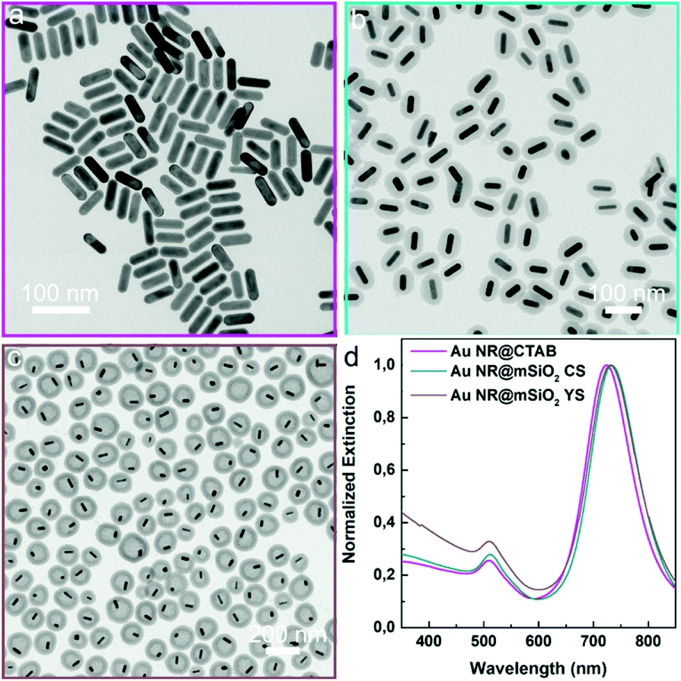 | ||
| Fig. 6 (a) BF-TEM image of Au NRs, (b) BF-TEM image of Au NR@mSiO2 core–shell NPs, (c) BF-TEM image of Au NR@hollow–mSiO2 yolk–shell NPs, and (d) UV-VIS spectra of Au NR, Au NR@mSiO2 core–shell and yolk–shell NPs. | ||
Moreover, even without using any Au NPs as seeds, hollow mSiO2 NSs were still obtained (Fig. S10b†). In addition, both Au NR@mSiO2 core–shell and yolk–shell particles can be acquired by simply tuning the concentration of the Au NRs (Fig. 6b and c), which further corroborates our findings on the influence of the concentration of the Au NTs. The LSPR band of Au NRs with and without mSiO2 are shown in Fig. 6d, and similarly to the case of the Au NTs, the mSiO2 shells caused the LSPR bands of Au NRs to be slightly red-shifted in comparison to Au NR–CTAB. Lastly, the synthesis method demonstrated here was proven to be equally applicable for preparing Au NT@M (Ag, Pd, & Pt)@hollow–mSiO2 yolk–shell NPs (Fig. S11–S14†). Thus, it is safe to conclude that the method will work well for synthesizing yolk–shell NPs of mSiO2 and other metal NPs, as long as the metal surface is protected by CTAC or CTAB molecules and it is even likely that also other NPs, as long as their ligands can be replaced with CTAB/CTAC can be used as cores as well.
Conclusions
In summary, we have developed a scalable single-step coating method for the synthesis of Au NP@hollow–mSiO2 yolk–shell structures. It features a single-step coating, using CTAB or CTAC as pore inducing agent and its suitability for various shapes of Au NPs, tunability of the hollow space between the core and the shell, and opportunity for scaled-up synthesis have been demonstrated. In addition, we showed that the Au NP@hollow–mSiO2 yolk–shell structure forms via a growth-etching synergistic mechanism that was revealed by time-resolved TEM images. Compared to the LSPR band of Au NT–CATC, the LSPR bands of Au NT@mSiO2 core–shell particles and Au NT@hollow–mSiO2 particles show a slight red shift and mild etching allows the metal NPs to be freely diffusing in the mesoporous silica shell. Considering the generality of this method, it can very likely also be used for synthesizing mSiO2 based shell yolk–shell particles for other kinds of NPs cores, e.g., semiconductors quantum dots, upconversion rare earth doped NPs, and metallic oxide NPs. As such, this versatile synthesis method of yolk–shell particles helps realizing applications of these NPs in catalysis, targeted drug delivery, in vivo imaging, and sensing.Experimental
Synthetic procedures of Au NT@mSiO2
A volume of 10 mL of Au NTs storage solution was centrifuged at 9000 rpm for 10 minutes. After centrifuging, the suspension was removed and the sediment was suspended in 10 mL of deionized water and transferred into a 20 mL glass vial. 150 μL of 0.1 M CATC and 100 μL of 0.10 M NaOH were added into above Au NTs solution. Lastly, 200 μL of 20 vol% TEOS in methanol was injected into the above solution in one shot under stirring. The solution was stirred for 45–48 hours. More details of changing the reaction conditions synthesis and their corresponding figures are shown in Table S1 in ESI.†More details of Experimental section, e.g. chemicals, characterization, and liquid-cell TEM experiments, can be found in ESI.†
Conflicts of interest
There are no conflicts to declare.Acknowledgements
We thank Dr Da Wang for the useful discussions. X. X. and A. v. B. acknowledge financial support from EU H2020-MSCA-ITN-2015 project ‘MULTIMAT’ (project number: 676045). M. v. H. acknowledges the European Research Council for an ERC-CoG grant (NANO-INSITU, grant no. 683076).References
- C. Boerigter, R. Campana, M. Morabito and S. Linic, Nat. Commun., 2016, 7, 10545 CrossRef PubMed.
- Z. Bian, T. Tachikawa, P. Zhang, M. Fujitsuka and T. Majima, J. Am. Chem. Soc., 2014, 136, 458–465 CrossRef PubMed.
- Y. C. Tsao, S. Rej, C. Y. Chiu and M. H. Huang, J. Am. Chem. Soc., 2014, 136, 396–404 CrossRef PubMed.
- M. Dunwell, Q. Lu, J. M. Heyes, J. Rosen, J. G. Chen, Y. Yan, F. Jiao and B. Xu, J. Am. Chem. Soc., 2017, 139, 3774–3783 CrossRef PubMed.
- J. Duan, L. Bai, K. Xu, Q. Fang, Y. Sun, H. Xu, K. C. Leung and S. Xuan, J. Hazard. Mater., 2020, 15, 121276 CrossRef PubMed.
- X. Zhu, H. K. Yip, X. Zhuo, R. Jiang, J. Chen, X. M. Zhu, Z. Yang and J. Wang, J. Am. Chem. Soc., 2017, 139, 13837–13846 CrossRef PubMed.
- G. Bodelon, V. Montes-Garcia, V. Lopez-Puente, E. H. Hill, C. Hamon, M. N. Sanz-Ortiz, S. Rodal-Cedeira, C. Costas, S. Celiksoy, I. Perez-Juste, L. Scarabelli, A. La Porta, J. Perez-Juste, I. Pastoriza-Santos and L. M. Liz-Marzan, Nat. Mater., 2016, 15, 1203–1211 CrossRef PubMed.
- Y. Liu, Z. Wang, Y. Liu, G. Zhu, O. Jacobson, X. Fu, R. Bai, X. Lin, N. Lu, X. Yang, W. Fan, J. Song, Z. Wang, G. Yu, F. Zhang, H. Kalish, G. Niu, Z. Nie and X. Chen, ACS Nano, 2017, 11, 10539–10548 CrossRef CAS PubMed.
- Y. Zhao, Y. Huang, H. Zhu, Q. Zhu and Y. Xia, J. Am. Chem. Soc., 2016, 138, 16645–16654 CrossRef CAS PubMed.
- P. Li, Y. Li, Z. K. Zhou, S. Tang, X. F. Yu, S. Xiao, Z. Wu, Q. Xiao, Y. Zhao, H. Wang and P. K. Chu, Adv. Mater., 2016, 28, 2511–2517 CrossRef CAS PubMed.
- H. Jing, Q. Zhang, N. Large, C. Yu, D. A. Blom, P. Nordlander and H. Wang, Nano Lett., 2014, 14, 3674–3682 CrossRef CAS PubMed.
- S. Schlucker, Angew. Chem., Int. Ed., 2014, 53, 4756–4795 CrossRef.
- S.-Y. Ding, J. Yi, J.-F. Li, B. Ren, D.-Y. Wu, R. Panneerselvam and Z.-Q. Tian, Nat. Rev. Mater., 2016, 1, 16021 CrossRef CAS.
- X. Xie, G. Gao, S. Kang, Y. Lei, Z. Pan, T. Shibayama and L. Cai, Nanotechnology, 2017, 28, 245602 CrossRef.
- H. Huang, L. Zhang, Z. Lv, R. Long, C. Zhang, Y. Lin, K. Wei, C. Wang, L. Chen, Z. Y. Li, Q. Zhang, Y. Luo and Y. Xiong, J. Am. Chem. Soc., 2016, 138, 6822–6828 CrossRef CAS.
- D. Andren, L. Shao, N. Odebo Lank, S. S. Acimovic, P. Johansson and M. Kall, ACS Nano, 2017, 11, 10053–10061 CrossRef CAS.
- H. A. Atwater and A. Polman, Nat. Mater., 2010, 9, 205–213 CrossRef CAS.
- X. Huang, Y. Li, Y. Chen, H. Zhou, X. Duan and Y. Huang, Angew. Chem., Int. Ed., 2013, 52, 6063–6067 CrossRef CAS PubMed.
- S. Linic, U. Aslam, C. Boerigter and M. Morabito, Nat. Mater., 2015, 14, 567–576 CrossRef CAS.
- M. L. Personick and C. A. Mirkin, J. Am. Chem. Soc., 2013, 135, 18238–18247 CrossRef CAS.
- Y. Xia, X. Xia and H. C. Peng, J. Am. Chem. Soc., 2015, 137, 7947–7966 CrossRef CAS PubMed.
- Q. Zhang, Y. Zhou, E. Villarreal, Y. Lin, S. Zou and H. Wang, Nano Lett., 2015, 15, 4161–4169 CrossRef CAS.
- L. Polavarapu, D. Zanaga, T. Altantzis, S. Rodal-Cedeira, I. Pastoriza-Santos, J. Perez-Juste, S. Bals and L. M. Liz-Marzan, J. Am. Chem. Soc., 2016, 138, 11453–11456 CrossRef CAS.
- X. Xie, G. Gao, S. Kang, T. Shibayama, Y. Lei, D. Gao and L. Cai, Adv. Mater., 2015, 27, 5573–5577 CrossRef CAS.
- X. Xie, M. A. van Huis and A. van Blaaderen, Nanoscale, 2021, 13, 2902–2913 RSC.
- W. Albrecht, T. S. Deng, B. Goris, M. A. van Huis, S. Bals and A. van Blaaderen, Nano Lett., 2016, 16, 1818–1825 CrossRef CAS PubMed.
- Y. Bai, C. Gao and Y. Yin, Nanoscale, 2017, 9, 14875–14880 RSC.
- A. Guerrero-Martinez, J. Perez-Juste and L. M. Liz-Marzan, Adv. Mater., 2010, 22, 1182–1195 CrossRef CAS.
- S. H. Joo, J. Y. Park, C. K. Tsung, Y. Yamada, P. Yang and G. A. Somorjai, Nat. Mater., 2009, 8, 126–131 CrossRef CAS PubMed.
- Z. Teng, W. Li, Y. Tang, A. Elzatahry, G. Lu and D. Zhao, Adv. Mater., 2019, 31, 1707612–1707636 CrossRef.
- I. Gorelikov and N. Matsuura, Nano Lett., 2008, 8, 369–373 CrossRef CAS.
- W. Zhu, Z. Chen, Y. Pan, R. Dai, Y. Wu, Z. Zhuang, D. Wang, Q. Peng, C. Chen and Y. Li, Adv. Mater., 2019, 31, 1800426–1800456 CrossRef PubMed.
- Z. Wang, Z. M. Chang, D. Shao, F. Zhang, F. Chen, L. Li, M. F. Ge, R. Hu, X. Zheng, Y. Wang and W. F. Dong, ACS Appl. Mater. Interfaces, 2019, 11(38), 34755–34765 CrossRef CAS PubMed.
- B. Li and H. C. Zeng, Adv. Mater., 2019, 31, 1801104–1801132 CrossRef.
- D. Mao, J. Wan, J. Wang and D. Wang, Adv. Mater., 2019, 31, 1802874–1802893 CrossRef.
- L. R. Rowe, B. S. Chapman and J. B. Tracy, Chem. Mater., 2018, 30, 6249–6258 CrossRef CAS.
- Y. Chen, H. R. Chen and J. L. Shi, Acc. Chem. Res., 2014, 47, 125–137 CrossRef CAS.
- X. W. D. Lou, L. A. Archer and Z. Yang, Adv. Mater., 2008, 20, 3987–4019 CrossRef CAS.
- Y. Si, M. Chen and L. Wu, Chem. Soc. Rev., 2016, 45, 690–714 RSC.
- X. Wang, J. Feng, Y. Bai, Q. Zhang and Y. Yin, Chem. Rev., 2016, 116, 10983–11060 CrossRef CAS PubMed.
- Z. Teng, S. Wang, X. Su, G. Chen, Y. Liu, Z. Luo, W. Luo, Y. Tang, H. Ju, D. Zhao and G. Lu, Adv. Mater., 2014, 26, 3741–3747 CrossRef CAS.
- A. A. Volkert, M. C. S. Pierre, B. Shrestha and A. J. Haes, RSC Adv., 2015, 5, 3774–3780 RSC.
- K. Watanabe, T. A. J. Welling, S. Sadighikia, H. Ishii, A. Imhof, M. A. van Huis, A. van Blaaderen and D. Nagao, J. Colloid Interface Sci., 2020, 566, 202–210 CrossRef CAS.
- T.-S. Deng, J. E. S. van der Hoeven, A. O. Yalcin, H. W. Zandbergen, M. A. van Huis and A. van Blaaderen, Chem. Mater., 2015, 27, 7196–7203 CrossRef CAS.
- M. Priebe and K. M. Fromm, Chem. – Eur. J., 2015, 21, 3854–3874 CrossRef CAS.
- Q. Fan, H. Yang, J. Ge, S. Zhang, Z. Liu, B. Lei, T. Cheng, Y. Li, Y. Yin and C. Gao, Research, 2020, 2020, 2131806 CAS.
- D. Nagao, C. M. van Kats, K. Hayasaka, M. Sugimoto, M. Konno, A. Imhof and A. van Blaaderen, Langmuir, 2010, 26, 5208–5212 CrossRef CAS.
- L. Scarabelli, M. Coronado-Puchau, J. J. Giner-Casares, J. Langer and L. M. Liz-Marzán, ACS Nano, 2014, 8, 5833–5842 CrossRef CAS PubMed.
- D. Shen, J. Yang, X. Li, L. Zhou, R. Zhang, W. Li, L. Chen, R. Wang, F. Zhang and D. Zhao, Nano Lett., 2014, 14, 923–932 CrossRef CAS.
- A. M. Vinogradov, A. S. Tatikolov and S. M. B. Costa, Phys. Chem. Chem. Phys., 2001, 3, 4325–4332 RSC.
- B. Thierry, J. Ng, T. Krieg and H. J. Griesser, Chem. Commun., 2009, 1724–1726 RSC.
- P. Selvam, S. K. Bhatia and C. G. Sonwane, Ind. Eng. Chem. Res., 2001, 40, 3237–3261 CrossRef CAS.
- Y. Han, Z. Guo, S. Teng, H. Xia, D. Wang, M.-Y. Han and W. Yang, Chem. Mater., 2019, 31(18), 7470–7477 CrossRef CAS.
- Y. J. Wong, L. Zhu, W. S. Teo, Y. W. Tan, Y. Yang, C. Wang and H. Chen, J. Am. Chem. Soc., 2011, 133, 11422–11425 CrossRef CAS.
- K. Watanabe, H. Ishii, M. Konno, A. Imhof, A. van Blaaderen and D. Nagao, Langmuir, 2017, 33, 296–302 CrossRef CAS.
- H. Chen, M. Tian, L. Zhao, F. Wang, L. Sun, J. Wang and C. Yan, Nano Today, 2010, 494–505 CrossRef.
Footnote |
| † Electronic supplementary information (ESI) available. See DOI: 10.1039/d1nr01242h |
| This journal is © The Royal Society of Chemistry 2021 |