
 Open Access Article
 This Open Access Article is licensed under a
 Creative Commons Attribution 3.0 Unported Licence
Novel PET-pperated rosamine pH-sensor dyes with substitution pattern-tunable pKa values and temperature sensitivity†
Elina
Andresen
ab,
Sebastian
Radunz
a and
Ute
Resch-Genger
 *a
*a
aFederal Institute for Materials Research and Testing (BAM), Division Biophotonics, Department 1, Richard-Willstaetter-Straße 11, D-12489 Berlin, Germany. E-mail: ute.resch@bam.de
bHumboldt-Universität zu Berlin, Department of Chemistry, Brook-Taylor-Straße 2, 12489 Berlin, Germany
First published on 9th July 2021
Abstract
We present the synthesis and characterization of a family of regioisomerically pure pH-sensitive rosamine fluorophores consisting of xanthene fluorophore cores, which determine the dyes’ photophysical properties such as excitation/emission wavelength, fluorescence quantum yield, and fluorescence lifetime, and differently substituted phenol moieties. The hydroxyl substituent of the phenol moiety introduces a pH sensitivity of the dyes’ fluorescence exploiting a photoinduced electron transfer (PET), that leads to a protonation-induced switching ON of the rosamine emission. Rational tuning of the pKa value of the rosamine fluorescence between 4 to 9 is achieved by altering the substitution pattern and degree of bromination of the phenolic subunits. Additionally, a temperature sensitivity of the fluorescence quantum yield is introduced or suppressed based upon the degree of rigidity of the xanthene scaffold.
1 Introduction
Luminescence-based sensing and imaging plays a significant role in the life sciences because it enables the non-invasive and spatially resolved detection, monitoring, and quantification of biomolecular interactions or neutral and ionic targets that have no intrinsic color or luminescence with the aid of specifically designed fluorescent probes.1–5 Signaling of specific interactions or the presence of a target or analyte is done by spectroscopically measurable changes in the probes’ absorption and/or fluorescence properties. The readout parameters include changes in intensity and/or spectral position of the absorption/excitation and emission bands.Fluorescent probes that undergo reversible spectroscopic changes are often also termed fluorescent (chemical) sensors, while molecules that reveal irreversible changes in their optical properties caused by the breakage of chemical bonds are referred to as chemodosimeters.6–10
Of particular interest are fluorescent probes for biologically, health, and corrosion-relevant analytes such as pH.11–15 Here, photoinduced electron transfer (PET) – based probes which reveal protonation-induced changes in fluorescence quantum yield and lifetime, while the spectral position of their absorption and emission bands is not or only barely affected, are highly desired Such probes consist of a fluorophore moiety and an electronically decoupled protonation side. Protonation-induced spectral changes in absorption and emission together with relatively moderate intensity changes can be realized, if the (de)protonable group is part of the π-electron system of a charge transfer (CT)-operated chromophore. These modifications are caused by changes in the electron density of the (de)protonable group, which affects the light-induced intramolecular CT process.16–19
For the design of PET probes, fluorophores like fluoresceins,20 naphthalimides,21 BODIPYs22–24 and aza-BODIPYs,25 are combined with electron donating (de)protonable moieties with non-bonding electron pairs provided by nitrogen or oxygen atoms. Electronic excitation of these PET probes leads to an electron transfer from the electron rich receptor to the electronically decoupled excited fluorophore promoting fluorescence quenching. Protonation of the donor receptor changes the energetic position of its HOMO, thereby preventing PET and reversibly switching ON the fluorophore's emission.
Xanthene dyes like fluoresceins, rhodamines, and rosamines are particularly attractive fluorophores for the design of fluorescent probes because of their excellent photophysical properties.26,27 This includes high molar absorption coefficients and high fluorescence quantum yields in the visible or red spectral region as well as a relatively high photostability. Rhodamine fluorophores, which consist of a planar xanthene ring 3,6-disubstituted by in-plane amino groups and a nearby perpendicular 2′-carboxyphenyl ring in position 9 (see Fig. 1A), have been employed in many different probes and sensing applications.28–31 A previously developed strategy to design pH-sensitive rhodamine dyes exploits protonation-induced structural changes, i.e., the switching from the spirocyclic to the ring-open form of certain rhodamine derivatives.32–41 These so called rhodamine spirolactams (RSLs), which are readily accessible by amidation of the carboxylic group of commercially available rhodamines are colorless and non-fluorescent in their spirocyclic form. Protonation of the amide nitrogen induces the opening of the spirocyclic ring system, yielding the fully conjugated and highly fluorescent rhodamine dye. However, these RSLs reveal a limited pKa tunability as substituents of different bulkiness on the carboxylic group of rhodamine B or rhodamine 6G can introduce a steric hindrance of the signal-generating ring-opening mechanism.34,40 This can considerably slow down their response to pH.39
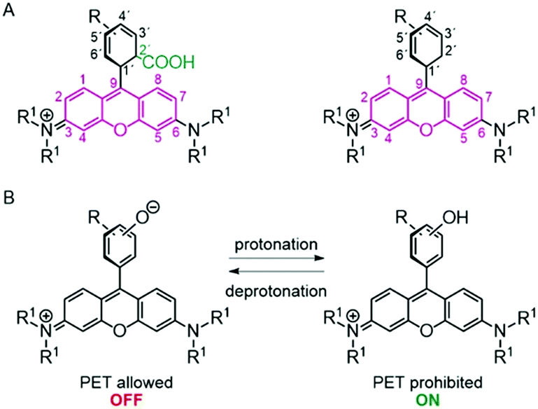 | ||
| Fig. 1 (A) General structures of rhodamine and rosamine dyes with the xanthene core being highlighted in pink. (B) Mechanism of pH sensing of a rosamine dye via intramolecular photoinduced electron trasfer (PET). | ||
Another potentially favorable property of rhodamines bearing sterically non-hindered unbridged dialkylamino substituents can be the sensitivity of their fluorescence intensity or quantum yield to temperature.42–44 This has been utilized for luminescence-based temperature sensors42 but could be also disadvantageous for other applications. Only rhodamines with rigidized, i.e., fused cyclic rings on their xanthene nitrogen atoms like rhodamine 101 or rhodamine 6G reveal a temperature-independent fluorescence.
Thus, alternative concepts for designing fluorescent pH-probes utilizing xanthene dyes are of great interest. Rosamines, which are xanthene-type dyes lacking the carboxylic group in the 2′-position (see Fig. 1A), are less well studied, despite their similarly attractive optical properties. Moreover, they can be easily synthesized and isolated as single regioisomers.45 Hence, they can provide an ideal platform for the rational design of stimuli-responsive fluorophores. So far, only few pH-sensitive rosamine-based probes have been reported that utilize PET.46–48 These dyes typically consist of a fluorophore and a nitrogen-containing subunit, either a piperazonium group46 or an amine-containing side chain,47 located in close proximity to the xanthene core.
To the best of our knowledge, the only series of pH-sensitive rosamine dyes (referred to as rhodamines by the authors) bearing a phenolate group as pH sensitive subunit was reported by Collot's group.48 These dyes, called H-Rubies, are derived from rhodamine X and possess different pKa values depending on the substitution pattern of the phenolic subunit. However, these dyes were not systematically spectroscopically assessed and a systematic tuning of their pKa-was not explored.
Herein, we present a series of novel pH-sensitive rosamine dyes with tuneable pKa values, introduced by altering the substitution pattern of the phenolic moiety. Tuning of their photophysical properties was achieved by modulation of the xanthene unit. Subsequently, the spectroscopic properties of this new family of sensor dyes and their pH-responsivity are systematically assessed as well as their potential as temperature sensors. To the best of our knowledge, this presents the first example of systematic pKa-tuning for PET-based phenol-modulated pH-sensitive rosamine-based dyes.
2 Experimental section
2.1 Materials
3-Hydroxybenzaldehyde (≥99%) and p-toluenesulfonic acid monohydrate (≥98%) were purchased from Sigma-Aldrich. 8-Hydroxyjulolidine (97%) and p-chloranil (97%) were obtained from Alfa Aesar,3-diethyl-aminophenol (98%) was purchased from J&K, and 4-hydroxy-benzaldehyde (≥99%) was obtained from Roth. Propionic acid (99.5%) was purchased from Honeywell and chloroform, and dichloromethane and methanol from Carl Roth GmbH. All chemicals were used without further purification. Brominated hydroxybenzaldehydes were provided by S. Radunz; their synthesis and characterization is described in a previous work.22 ALUGRAM Xtra SIL G/UV254 plates by Macherey-Nagel were used for thin-layer chromatography. Isolation of products by chromatography was performed with silica from Macherey-Nagel Silica 60 M (0.04–0.063 mm).2.2 General procedure for rosamine synthesis
To a solution of aldehyde (1 eq.) in propanoic acid 3-diethylaminophenol or 8-hydroxyjulolidine (2 eq.) and p-TsOH (1 eq.) were added. The solution was protected from light and stirred at 80 °C for 2 h. After cooling to room temperature (rt), chloranil (1.1 eq.) was added to the reaction mixture that was then stirred overnight at rt. The resulting dark purple solution was evaporated to dryness and the crude product was purified by column chromatography on silica gel (DCM/MeOH, 98/2 to 90/10) yielding the desired compound as a purple or violet solid. For detailed synthesis and characterisation of the dyes can be found in the ESI.†2.3 NMR spectroscopy and mass spectrometry
NMR spectra were recorded on a JEOL Eclipse+ 500 (1H 500 MHz, 13C 126 MHz) and BRUKER AVANCE 700 (1H 700 MHz, 13C 176 MHz) spectrometer at 25 °C. The chemical shifts δ are calibrated on the respective solvent peak as internal standard (1H: δ(CD3OH) = 3.31 ppm; 13C: δ(CD3OH) = 77.16 ppm). All shifts are reported in in parts per million (ppm) and NMR multiplicities.2.4 Optical spectroscopy
Absorption measurements were carried out on a calibrated Varian Cary 5000 UV-/VIS-/NIR spectrophotometer with a scan rate of 600 nm s−1 and a slit width of 2 nm using a baseline correction (air/air) and a solvent sample as a reference.Fluorescence spectra were recorded on a calibrated FLS920 spectrofluorometer from Edinburgh Instruments with an integration time of 0.3 s and slit widths of 6.0 nm of the excitation and emission monochromators. The excitation wavelength was set to 540 nm for dyes 1a–c*, dyes 2a,b and dye 3 and to 570 nm for dyes 1d and 2c, respectively.
All optical measurements were done with 1 cm quartz glass cuvettes from Hellma GmbH.
Fluorescence quantum yields were determined absolutely using a Quantaurus-QY integrating sphere spectrometer C11347-01 from Hamamatsu Photonics K. K. using special long-neck quartz cells provided by the instrument manufacturer. The absorbance of the sample solutions was kept below 0.1 at the excitation wavelength (λex = 540 nm). The quantum yield values of the dyes given were obtained by averaging five independent measurements. To determine the fluorescence quantum yield of the protonated species, 5 μl of concentrated HCl (12 M) were added to 3 ml of a 1 × 10⁻⁶ M ethanolic solution of the dyes.
The determination of fluorescence lifetimes was carried out with a FLS920 spectrofluorometer from Edinburgh Instruments using a femtosecond supercontinuum laser. The repetition rate was set to 10 MHz. The fluorescence lifetimes were measured for methanolic dye solutions with concentrations of 5 × 10⁻⁶ M. The excitation wavelength was set to 540 nm for dyes 1a–c*, dyes 2a, b, and dye 3 and to 570 nm for dyes 1d and 2c, respectively. All fluorescence decay curves were measured in 1 cm quartz cuvettes from Hellma GmbH and were evaluated using the software FAST (Edinburgh Instruments) and applying monoexponential fits to obtain the provided fluorescence lifetimes.
pH-titration experiments were carried out with solutions of the dyes in a 1/2 (v/v) mixture of methanol and aqueous buffer (borate-citrate, 25 mM) with dye concentrations of 1 × 10⁻⁶ M, thereby also suppressing a possible dye dimerization and self-quenching. The pH values of the used buffer solutions were previously adjusted with a 780 pH meter using a glass electrode from Deutsche METROHM GmbH & Co. KG and were verified with a pH 211 microprocessor pH meter from Hanna Instruments. The 780 pH meter was calibrated in a three-point calibration with standard buffers of pH 10.01, 7.01, and 4.01 (Hanna Instruments) at 25 °C. For the determination of the pKa-values, 5 μl of the methanolic stock solutions of the dyes (2 × 10⁻³ M) diluted in 2 ml methanol, were added to 1 ml buffer, resulting in a 1 × 10⁻⁶ M dye solution. Successively, defined amounts of concentrated NaOH were added and the absorption and fluorescence emission spectra were collected at the respective pH. pKa-values were determined from 3 independent samples and measurements applying a sigmoidal curve fit (see ESI,† eqn (S1)).
The temperature dependent fluorescence intensity measurements were performed in triplicate in from the temperature range of 10 °C to 50 °C in 5 °C steps (using equilibration times of 5 min and stirring the solutions in between the measurements). After heating to 50 °C the sample was cooled down to 10 °C and the fluorescence intensity was measured again to confirm the thermal stability of the studied rosamine dyes.
3. Results and discussion
3.1 Design and synthesis of pH-sensitive rosamines with tunable pKa-values
A series of novel pH sensitive rosamine dyes with tunable pKa values was obtained in overall yields of up to 30% by the condensation of differently substituted benzaldehydes with 3-aminophenol derivatives (see Fig. 2A and B). The condensation was carried out using propionic acid and a catalytic amount of p-toluenesulfonic acid (p-TsOH), followed by oxidation with chloranil. The rosamine scaffold was derived from rhodamine B (RhB), rhodamine 101 (Rh101), and tetramethylrhodamine (TMR), which differ in their substitution pattern at the xanthene ring systems and thus, in their spectroscopic properties. By using differently brominated ortho- and para-hydroxyphenyl as pH-sensitive moiety, a set of pH-responsive rosamines (1a-2d) was obtained with pKa values varying from 4 to 9 (Fig. 2B). Also, a pH-inert reference dye 3 was synthesized consisting of a xanthene and unsubstituted phenolic unit. | ||
| Fig. 2 (A) General synthetic route and (B) chemical structures of the synthesized pH-sensitive fluorescent rosamine probes. | ||
3.2 Structure/property relationship of the rosamine dyes
Subsequently, the influence of the substitution pattern of the phenolic subunits and the nitrogen atoms at the xanthene core on the pH-dependent spectroscopic behavior of the dyes was examined. As previously stated, the former introduces the desired pH-sensitivity and tunability of the pKa value, while the latter determines the photophysical properties of the dyes such as the spectral position of the absorption and emission maxima (λabs(max), λem(max)), the fluorescence quantum yields (ϕ), and the fluorescence lifetimes (τ). An overview of the spectroscopic properties of the new rosamine compounds in methanol is provided in Table 1. The molar absorption coefficients ε of the rosamines are in the order of 38![[thin space (1/6-em)]](https://www.rsc.org/images/entities/char_2009.gif) 000–90000 M−1 cm−1 and the ϕ values amount to 30–80% in methanol. Fig. 3A shows a comparison of the absorption and emission spectra of compounds bearing alkyl groups or fully rigidized xanthene scaffolds. For the alkyl substituted compounds, similar absorption and emission spectra with absorption and emission maxima at 555 nm and 580 nm are observed. The absorption and emission spectra of the compounds with a fused xanthene nitrogen are bathochromically shifted by 20 nm yielding absorption and emission maxima at 583 nm and 602 nm, respectively. This is ascribed to the enhanced delocalization of the electron density of the nitrogen electron pairs favored by the restricted rotation around the C–N bonds caused by the fused rings of compound 1d. The influence of the halogen substituents on the phenyl ring is illustrated in Fig. 3B. This figure compares the absorption and emission spectra of the non-halogenated rosamine reference dye 3 with those of the para-hydroxy substituted non-, mono- and di-brominated rosamine dyes 1a, 1b, and 1c. Apparently, introducing a hydroxy group on the phenyl ring does not affect the rosamine absorption and emission spectra, while each bromine atom induces a red shift of 5 nm of the absorption and emission maxima. The absorption maximum of 3 at 551 nm is shifted to 556 nm for 1a and to 561 nm for 1b and 1c and the emission maxima from 572 nm to 577 nm and 583 nm, respectively. Similar shifts in absorption and emission results also for the meta-hydroxy substituted rosamine derivatives 2a–c in dependence of the degree of bromination of the phenyl moiety (see Table 1). The very similar spectral properties of 1a–c and 2a–b as well as 1d and 2c indicate more or less complete electronic decoupling of the rosamine chromophore and the phenyl moiety inducing pH-sensitivity. Deprotonation of the hydroxyl group of the pH-responsive rosamines leads to a hypsochromic shift in absorption of about 10 nm whereas the absorption spectrum of the reference dye 3 remains unaffected by pH (see ESI,† Fig. S2–S10). Such a protonation-induced hypsochromic shift of the main absorption band is a consequence of the charge localization originating from an aromatic destabilization in the molecules and was observed for different pH sensitive fluorophores.46–49 Consequently, these changes are attributed to an equilibrium between the cationic form (protonated hydroxyl group) and zwitterionic form (deprotonated hydroxyl group) of the rosamines introduced by deprotonation of the hydroxylic group and the increased electron density (enhanced electron donor strength) – of the negatively charged hydroxylic group located in the immediate proximity of the rhodamine core. The ϕ values of the rosamines are reduced by 30% compared to their parent rhodamine dyes (ΦEtOH(RhB) = 69%, ΦEtOH(TMR) = 68%, ΦEtOH(Rh101) = 96% at 25 °C).50 (see Table 1) This diminution in ϕ is ascribed to the absence of the carboxylic group in 2′ position, which permits rotation of the phenolic moiety, thereby opening further non-emissive relaxation pathways. A similar behavior was also observed by Mottram et al.51 The rosamine dyes reveal mono-exponential fluorescence decay kinetics in methanol with τf values in the nanosecond (ns) range. The rosamine dyes 1d and 2c with fused xanthene nitrogen atoms possess the longest lifetimes of around 4 ns, whereas the rosamines bearing N-alkyl groups have fluorescence lifetimes of less than two ns (Fig. 3C). Bromination of the phenol group is accompanied by a diminution in τf as reflected, e.g. by a comparison of dye 1c and non-brominated 1a (see ESI,† Fig. S19). This trend is ascribed to the heavy-atom effect of bromine. Finally, the pH dependence of the fluorescence decay kinetics of the pH-sensitive rosamine dyes was examined. Therefore, the fluorescence decay curves were measured at selected pH values exemplary for dyes 1c and 1d (see ESI,† Fig. S23). The results indicate that no significant changes in the decay kinetics and fluorescence lifetimes of the dyes occur upon deprotonation.
000–90000 M−1 cm−1 and the ϕ values amount to 30–80% in methanol. Fig. 3A shows a comparison of the absorption and emission spectra of compounds bearing alkyl groups or fully rigidized xanthene scaffolds. For the alkyl substituted compounds, similar absorption and emission spectra with absorption and emission maxima at 555 nm and 580 nm are observed. The absorption and emission spectra of the compounds with a fused xanthene nitrogen are bathochromically shifted by 20 nm yielding absorption and emission maxima at 583 nm and 602 nm, respectively. This is ascribed to the enhanced delocalization of the electron density of the nitrogen electron pairs favored by the restricted rotation around the C–N bonds caused by the fused rings of compound 1d. The influence of the halogen substituents on the phenyl ring is illustrated in Fig. 3B. This figure compares the absorption and emission spectra of the non-halogenated rosamine reference dye 3 with those of the para-hydroxy substituted non-, mono- and di-brominated rosamine dyes 1a, 1b, and 1c. Apparently, introducing a hydroxy group on the phenyl ring does not affect the rosamine absorption and emission spectra, while each bromine atom induces a red shift of 5 nm of the absorption and emission maxima. The absorption maximum of 3 at 551 nm is shifted to 556 nm for 1a and to 561 nm for 1b and 1c and the emission maxima from 572 nm to 577 nm and 583 nm, respectively. Similar shifts in absorption and emission results also for the meta-hydroxy substituted rosamine derivatives 2a–c in dependence of the degree of bromination of the phenyl moiety (see Table 1). The very similar spectral properties of 1a–c and 2a–b as well as 1d and 2c indicate more or less complete electronic decoupling of the rosamine chromophore and the phenyl moiety inducing pH-sensitivity. Deprotonation of the hydroxyl group of the pH-responsive rosamines leads to a hypsochromic shift in absorption of about 10 nm whereas the absorption spectrum of the reference dye 3 remains unaffected by pH (see ESI,† Fig. S2–S10). Such a protonation-induced hypsochromic shift of the main absorption band is a consequence of the charge localization originating from an aromatic destabilization in the molecules and was observed for different pH sensitive fluorophores.46–49 Consequently, these changes are attributed to an equilibrium between the cationic form (protonated hydroxyl group) and zwitterionic form (deprotonated hydroxyl group) of the rosamines introduced by deprotonation of the hydroxylic group and the increased electron density (enhanced electron donor strength) – of the negatively charged hydroxylic group located in the immediate proximity of the rhodamine core. The ϕ values of the rosamines are reduced by 30% compared to their parent rhodamine dyes (ΦEtOH(RhB) = 69%, ΦEtOH(TMR) = 68%, ΦEtOH(Rh101) = 96% at 25 °C).50 (see Table 1) This diminution in ϕ is ascribed to the absence of the carboxylic group in 2′ position, which permits rotation of the phenolic moiety, thereby opening further non-emissive relaxation pathways. A similar behavior was also observed by Mottram et al.51 The rosamine dyes reveal mono-exponential fluorescence decay kinetics in methanol with τf values in the nanosecond (ns) range. The rosamine dyes 1d and 2c with fused xanthene nitrogen atoms possess the longest lifetimes of around 4 ns, whereas the rosamines bearing N-alkyl groups have fluorescence lifetimes of less than two ns (Fig. 3C). Bromination of the phenol group is accompanied by a diminution in τf as reflected, e.g. by a comparison of dye 1c and non-brominated 1a (see ESI,† Fig. S19). This trend is ascribed to the heavy-atom effect of bromine. Finally, the pH dependence of the fluorescence decay kinetics of the pH-sensitive rosamine dyes was examined. Therefore, the fluorescence decay curves were measured at selected pH values exemplary for dyes 1c and 1d (see ESI,† Fig. S23). The results indicate that no significant changes in the decay kinetics and fluorescence lifetimes of the dyes occur upon deprotonation.
| Dye | ε (λmax) (MeOH) [M−1 cm−1] | λ abs-ON (MeOH) [nm] | λ em (MeOH) [nm] | Φ fl (EtOH)(abs.) [%] | τ fl (MeOH)a [ns] | pKab (Em) |
|---|---|---|---|---|---|---|
|
a Mono exponential fit (global analysis fi (%) = 100.0).
b In H2O/MeOH 2:1 vol%, 25 mM buffered solution).
|
||||||
| 1a | 81000 |
551 | 570 | 35 | 1.73 | 8.99 |
| 1b | 93000 |
556 | 577 | 34 | 1.66 | 7.37 |
| 1c | 86000 |
561 | 580 | 27 | 1.56 | 5.51 |
| 1c * | 58000 |
555 | 578 | 32 | 1.98 | 5.54 |
| 1d | 70000 |
582 | 600 | 70 | 4.13 | 5.87 |
| 2a | 65500 |
553 | 576 | 35 | 1.72 | 9.42 |
| 2b | 53000 |
565 | 580 | 33 | 1.64 | 8.71 |
| 2c | 57000 |
600 | 620 | 56 | 4.44 | 5.14 |
| 3 | 76000 |
555 | 575 | 32 | 1.65 | n. a. |
 | ||
| Fig. 3 Influence of (A) the xanthene moiety and (B) the bromination degree on the spectral position of the absorption and emission maxima. (C) Luminescence decay kinetics of different xanthene moieties. | ||
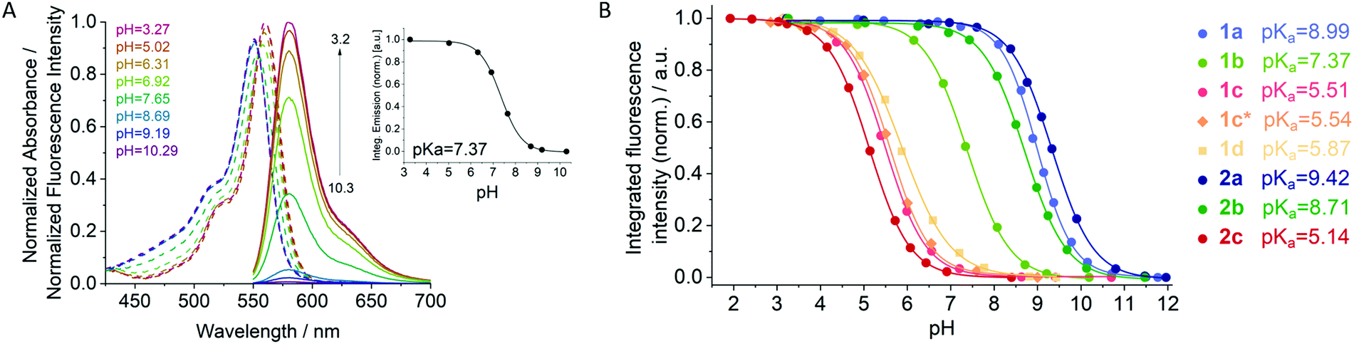 | ||
| Fig. 4 (A) pH-dependent normalized absorption (dashed lines) and emission (solid lines) spectra of 1b in a mixture of methanol-aqueous buffer solution (λex = 540 nm). The insert shows a typical pH titration curve in the insert; (B) pH-titration curves (dose response equation fit) of all synthesized dyes illustrating the broad pH range covered by the novel rosamine dyes. | ||
Moreover, as shown in Fig. 4B, we could successfully expand the concept of halogenating phenolic subunits to tune the pKa of fluorescent probes utilized for different classes of fluorophores such as BODIPY dyes21,22 to rosamine dyes. Stepwise bromination of the phenolic aryl moiety in position 9 of the xanthene core clearly provides substitution pattern control of the pKa of this dye class with the degree of halogenation strongly influencing the acidity of the pH-sensitive hydroxy group due to its electron withdrawing effect. The results of the fluorometric pH titration experiments of the rosamine dyes are shown in the ESI† (Fig. S3–S10) and the pH titration curves used to derive the pKa values are summarized in Fig. 4B. Unsubstituted compounds 2a and 1a exhibit the most basic pKa values of 9.42 and 8.99, respectively. Monobromination lowers the pKa values to 8.71 for 2b and to 7.37 for 1b. This demonstrates that substitution in ortho-position to the hydroxylic group (in the case of 1b) has a stronger influence on the pKa value than halogenation in meta-position (for 2b). Further halogenation leads to more acidic pKa values of 5.51 for the di-brominated compound 1c. The lowest pKa value of 5.14 is observed for the tri-brominated compound 2c. While halogenation of the subunits plays an important role for the tuning of the pKa value, a comparison of compounds 1c, 1c*, and 1d with different substitution pattern of the xanthene core, which exhibit pKa values of 5.51, 5.54, and 5.87, respectively, shows that the substitution pattern of the xanthene core barely affects the pKa value of the sensor dye.
Subsequently, the reversibility of the PET-triggered ON/OFF-switching of the rosamine fluorescence was investigated that accompanies the protonation/deprotonation of the hydroxyl group. Therefore, the pH of the dyes solutions was reversibly changed from pH of 2 to 10 in 10–12 cycles. As shown in the ESI,† in Fig. S12 and S13, all dyes reveal an excellent reversibility of their fluorescence with their initial fluorescence intensity at a pH of 2 being completely restored even after 12 deprotonation/protonation cycles. The observed slight decrease in fluorescence intensity is attributed to dilution effects and not to a loss in reversibility.
As some rhodamine dyes show a temperature sensitive fluorescence intensity, we also performed temperature dependent studies with the differently substituted rosamine dyes to clarify whether the derived pKa values are temperature- dependent. Such a temperature dependence could hamper their suitability for pH-sensing. Representatively, the pKa value of compound 1c was determined at 20 °C and 40 °C. The obtained values of 5.50 and 5.48 for 20 °C and 40 °C underline the temperature independence of the pH-sensing behavior (see ESI,† Fig. S11).
Temperature sensitivity of the rosamine dyes
As the substitution pattern, of the xanthene scaffold of the parent rhodamine dyes, i.e., the degree of rigidity of the amino substituents can introduce a temperature dependence of the fluorescence,44,52–54 the influence of temperature on fluorescence intensity was investigated for selected rosamine dyes. Our rosamine dyes can be classified into four groups with respect to their structural pattern, i.e., the substitution pattern of the amino groups at the xanthene core and the phenolic moiety. This includes the following classes: (i) rigid-rigid (rigidized amino groups on the xanthene core and hindered rotation of the phenolic subunit (2c)), (ii) free-rigid (freely rotatable amino groups on the xanthene core and hindered rotation of the phenolic subunit (2b)), (iii) rigid-free (rigidized amino groups on the xanthene core and free rotation of the phenolic subunit (1d)), and (iv) free-free (freely rotatable amino groups on the xanthene core and non-hindered rotation of the phenolic subunit (1c)). Subsequently, we studied the temperature dependence of the fluorescence of representative rosamine dyes from each structural class. The changes of the integrated fluorescence intensities of the studied dyes in dependence of the temperature in a temperature interval from 10 °C to 50 °C are highlighted in Fig. 5. The corresponding absorption and emission spectra of the four dyes are provided in the ESI† (see Fig. S14–S17). For a better comparison of the different rosamines, the fluorescence intensities were normalized to the fluorescence intensity at 10 °C and a linear fit of the temperature-dependent changes using the least-mean-squares method was applied. This yielded fluorescence intensity changes of approximately −1.59%, −1.57%, −0.49%, and −0.15% per °C for 1c, 2b, 1d, and 2c, respectively. The absorption spectra of the dyes display only a small temperature-dependent decrease of the absorption maximum by 0.5–1%. Dye 2c, representing the rigid–rigid class (i) reveals a quasi-temperature insensitive fluorescence. This is ascribed to the rigidization of the xanthene anilines as fused cyclic rings and the sterically restricted rotation of the phenolic subunit induced by substitution in positions 2′and 6′. However, reducing the overall rigidity of the dyes by either reducing the rigidity of the amine scaffold at the xanthene core (class i) or enabling a rotation of the phenolic PET-subunit (class iii) results in an enhanced temperature sensitivity favored by the thereby opened non-radiative relaxation pathways. For example, for dye 1d, representing (class iii), a slight decrease in fluorescence intensity with increasing temperature is accordingly observed. Dyes 2b and 1c (classes (ii) and (iv)) reveal a similar, yet more pronounced temperature sensitivity. This behavior is ascribed to the free rotation of the alkylated amines on the xanthene core, enabling an electron transfer from the amino groups to the xanthene ring, that leads to a fast non-radiative deactivation to the ground state ascribed to a twisted intermolecular charge-transfer (TICT) process.19 Notably, the degree of rotational hindrance of the phenolic group has only a negligible effect. | ||
| Fig. 5 Integrated fluorescence intensity of dyes 1c, 1d, 2b and 2c as a function of temperature. | ||
4 Conclusions
In summary, we developed a simple and efficient strategy for the synthesis of a series of moderately to strongly fluorescent rosamine dyes with pKa values tunable between 5.1 to 9.5 that can be utilized for pH sensing in the pH window of 4 to 10. The rational design of these sensor molecules relies on a phenolic subunit electronically decoupled from the xanthene core. The latter determines the spectral features of the fluorescent probes while the protonation state of the former controls the dye's fluorescence via an intramolecular photoinduced electron transfer (PET). Protonation of the phenol group inhibits the fluorescence quenching PET, leading to a pH dependent switching ON of the rosamine fluorescence. Tuning of the pKa values of these pH-sensitive rosamines could be achieved by systematically introducing electron withdrawing groups like bromine atoms to the phenolic PET-inducing subunit. This strategy has been previously utilized by us also for a set of BODIPY dyes.22 Moreover, a temperature-dependent fluorescence response of the rosamines can be promoted or suppressed in dependence of the rigidity of the dye scaffold, as reported for the rhodamine parent dyes. Thereby, rosamine dyes with a rigid scaffold as dye 2c and thus suppressed temperature sensitivity, are excellent candidates for pH sensors. In addition, our findings provide new insights into a class of molecules that are responsive to two parameters, here pH and temperature, by different mechanisms. This could be interesting in the future, e.g. for the design of logic gates.55,56Currently, we are equipping our pH-responsive rosamines with a linker group on the phenol moiety. This will enable the facile coupling of these promising fluorophores to biomolecules such as antibodies or nanoparticles bearing reactive surface groups to construct pH-responsive bioconjugates and nanosensors for bioimaging applications.
Conflicts of interest
There are no conflicts to declare.Acknowledgements
This work was funded by the German Research Council DFG (grant SCHA 1009/17-1) and Bundesanstalt für Materialforschung und -prüfung (BAM). NMR and MS measurements were performed at FU Berlin.References
- J. Wu, W. Liu, J. Ge, H. Zhang and P. Wang, Chem. Soc. Rev., 2011, 40, 3483–3495 RSC.
- T. Ueno and T. Nagano, Nat. Methods, 2011, 8, 642–645 CrossRef CAS PubMed.
- H. Zhu, J. Fan, J. Du and X. Peng, Acc. Chem. Res., 2016, 49, 2115–2126 CrossRef CAS PubMed.
- K. Kikuchi, Chem. Soc. Rev., 2010, 39, 2048–2053 RSC.
- T. Terai and T. Nagano, Pflugers Arch, 2013, 465, 347–359 CrossRef CAS PubMed.
- Y. Yang, Q. Zhao, W. Feng and F. Li, Chem. Rev., 2013, 113, 192–270 CrossRef CAS PubMed.
- K. Kaur, R. Saini, A. Kumar, V. Luxami, N. Kaur, P. Singh and S. Kumar, Coord. Chem. Rev., 2012, 256, 1992–2028 CrossRef CAS.
- D. T. Quang and J. S. Kim, Chem. Rev., 2010, 110, 6280–6301 CrossRef CAS PubMed.
- M. J. Culzoni, A. Muñoz de la Peña, A. Machuca, H. C. Goicoechea and R. Babiano, Anal. Methods, 2013, 5, 30–49 RSC.
- S. Suganya, S. Naha and S. Velmathi, ChemistrySelect, 2018, 3, 7231–7268 CrossRef CAS.
- J. Han and K. Burgess, Chem. Rev., 2010, 110, 2709–2728 CrossRef CAS PubMed.
- D. Wencel, T. Abel and C. McDonagh, Anal. Chem., 2014, 86, 15–29 CrossRef CAS PubMed.
- J.-T. Hou, W. X. Ren, K. Li, J. Seo, A. Sharma, X.-Q. Yu and J. S. Kim, Chem. Soc. Rev., 2017, 46, 2076–2090 RSC.
- S. Wen, L. Xiaohua and M. Huimin, Methods Appl. Fluoresc., 2014, 2, 042001 CrossRef PubMed.
- A. Steinegger, O. S. Wolfbeis and S. M. Borisov, Chem. Rev., 2020, 120, 12357–12489 CrossRef CAS PubMed.
- Y. Q. Li, J. L. Bricks, U. Resch-Genger, M. Spieles and W. Rettig, J. Phys. Chem. A, 2006, 110, 10972–10984 CrossRef CAS PubMed.
- K. Rurack and U. Resch-Genger, Chem. Soc. Rev., 2002, 31, 116–127 RSC.
- Y. Fu and N. S. Finney, RSC Adv., 2018, 8, 29051–29061 RSC.
- Z. R. Grabowski, K. Rotkiewicz and W. Rettig, Chem. Rev., 2003, 103, 3899–4032 CrossRef PubMed.
- K. M. K. S. Ha Na Kim and Juyoung Yoon, Tetrahedron Lett., 2011, 52, 2340–2343 CrossRef.
- J. Qi, D. Liu, X. Liu, S. Guan, F. Shi, H. Chang, H. He and G. Yang, Anal. Chem., 2015, 87, 5897–5904 CrossRef CAS PubMed.
- S. Radunz, H. R. Tschiche, D. Moldenhauer and U. Resch-Genger, Sens. Actuators, B, 2017, 251, 490–494 CrossRef CAS.
- R. Gotor, P. Ashokkumar, M. Hecht, K. Keil and K. Rurack, Anal. Chem., 2017, 89, 8437–8444 CrossRef CAS PubMed.
- Y. S. Marfin, M. V. Shipalova, V. O. Kurzin, K. V. Ksenofontova, A. V. Solomonov and E. V. Rumyantsev, J. Fluoresc., 2016, 26, 2105–2112 CrossRef CAS PubMed.
- C. Staudinger, J. Breininger, I. Klimant and S. M. Borisov, Analyst, 2019, 144, 2393–2402 RSC.
- R. Zhang, F. Yan, Y. Huang, D. Kong, Q. Ye, J. Xu and L. Chen, RSC Adv., 2016, 6, 50732–50760 RSC.
- L. D. Lavis and R. T. Raines, ACS Chem. Biol., 2008, 3, 142–155 CrossRef CAS PubMed.
- K. Othmer, Encyclopedia of Chemical Technology, 2000, pp. 1–19, DOI:10.1002/0471238961.2401142023090708.a01.pub2.
- X. Chen, T. Pradhan, F. Wang, J. S. Kim and J. Yoon, Chem. Rev., 2012, 112, 1910–1956 CrossRef CAS PubMed.
- H. Zheng, X.-Q. Zhan, Q.-N. Bian and X.-J. Zhang, Chem. Commun., 2013, 49, 429–447 RSC.
- S. Ma, Y. Wang, M. She, S. Wang, Z. Yang, P. Liu, S. Zhang and J. Li, Rev. Anal. Chem., 2017, 36, 20160024 Search PubMed.
- W. Zhang, B. Tang, X. Liu, Y. Liu, K. Xu, J. Ma, L. Tong and G. Yang, Analyst, 2009, 134, 367–371 RSC.
- Q. A. Best, R. Xu, M. E. McCarroll, L. Wang and D. J. Dyer, Org. Lett., 2010, 12, 3219–3221 CrossRef CAS PubMed.
- L. Yuan, W. Lin and Y. Feng, Org. Biomol. Chem., 2011, 9, 1723–1726 RSC.
- Z. Li, S. Wu, J. Han and S. Han, Analyst, 2011, 136, 3698–3706 RSC.
- M. Tian, X. Peng, J. Fan, J. Wang and S. Sun, Dyes Pigm., 2012, 95, 112–115 CrossRef CAS.
- A. Bender, Z. R. Woydziak, L. Fu, M. Branden, Z. Zhou, B. D. Ackley and B. R. Peterson, ACS Chem. Biol., 2013, 8, 636–642 CrossRef CAS PubMed.
- H.-S. Lv, S.-Y. Huang, Y. Xu, X. Dai, J.-Y. Miao and B.-X. Zhao, Bioorg. Med. Chem. Lett., 2014, 24, 535–538 CrossRef CAS PubMed.
- W. L. Czaplyski, G. E. Purnell, C. A. Roberts, R. M. Allred and E. J. Harbron, Org. Biomol. Chem., 2014, 12, 526–533 RSC.
- S. G. Stratton, G. H. Taumoefolau, G. E. Purnell, M. Rasooly, W. L. Czaplyski and E. J. Harbron, Chem. – Eur. J., 2017, 23, 14064–14072 CrossRef CAS PubMed.
- H. N. Kim, M. H. Lee, H. J. Kim, J. S. Kim and J. Yoon, Chem. Soc. Rev., 2008, 37, 1465–1472 RSC.
- S. Uchiyama, A. Prasanna de Silva and K. Iwai, J. Chem. Educ., 2006, 83, 720 CrossRef CAS.
- K. Kemnitz and K. Yoshihara, J. Phys. Chem., 1991, 95, 6095–6104 CrossRef CAS.
- D. Moreau, C. Lefort, R. Burke, P. Leveque and R. P. O’Connor, Biomed. Opt. Express, 2015, 6, 4105–4117 CrossRef CAS PubMed.
- M. Gemma, P. Irene Pérez, F. Nicholas, G. D. Peter, R. B. Olivier and A. Manfred, Methods Appl. Fluoresc., 2015, 3, 045002 CrossRef PubMed.
- D. Aigner, S. M. Borisov, F. J. Orriach Fernández, J. F. Fernández Sánchez, R. Saf and I. Klimant, Talanta, 2012, 99, 194–201 CrossRef CAS PubMed.
- R. Sun, X.-D. Liu, Z. Xun, J.-M. Lu, Y.-J. Xu and J.-F. Ge, Sens. Actuators, B, 2014, 201, 426–432 CrossRef CAS.
- G. Despras, A. I. Zamaleeva, L. Dardevet, C. Tisseyre, J. G. Magalhaes, C. Garner, M. De Waard, S. Amigorena, A. Feltz, J.-M. Mallet and M. Collot, Chem. Sci., 2015, 6, 5928–5937 RSC.
- Y. Chen, D. Qi, L. Zhao, W. Cao, C. Huang and J. Jiang, Chem. – Eur. J., 2013, 19, 7342–7347 CrossRef CAS PubMed.
- R. F. Kubin and A. N. Fletcher, J. Lumin., 1982, 27, 455–462 CrossRef.
- L. F. Mottram, S. Forbes, B. D. Ackley and B. R. Peterson, Beilstein J. Org. Chem., 2012, 8, 2156–2165 CrossRef CAS PubMed.
- V. M. Chauhan, R. H. Hopper, S. Z. Ali, E. M. King, F. Udrea, C. H. Oxley and J. W. Aylott, Sens. Actuators, B, 2014, 192, 126–133 CrossRef CAS PubMed.
- S. Claucherty and H. Sakaue, Sens. Actuators, B, 2017, 240, 956–961 CrossRef CAS.
- Y. Wu, J. Liu, J. Ma, Y. Liu, Y. Wang and D. Wu, ACS Appl. Mater. Interfaces, 2016, 8, 14396–14405 CrossRef PubMed.
- A. Diacono, M. C. Aquilina, A. Calleja, G. Agius, G. Gauci, K. Szaciłowski and D. C. Magri, Org. Biomol. Chem., 2020, 18, 4773–4782 RSC.
- S. Uchiyama, N. Kawai, A. P. de Silva and K. Iwai, J. Am. Chem. Soc., 2004, 126, 3032–3033 CrossRef CAS PubMed.
Footnote |
| † Electronic supplementary information (ESI) available. See DOI: 10.1039/d1nj02505h |
| This journal is © The Royal Society of Chemistry and the Centre National de la Recherche Scientifique 2021 |