Context contribution to the intermolecular recognition of human ACE2-derived peptides by SARS-CoV-2 spike protein: implications for improving the peptide affinity but not altering the peptide specificity by optimizing indirect readout†
Peng
Zhou
 *abc,
Heyi
Wang
ab,
Zheng
Chen
ab and
Qian
Liu
ab
aCenter for Informational Biology, University of Electronic Science and Technology of China (UESTC) at Qingshuihe Campus, No. 2006 Xiyuan Ave West Hi-Tech Zone, Chengdu 611731, China. E-mail: p_zhou@uestc.edu.cn; Fax: +86 28 61830654; Tel: +86 28 61830670
bSchool of Life Science and Technology, University of Electronic Science and Technology of China (UESTC) at Shahe Campus, Chengdu 610054, China
cCenter for Information in BioMedicine, University of Electronic Science and Technology of China (UESTC) at Qingshuihe Campus, Chengdu 611731, China
First published on 28th October 2020
Abstract
Severe acute respiratory syndrome coronavirus 2 (SARS-CoV-2) is an etiological agent of the current rapidly growing outbreak of coronavirus disease (COVID-19), which is straining health systems around the world. Disrupting the intermolecular association of SARS-CoV-2 spike glycoprotein (S protein) with its cell surface receptor human angiotensin-converting enzyme 2 (hACE2) has been recognized as a promising therapeutic strategy against COVID-19. The association is a typical peptide-mediated interaction, where the hACE adopts an α1-helix, which can form a two-helix bundle with the α2-helix, to pack against a flat pocket on the S protein surface. Here, we demonstrate that the protein context of full-length hACE plays an essential role in supporting the hACE2 α1-helix recognition by viral S protein. Energetic analysis reveals that the α1-helical peptide (αHP) and also the two-helix bundle peptide (tBP) cannot bind effectively to S protein when they are split from the hACE protein. The context contributes moderately and considerably to the direct readout (DR) and indirect readout (IR) of peptide recognition, respectively. Dynamics simulation suggests that the two free peptides exhibit a large intrinsic disorder without the support of protein context, which would incur a considerable entropy penalty upon binding to S protein. To restore the IR effect lost by splitting peptides from hACE, we herein propose employing hydrocarbon stapling and cyclization strategies to constrain the free αHP and tBP peptides into their native ordered conformations, respectively. The stapling and cyclization are carefully designed in order to avoid influencing the peptide DR effect, which has been demonstrated to improve the peptide binding affinity (but not specificity) to S protein. The stapling/cyclization-imposed conformational constraint can effectively minimize the unfavorable IR effect (i) by reducing the peptide flexibility and entropy cost upon their binding to S protein, and (ii) by helping peptide pre-folding into their native state to facilitate the conformational selection by S protein.
1. Introduction
Severe acute respiratory syndrome coronavirus 2 (SARS-CoV-2), the virus that causes coronavirus disease 2019 (COVID-19), is the seventh coronavirus known to infect humans and is one of the three coronaviruses (SARS-CoV, MERS-CoV and SARS-CoV-2) that can cause severe disease.1 With more than 38![[thin space (1/6-em)]](https://www.rsc.org/images/entities/char_2009.gif) 404
404![[thin space (1/6-em)]](https://www.rsc.org/images/entities/char_2009.gif) 000 confirmed cases and more than one million deaths to date (Oct., 2020), COVID-19 has become an ongoing global health emergency, without any effective drug available for treatment at the moment.2 SAR-CoV2 is an enveloped, non-segmented, positive sense RNA virus with a virion diameter of about 65–125 nm, containing a positive-sense single-stranded genomic RNA, approximately 30 kb in length, and provided with crown-like spikes on the outer surface. Structurally, SARS-CoV-2 has four main structural proteins, i.e. spike (S) glycoprotein, small envelope (E) glycoprotein, membrane (M) glycoprotein and nucleocapsid (N) protein, and also several accessory proteins,3 of which the S protein plays a central role in mediating membrane fusion and virus entry by binding to its receptor human angiotensin-converting enzyme 2 (hACE2) on the host cell surface.4 On mature viruses, the S protein is present as a trimer; each monomer is about 180
000 confirmed cases and more than one million deaths to date (Oct., 2020), COVID-19 has become an ongoing global health emergency, without any effective drug available for treatment at the moment.2 SAR-CoV2 is an enveloped, non-segmented, positive sense RNA virus with a virion diameter of about 65–125 nm, containing a positive-sense single-stranded genomic RNA, approximately 30 kb in length, and provided with crown-like spikes on the outer surface. Structurally, SARS-CoV-2 has four main structural proteins, i.e. spike (S) glycoprotein, small envelope (E) glycoprotein, membrane (M) glycoprotein and nucleocapsid (N) protein, and also several accessory proteins,3 of which the S protein plays a central role in mediating membrane fusion and virus entry by binding to its receptor human angiotensin-converting enzyme 2 (hACE2) on the host cell surface.4 On mature viruses, the S protein is present as a trimer; each monomer is about 180![[thin space (1/6-em)]](https://www.rsc.org/images/entities/char_2009.gif) kDa and contains two subunits, S1 and S2, mediating attachment and membrane fusion, respectively. The viral S protein is a main target for neutralization antibodies and therapeutic agents. Recently, the pan-coronavirus fusion inhibitor peptides EK1 and EK1C4 have been successfully reported to target the heptad repeat 1 (HR1) region of the spike S2 subunit with high inhibitory potency at the nanomolar level; these peptides were very efficient to combat the viral membrane fusion event and exhibited broad fusion inhibitory activity against multiple coronaviruses.5,6
kDa and contains two subunits, S1 and S2, mediating attachment and membrane fusion, respectively. The viral S protein is a main target for neutralization antibodies and therapeutic agents. Recently, the pan-coronavirus fusion inhibitor peptides EK1 and EK1C4 have been successfully reported to target the heptad repeat 1 (HR1) region of the spike S2 subunit with high inhibitory potency at the nanomolar level; these peptides were very efficient to combat the viral membrane fusion event and exhibited broad fusion inhibitory activity against multiple coronaviruses.5,6
The receptor-binding domain (RBD) of viral S protein forms a wide pocket on its surface that can accommodate the recognition motif (RM) of hACE2. Crystallographic analysis revealed that the hACE2 RM motif consists of two α-helices (α1 and α2, residues 21–54 and 55–84, respectively), two β-strands (β1 and β2, residues 346–353 and 354–362, respectively) and a random coil (residues 323–345), in which the α1-helix is the major contact site of S protein.7 Therefore, the hACE2-spike complex can be considered to undergo a peptide-mediated interaction (PMI)8 where the α1-helix serves as the core peptide region to mediate the recognition and binding of hACE2 to the globular RBD domain of S protein. Recently, Han and Král found that the α1-helix (and the whole RM motif) can be split from the full-length hACE2 protein to derive a number of polypeptide segments that can partially maintain their binding capability to S protein.9 Therefore, these peptides can be regarded as self-inhibitory peptides (SIPs)10 to competitively disrupt the native interaction of hACE2 with S protein as potential therapeutics against COVID-19. However, they also found that the split α1-helical region, which mostly contributes to complementarity and conformational matching, is unstable when rebinding to S protein, exhibiting a structural deformation and unfolding, and thus would be considerably unfavorable to the binding. It is known that the domain–peptide relationship is evolutionarily driven since the modular domains with independent folding often mediate interactions between their parent proteins and the short peptide segments in their partners. The evolution often uses these domains and their counterparts without structural variations. In particular, these linear peptide segments appear to have developed how to manage the functional diversity of domain–peptide interactions by matching specificity and affinity.11
Previously, we have systematically examined a variety of biologically functional PMIs and revealed that the protein context plays an important role in many PMIs, which can reduce the peptide flexibility and disorder in the unbound state, help the peptide conformational selection to fit the active pocket of their partner proteins, and enhance the peptide packing tightness against the partners.12 Recently, we also found that the context factor plays a crucial role in the intramolecular self-binding peptide (SBP) recognition by the SH3 domain in c-Src kinase, where the SBP possesses an atypical polyproline-II (PPII) motif that is not the classical target of the SH3 domain.13 Therefore, it is supposed that the binding behavior of the isolated α1-helix and its two-helix bundle with α2-helix to S protein would be largely influenced (or impaired) due to the lack of the support of the hACE2 protein context. In this study, we attempted to ascertain the contribution of the protein context to hACE2-derived peptide recognition by S protein at structural, energetic and dynamic levels. We divided the contribution into two aspects of direct and indirect readouts;14 the former represents the nonbonded interaction and solvent effect between the S protein and peptides, while the latter is characterized by the flexibility change, conformational constraint and entropy cost upon α1-helix binding to S protein. We considered that the context not only improves direct readout (DR), but also benefits indirect readout (IR). We also proposed employing hydrocarbon stapling and cyclization strategies to improve the binding capability of isolated peptides to S protein by optimizing the IR effect, which is expected to help the rational design of SIPs to specifically target and competitively disrupt hACE2–spike interaction in SARS-CoV-2 infection.
2. Materials and methods
2.1. Molecular dynamics simulation
The cryo-EM structure (PDB: 6M17) was retrieved from the PDB database.15 The structure is a C2-symmetric system that contains two sodium-dependent neutral amino acid transporters B(0)AT1, two hACE2 proteins and two spike RBD domains. The two spike RBD domains are spatially separated from each other by a large distance; each can only directly interact with a hACE2 protein and their contact interface is also far away from other components of the system. Therefore, the binding of a spike RBD domain to a hACE2 protein can be regarded as an independent molecular event in the huge cryo-EM complex architecture, which is not influenced by other components essentially. Here, the complex structure of a spike RBD domain with a hACE2 protein was extracted from the cryo-EM structure to define an independent hACE2-spike RBD complex (Fig. 1). In addition, the complex structures of the spike RBD domain with hACE2-derived peptides are also directly picked from the cryo-EM structure. Subsequently, these structures were used as starts to perform atomistic molecular dynamics (MD) simulations, which were carried out using AMBER force field,16 basically following the protocol described in our previous works.17–20 An explicit TIP3P water box was added around the solute with a 12 Å buffer extension at each dimension. Charged counterions were placed based on the coulombic potential to keep the whole system electroneutral. In the energy minimization phase, first, only the water molecules and counterions were minimized for 3000 steps by using the steepest descent method while keeping the protein atoms restricted by the harmonic constraints of the force constant 5 kcal mol−1 Å−2. Second, a 6000-step optimization of steepest descent (1000 steps) and conjugate gradient (5000 steps) minimizations were conducted to eliminate bad atomic overlaps and bond distortions involved in the starting crude structures.21 In the dynamics simulation phase, the system was heated from 0 to 300 K over 50 ps in the NVT ensemble, followed by constant temperature equilibration at 300 K for 1 ns. Subsequently, MD production simulations were carried out in the NPT ensemble under periodic boundary conditions. The complex system and free peptide were simulated for 200 and 500 ns to obtain sufficient relaxation, respectively. It is worth noting that the MD method is not fully accurate and reliable, and can only provide a semiquantitative or qualitative insight into the dynamic behavior and thermodynamic properties of the hACE2 peptide–spike RBD domain interaction. The wet-lab biophysical studies such as solution NMR analysis can be further used as complementary strategies to validate the MD findings.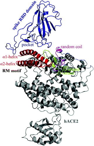 | ||
| Fig. 1 Cryo-EM structure of hACE2 in complex with the RBD domain of viral S protein (PDB: 6M17). The hACE2 RM motif consists of two α-helices, two β-strands and a random coil, in which the α1-helix is the major contact site of S protein. | ||
2.2. Conformational clustering analysis
Clustering data analysis was used to characterize the conformational space of the investigated systems during MD simulations.13 A total of 1000 frames were collected evenly over the stable trajectory of MD equilibrium simulations. The Average-Linkage algorithm22 was employed to produce a number of clusters using the pairwise root-mean-square-deviations between frames as a metric comparing all the atoms from residues of the simulated system under different conditions. Structural files were dumped for the average and representative structures from the clusters.2.3. Binding energetics calculation
Energetics calculations were performed over conformational snapshots derived from the collected frames using molecular mechanics/Poisson–Boltzmann surface area (MM/PBSA) and normal mode analysis (NMA). The total binding energy (ΔGttl) of hACE2 or its derived peptides to viral S protein can be decomposed as follows:23 | (1) |
3. Results and discussion
3.1. Context effect on the direct readout of hACE2-derived peptide binding to S protein
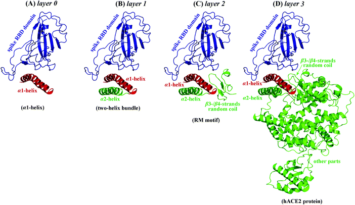
Structural analysis of the hACE2-spike complex (PDB: 6M17) revealed that in total 15 residues in the hACE2 RM motif are in contact with S protein, of which ten are from the α1-helix, one from the α2-helix, and the other four from β3-/β4-strands and random coil,9 suggesting that the hACE2 mainly engages the α1-helix to interact with the S protein. Therefore, it is suggested that the hACE2-spike interaction can be regarded as a PMI, where the interaction is mediated primarily by binding the α1-helical peptide of hACE2 to the globular RBD domain of the S protein. The nonbonded interactions across the complex interface of hACE2 protein with the spike RBD domain were analyzed using the in-house 2D-GraLab program26 based on the complex structure. In total four hydrogen bonds were identified and schematically shown in the ESI,† Fig. S1. It was found that (i) the complex interaction is not very hydrogen bond-rich and other nonspecific forces such as hydrophobic contacts and van der Waals packing may contribute significantly to the complex stability, and (ii) the four identified hydrogen bonds are all located in the α1-helical region of hACE2 protein, suggesting that this helix plays an important role in recognition specificity. According to our previous findings,14 a protein–peptide binding event can be divided into two thermodynamically independent steps, namely, direct readout (DR) and indirect readout (IR). The DR represents the intermolecular association between the S protein and α1-helical peptide, which is composed of interaction energy ΔEint and solvent effect ΔGsol (i.e. ΔUDR = ΔEint + ΔGsol), while the IR involves the flexibility, vibration and conformational constraint of the α1-helical peptide upon binding to S protein, which is roughly defined as the entropy penalty, if ignoring strain energy as the peptide is a very flexible molecule that usually has only a low strain energy (i.e. ΔUIR ≅ −TΔS).Here, we first discussed the context contribution to the DR of α1-helical peptide recognition by S protein. The context can be defined at different layers of the hACE2 protein components surrounding the α1-helix, namely, layer 0: nothing except the α1-helix; layer 1: α2-helix, which forms a two-helix bundle with the α1-helix; layer 2: α2-helix, β3-/β4-strands and random coil, which forms the RM motif of hACE2 with the α1-helix; layer 3: layer 2 plus other parts of hACE2, which forms the whole hACE2 protein with the α1-helix (Fig. 2). The complex systems of S protein with α1-helix plus different layers were picked up from the cryo-EM structure (PDB: 6M17); each of them was then subjected to 200 ns MD simulations, followed by post MM/PBSA analysis to characterize the DR effect involved in this complex binding, which can be decomposed into interaction energy (ΔEint) and solvent effect (ΔGsol) upon binding. As listed in Table 1, the α1-helix can interact effectively with S protein by itself (ΔEint = −156.7 kcal mol−1), which, however, would be largely counteracted by the solvent effect (ΔGsol = 118.3 kcal mol−1). Consequently, the DR energy of α1-helix binding to S protein is relatively weak (ΔGDR = −38.4 kcal mol−1). Further adding layers 1, 2 and 3 to the α1-helix can only modestly or moderately improve the DR of the system, with ΔGDR increased by ΔΔGDR = −7.4, −24.1 and −39.9 kcal mol−1, respectively. Although these layer sections are significantly larger than the α1-helix, their contributions to the DR energy increase of the α1-helix is considerably lower than or just comparable with the α1-helix itself (ΔUDR = −38.4 kcal mol−1), confirming that the α1-helix is the key recognition element of hACE2 by S protein, which is primarily responsible for mediating the direct intermolecular interaction between hACE2 and S protein. Even so, the context effect cannot be totally ignored. For example, although the contributions of layer 1 (α2-helix) and layer 2 (RM motif) contexts to the α1-helix DR are not very significant ΔGDR (ΔΔGDR = −7.4 and −24.1 kcal mol−1, respectively), layer 3 (other parts of the whole hACE2 protein) appears to have a substantial effect on the α1-helix DR (ΔΔGDR = −39.9 kcal mol−1), suggesting that the long-range contribution of protein context should also play an important role in α1-helix interaction with S protein.
| Context | System | Description | ΔEint | ΔΔEinta | ΔGsol | ΔΔGsola | ΔUDR | ΔΔUDRa |
|---|---|---|---|---|---|---|---|---|
| a Relative to α1-helix. | ||||||||
| Layer 0 | α1-helix + layer 0 | α1-helix | −156.7 | 0 | 118.3 | 0 | −38.4 | 0 |
| Layer 1 | α1-helix + layer 1 | two-helix bundle | −171.4 | −14.7 | 125.6 | 7.3 | −45.8 | −7.4 |
| Layer 2 | α1-helix + layer 2 | RM motif | −198.5 | −41.8 | 136.0 | 17.7 | −62.5 | −24.1 |
| Layer 3 | α1-helix + layer 3 | hACE2 protein | −227.5 | −80.8 | 149.2 | 23.6 | −78.3 | −39.9 |
3.2. Context effect on the indirect readout of hACE2-derived peptide binding to S protein
The α1-helix possesses a typical α-helical conformation in the full-length hACE2 protein, which matches well with the active pocket of S protein. In the context of hACE2 protein, the α1-helix is always maintained in an ordered state and forms a two-helix bundle with α2-helix that is integrated into the protein. In order to observe the conformational effect of protein context on the α1-helix and two-helix bundle, both are split from the hACE2 to explore their structural dynamics behavior in the solvent. It is known that the structural organization of a peptide in solution depends on the long-range interactions between its amino acid components as well as the equilibrium condition generated by the thermodynamic characteristics of interaction between the peptide and aqueous solvent under the given conditions of temperature and pressure.27–29 Here, in order to observe the conformational effect of protein context on the α1-helix and two-helix bundle, both are split from the hACE2 and then subjected to 500 ns MD simulations, during which their RMSD fluctuation profile and conformational change are recorded (Fig. 3). As might be expected, with the lack of the constraint from protein context the two peptides cannot maintain an ordered helical state and would gradually become disordered along the simulations. As can be seen, the α1-helix first exhibits a loose unfolding at its termini (250 ns) and then is totally disordered after the simulations (500 ns) (A). Therefore, the peptide is intrinsically disordered in the free state and cannot retain a helical conformation without the support of protein context. In addition, the two-helix bundle is gradually unstructured over the simulations, with a large unfolding at its two ends and the linker between the α1-helix and α2-helix (B). It is also suggested that the two-helix bundle is not fully disordered in the free state and partially structured in its local regions. As can be seen, the structured regions cover the central sections of the two helices; they are packed against each other to help maintain the local helical conformation, indicating a preliminary context effect that existed in the interior of the free two-helix bundle.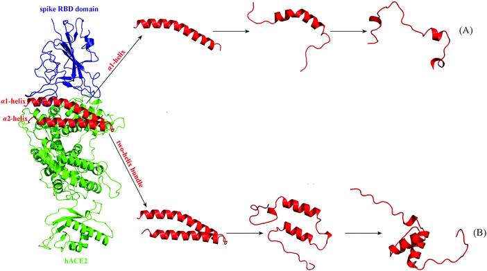 | ||
| Fig. 3 The α1-helix (A) and two-helix bundle (B) are split from the hACE2 complex interface with S protein and then subjected to 500 ns MD simulations. | ||
Configurational entropy has been widely used to characterize the conformational flexibility and freedom degree of the biomolecular system, which primarily includes vibrational and conformational effects. Here, the configurational entropy changes upon the binding of the α1-helix and two-helix bundle to S protein were calculated using NMA analysis based on their MD simulations in the full-length hACE2 protein and in the free state, which can be regarded as indirect readout (ΔUIR) and are generally larger than 0, an unfavorable entropy penalty to the binding. The calculated ΔUIR values are visualized as a histogram in Fig. 4. Evidently, both the peptides have a moderate entropy penalty upon binding to S protein when they are embedded in hACE2 protein, with ΔUIR = 21.8 and 28.9 kcal mol−1, respectively. However, the penalty effect would increase considerably when splitting the two peptides from hACE2 protein, with ΔUIR = 33.4 and 46.1 kcal mol−1, respectively, confirming that the protein context can impose a strong conformational constraint on the two peptides. In addition, the α1-helix has a generally smaller penalty than the two-helix bundle, no matter whether they are in the free state or in protein context; this is expected since the peptide flexibility is roughly increased linearly with their length30 and thus the penalty effect is also related to their size. In this respect, the protein context should play an essential role in (i) the restriction of intrinsically disordered peptides into native ordered conformation (helping conformational selection) and (ii) the reduction of entropy cost upon peptide binding (impairing the IR penalty effect); both would be favorable for the α1-helix recognition by S protein.
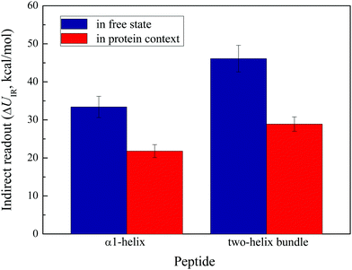 | ||
| Fig. 4 The indirect readout (ΔUIR) of α1-helix and two-helix bundle in the free state and protein context. | ||
3.3. Rational redesign of hACE2-derived peptides by optimizing indirect readout
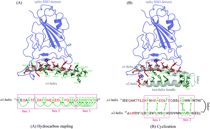
Traditionally, researchers used to improve peptide affinity by optimizing the intermolecular interaction (i.e. DR) between the peptide and its protein receptor; this is commonly fulfilled by residue mutation on the peptide sequence. However, a recent study found that the mutation would not only change the peptide affinity but also alter the peptide specificity, which may therefore cause the peptide to be untargeted.31–34 Instead of enhancing the favorable DR effect, we herein attempted to improve the peptide affinity by reducing the unfavorable IR effect. Since the IR is an intrinsic attribute of the peptide itself, its optimization would not change substantially the direct interaction of hACE2-derived peptides with S protein, thus storing the peptide recognition specificity and selectivity. As discussed above, the IR is primarily associated with the flexibility, conformation and entropy of peptides and hACE2 protein context can provide support to constrain the peptides into native conformation and to decrease the peptide flexibility and entropy cost upon binding to S protein.Here, we employed hydrocarbon stapling and cyclization strategies to fulfill this purpose for free α1-helical peptide (αHP) and two-helix bundle peptide (tBP) splitting from the full-length hACE2 protein, respectively. The αHP is natively a typical α-helix, of which the helical conformation can be readily restricted by stapling all-hydrocarbon bridges across its i and i + 4 residues; the tBP is natively a typical helical hairpin, which can be readily cyclized by adding a disulfide bridge across the α1-helix and α2-helix. Here, the peptide residues selected to anchor the hydrocarbon and disulfide bridges should point out of the active pocket of S protein so that chemical modification on them would not influence the direct protein–peptide interaction. In addition, the location of two anchor residues on the peptide sequence should be spanned by 4 amino acids (i, i + 4) for αHP stapling or be spatially vicinal to each other for tBP cyclization. Consequently, the schemes used to select the anchor residues for αHP and tBP peptides are shown in Fig. 5, in which the peptide residues that directly interact with S protein are shown as red sticks, and those that are considered as potential candidate anchors of stapling and cyclization are shown as green sticks.
For αHP peptide (Fig. 5A), a number of green anchor residues distributing along the peptide can be used to design hydrocarbon bridges; they can be divided into three box regions in terms of their location in the peptide sequence, that is, box 1, box 2 and box 3 cover the anchor residues in the N-terminal, middle and C-terminal regions of the peptide, respectively. According to the MD simulations of free αHP peptide (see Fig. 3A), the peptide is primarily disordered at its two termini, where the helical conformation is fast unfolded without context support during the simulations. Therefore, we considered to staple two hydrocarbon bridges separately in box 1 and box 3 of the peptide sequence. Box 1 only contains two residues (i.e. residues 22 and 26) that can be used to perform the stapling, while box 3 has a number of staplable residues (from Ala46 to Asn53). Therefore, four stapled αHP peptides (i.e. αHP1–4) were rationally designed, each including a stapling across residues 22 and 26 as well as a stapling within the residue range 46–53. In addition, the unstapled α1-helical peptide, termed αHP0, was used as the control (Table 2).
| Peptide | Form | Modification | ΔUDR | ΔUIR | ΔGttl |
|---|---|---|---|---|---|
| αHP0 | Unstapled | — | −38.4 | 33.4 | −5.0 |
| αHP1 | Stapled | [22 ↔ 26; 46 ↔ 50] | −36.8 | 28.6 | −8.2 |
| αHP2 | Stapled | [22 ↔ 26; 47 ↔ 51] | −37.4 | 28.0 | −9.4 |
| αHP3 | Stapled | [22 ↔ 26; 48 ↔ 52] | −36.9 | 26.3 | −10.6 |
| αHP4 | Stapled | [22 ↔ 26; 49 ↔ 53] | −37.1 | 27.3 | −9.8 |
| tBP0 | Uncyclized | / | −45.8 | 46.1 | 0.3 |
| tBP1 | Cyclized | [32 ↔ 76] | −47.1 | 31.9 | −15.2 |
| tBP2 | Cyclized | [36 ↔ 72] | −46.4 | 33.7 | −12.7 |
| tBP3 | Cyclized | [40 ↔ 69] | −45.8 | 34.2 | −11.6 |
For tBP peptide (Fig. 5B), its native conformation is folded into a helical hairpin, in which the α1-helix is primarily responsible for interacting with S protein. However, according to the above dynamic and energetic analyses the α2-helix can also play a context role to support the interaction of α1-helix with S protein, albeit it almost does not contact the protein. Therefore, we herein considered to constrain the helical hairpin conformation of free tBP peptide by adding a disulfide bridge across the α1-helix and α2-helix. As can be seen in Fig. 5B, the potential anchors include six residue pairs; they are divided into box 1 and box 2 in terms of their location. As a straightforward observation, the three box 1 residue pairs are good candidates since they can effectively constrain the peptide terminal conformation, whereas the three box 2 residue pairs are too close to the linker region between the α1-helix and α2-helix, which are unable to address conformational constraint on the peptide termini. Therefore, we herein considered to define a disulfide bridge at the three box 1 residue pairs, consequently resulting in three cyclized peptides (i.e. tBP1–3). In addition, the uncyclized two-helix bundle peptide, termed tBP0, was used as the control (Table 2).
Next, the direct readout (ΔUDR), indirect readout (ΔUIR) and total binding energy (ΔGttl) of unstapled αHP0 and stapled αHP1–αHP4 peptides as well as uncyclized tBP0 and cyclized tBP1–tBP3 peptides to S protein were calculated using MM/PBSA/NMA energetic analysis based on MD simulations, which are tabulated in Table 2 and compared in Fig. 6. As might be expected, the stapling and cyclization can considerably reduce the unfavorable IR effect of peptide binding, with ΔUIR decreasing by ∼6 and ∼13 kcal mol−1, respectively, whereas the peptide IR effect is influenced modestly upon stapling and cyclization, with ΔUDR change <2 kcal mol−1. Consequently, the peptide binding capability is improved substantially, with ΔGttl increasing from −5.0 kcal mol−1 for αHP0 to −8.2, −9.4, −10.6 and −9.8 kcal mol−1 for αHP1, αHP2, αHP3 and αHP4, respectively, and from 0.3 kcal mol−1 for tBP0 to −15.2, −12.7 and −11.6 kcal mol−1 for tBP1, tBP2 and tBP3, respectively. The tBP peptides seem to generally have a higher affinity than the αHP peptides, confirming that the α2-helix can serve as a local context to support the interaction of the α1-helix with S protein. The energetic decomposition also revealed that the stapling and cyclization can effectively improve the peptide affinity primarily by decreasing the unfavorable IR but not increasing the favorable DR, and the chemical modifications would therefore not shift the peptide recognition specificity for S protein.
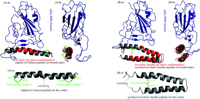
In order to examine conformational constraint imposed by hydrocarbon stapling and cyclization, the αHP3 and tBP1 peptides were selected and their structures in the free state and in complex with S protein were subjected to long-term MD simulations for sufficient relaxation and equilibrium. In the bound state, the equilibrium structures of the two peptides were superposed onto their respective native conformations (in full-length hACE2 protein context), as shown in Fig. 7(A-ab) and (B-ab), respectively. Evidently, the two peptides exhibit a good agreement with their native conformations (RMSD = 0.41 and 0.53 Å, respectively), confirming that, with stapling and cyclization, the two peptides can adopt a similar binding mode with them in the protein context to interact with S protein; stapling and cyclization can be considered as alternative strategies to mimic the constraint effect of protein context on peptide conformation, thus helping the proper recognition of peptides by S protein. In addition, the two free peptides can also be maintained in native conformations, despite their lack of context support. As seen in Fig. 7(A-c), the stapled αHP3 peptide is well folded into an α-helix in the free state, which is basically consistent with its native conformation in hACE2 protein. As compared to the totally disordered structure of unstapled αHP0 peptide (Fig. 3A), the stapled αHP3 peptide is considerably structured, with only minor unfolding in its middle region, where it has no hydrocarbon bridge imposed. Similarly in Fig. 7(B-c), the cyclized tBP1 peptide can spontaneously be structured into two-helix bundle conformations in the free state, with only moderate unfolding at its two ends, which is very ordered as compared to the uncyclized tBP0 peptide (Fig. 3B). The stapling/cyclization-imposed conformational constraint is expected to effectively minimize the unfavorable IR effect of peptides by reducing peptide flexibility and the entropy penalty upon their binding to S protein, and helping peptide pre-folding into their native state to facilitate the conformational selection by S protein.
Conflicts of interest
The authors declare that they have no conflicts of interest with the contents of this article.Acknowledgements
This work was supported by the National Natural Science Foundation of China (No. 31671361).References
- K. G. Andersen, A. Rambaut, W. I. Lipkin, E. C. Holmes and R. F. Garry, Nat. Med., 2020, 26, 450–452 CrossRef CAS.
- J. Zheng, Int. J. Biol. Sci., 2020, 16, 1678–1685 CrossRef CAS.
- I. A. Ysrafil, Diabetes Metab. Syndr., 2020, 14, 407–412 CrossRef.
- J. Shang, Y. Wan, C. Luo, G. Ye, Q. Geng, A. Auerbach and F. Li, Proc. Natl. Acad. Sci. U. S. A., 2020, 117, 11727–11734 CrossRef CAS.
- S. Xia, L. Yan, W. Xu, A. S. Agrawal, A. Algaissi, C. T. K. Tseng, Q. Wang, L. Du, W. Tan, I. A. Wilson, S. Jiang, B. Yang and L. Lu, Sci. Adv., 2019, 5, eaav4580 CrossRef CAS.
- S. Xia, M. Liu, C. Wang, W. Xu, Q. Lan, S. Feng, F. Qi, L. Bao, L. Du, S. Liu, C. Qin, F. Sun, Z. Shi, Y. Zhu, S. Jiang and L. Lu, Cell Res., 2020, 30, 343–355 CrossRef CAS.
- R. Yan, Y. Zhang, Y. Li, L. Xia, Y. Guo and Q. Zhou, Science, 2020, 367, 1444–1448 CrossRef CAS.
- E. Petsalaki and R. B. Russell, Curr. Opin. Biotechnol, 2008, 19, 344–350 CrossRef CAS.
- Y. Han and P. Král, ACS Nano, 2020, 14, 5143–5147 CrossRef CAS.
- N. London, B. Raveh, D. Movshovitz-Attias and O. Schueler-Furman, Proteins, 2010, 78, 3140–3149 CrossRef CAS.
- A. Kelil, E. D. Levy and S. W. Michnick, Proc. Natl. Acad. Sci. U. S. A., 2016, 113, E3862–E3871 CrossRef CAS.
- P. Zhou, Q. Miao, F. Yan, Z. Li, Q. Jiang, L. Wen and Y. Meng, Mol. Omics, 2019, 15, 280–295 RSC.
- P. Zhou, F. Yan, Q. Miao, Z. Chen and H. Wang, J. Biomol. Struct. Dyn., 2020 DOI:10.1080/07391102.2019.1709547.
- H. Yu, P. Zhou, M. Deng and Z. Shang, J. Chem. Inf. Model., 2014, 5, 2022–2032 CrossRef.
- H. M. Berman, J. Westbrook, Z. Feng, G. Gilliland, T. N. Bhat, H. Weissig, I. N. Shindyalov and P. E. Bourne, Nucleic Acids Res., 2000, 28, 235–242 CrossRef CAS.
- J. A. Maier, C. Martinez, K. Kasavajhala, L. Wickstrom, K. E. Hauser and C. Simmerling, J. Chem. Theory Comput., 2015, 11, 3696–3713 CrossRef CAS.
- P. Zhou, S. Zhang, Y. Wang, C. Yang and J. Huang, J. Biomol. Struct. Dyn., 2016, 34, 1806–1817 CrossRef CAS.
- C. Yang, S. Zhang, P. He, C. Wang, J. Huang and P. Zhou, J. Chem. Inf. Model., 2015, 55, 329–342 CrossRef CAS.
- C. Yang, S. Zhang, Z. Bai, S. Hou, D. Wu, J. Huang and P. Zhou, Mol. BioSyst., 2016, 12, 1201–1213 RSC.
- Z. Bai, S. Hou, S. Zhang, Z. Li and P. Zhou, J. Chem. Inf. Model., 2017, 57, 835–845 CrossRef CAS.
- C. Yang, C. Wang, S. Zhang, J. Huang and P. Zhou, Mol. Simul., 2015, 41, 741–751 CrossRef CAS.
- L. Saíz-Urra, M. A. Cabrera and M. Froeyen, J. Mol. Graphics Modell., 2011, 29, 726–739 CrossRef.
- Z. Li, F. Yan, Q. Miao, Y. Meng, L. Wen, Q. Jiang and P. Zhou, J. Theor. Biol., 2019, 469, 25–34 CrossRef CAS.
- P. A. Kollman, I. Massova, C. Reyes, B. Kuhn, S. Huo, L. Chong, M. Lee, T. Lee, Y. Duan, W. Wang, O. Donini, P. Cieplak, J. Srinivasan, D. A. Case and T. E. Cheatham, Acc. Chem. Res., 2000, 33, 889–897 CrossRef CAS.
- D. Case, Curr. Opin. Struct. Biol., 1994, 4, 285–290 CrossRef CAS.
- P. Zhou, F. Tian and Z. Shang, J. Comput. Chem., 2009, 30, 940–951 CrossRef CAS.
- M. Hao and H. A. Scheraga, Acc. Chem. Res., 1998, 31, 433–440 CrossRef CAS.
- H. Edelhoch and J. C. Osborne, Adv. Protein Chem., 1976, 30, 183–250 CrossRef CAS.
- M. Schaefer, C. Bartels and M. Karplus, J. Mol. Biol., 1998, 284, 835–848 CrossRef CAS.
- P. Zhou, S. Hou, Z. Bai, Z. Li, H. Wang, Z. Chen and Y. Meng, Artif. Cells Nanomed. Biotechnol., 2018, 46, 1122–1131 CrossRef CAS.
- B. S. Knudsen, J. Zheng, S. M. Feller, J. P. Mayer, S. K. Burrell, D. Cowburn and H. Hanafusa, EMBO J., 1995, 14, 2191–2198 CrossRef CAS.
- S. H. Gee, S. Quenneville, C. R. Lombardo and J. Chabot, Biochemistry, 2000, 39, 14638–14646 CrossRef CAS.
- A. Udyavar, R. Alli, P. Nguyen, L. Baker and T. L. Geiger, J. Immunol., 2009, 82, 4439–4447 CrossRef.
- H. Qian, P. He, F. Lv and W. Wu, Int. J. Biol. Macromol., 2019, 129, 13–22 CrossRef CAS.
Footnote |
| † Electronic supplementary information (ESI) available. See DOI: 10.1039/d0mo00103a |
| This journal is © The Royal Society of Chemistry 2021 |

