Engineered two-dimensional nanomaterials: an emerging paradigm for water purification and monitoring

Minxiang Zeng *a, Mingfeng Chen a, Dali Huang b, Shijun Lei a, Xuan Zhang c, Ling Wang *c and Zhengdong Cheng *abde
aArtie McFerrin Department of Chemical Engineering, Texas A&M University, College Station, TX 77843, USA. E-mail: glennzeng@tamu.edu; zcheng@tamu.edu
bDepartment of Materials Science & Engineering, Texas A&M University, College Station, TX 77843, USA
cSchool of Materials Science & Engineering, Tianjin University, Tianjin 300350, China. E-mail: lwang17@tju.edu.cn
dMary Kay O’Connor Process Safety Center, Texas A&M University, College Station, TX 77843-3122, USA
eDepartment of Chemical Engineering, Texas A&M University at Qatar, PO Box 23874, Education City, Doha, Qatar

Received 21st August 2020 , Accepted 4th November 2020

First published on 4th November 2020


Abstract

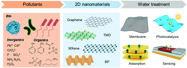
Water scarcity has become an increasingly complex challenge with the growth of the global population, economic expansion, and climate change, highlighting the demand for advanced water treatment technologies that can provide clean water in a scalable, reliable, affordable, and sustainable manner. Recent advancements on 2D nanomaterials (2DM) open a new pathway for addressing the grand challenge of water treatment owing to their unique structures and superior properties. Emerging 2D nanostructures such as graphene, MoS2, MXene, h-BN, g-C3N4, and black phosphorus have demonstrated an unprecedented surface-to-volume ratio, which promises ultralow material use, ultrafast processing time, and ultrahigh treatment efficiency for water cleaning/monitoring. In this review, we provide a state-of-the-art account on engineered 2D nanomaterials and their applications in emerging water technologies, involving separation, adsorption, photocatalysis, and pollutant detection. The fundamental design strategies of 2DM are discussed with emphasis on their physicochemical properties, underlying mechanism and targeted applications in different scenarios. This review concludes with a perspective on the pressing challenges and emerging opportunities in 2DM-enabled wastewater treatment and water-quality monitoring. This review can help to elaborate the structure–processing–property relationship of 2DM, and aims to guide the design of next-generation 2DM systems for the development of selective, multifunctional, programmable, and even intelligent water technologies. The global significance of clean water for future generations sheds new light and much inspiration in this rising field to enhance the efficiency and affordability of water treatment and secure a global water supply in a growing portion of the world.


image file: d0mh01358g-p1.tif

Minxiang Zeng

Minxiang Zeng received his BS Degree from the University of Science and Technology of China in 2014. He completed his PhD in Chemical Engineering from Texas A&M University in 2018. He is currently a Postdoctoral Researcher at the University of Notre Dame. His research interests include the self-assembly of two-dimensional materials, additive manufacturing of functional devices, and development of sustainable water technologies.

image file: d0mh01358g-p2.tif

Mingfeng Chen

Mingfeng Chen is a PhD student in Guangdong University of Technology and was a visiting student in Zhengdong Cheng's group at Texas A&M University. His research focuses on the phase transition of 2D nanoplatelets, orientation control of liquid crystals, ferronematic structure of magnetic nanoplatelets, and 2D material-based functional hydrogels.

image file: d0mh01358g-p3.tif

Dali Huang

Dali Huang is currently a PhD student in the Department of Materials Science & Engineering and Mary Kay O’Connor Process Safety Center at Texas A&M University. His research interests include 2D colloidal nanoparticles in photonic crystals, liquid crystals, and advanced functional materials.

image file: d0mh01358g-p4.tif

Shijun Lei

Shijun Lei completed his PhD at Tianjin University, under the supervision of Dr Beidou Xi from the Chinese Research Academy of Environmental Science and Dr Guanyi Chen from Tianjin University in 2020. He served as a visiting PhD student in the group of Dr Zhengdong Cheng at Texas A&M University from 2017 to 2019. He is currently a Postdoctoral Researcher in the group of Dr Yuan Hu at State Key Laboratory of Fire Science, University of Science and Technology of China. His research interests include carbon-based nanomaterials for oil–water separation, solar-powered water vapor generation, and solar distillation for seawater desalination/wastewater treatment.

image file: d0mh01358g-p5.tif

Ling Wang

Ling Wang is a full Professor at the School of Materials Science and Engineering of Tianjin University. He received his PhD degree in Materials Science from the University of Science and Technology Beijing in 2013. Prior to joining Tianjin University, he worked as a Postdoctoral Research Fellow at the Advanced Materials and Liquid Crystal Institute of Kent State University (USA), and Senior Research Fellow at the Artie McFerrin Department of Chemical Engineering of Texas A&M University (USA). His research interests focus on the design, synthesis and properties of smart soft materials, bioinspired materials and functional nanomaterials, and their emerging applications in diverse fields ranging from soft robotics, adaptive camouflage, and additive manufacturing to energy and safety issues.

image file: d0mh01358g-p6.tif

Zhengdong Cheng

Zhengdong Cheng is a Professor at the Artie McFerrin Department of Chemical Engineering at Texas A&M University. He received his PhD Degree in Physics from Princeton University, followed by Postdoctoral Fellowships at Harvard University, ExxonMobil Research & Engineering, and the Princeton Materials Institute. His research focuses on the self-organization of anisotropic particles, water treatment/monitoring, clean and sustainable energy systems, and the application of microfluidics in bio-encapsulation.


1. Introduction

Water is of paramount significance for all known forms of life, and plays an important role in human civilization, social development, and even space exploration.1–4 On Earth, water mainly exists in form of salty seawater, where fresh water only accounts for around 3% of the total water resources with mostly groundwater, icecaps, and glaciers (Fig. 1a and b).5–7 As the fresh water demand rockets with the growth of the global population, it has long been a grand challenge to provide clean water affordably and sustainably.8 Freshwater scarcity has been intensified by water pollution, which is either from naturally occurring contaminants or human activities. Various types of diseases and even lethal effects have been reported from common contaminants such as pesticides,9 pharmaceuticals,10 volatile organic compounds (VOCs),11 pathogenic microorganisms,12 and heavy metals and metalloids (Fig. 1c).13,14 In 2015, 29% of the global population lived without a safe freshwater system, and contaminated water causes nearly half a million diarrheal deaths annually.15,16 Moreover, water pollution not only prevails in underdeveloped water-stressed areas, but also has been increasingly witnessed in developed countries such as the United States. Thus, to address this grand challenge, many wastewater treatment technologies, such as coagulation, flocculation, screening, electrolysis, centrifugation, aerobic and anaerobic treatments, have been traditionally employed. However, most of these processes involve bulky/expensive systems or labour-intensive operation.17 Thus, there is a huge demand for innovations in conventional water processes globally, such as banning the use of Cl2 for water disinfection because of the potential generation of carcinogens.18
image file: d0mh01358g-f1.tif
Fig. 1 Challenges of freshwater scarcity and emerging water treatment technologies. (a) Water distribution on Earth. Credit: U.S. Geological Survey.47 (b) Estimated use of water in the USA (2015). Credit: U.S. Geological Survey.7 (c) Schematic illustration of water treatment processes leveraging the versatile behavior of 2D nanomaterials in membrane separation, adsorption, photocatalysis, and pollutant sensing.

The 21st century has brought to the field of water treatment exciting new opportunities in nanoscience and nanotechnology. Various nanosystems, including zero-dimensional (0D) nanoparticles,19,20 one-dimensional (1D) nanowires/nanorods,21–24 two-dimensional (2D) nanoplates/nanosheets,25–27 three-dimensional (3D) nanostructures,28 and their functional composites,29–34 have garnered research interest in water treatment and monitoring. Among them, 2D nanomaterials (2DM) are arguably the thinnest materials and may possess the largest surface-to-volume ratio,35 which promises ultralow material use, ultrafast processing time, and ultrahigh treatment efficiency.36,37 In addition, the unique shape of 2D nanomaterials also enables highly anisotropic physical and chemical properties compared to 1D and 3D nanosystems.35 Through control of their size, thickness, and nanostructure, 2D nanomaterials have led to innovations in multiple water treatment systems with exceptional catalytic, adsorptive, and separation performances. These systems include atomic-thin graphene membranes,38,39 high-efficiency oil adsorbents,40 ultrafast visible light photocatalysts,41,42 and other smart/self-healing structures.43,44 Thus far, numerous 2D nanomaterials have been designed and developed through the bottom-up method from small-molecule precursors or top-down exfoliation method from bulk crystals.45Fig. 2 presents several prominent examples of 2D nanomaterials that have emerged in the field of water purification/monitoring, such as graphene (Gr), graphitic carbon nitride (g-C3N4), hexagonal boron nitride (h-BN), metal carbides/carbonitrides/nitrides (MXene), transition metal dichalcogenides (TMDs), transition metal oxides (TMOs), black phosphorus (BP), metal organic frameworks (MOFs), covalent organic frameworks (COFs), 2D nanoclays, and many other 2D nanostructures. Graphene is a single-layer graphite, where the sp2 C atoms are arranged in a “honeycomb” lattice. Similar to graphene, h-BN and g-C3N4 can be viewed as doped graphene by B and N atoms, though much more defective sites are commonly found in g-C3N4. MXene and TMDs have an ultrathin nanostructure with the chemical formula of Mn+1XnTx (n = 1–3) and MY2, respectively, where M is a transition metal (e.g., Ti, Mo, W, and Zr); X is C and/or N atom; T is the surface termination element (O, OH, and F); Y is a chalcogen (S, Se, Te, etc.). TMOs, similar to TMDs, are transition metal-based 2D nanomaterials, although TMOs possess larger bandgaps. 2D MOFs are hybrid materials with metal ions or clusters interconnected by molecular linkers to form two-dimensional crystals. 2D COFs, known as “organic zeolites”, are porous crystalline polymer nanosheets, in which their organic molecular building units are covalently bonded. Clay nanoplates such as kaolinite, montmorillonite and layered double hydroxides (LDHs) are naturally occurring 2D nanomaterials with layers of octahedral hydroxide and/or tetrahedral silicate.46


image file: d0mh01358g-f2.tif
Fig. 2 Different 2D nanomaterials and their schematic nanostructures. Reproduced with permission.51 Copyright 2019, The Royal Society of Chemistry.

Despite the tremendous advancements in the literature regarding 2DM-based water treatment technology, to date, very few have made it to market, and thus the social benefit of 2DM in the water sector has yet to meet its initial promise.37,48–50 A question that this review seeks to answer is what are the practical barriers preventing 2D nanomaterials from large-scale applications, and more importantly, what are the next directions needed to be prioritized. In addition, the rich chemical diversity of 2D nanomaterials offers numerous opportunities, but also presents a great challenge for comprehensively understanding and predicting their performance in water cleaning, particularly considering their complicated physicochemical response to the aqueous environment in the colloidal state. Therefore, we aim to provide a framework to better understand how the fundamental structures of 2D nanomaterials affect their collective behaviour, material properties, and corresponding applications in water treatment. This review will elaborate these connections in various application-targeted scenarios and discuss the chemical properties and molecular interaction of 2DM at the colloidal level (interaction radius between approximately 1 and 1000 nm). Initially, the state-of-the-art advances in functional membrane-based water treatment technologies including ultrathin monolayer membranes, stacked multilayer membranes, and solar desalination membranes are discussed. The discussion on 2DM-based adsorbents is sequentially presented, highlighting recent developments in the adsorption of oil spills, organic pollutants, heavy metal ions, and other contaminants. Then, various types of 2DM-based photocatalytic systems leveraging light in water decontamination are introduced. Since sensors are used to quantitatively determine the effectiveness of water decontamination, we expand our discussion to the 2DM-related detection of pollutants. Finally, promising directions and emerging trends of 2DM technologies are considered together with their possible negative implications. This review aims to summarize the typical scenarios of targeted applications, help to elaborate the structure-to-property relationship of 2DM systems, and provide an impetus toward the fast development of this highly interdisciplinary research field involving chemistry, physics, hydrology, biology, microbiology, materials science, chemical engineering, civil engineering, ocean engineering, and others.

2. Functional membranes with 2D nanomaterials

Since the primary objective of water treatment is the removal of undesired components from water, functional membranes offer a physical barrier to separate these components from water based on their size or permeability.52–55 Also, since there is a typical trade-off between water permeability and selectivity, the development of functional membranes that maintain high permeability and high selectivity simultaneously is particularly challenging based on traditional membrane materials. It is well-known that the permeability is inversely proportional to the membrane thickness. Therefore, 2D nanomaterials with a thickness of one to a few atoms have emerged as promising building units for the development of next-generation separation membranes. In particular, introducing 2D nanomaterials into functional membrane design allows the realization of atomic-thin separation structures, which can selectively facilitate water molecules to pass through. These 2DM-based functional membranes can maintain relatively high permeation without serious sacrifice of the separation selectivity.56 Based on the phase of water that permeates the membranes (i.e., either gas phase or liquid phase), separation membranes can be categorized into membrane filtration (liquid phase of water) and solar membrane distillation (gas phase of water). Filtration membranes primarily rely on the design of the hydrodynamic size of their pores for size-selective separation (adsorption selective separation will be discussed in detail in the next section),57,58 while the design of solar desalination membrane involves several factors such as water evaporation enthalpy, hydrodynamic water transport, and light–matter interaction.59,60 In this part, we mainly introduce filtration membranes and solar desalination membranes based on engineered two-dimensional nanomaterials.

2.1. Filtration membrane

In size-selective separation, the purification of water by membranes relies on the effect of the excluded volume repulsion between the membrane pores and undesired pollutant molecules. Therefore, two main strategies (i.e., monolayer membranes and stacked multilayer membranes) have been studied to fabricate 2DM-based filtration membranes with a controlled pore size. Since the membrane permeation rate is inversely proportional to membrane thickness, the atomic thickness of 2D nanomaterials (e.g., tgraphene ≈ 0.34 nm) helps to achieve higher water permeability and less energy consumption than the commercial polyamide membranes (d ≈ 100 nm).61 Furthermore, since the structural voids of many 2D nanosheets (e.g., graphene or graphene oxide (GO)) are too small for water molecules to permeate, additional modifications are commonly required. By introducing nanopores with a controlled size by electron beam irradiation, ion/plasma etching, and electrochemical oxidation (as shown in Fig. 3a), membranes with a 2DM monolayer are promising alternatives to conventional separation membranes. Several examples of 2DM-based functional membranes are listed in Table 1.
image file: d0mh01358g-f3.tif
Fig. 3 Single-layer filtration membrane from 2D nanomaterials. (a) Popular strategies for the fabrication of single-layer nanoporous membranes. (b) Transmission electron microscopy (TEM) image of a nanopore on graphene. (c) TEM image of multiple graphene nanopores forming a porous structure. Scale bars in (b) and (c) are 2 and 10 nm, respectively. Reproduced with permission.73 Copyright 2008, the American Institute of Physics. (d) Nanopores in graphene by ion bombardment. Reproduced with permission.74 Copyright 2014, the American Chemical Society. (e) TEM image of MoS2 nanopore with a diameter of 5 nm. (f) Optical microscopic image of the single-layer nanoporous MoS2 device. (e and f) Reproduced with permission.75 Copyright 2016, Springer Nature. (g) Schematic illustration of water purification by nanoporous graphene. Reproduced with permission.76 Copyright 2012, the American Chemical Society. (h) Photo of nanoporous graphene membrane prepared by ion bombardment and oxidative etching. Reproduced with permission.74 Copyright 2014, the American Chemical Society. (i) Modeled MoS2 nanopore for membrane desalination. Reproduced with permission.77 Copyright 2016, the American Chemical Society.
Table 1 Examples of 2DM-based functional membranes for water treatment
2DM Pollutant Flux/permeance Efficiency Comments Ref.
a RB5 stands for reactive black 5; RhB stands for rhodamine B and LbL stands for layer-by-layer.
Gr KCl 106 g m−2 s−1 ∼100% Nanometer-sized pores in a graphene monolayer. 39
GO Congo red 8.4 kg m−2 h−1 99.5% Separates ions and organics. 62
GO RB5a 37.5 kg m−2 h−1 99.2% Surface zwitterionic-functionalized graphene oxide. 63
GO MgCl2 4.2 L m−2 h−1 93.9% LbL assembly of graphene oxide and polyethylenimine.a 64
GO MgSO4 2 L h−1 m−2 bar−1 91.1% Moderate salt rejection of NaCl (60%). 65
rGO Na2SO4 60.6 L m−2 h−1 93.57% Low salt rejection of NaCl (36.6%). 66
MoS2 Ag+ 160 L h−1 m−2 bar−1 ∼99% (0.2 ppm) High removal capacity of ∼4000 mg g−1. 67
MoS2 Evans blue 245 L h−1 m−2 bar−1 89% High structural stability up to 1.0 MPa. 68
WS2 Evans blue 730 L m−2 h−1 bar−1 90% Nanostrands forming fluidic channels in the WS2 membrane. 69
MXene Evans blue 1084 L m−2 h−1 bar−1 90% Colloidal Fe(OH)3 used as a distance holder. 70
h-BN CH2Cl2 2.65 L m−2 s−1 Effective treatment of oil-polluted water. 71
g-C3N4 RhBa 957 L m−2 h−1 bar−1 60% Integration of filtration and visible light photocatalysis. 72


Theoretical works have predicted that nanoporous monolayers are highly efficient in water treatment.76 Driven by a pressure difference, the nanoporous graphene monolayer forms a physical “mesh”, which selectively allows water molecules to pass through the membrane (Fig. 3g). Molecular dynamics calculations showed that nanoporous graphene may achieve an excellent water flux of 66 L cm−2 day−1 MPa−1 and salt removal efficiency of up to 99%.78 In these simulations, the nanopores of graphene monolayer play an essential role in water desalination, enabling H2O molecules to selectively pass through. By controlling the size and functional groups of the pores on 2D nanomaterials, some porous monolayers may achieve a water permeation rate several orders of magnitude higher than that of the current RO membranes (Fig. 3i).77,79

Experimentally, early studies on graphene have suggested that an electron beam (e-beam) may be used to generate nanopores on its 2D sheets with nanometer precision (Fig. 3b and c).73 Nanopores with a controlled size have been realized on graphene using the focused e-beam technique.80 O’Hern et al. fabricated nanoporous graphene monolayers via the transfer of CVD graphene on porous polycarbonate.81 Scanning transmission electron microscopy (STEM) showed that ∼83% of the pores were relatively small with a diameter of less than 10 nm. These nanopores on graphene enabled the selective transport of several molecules, including potassium chloride, tetramethylammonium chloride, and tetramethylrhodamine dextran (70[thin space (1/6-em)]000 Da). The graphene composite membrane enabled KCl and tetramethylammonium chloride to pass through, while rejecting tetramethylrhodamine dextran. To fabricate dense pores on graphene with controlled sizes, Surwade et al. developed a single-layer membrane via ambient pressure CVD (APCVD) on copper.39 During the fabrication process, nanopores were introduced on graphene and tuned via the oxygen plasma etching technique. The size-controlled nanopores improved the membrane selectivity and allowed water to pass through, while rejecting other species such as Li+, Na+, and K+. In particular, this monolayer membrane achieved excellent salt rejection (∼100%) with high water permeability. It is worth mentioning that the selectivity of the low-porosity membrane exceeded five orders of magnitude; however, it decreased at high porosities, possibly because of the size change of the nanopores. Under an external pressure, a flux of 106 g m−2 s−1 at 40 °C was achieved. Based on the estimated hole density of approximately 1/100 nm2, the permeation rate at one nanopore could reach a value of three molecules per picosecond.

The ion bombardment technique has also been applied for the fabrication of nanoporous graphene membranes (Fig. 3).82 Compared with conventional pore generation strategies, which mainly rely on direct oxidative processes,83–85 ion-bombardment-based techniques (for example, argon ion irradiation) can generate pores with a controlled size and density.82 For example, O’Hern et al. fabricated a monolayer graphene membrane with high-density nanopores via a two-step method (Fig. 3d).74 In the first step, gallium ion bombardment was used to generate isolated reactive defects. In the second step, these defects were enlarged using oxidative etching into permeable nanopores (0.40 ± 0.24 nm). After the two-step treatment, a nanoporous graphene membrane with a pore density of over 1012 cm−2 was prepared, as shown in Fig. 3h. Interestingly, the authors found that the nanopores by short-time etching were cation selective, which is likely due to the formation of negatively charged groups (e.g., –COOH). However, a longer oxidation time led to a larger pore size, and thus the electrostatic effect determining the K+/Cl selectivity weakened. Owing to the size exclusion effect of the graphene membrane, the authors also observed that the membrane pores allowed small-sized salt to pass through, while hampering the transport of larger organic species.

As an economic alternative, electrochemical oxidation has also been reported for the formation of nanopores on 2D nanosheets (Fig. 3). For example, Feng et al. established a facile approach to generate nanoholes on MoS2 nanosheets by applying electrochemical reaction (ECR).86 The formation of a nanopore starts at the lattice defects of MoS2 by the ECR process, which subsequently enlarges these defects into nanoscale holes by removing atoms or unit cells from single-layer MoS2. The step-like feature in the ionic current gives direct feedback on the pore dimensions, which can be calculated from a conductance vs nanopore size model.86 Interestingly, Feng et al. took advantage of the superior transport property of porous MoS2 and prepared single-layer MoS2 nanopower generators (Fig. 3e and f). The authors observed an osmotically driven current generated from a salt gradient with an estimated power density of up to 106 watts per m2, which is likely due to the high efficiency of the atomically thin membrane of MoS2.75

Although monolayer 2DM membranes possess several unique advantages including high water permeability, one practical barrier preventing their application is the difficulty of fabricating leak-free, large-area monolayer membranes with precise pores on an industrial scale.74,87,88 By contrast, commercial nanofiltration membranes made from polymers or polymer composites often have low production cost and high scalability. Thus, to reduce the fabrication cost and improve the scalability, novel separation membranes based on stacked nanosheets have been proposed (Fig. 4). Owing to their highly anisotropic feature, 2D nanosheets have been readily assembled into lamellar structures by external assembly methods (e.g., vacuum filtration and layer-by-layer (LbL) assembly), as shown in Fig. 4a.89 Owing to their solution processable feature, these fabrication methods are compatible with low-cost nanosheets, which can be synthesized in large quantities via liquid-phase exfoliation strategies. Therefore, the layered stacking of 2DM promises the low-cost and industrial-scale manufacturing of high-performance separation membranes.90,91


image file: d0mh01358g-f4.tif
Fig. 4 Common methods for the fabrication of multilayer filtration membranes from 2D nanomaterials. (a) Schematic illustration of nanoporous membranes based on stacked nanosheets. Reproduced with permission.89 Copyright 2014, AAAS. (b) MXene membrane fabricated by vacuum filtration. Reproduced with permission.70 Copyright 2017, Wiley-VCH. (c) GO-based separation membranes by LbL assembly. Reproduced with permission.79 Copyright 2013, the American Chemical Society.

During vacuum-assisted filtration, the nanosheet concentration increases continuously, and thus they tend to assemble into an ordered lamellar structure. Li et al. developed a lamellar MXene-based membrane using vacuum filtration (Fig. 4b).70 The MXene membrane exhibited a high water flux (>1000 L m−2 h−1 bar−1) and good rejection efficiency (>90%) for pollutants bigger than 2.5 nm. Han et al. reported a graphene-based nanofiltration membrane by filtering chemically converted graphene (CCG) on porous substrates.92 This graphene-based membrane demonstrated an excellent rejection efficiency (>99%) for dye molecules and inferior efficiency (20–60%) for salts. The authors attributed the separation performance to the effect of electrostatic interaction and size sieving. Tsou et al. showed that the type of deposition method can change the morphology of layered GO membranes.93 They compared the effect of the vacuum-, pressure-, and evaporation-assisted self-assembly techniques on the prepared GO membrane. Under the tested conditions, the authors found that pressure-assisted filtration techniques help to induce an ordered laminate structure of GO, leading to an excellent separation performance. In addition to the deposition methods, the deposition rate of GO through vacuum filtration is another crucial factor in the membrane performance.94 Specifically, it was found that a slow deposition of GO nanosheets can adjust the separation efficiency and water flux of GO-based separation membranes. The authors suggested that the interlayer spacing of GO laminates can be engineered by changing the deposition rate.

Since GO nanosheets are intrinsically hydrophilic, GO membranes that are solely vacuum-filtered may lack sufficient bonding between GO nanosheets, and thus may suffer from structural damage under the cross-flow conditions commonly encountered in industrial membrane operations.89 Thus, to improve the structural stability of 2DM stacked membranes, tremendous efforts have been made in LbL assembly strategy to introduce an interlayer bonding by electrostatic attraction or/and covalent linkage during membrane fabrication. Multiple molecular interactions, including coordination interactions, covalent bonding, charge-transfer interactions, and biologically specific interactions, have been increasingly investigated to facilitate LbL assembly for the stabilization of the functional multilayers.89,95 During the LbL process, 2D nanosheets are first dispersed in a stable colloidal dispersion, followed by dip, spin, or spray assembly. 2D graphene oxide nanosheets43,96,97 with rich charged groups have been widely reported for the fabrication of 2DM-based stacked membranes via LbL assembly techniques. For example, Hu et al. developed a separation membrane via the LbL assembly of graphene oxide, where 1,3,5-benzenetricarbonyl trichloride was used as the cross-linker (Fig. 4c).79 The authors found that the cross-linkers considerably improved the structural stability of the stacked GO monolayers. This GO membrane showed a water flux in the range of 80 to 276 LMH per MPa (depending on the number of GO layers), which is roughly 4–10 times better than that of many conventional nanofiltration systems.79

For most multilayer membranes, the interlayer spacing of stacked nanosheets has been regarded as one of the most crucial factors in determining their performance.89,99,100 Taking GO nanosheets as an example, the average interlayer spacing can reach ∼0.9 nm in an ionic solution due to the hydration effect, allowing ions with a hydrodynamic radius of 0.45 nm or smaller to permeate, while rejecting larger-sized molecules.101 Therefore, desalination requires that the interlayer distance of nanosheets to be <0.7 nm for the separation of hydrated Na+ (with a hydrodynamic radius of 0.36 nm) from water (Fig. 5a).89 This small spacing can be achieved by covalently bonding GO with small molecules to overcome the hydration force in an aqueous environment. By contrast, a medium-sized GO spacing (0.7 to 2 nm) can be useful for the separation of pollutants and fuel purification. If even larger-sized particles were adopted as spacers, GO membranes with an interlay distance of >2 nm can be obtained for possible application in biomedical processes (e.g., artificial kidneys and dialysis), in which the separation of large biomolecules is important. Long et al. investigated the transport properties of a metal-ion-decorated GO membrane.102 By using LiCl, NaCl, KCl, MgCl2, CaCl2, SrCl2, Ca(NO3)2, MnCl2, and YCl3 to functionalize the surface of GO, the authors systematically evaluated the effect of different salts on the permeation rate of organic molecules on the metal-ion-decorated GO membrane. They observed that the Ca2+–/Mg2+–GO membranes showed high selectivity for organic solvents, such as acetone, acetonitrile, and benzene. They attributed the selective permeability of the metal-ion-decorated GO membrane to the relative strength of the cation–π interaction, electrostatic force, and hydrogen bonding. In addition to the use of interlayer spacing, Wang et al. reported a 2D multilayer membrane with both artificial nanopores and self-supporting spacers (Fig. 5b).98 The authors used porous g-C3N4 nanosheets as building blocks and unstripped fragments as self-supporting spacers to fabricate a separation membrane, providing a water transport pathway that tolerates pH and pressure changes during the water treatment. A porous g-C3N4 membrane with a thickness of 190 nm showed a salt rejection of 90% with a decent water flux of 11.6 L m−2 h−1 bar−1. Recently, to suppress the swelling effect of 2D nanomaterials, which adversely changes their ion sieving performance, Ding et al. leveraged the intercalation of Al3+ ions for the fabrication of non-swelling MXene membranes. As shown in Fig. 5c, the as-prepared MXene membrane could effectively reject NaCl from seawater, while maintaining strong structural stability with high water fluxes (∼1.1–8.5 L m−2 h−1).99


image file: d0mh01358g-f5.tif
Fig. 5 Pore size engineering for the separation of different compounds. (a) Tunable separation capability of 2D multilayer membrane by tailoring the interlayer spacing. Reproduced with permission.89 Copyright 2014, AAAS. (b) Diagram showing water transport through g-C3N4 nanosheets via water nanochannels including intrinsic nanopores (P1), artificial nanopores (P2), and self-supporting spacers. Reproduced with permission.98 Copyright 2017, Wiley-VCH. (c) Schematic illustration of ion sieving with multilayer MXene membrane. Reproduced with permission.99 Copyright 2020, Springer Nature.

However, despite the significant advances in 2DM membranes, major challenges, e.g. large-scale fabrication of these membranes with long-term stability, still exist.103 Fouling is a common issue that prevents the long-term operation of many membranes since it increases the energy consumption and reduces the water recovery of desalination processes.58,104 Thus, to mitigate fouling/biofouling, several nanostructure-based materials have been proposed, such as effectively inactivating bacteria on direct contact.105,106 Combining GO nanosheets with other antibacterial nanomaterials (such as silver nanoparticles) appears to be a promising approach to ensure the inactivation of bacteria.107 In addition, self-cleaning and self-repair design may further facilitate the practical use of 2DM membranes in water treatment.108

2.2. Solar desalination membrane

The ability to generate desalinated water directly using sunlight, solar steam desalination has emerged as an encouraging solution toward addressing the increasingly pressing global freshwater shortage.109 Different from separation membranes, which are primarily based on size selective separation, desalination membranes leverage the difference in evaporation enthalpy between water and pollutants. Fig. 6a presents a typical solar desalination device, consisting of four components, including a seawater reservoir, solar desalination membrane, condenser, and freshwater collector. Among these components, the solar desalination membrane plays a key role in converting solar energy into heat for steam generation. Black 2D nanomaterials, such as graphene and MXene, have been investigated for solar water evaporation applications due to their outstanding photon-harvesting ability, low molar specific heat, and tunable thermal conductivity by chemical doping.60 In contrast to the popular lamellar design in filtration membranes, high porosity and even vertically aligned structure of 2D sheets have been proposed to promote the water transport. For example, a vertically aligned graphene sheet membrane (VA-GSM) was fabricated to convert solar energy into heat for desalinated water production.110 Compared to graphene with a lamellar alignment (Fig. 6b) and random structure (Fig. 6c), the VA-GSM (Fig. 6d) demonstrated unique advantages for water transport, and thus showed a higher solar desalination performance. The VA-GSM showed a high vapour generation rate of 1.6 and 6.3 kg m−2 h−1 under 1 and 4 sun illumination with an excellent efficiency of up to 86.5% and 94.2%, respectively.110 To maximize the use of solar energy and reduce thermal loss, efficient solar absorption and effective heat management have been regarded as the two essential factors for solar desalination.111–121 Efficient solar absorption has been achieved in a variety of advanced functional materials,116,119,120 and an ideal sunlight absorber will minimize the effect of reflectance or transmittance in a broad wavelength range. To date, several strategies have been investigated to develop highly efficient absorbers, including effective antireflection,120,121 strong light coupling for efficient absorption,122 and high density of optical modes.115,123 Due to the low-cost and environment-friendly feature of carbon-based nanomaterials, they are increasingly studied for membrane desalination and solar steam desalination.124–130 However, despite their ability to absorbing light by the π-band optical transitions, conventional graphitic materials suffer from roughly 5–10% reflection at the air–dielectric interface.131 Thus, to overcome this limitation, the design of nanostructured carbon materials appears to be an effective approach, e.g., porous graphene.132–136 These nanostructures can on-demand confine sunlight to small regions by resonant recirculation, increasing the interaction of light within the materials.137 Moreover, plasmon resonance techniques have also been studied to increase the absorption bandwidth.138–144 On the surface of a metal, light can trigger the collective excitation of electrons if its frequency matches the oscillation frequency of delocalized electrons. As shown in Fig. 6e–h,145 these excited hot electrons interact coherently with the incident electromagnetic field, leading to heat generation in materials.146–148 During this process, the hot carriers complete the energy redistribution through electron–electron scattering processes, generating a considerable amount of heat, which can be further transferred to surrounding materials by thermal conduction.149
image file: d0mh01358g-f6.tif
Fig. 6 Solar desalination membranes from 2D nanomaterials. (a) Schematic demonstration of a solar desalination device. Reproduced with permission.109 Copyright 2016, Springer Nature. Water transport channels based on lamellar (b), random (c), and vertically aligned structures (d). Reproduced with permission.110 Copyright 2017, the American Chemical Society. Plasmon-induced hot carrier phenomena of metallic nanoparticles involving photoexcitation (e), Landau damping (f), carrier relaxation (g), and subsequent thermal dissipation processes (h). Reproduced with permission.145 Copyright 2015, Springer Nature. (i) Schematic of plasmonic colloidosomes prepared from metal precursors. Reproduced with permission.150 Copyright 2018, Elsevier B.V. (j) Schematic of solar steam production in hierarchically nanostructured gel (HNG). Reproduced with permission.151 Copyright 2018, Springer Nature.

Experimentally, various plasmonic components integrated with 2D nanomaterials have been realized on membrane substrates. As presented in Fig. 6i, Wang et al. designed a solar desalination membrane using graphene oxide and a multiscale plasmonic nanostructure, in which Ag/Au nanocubes were used as assembly blocks to build plasmonic colloidosomes.150 Owing to the plasmonic coupling of the metal nanoparticles, the authors reported an excellent efficiency of 92% at 10 kW m−2 and steam generation rate of 12.96 kg m−2 h−1 for the solar desalination membrane.

Since the average solar energy on Earth is commonly around or even less than 1 kW m−2 (1 sun condition), effective heat management can be equally important. Although a higher efficiency can be achieved by applying a concentrated solar flux,113,152,153 it will be advantageous if high efficiency can be achieved under 1 kW m−2 or even weaker light intensity. Thus, to suppress undesirable heating loss such as water heating and parasitic thermal loss during solar steam generation,154 heat localization is an efficient strategy for solar energy utilization since it can restrict heat to small quantity of water for effective evaporation.112,155,156 In addition, from dispersed particles,157,158 to porous carbon films,154,159 and thin metallic absorbers,160–162 an increasing trend of using a confined region for localized heating has been observed for reducing heat loss to the bulk water and surrounding environment. Zhao et al. developed an energy confinement approach by hierarchical water pathways in a hydrogel (Fig. 6j).151 This hierarchically nanostructured gel (HNG) showed an excellent evaporation rate of 3.2 kg m−2 h−1, producing 18–23 litres of H2O per square metre of HNG per day from brine water. Li et al. reported a GO-based solar desalination device and found that an efficient water supply and suppressed heat loss could be achieved simultaneously by confining the water path to a 2D geometry (Fig. 7a and b). Due to the suppressed heat loss, the authors demonstrated that the exceptional performance of solar desalination can be independent of the amount of water and effective water evaporation can be achieved even in the absence of thermal insulation in the container.156


image file: d0mh01358g-f7.tif
Fig. 7 Strategies to improve the efficiency of solar desalination systems using 2D nanomaterials. (a and b) Enhanced thermal management by 2D path of water supply. Reproduced with permission.156 Copyright 2016, the National Academy of Sciences. (c) Schematic of bifunctional MoS2/C microbeads. Reproduced with permission.163 Copyright 2018, Wiley-VCH. (d) Infrared thermal images of chemically exfoliated MoS2/bacterial nanocellulose (Ce-MoS2/BNC) and reference samples. Surface temperature of bare water, Ce-MoS2/BNC, and bulk-MoS2/BNC aerogels (e) and camera image showing the water steam at the surface of Ce-MoS2/BNC bilayer aerogel under light irradiation (f). Reproduced with permission.166 Copyright 2018, Elsevier B.V. Photographic images and solar steam generation of the hydrophilic and hydrophobic 2D MXene membranes before (g and i) and after 24 h (h and j). Schematic showing the solar desalination process with (k) and without salt formation (l). Reproduced with permission.170 Copyright 2018, The Royal Society of Chemistry.

For 2DM-based solar desalination membranes, graphene, TMDs, and MXene have been demonstrated as advantageous photothermal materials. Since TMDs contain active sulphide bonds and can adsorb heavy metal ions, Li et al. showed that MoS2-based photothermal devices not only produce a high evaporation rate (1.95 kg m−2 h−1) at 1 sun, but also can reduce Hg from 200 to 1 ppb (Fig. 7c).163 In addition, chemical exfoliation of MoS2 enables a partial phase transition from the 2H phase to the 1T phase, leading to a higher visible light absorption of MoS2.164,165 Ghim et al. proposed chemically exfoliated MoS2 (Ce-MoS2) as a photothermal material for solar desalination (Fig. 7d–f).166 The authors used bacterial nanocellulose (BNC) as the support material for heat localization and demonstrated that the chemically exfoliated MoS2/BNC bilayer aerogel (96%) outperformed the unexfoliated MoS2/BNC (∼90%) and bare BNC (∼10%).166 In addition to TMDs, MXene has also been suggested as a promising photothermal material because of its high light-to-heat conversion efficiency.167 An MXene-based solar desalination membrane was developed by vacuum filtration on a porous substrate, exhibiting a decent solar conversion efficiency of 84% under 1 sun condition.168 However, despite the tremendous efforts in designing and developing 2DM-based desalination membranes, some technical challenges still exist. For instance, the long-term stability of many solar desalination membranes is limited in highly saline water because salt continuously accumulates on the top of the membrane, and thus decreases the light absorption and water supply, leading to a decrease in the water evaporation performance.109,156,169 To address this issue, Zhao et al. reported an MXene-based solar desalination membrane with a hydrophobic surface (Fig. 7g–l).170 Using trimethoxy(1H,1H,2H,2H-perfluorodecyl)silane as the surface modifier, this membrane showed strong hydrophobicity under high salinity conditions, and thus demonstrated good stability under one sun over 200 h.170 Another technical challenge comes from the generated water vapour, which can interfere with the light collection of solar desalination membranes. This issue may be circumvented by integrating solar steam generation with the conventional membrane distillation process using advanced design of distillation geometries.171 In conventional membrane distillation, a temperature difference is established between the input fluid and distilled water for generating a vapour pressure difference, which enables water vapour to pass through the membrane from the input side to the distilled side. By modifying the membrane with light-absorbing nanoparticles, solar energy can be used to replace conventional heating of the input fluid for the distillation process. This process integration of solar-driven steam generation with membrane distillation may profoundly improve the conventional membrane distillation process. Therefore, in addition to the material aspect, judicious system design and innovation, such as introducing Janus membranes or enhanced convection,172–177 will be highly desirable for off-grid, large-scale applications.

3. Functional adsorbents with 2D nanomaterials

Although functional membranes can be readily adapted into the workflow of wastewater treatment, adsorption-based technology, due to its low capital expense and simple procedure, is another promising option for wastewater treatment, particularly for oil spill mitigation and heavy metal collection. In particular, 2D nanomaterials have received broad interest for contaminant adsorption applications. Since the adsorption of pollutants is essentially an interfacial process, 2D nanomaterials with a large surface area that enables high levels of surface/interface interactions may possess exceptional adsorption efficiency. The plausible adsorption or interaction mechanisms and selective adsorptions are summarized in Fig. 8, including hydrophobic interaction, π–π stacking, H-bonding, ion/ligand exchange, Lewis acid/base and electrostatic interactions. In this section, the recent developments in 2DM-based adsorbents are discussed to address the challenges of oil spills, heavy metals, anions, and hydrophilic organic pollutants.
image file: d0mh01358g-f8.tif
Fig. 8 Main mechanisms of pollutant adsorption on 2D functional nanomaterials.

3.1. Oil spill adsorbents

Frequent oil spills have resulted in serious harm to the marine ecosystems and huge loss of valuable resources.178,179 Thus, to quickly eliminate oil contaminants, efficient and environmentally friendly oil adsorbents are needed. There are several desirable features for an ideal oil spill adsorbent as follows: (1) high-speed clean-up of oil spills to reduce the oil diffusion; (2) the ability to restore the oil spill at a high oil water separation efficiency; (3) negligible impacts to ocean creatures; (4) applicable in a large area and easy to operate; and (5) work effectively under severe ocean conditions. However, conventional oil recovery processes, such as the use of chemical dispersants,180 emulsifiers,181–183 solidifiers,184in situ burning185 and skimmers,186 have their own shortcomings, and thus new materials toward oil spill mitigation are desirable.

2DM-based advanced sorbents with oleophilicity and hydrophobicity have become promising candidates for fast oil spill mitigation. Taking advantage of conductive reduced graphene oxide (rGO) nanosheets, Ge et al. reported that a graphene-wrapped sponge (GWS) was designed for viscous crude clean-up (Fig. 9a and b).40 After applying a current, the Joule-heated graphene-wrapped sponge showed hydrophobic characteristics and oleophilic features (Fig. 9c and d). As shown in Fig. 9d, the highly viscous crude oil easily spread over the surface of the heated GWS. This can be explained by the fact that the Joule heating of GWS reduced the oil viscosity, which considerably increased the oil-diffusion rate in the GWS pores, speeding up the oil adsorption. It was observed that the adsorption time was shortened by 5.4% of the original time. In addition, compared with the unheated sponge, the heated one showed a much higher oil recovery rate due to the reduced viscosity of the crude oil.


image file: d0mh01358g-f9.tif
Fig. 9 Removal of oil pollutant by hydrophobic 2D nanomaterials. (a) Hydrophobic modification of 2DM, enabling effective oil adsorption. (b) Demonstration of the GWS fabrication process. (c) GWS device showing both hydrophobic and conductive features. The inset image demonstrates the water contact angle of GWS. (d) Oil droplet permeating behavior on GWS with (lower row) or without (upper row) external voltage. (b–d) Reproduced with permission.40 Copyright 2017, Springer Nature. (e) Structure of functional hydrophobic 2D COFs. (f) TEM image of COF nanosheets. Scale bar is of 200 nm. (g) COF-based composites showing strong hydrophobicity. (e–g) Reproduced with permission.187 Copyright 2017, The Royal Society of Chemistry.

Compared with hydrophobic structures, superhydrophobic materials with even higher contact angles (CA > 150°) have shown further improvement in adsorbing oil pollutants from water due to their exceptional water/oil selectivity. The functional groups and geometric morphology of a surface directly affect the wetting behavior of the testing liquids on it.188,189 In general, pristine nanosheets require surface functionalization to become superhydrophobic. Jayaramulu et al. reported a highly fluorinated graphene oxide, which exhibited a low oil CA of 0° and large water CA of 162°. Thus, this fluorinated graphene oxide showed high sorption selectivity and good absorbencies.190 Taking advantage of the versatile chemistry of 2D COFs (Fig. 9e–g), superhydrophobic COFs were used as coatings with excellent water repellency (water CA of 163°).187 It was found that the coatings still retained superhydrophobicity under harsh conditions from pH = 1 to pH = 14, and temperature from 0 °C to 80 °C. In addition to the low energy surface by chemical modification, high surface roughness also plays a role in improving the superhydrophobicity. According to Wenzel's model,189 the apparent contact angle, θ*, at the stable equilibrium state can be estimated by the equation: cos[thin space (1/6-em)]θ* = R × cos[thin space (1/6-em)]θ, where θ is the contact angle, which corresponds to an ideal flat surface. R is the surface roughness ratio of the sample.

Since most flat surfaces do not exhibit sufficient hydrophobicity by only low energy coatings,191,192 controlled manufacturing of the microstructures is critical to enhance surface roughness and achieve superhydrophobicity. For instance, Jiang et al. developed microstructures on plain surfaces by introducing polymers/carbon nanotubes.193,194 Yu et al. synthesized hydrophobic aerogels with 1D carbonaceous fibers195,196 and 2D graphene sheets197 as building blocks. Besides graphene oxides, superhydrophobic and superoleophilic h-BN composite materials have also been realized via the self-assembly of exfoliated h-BN nanosheets.71 The as-prepared composite material can be used to effectively separate various water/oil emulsions, in which droplets can be between 10−6 m to 10−9 m in size with exceptional separation efficiency, even under severe high flux rate.

To investigate the correlation between surface roughness and nanoscale building blocks, Du et al. designed several microstructures of various dimensions including zero, one, two dimensions and 1D–2D and 0D–1D composites on commercial melamine foams (Fig. 10a–g).198 Experiments and simulations were performed to explain the dimension-to-wettability relation. Among these composites, the authors found the highest surface roughness in the hierarchical structure of the 1D/2D nanomaterials, in which CNTs were conformally deposited on Co(OH)2 sheets. Indeed, the hierarchical structure allows larger surface interaction, and thus amplifies the intrinsic hydrophobicity. Since 2D nanomaterials possess a large surface area and tunable surface chemistry that enable facile co-assembly or post-functionalization with other building blocks, numerous hierarchical nanostructures made from 2D nanomaterials have been extensively demonstrated in the recent success in energy storage,199 high-performance catalysts,200 and bio-inspired structures.201–203 Taking advantage of the barrier properties of 2D zirconium phosphate nanoplates (ZrP), Cheng's group proposed a hierarchical structure with self-healing and superhydrophobic features (Fig. 10h and i).204 With the hierarchical porous platform textile fibers, the 2D ZrP nanoplatelets with a superhydrophobic coating could remove oil from an oil–water mixture with high separation efficiency (99.9%). After chemical etching, the hybrid film was capable of autonomously and repeatedly recovering its hydrophobicity since the layered structure protected the healing agent, which could gradually diffuse to the surface to reduce the surface energy.


image file: d0mh01358g-f10.tif
Fig. 10 Removal of oil pollutants based on hierarchical structure of 2D nanomaterials. (a) Schematic illustration of dimension control on microstructures. (b) Scanning electron microscopy (SEM) images of melamine foams with 2D particle coating. The inset shows the optical image of the coated foam. (c) SEM images and camera image (inset) of melamine foams with 1D/2D particle coating. (d) Stepwise modulation of the wettability of materials by engineering the dimension of the microstructures. (e) Water contact angles of various coated foams. (f) Water droplet possesses an effective roughness (Rf,e) close to the intrinsic surface roughness (Rf,0) on a less rough surface. (g) Effective roughness Rf,e much smaller than Rf,0 on an extremely rough surface. Reproduced with permission.198 Copyright 2016, Wiley-VCH. (h) Schematic illustration of hierarchical structure in ZrP-coated membrane. (i) Contact of various liquid droplets on membrane surface before and after the healing process, demonstrating self-healing behavior. Reproduced with permission.204 Copyright 2018, the American Chemical Society.

3.2. Hydrophilic organic pollutant adsorbents

Hydrophilic organic molecules, such as detergents, biphenyls, fertilizers, pesticides, and pharmaceuticals, are common water contaminants.205 Thus, to effectively remove these hydrophilic species from water, judicious design of 2DM with electrostatic attraction, van der Waals force, and hydrogen bonding are important approaches for developing 2DM-based adsorbents. In particular, water-soluble dyes have been extensively studied as model pollutants for investigating the decontamination behavior of adsorbents for the following two reasons: (1) dye pollution exists in the textile, rubber, plastic, printing and other industries, which easily results in water body contamination and (2) importantly, the ability of dyes to absorb a certain wavelength of light allows the pollutant concentration to be monitored in a simple, straightforward, and accurate manner. Therefore, the removal of dyes by 2DM-based adsorbents is highlighted herein.

Li et al. reported a water-stable graphene-like MOF (BUC-17) for the adsorption of dyes from simulated wastewater, showing excellent adsorption properties at room temperature.207 Ahmad et al. reported a 2D MOF material for the removal of rhodamine B (RhB), methyl orange (MO), methylene blue (MB), and Congo red (CR), as shown in Fig. 11.206 Adsorption was selective for the size, shape, and ionic strength of the dye. In addition, MoS2 nanosheets have also been developed into adsorbent materials, which have a good adsorption capacity for various organic dyes, including MB, MG, RhB, CR and fuchsin acid.208,209 Similar dye-adsorption behavior has been observed for other 2D nanomaterials, such as graphene-based adsorbents.210 The dye adsorption efficiency of 2D nanomaterials typically originates from van der Waals interaction, covalent bonding, or electrostatic attraction.208,209 Since colloidal MoS2 typically shows a negative charge, the adsorption of cationic species on MoS2 is much more effective than that of anionic molecules, which demonstrates the important role of electrostatic attractions.209


image file: d0mh01358g-f11.tif
Fig. 11 Removal of hydrophilic organic pollutants by 2D nanomaterials. (a) Crystal structure of 2D MOF (Cu(II)-5N3IP). (b) 2D MOF sheets in the (001) plane. One sheet is shown in red. (c) Solvent-accessible voids in the Kagomé-type structure. (d) SEM of 2D Cu(II)-5N3IP. (e) Adsorption of Congo red (CR) using Cu(II)-5N3IP. Reproduced with permission.206 Copyright 2018, The Royal Society of Chemistry.

One popular strategy to preserve the high surface area of 2D sheets in devices is to integrate 2DM on a 3D framework. A 3D sponge skeleton can adhere exfoliated 2D nanoplates by van der Waals interactions.211 Besides, a three-dimensional flower-like nanostructure of 2D MoS2 nanosheet was successfully fabricated using a hydrothermal process.209,212 The specific surface area of MoS2 nanoflowers could reach above 100 m2 g−1.209 The flower structure of molybdenum selenide with a good adsorption performance could be achieved by a self-assembly hydrothermal method.212 Hydrothermal strategies based on combining 2D nanomaterials and other functional materials are popular to prepare novel composite adsorbents. Magnetic Fe3O4 can be integrated with 2D nanomaterials to form heterostructures with strong adsorption capability, in which the composite can be easily recovered using a magnetic field.213

3.3. Heavy metal ion adsorbents

Heavy metals are toxic pollutants commonly found in industry wastewater and poorly managed water resources.214 Some of the heavy metal ions that are known to be harmful to humans include Cd, Cr, Hg, and Pb.215–220 Above the allowable limit,221 cobalt will result in vomiting, nausea, asthma, and carcinoma. Thus, to remove heavy metal ions, several strategies (including optimizing surface area and enhancing bonding strength with metal ions) have been proposed to fabricate efficient 2DM-based adsorbents.

Single-layer and few-layer 2D nanomaterials have exceedingly high surface areas, which can facilitate the adsorption of heavy metals. For example, 2D TMDs (e.g., MoS2) can be utilized as high-performance adsorbents for removing several heavy metal ions such as Hg 2+ and Ag+.49 This adsorption process often involves the Lewis soft acid and soft base interaction. For example, MoS2 functions as an adsorbent for the removal of Hg vapour or Pb2+, Cd2+, Zn2+, and Co2+ ions.49 Non-chalcogenide 2D nanosheets can also function as effective adsorbents for the removal of heavy metals. Chengchun et al. prepared h-BN-based structures, including porous BN whiskers,222 activated BN sheets,223 and three-dimensional carbon/BN structure, for the removal of heavy metals.224 The 2D fluorinated boron nitride nanoplates with high absorbency were demonstrated to be a capable adsorbents for heavy metal removal, and their water-cleaning capability was attributed to the surface defects and hydroxyl groups of BN.

Since many 2D nanomaterials are weakly bonded layered compounds by short range forces such as van der Waals forces, one of their unique characteristics is the possibility of fine-tuning their properties through tailoring their interlayer distance.225 Consequently, size-selective adsorption on the interlayer porosity of 2D nanomaterials is often achievable during the decontamination process. Wang et al. reported the synthesis of intercalated MXenes with a controlled interlayer distance.226 Their work showed that the larger interlayer space of hydrated Ti3C2Tx can enhance the adsorption performance with higher selectivity of radioactive U over other elements including Co, Ni, and Zn (Fig. 12a).226 This intercalation process of Ti3C2Tx was confirmed by SEM analysis (Fig. 12b–d). As an example of designed framework structure, Chen et al. fabricated two-dimensional silicate RUB-15, which exhibited a high adsorption energy and efficient regeneration of heavy metal ions such as UO22+ ions (Fig. 12e–g).227 Very recently, Ping's group successfully integrated MoS2 nanosheets with one-dimensional silk nanofibrils for the removal of heavy metal ions and dyes (Fig. 12h).228


image file: d0mh01358g-f12.tif
Fig. 12 Heavy metal removal by 2D nanomaterials. (a) Hydrated intercalation synthesis of Ti3C2Tx MXene for the efficient adsorption of U(VI). SEM images of Ti3C2Tx (b), Ti3C2Tx–NaOH (c), and Ti3C2Tx–DMSO (d). Reproduced with permission.226 Copyright 2017, The Royal Society of Chemistry. (e) Schematic illustration of the 2D silicate RUB-15 at a plane view (110). (f) TEM image of RUB-15. (g) Capture of UO22+ ions by RUB15 through ion exchange with TMA cations. Reproduced with permission.227 Copyright 2017, The Royal Society of Chemistry. (h) Pb2+ rejection efficiency of MoS2/silk nanofibril membranes. Copyright 2017, the American Chemical Society.228

In addition, natural multilayer clay-based 2D nanomaterials have exchangeable interlayer cations such as vermiculite and montmorillonite (MMT), which can be used as low-cost adsorbents for large-scale heavy metal ion removal.230–233 In addition to vermiculite, titanate nanoplates234 and vanadate-based layered 2D nanomaterials235 have been demonstrated to remove toxic heavy metals. After the adsorption/intercalation process, a structural change in the thin layers due to the sorption of metal ions was observed.234,235 To accelerate the interaction of the adsorbent and heavy metals, surfactants are commonly applied to treat 2D clays.236 Montmorillonite was functionalized with the anionic surfactant sodium dodecyl sulfate (SDS), resulting in excellent heavy metal adsorption. During this process, MMT was first intercalated with SDS surfactant, which enables 2D nanoclays with a larger negative ζ for better heavy metal adsorption.237

Owing to their unique structure of pore walls and skeletons, two-dimensional COFs provide a porous framework for adsorbing heavy metal ions from the aqueous phase. Huang et al. synthesized an Hg2+ adsorbent TAPB–BMTTPA–COF using 1,3,5-tris(4-aminophenyl)benzene (TAPB) and 2,5-bis(methylthio)terephthalaldehyde (BMTTPA) (Fig. 13).229 This framework structure includes bigger pores than many MOF systems. The methyl sulfide terminal groups have active sites to adsorb the Hg2+ element. This COF material could tolerate strong acids or bases, while maintaining its capability, reusability, and selectivity. Thus, these results demonstrate the huge research opportunities of COFs and other framework materials for heavy metal adsorption.


image file: d0mh01358g-f13.tif
Fig. 13 2D COF for heavy metal removal. (a) Top side views of 1,3,5-tris(4-aminophenyl)benzene (TAPB)–2,5-bis(methylthio)terephthalaldehyde (BMTTPA) COF with mesoporous channels. (b) Chemical structure of TAPB–BMTTPA–COF. (c) Cycle performance of COF on Hg2+ removal. (d) TEM image of TAPB–BMTTPA–COF. (e) 13C NMR spectra of pristine (black curve) and Hg2+-adsorbed TAPB–BMTTPA–COF (blue curve). (f) Capture efficiency of various metals. Reproduced with permission.229 Copyright 2017, the American Chemical Society.

3.4. Anion and oxyanion adsorbents

Many toxic elements, including some heavy metals, exist as an oxyanion in aqueous solution, forming negative charges, and thus the electrostatic attraction of some 2DM-based adsorbents should be redesigned. Arsenic has long been regarded as a deadly poison because of its lethality, and the toxicity of As derivatives decreases from left to right in the order of (1) arsenite, (2) arsenate, (3) monomethyl arsenic acid, and (4) dimethyl arsenic acid.205 In addition, NO2/NO3, F, and C2O42− also have some detrimental effects at high concentration. For example, high concentrations of nitrate (NO3) can cause children to develop blue infant disease.238 Some anions such as SO42−, PO43−, and Cl in high concentrations can change the taste of water and have adverse effects. Thus, to eliminate anions and oxyanions, ion exchange, anion intercalation, and other mechanisms have been adopted for the design and development of 2DM-based adsorbents.

The large surface areas of thin 2D nanomaterials not only accelerate the adsorption kinetics, but also result in higher adsorption capacity and chemical reactivity compared to many other traditional nanomaterials. Another characteristic of layered 2D nanomaterials (such as 2D clays) is their ion exchange properties, which are related with their interlayer space. For instance, a layered double hydroxide (LDH) is a 2D clay consisting of brucite-like layers with positive charges. It has been reported that LDHs were utilized for the removal of contaminants such as PO43−,241 F,242 and an anionic herbicide.243 The adsorption of LDHs toward anions mainly relies on their cations, and thus a high content of trivalent cations in LDHs typically leads to a good adsorption ability.241 On the other hand, some molecules such as sodium dodecyl sulfate can also be incorporated into the LDH gap by ion exchange.244 It has been reported that chloride ions can be embedded in LDHs to effectively remove As(V) from polluted groundwater.245 The As(V) adsorption capacity of LDHs was about 90 mg g−1, while >98% of As was eliminated from contaminated water. It was shown that the MoS42− intercalated LDH (MoS4-LDH) has excellent removal efficiency (>93%) of As(V) in 60 s.246 This adsorption/intercalation of HAsO42− into LDH induced a decrease in the interlayer distance of MoS4-LDH.

Furthermore, the efficient removal of As(III) on Fe(III) modified montmorillonite was reported for anion removal.239 As shown in Fig. 14a, the authors employed a low-temperature clay intercalation approach using FeCl3 followed by treatment of NH4OH solution. This nanoclay adsorbent showed a fast arsenite removal with 55% of As(III) removed within 30 s. Recently, two-dimensional MOFs (e.g., FIR-53) with abundant nanotubular channels were used for the removal of Cr2O72−, in which FIR-53 presented quick adsorption kinetics, high regeneration ability, and high selectivity for Cr2O72− over Cl, Br, and NO3 ions.247 However, the selectivity showed a decrease upon the introduction of coexisting ions. It is noteworthy that some 2DM adsorbents were found to be effective to remove same-charged ions. For example, in addition to their good adsorption performance for cations, 2D MXene (Ti3C2Tx) nanosheets also exhibited excellent removal capacity for anions Cr2O72− (Fig. 14b and c).240 The XPS study demonstrated that Cr(VI) ions were reduced when adsorbed on the surface of Ti3C2Tx. This is likely due to the metallic property of MXene and high oxidative capability of Cr2O72−. Additional examples of 2DM-based adsorbents can be found in Table 2.


image file: d0mh01358g-f14.tif
Fig. 14 2D nanoadsorbents for inorganic anion removal. (a) Schematic illustration of the modification of MMT with iron(III) species. Reproduced with permission.239 Copyright 2018, Elsevier B.V. (b) TEM image of functionalized Ti3C2Tx nanosheets after exfoliation. (c) Dosage effect of functionalized Ti3C2Tx nanosheets on the removal of Cr(VI) (pH = 5). Inset is a photograph showing the change in colour in the case of 0.4 g L−1 MXene nanosheets. Reproduced with permission.240 Copyright 2015, the American Chemical Society.
Table 2 Various 2DM-based adsorbents for water treatment
Pollutant Composite Synthesis Time Removing capability Ref.
Pb(II) GO/poly(N-vinylcarbazole) Sonication mixing 24 h 887.98 mg g−1 248
Pb(II) MXene (Ti3C2Tx) Chemical etching 2 h ∼30 mg g−1 249
Pb(II) GO/modified silica Multi-step method 24 h 333 mg g−1 250
Pb(II) MoS2/CeO2/biochar Pyrolysis 24 h 263.6 mg g−1 251
Cd(II) GO/polyamidoamine Grafting method 360 min 253.81 mg g−1 252
Cu(II) GO/chitosan Electrostatic self-assembly 90 min 425 mg g−1 253
As(III) GO/MnFe2O4 In situ growth 120 min 146 mg g−1 254
U(VI) GO/amino siloxane oligomer Cross-linking reaction 30 min 310.6 mg g−1 255
Cr(VI) rGO/NiO In situ growth 198 mg g−1 256
RhB MoS2-glue sponge Dip-coating method 60 min 127.39 mg g−1 257
Rh6G MoS2/CuS Hydrothermal method 240 min 211.18 mg g−1 258
Methylene blue 1T-WS2 Hydrothermal method 2 min 210.2 mg g−1 259
Pump oil Polyimide/MXene Freeze dry 30 min 5.8 g g−1 260


In addition to the judicious design of surface interactions within the aqueous phase, the colloidal stability of 2DM-based adsorbents is important to prevent the aggregation of 2D nanomaterials, which inevitably will reduce their effective surface area for adsorption. When colloidal 2D nanomaterials are dispersed in water, the Derjaguin–Landau–Verwey–Overbeek (DLVO) theory can provide useful information on the trend of nanoparticle aggregation.51 According to the DLVO theory, the design of electrostatic repulsion and van der Waals attraction may lead to excellent colloidal stability of 2D nanomaterials in water, maintaining the effective adsorption of aqueous pollutants during the contact time. Similar to aggregation, gravity can also negatively impact the colloidal stability of 2DM nano-adsorbents due to the sedimentation (ρparticle > ρwater) or creaming effect (ρparticle < ρwater). For a spherical particle, the terminal sedimentation (creaming) velocity, U0, can be estimated using Stokes' law, in which U0 is related to the particle size, density, gravity force, and solvent viscosity.51,261

image file: d0mh01358g-t1.tif
where D is the particle diameter, and μ, g, and Δρ represent the viscosity, gravity constant, and density difference between the solution and particles, respectively.

In pursuit of developing high-performance 2DM adsorbents, the terminal sedimentation rate needs to be prudently considered since fast sedimentation leads to the removal of adsorbents before effective decontamination, while ultra-slow sedimentation may increase the recovery cost in separating used adsorbents from water. More importantly, the judicious design of the dimensions of nano-adsorbents and comprehensive understanding of their nanosheet behaviour in colloidal dispersions can be crucial for the practical application of pollutant adsorbents in lakes and offshore conditions, where homogeneous stirring is often inaccessible. Overall, considering the broad chemical and physical diversity of 2D nanomaterials, more research is needed for the understanding of their molecular levels and adjustment of their surface chemistry to precisely control their adsorption behaviour.

4. Photocatalytic wastewater treatment with 2D nanomaterials

To solve environmental issues with minimal energy input, photocatalysis using solar energy to clean water has been researched broadly and considered as one of the most sustainable approaches.262–264 Generally, photocatalysis involves one or several photo-reaction processes catalysed by a photocatalyst, where free electrons are excited to the conduction band (CB) and holes are left in the valence band (VB). These electron–hole pairs play an important role in photocatalytic applications, including water splitting, pollutant degradation, and carbon dioxide reduction.265,266 However, the excited photogenerated charge carriers are not stable and easily recombine, which reduces the conversion efficiency of photocatalysis.264 Due to the unique structures of 2D nanomaterials and their nanocomposites, 2DM-based photocatalysts are encouraging candidates to provide signature features such as diverse host–guest species, abundant surface-active sites, and high specific surface areas.267 Recently, several 2DM-based photocatalysts, including graphene, carbon nitride, metal oxide and chalcogenide nanosheets, have been investigated for various applications. According to the pollutant type, the application of 2DM-based photocatalysts can be mainly divided into three main categories: biological disinfection, organic decontamination, and inorganic pollutant removal. The nanosheet size/thickness, chemical structure, and edge morphologies of 2D nanomaterials are important for applying these materials in photocatalytic processes. Here, we consider the synthetic strategies of 2DM-based photocatalysts and critically evaluate their performance in the above categories. Other efficient catalyst systems (e.g., electrocatalysts) have been developed for water treatment, and details can be found in recent topical reviews.268–274Table 3 presents several recent examples of 2DM-based photocatalysts for water treatment.
Table 3 Various 2DM-based photocatalysts for water treatment
Pollutant Photocatalyst Synthesis Design Photocatalytic conditions
Formaldehyde TiO2/graphene composites Solvothermal method 0D/2D UV light in air275
Methylene blue ZnO/rGO Ultrasonic treatment 0D/2D UV light irradiation276
Methylene blue CdSe/TiO2 Hydrothermal method 0D/2D Visible light irradiation277
Rhodamine B Carbon quantum dots/TiO2 nanosheet Assembly method 0D/2D Visible light irradiation278
Rhodamine B TiO2 nanorod/SnS2 nanosheet Hydrothermal method 1D/2D UV and visible light irradiation279
Methyl orange Ag2CO3/g-C3N4 nanosheets Ex situ assembly 0D/2D visible light irradiation280
Methyl orange Bi4Ti3O12 nanofiber/BiOCl nanosheet Electrospinning and solvothermal 1D/2D Xenon lamp Irradiation281
Methyl orange Copper doped SnS2 nanosheet/rGO Cysteine-assisted hydrothermal 2D/2D Visible light irradiation282
Cr(VI) CdS/rGO Microwave 0D/2D Visible light irradiation283
Cr(VI) g-C3N4/N-doped graphene/MoS2 Multi-step method 2D/2D Visible light irradiation284
NH3 WS2/MoS2 Liquid exfoliation 2D/2D Visible light irradiation285
CO2 TiO2 nanosheets and graphene Layer-by-layer assembly 2D/2D Xenon lamp Irradiation286
Bacteria (E. coli) AgBr/g-C3N4 Adsorption–deposition method 0D/2D Visible light irradiation287
Bacteria (E. coli) Ag/ZnO/g-C3N4 Adsorption–deposition method 0D/2D Visible light irradiation288
Bacteria (S. aureus) MnO2/g-C3N4 Multi-step method 2D/2D Visible light irradiation289
Virus (bacteriophage MS2) Ag/MoS2 Multi-step method 0D/2D Low-power LED290


4.1. Photocatalysts for bio-disinfection

Virus and bacteria are pathogens that can cause diseases and threaten human life. Water pollution due to these pathogens is a common phenomenon in some developing countries, and thus the decontamination of water is paramount. By using strategies such as bandgap engineering and nanocompositing, several 2D nanosystems have been investigated and developed for the removal of these contaminants from water.287–290 During a typical photocatalytic process, the photo-exited charge carriers help to generate reactive oxygen species (ROS), which can deactivate bacteria (Fig. 15a and b).291 For example, as an extension of conventional TiO2 photocatalysts, TiO2–Bi2WO6 nanosheets (TBWO) were synthesized by Jia et al. using a two-step hydrothermal method. Bi2WO6 was grafted on the surfaces of TiO2, which has the ability to absorb visible light. Therefore, TBWO could induce the cell rupture and inactivate E. coli due to visible-light-driven photocatalysis.292 Liu et al. fabricated a novel MoS2 (FLV-MoS2) film by vertically aligning several layers of 2D platelets, which could absorb the whole spectrum range of visible light (which contains about 50% of solar energy) in the water disinfection process (Fig. 15c–e).42 To increase the bandgap of MoS2, the domain size was decreased. The results showed that the bandgap increased from 1.3 to 1.5 eV, which enabled FLV-MoS2 to inactivate bacteria in water by generating reactive oxygen species (ROS), enabling the deactivation of >99.999% bacteria in 20 min. In addition to metal oxides and dichalcogenides, a metal-free 2D g-C3N4 nanosheet photocatalyst was developed via a two-step template-free method (Fig. 15f–k).293 This porous g-C3N4 nanosheet possessed abundant surface reactive sites and better capability of charge transfer than the bulk g-C3N4. Therefore, the performance of organic pollutant degradation and visible-light-driven photocatalytic disinfection of the nanosheets were significantly improved. The unexfoliated g-C3N4 could only inactivate some E. coli within 4 h, whereas the g-C3N4 nanosheet could inactivate all the E. coli cells completely, confirming the advantage of bandgap engineering based on the dimension-control method.293
image file: d0mh01358g-f15.tif
Fig. 15 Photocatalysts based on 2D nanomaterials for the removal of bacteria. (a) Simplified schematic showing the photocatalytic process of 2DM photocatalysts. (b) Examples of the antibacterial mechanism of Ag/ZnO photocatalyst. Reproduced with permission.291 Copyright 2017, Elsevier B.V. (c) Schematic illustration of bacteria deactivation by FLV-MoS2 in water. (d) TEM image of FLV-MoS2 showing the vertically standing layers. (e) Disinfection performances of FLV-MoS2 compared with that of bulk MoS2 and horizontal nano-MoS2. (c–e) Reproduced with permission.42 Copyright 2016, Springer Nature. Photos and TEM images of E. coli before (f and i) and after photocatalysis for 2 h (g and j) and for 4 h (h and k) using porous g-C3N4 nanosheets. (f–k) Reproduced with permission.293 Copyright 2017, the American Chemical Society.

Compared with bacteria, viruses have been regarded as more dangerous pathogens due to their higher illness risk and lower infectious dose under a similar level of exposure.294 For example, exposure to one rotavirus particle can increase the chance of infection by up to 31%.295 In addition, viruses possess extremely small sizes and unique surface properties, and thus physical removal methods are often unavailable. For example, in water treatment, granular activated carbon adsorption was reported to be effective in removing protozoan parasites and bacteria, but not viruses.296 To deactivate viruses via photocatalysis, extensive efforts have also been made using a variety of 2D nanomaterials. Li et al. evaluated the virucidal performance and mechanism of g-C3N4.297 During the photocatalysis, the authors found that a shape distortion, protein oxidation, and RNA damage occurred, leading to virus death (Fig. 16a–d). The virus inactivation performance of g-C3N4 was also comparatively analysed with other visible-light-driven photocatalysts, namely TiO2, Bi2WO6 and Ag@AgCl, indicating that surface plasmon resonance and dissolved Ag+ of Ag nanoparticles may further improve the performance (Fig. 16e and f). Inspired by the discovery of the photocatalytic reduction of GO to rGO by WO3,298–300 Akhavan et al. added GO nanosheets to WO3 films to construct rGO–WO3 films, in which the composite film was shown to decrease the virus concentration from 2 × 10⁶ PFU mL−1 to <5 PFU mL−1 within 3 h under visible-light irradiation (Fig. 16g).301 To accelerate the disinfection process, Zhang et al. used the response surface methodology to optimize the operational conditions for photocatalysis.302 The optimal conditions of 135.40 mg L−1 (photocatalyst), 24.05 °C and 199.80 mW cm−2 (light power), were obtained with the disinfection time of 240 min (Fig. 16h–j). Bio-disinfection of 2DM by other mechanisms such as the photothermal effect and Fenton reaction can be found in another recent review.303


image file: d0mh01358g-f16.tif
Fig. 16 Photocatalysts based on 2D nanomaterials for virus removal. (a) Main mechanisms of virus disinfection by 2D photocatalysts, including RNA damage (b), protein oxidation (c), and shape distortion (d). (e) g-C3N4 photocatalyst for viral disinfection under visible light. (f) Virucidal performance of g-C3N4 compared with other nanomaterials. (b–f) Reproduced with permission.297 Copyright 2016, Elsevier B.V. (g) Virucidal performance of graphene–WO3 composites. Reproduced with permission.301 Copyright 2012, the American Chemical Society. (h) 3D response surface and (i) 2D contour plots of viral disinfection with light intensity (X1) and g-C3N4 loading (X2). (j) 2D contour plots of inactivation efficiency with light intensity (X1) and temperature (X3). (h–j) Reproduced with permission.302 Copyright 2018, Elsevier B.V.

4.2. Photocatalysts for removing organic pollutants

Compared with bio-disinfection, which only requires the deactivation of cell or proteins, removing organic pollutants requires the formation of sufficient reactive oxygen species that can destroy or mineralize organic contaminants completely.304 To develop an effective photocatalyst, the band structures of 2DM need to be carefully designed since they play a crucial role in the redox potentials of photogenerated electron–hole pairs.305 Compared with bulk 2D materials (Fig. 17a), monolayer or few-layer 2D nanoparticles (such as MoS2) possess a larger bandgap to shift the band edge position to generate a redox potential, which favours ROS, even though the photocatalysis of these nanosheets sometimes only uses a narrow range of the solar spectrum.49 Thus, to develop photocatalysts that utilize light in the desired wavelength range, several strategies, such as heterostructuring, element doping, and size control, have been utilized to modulate the band structure of 2D nanosheets.306 To date, 0D nanoparticles and 1D nanorods/nanowires/nanotubes have been composited with 2D nanomaterials.281,307–313 For example, some 2DM-based composites have been achieved by incorporating metal-containing nanoparticles, such as Pt,310 RuO2,314 Au,315 Rh(OH)3,316 into the space between the layers of metal oxide nanosheets. Recently, a new method that can intercalate metal nanoclusters, such as Pt, Pd, Rh, and Ru, into KCa2Nb3O10 nanosheets, was reported by Oshima et al.317 The prepared colloidal KCa2Nb3O10 nanosheets with intercalating Pt exhibited a high photocatalytic performance due to the co-catalysts of the well-dispersed Pt nanocluster between the 2D layers.
image file: d0mh01358g-f17.tif
Fig. 17 Photocatalysts based on 2D nanomaterials for the removal of organic pollutants. (a) Adsorption spectra of various materials compared with sunlight spectrum. Reproduced with permission.49 Copyright 2017, the American Chemical Society. (b) Schematic diagram of the growth mechanisms of hybrid nanocomposites. The black and silver colors represent MoS2 and TiO2, respectively. (c) SEM images of hybrid 2D composites of TiO2@MoS2 (1 and 2) and MoS2@TiO2 (3 and 4). (b and c) Reproduced with permission.322 Copyright 2015, The Royal Society of Chemistry.

Compared with 0D/2D and 1D/2D nanocomposites, 2D/2D heterostructures have unique coupling hetero-interfaces, facilitating the separation and transfer of photogenerated charge carriers, thereby leading to higher photocatalytic activity. Numerous studies have indicated that the properties of catalysts can be modulated by controlling the morphology and size of the semiconducting component in hybrid systems. For example, a systematic comparison study between 1D CNT–TiO2 composite photocatalysts and 2D graphene–TiO2 was conducted by Zhang et al.318 The interfacial contact of graphene–TiO2 sheets was shown to be superior in comparison with that of the CNT–TiO2 heterostructure.318 Yang et al. reported a comparative study of a TiO2 photocatalyst, where TiO2–graphene, TiO2–carbon nanotube, and TiO2–fullerene (C60) nanocomposite photocatalysts were prepared.319 The authors found that the three types of TiO2–carbon photocatalysts followed an analogous oxidation mechanism for the photocatalytic oxidation of benzyl alcohol. In addition, a graphene-based nanocomposite with different shapes of TiO2 nanomaterials, including TiO2 nanosheets, nanotubes, and nanoparticles, was developed.320 It was discovered that compared with the other two composites, the TiO2 nanosheet–rGO heterostructure possessed better photocatalytic performance for the degradation of 2,4-dichlorophenol and RhB. Through the analysis of the interfacial charge transfer kinetics, the 2D/2D hybrid structure was demonstrated to possess strong electronic coupling effects. The coupling effects can promote charge transfer across the interface of heterojunction, and thereby enhance the photocatalytic performance. Similar results were also obtained for graphene–CdS composite systems, including the graphene oxide–CdS (the shapes include nanoparticle, nanorod, and nanosheet) composites prepared by Bera et al.321 It was shown that the pollutant degradation by the composites varied upon engineering the shape of the CdS nanocrystals. The CdS nanosheet/rGO heterostructure showed the highest photocatalytic result compared with CdS nanoparticle/rGO and CdS nanorod/rGO.

In pursuit of an optimized heterostructure, Fu et al. constructed two types of MoS2/titania heterostructures (Fig. 17b), i.e., MoS2@TiO2 and TiO2@MoS2.322 The successful synthesis of these two nanocomposites was confirmed using SEM (Fig. 17c). The authors observed that TiO2@MoS2 exhibited a higher photocatalytic performance and better adsorption ability, which is primarily because of the large percentage of highly reactive (001) planes of TiO2 nanorods and the formation of heterojunctions. Meanwhile, MoS2@TiO2 exhibited a better performance in field emission due to the wide distribution of TiO2 nanoparticles, which led to sharp edges for emitting. Recently, the 1T and 2H phases of MoS2 were chemically exfoliated successfully by Bai and co-workers, which were used to form a co-catalyst with TiO2 nanocrystals.323 Their results indicated that the photocatalytic activity of the 1T phase of MoS2 is better than that of the 2H phase. The excellent performance of the 1T phase of MoS2 originates from the higher conductivity of this metallic phase, which promotes the charge transfer kinetics. The authors suggested that the 1T-phase MoS2 not only provides a high-mobility electron transfer path, but also possesses more reaction active sites on its basal plane.

In the context of the effective degradation of organic pollutants, oxidative species (e.g. H2O2) have been incorporated in the photocatalytic process to further improve the oxidation processes. For example, photo-Fenton systems, which contain hydrogen peroxide and Fe species, have been extensively studied. There are reports presenting that H2O2 can react with dissolved Fe species rapidly and the generated reactive hydroxyl radicals can oxidize organic compounds.324–327 Yoon et al. combined the photo-Fenton system with traditional photocatalytic reaction, developing an innovative Fe-doped C3N4/WO3 hybrid structure (Fig. 18a).328 The authors found that compared with the pristine g-C3N4 sample, the hybrid structure enabled an improved efficiency from 10% to 90% using visible-light irradiation. The mechanism was studied using electron paramagnetic resonance analysis (Fig. 18b). Compared to the Fe(III) counterpart, the Fe(II)-doped sample exhibited a higher initial catalytic activity, as suggested by the cyclic mechanism of [Fe2+ + H2O2 → Fe3+ + OH + OH˙].329,330 The Fe(II) concentration can be supplemented by the reduction of Fe(III) under visible-light irradiation.324,331 As another example, Lin et al. developed iron-doped graphitic C3N4 (Fc-CN) via the Schiff base reaction of ferrocene (Fc) and C3N4 (Fig. 18c).332 The results showed that the charge transfer/separation and the reaction of reversible photo-redox were significantly improved, which is due to the strong π conjugation between the aromatic C3N4 and Fc. Compared with C3N4, the obtained composited Fc-C3N4 showed considerably higher catalytic activity in activating hydrogen peroxide for the degradation of RhB (Fig. 18d and e). The photogenerated electrons from C3N4 can keep the bivalence state of Fe species in Fc effectively, which also leads to the activation of hydrogen peroxide.


image file: d0mh01358g-f18.tif
Fig. 18 Photocatalysts based on 2D nanomaterials by bandgap engineering. (a) Illustration of the photo-Fenton reaction on the 0D/2D hybrid photocatalyst with WO3 nanoparticles on C3N4 nanosheets. (b) Electron paramagnetic resonance (EPR) spectra of hydroxyl radicals trapped by dimethylpyrroline-N-oxide (DMPO). Results obtained with (1) C3N4/WO3, (2) Fe(II)–C3N4/WO3 and (3) Fe(III)–C3N4/WO3 under visible light illumination. (a and b) Reproduced with permission.328 Copyright 2017, Elsevier B.V. (c) Scheme for the synthesis of ferrocene-functionalized C3N4 (Fc-CN). (d) Degradation of RhB under various conditions. (e) Fc-CN dosage effect on decontamination performance. (c–e) Reproduced with permission.332 Copyright 2017, Elsevier B.V.

4.3. Photocatalysts for the removal of inorganic pollutants

Some inorganic pollutants can be converted into less toxic or nontoxic forms by oxidative or reductive photocatalysis. These processes may involve the oxidation of NH3 to N2, H2S to S, and the reduction of Cr(VI) to Cr(III). Prakash et al. (Fig. 19a) fabricated In2S3/CuS nanosheets, which photocatalytically converted toxic H2S into nontoxic S2O32− with the production of H2.333 Monolayer g-C3N4 was reported to have a good photocatalytic performance for the removal of total ammonia nitrogen (TAN) from contaminated water.334 The oxidation of ammonia can thermodynamically proceed on g-C3N4 since the corresponding redox levels of ammonia are fully covered by the bandgap range of g-C3N4 (Fig. 19b). More importantly, the photocatalytic performance of the exfoliated g-C3N4 was noticeably better than that of the bulk g-C3N4 (Fig. 19c). As another example, flower-like MoS2 was obtained via a hydrothermal method followed by further compositing with polyaniline (PANI).335 The authors demonstrated a maximum capacity for the removal of Cr(VI) of up to ∼600 mg g−1 under UV irradiation. This can be explained by the photocatalytic reduction of Cr(VI) to Cr(III), and following complexation with PANI. Deng et al. studied the photocatalytic oxidation of 2,4-dichlorophenol (2,4-DCP) and reduction of Cr(VI) simultaneously by doping phosphorus into porous g-C3N4 nanosheets (Fig. 19d–g).336 The results showed the 2,4-DCP oxidation and Cr(VI) reduction can be enhanced by dissolved oxygen and low pH value. The photocatalytic efficiency for the 2,4-DCP oxidation and Cr(VI) reduction reached 90% and 75%, respectively, and no notable reduction in photocatalytic activity was observed in 5 cycling experiments.
image file: d0mh01358g-f19.tif
Fig. 19 Photocatalysts based on 2D nanomaterials for the removal of inorganic pollutants. (a) Photocatalytic degradation of H2S by In2S3/CuS composites under visible light irradiation. Reproduced with permission.333 Copyright 2018, Elsevier B.V. (b) Illustration of the band structure of g-C3N4 with redox levels for ammonia oxidation. (c) Comparison of the photocatalytic performance of samples of single-layer g-C3N4 (SL g-C3N4), g-C3N4 nanosheets (NS g-C3N4), bulk g-C3N4, N-doped TiO2 (N-TiO2), and Bi2WO6 for pollutant removal under Xe lamp irradiation. (b and c) Reproduced with permission.334 Copyright 2014, the American Chemical Society. (d) Low- and (e) high-resolution of TEM images of P-doped C3N4 nanosheets (PCN-S), and (f) STEM of PCN-S and corresponding elemental mapping. (g) Schematic illustration of the simultaneous removal of Cr(VI) and 2,4-DCP using PCN-S photocatalyst. (d–g) Reproduced with permission.336 Copyright 2017, Elsevier B.V.

To apply photocatalysts in the decontamination of pollutants in rivers, lakes, and oceans, the photocatalytic performance of nanomaterials in unstirred or weakly stirred conditions is of great importance for practical implementation.337 Particle aggregation can occur due to the lack of photostability.338 During photocatalysis, oxidation and the subsequent desorption of ligands (e.g., thiols) lead to a reduction in steric stabilization. In addition, electrostatic repulsion can decrease when the surface charge of a photocatalyst diminishes or the charged solute is depleted. The uncontrolled aggregation seriously reduces the effective surface area of photocatalysts. As discussed in Section 3.4 (Stokes’ law), the terminal sedimentation rate is proportional to the square of the particle diameter, and thus uncontrolled aggregation leads to the fast removal of the photocatalyst from wastewater. This undesired aggregation may be mitigated by stirring the reaction mixture; however, the stirring of a lake may not be accessible in practice. Thus, to alleviate the detrimental effect of uncontrolled aggregation, judicious design of particle interaction, surface engineering of photocatalysts, and prudent selection of ligands have emerged as promising approaches.41,48 For example, self-assembly or controlled aggregation, as opposed to uncontrolled aggregation, may be beneficial for photocatalysis.339 In fact, the charge hopping among self-assembled nanoparticles alleviates the fast charge recombination. Park et al. demonstrated the improved photocatalytic reduction of Cr(VI) to Cr(III) by the controlled assembly of dye/TiO2 with bare TiO2.340 As another example, CdSe/CdS quantum dots were self-assembled with a Pt co-catalyst, leading to significantly improved electron transfer between particles with a good internal quantum efficiency of 65%.341

5. Water pollutant sensors with 2D nanomaterials

The accurate, sensitive, and rapid detection of pollutants is crucial in water treatment processes since it quantitatively determines the effectiveness of water decontamination. A large number of nanomaterials have been investigated for detecting and monitoring pollutants in water.342–344 Noteworthily, the ability of 2D nanomaterials to respond to external stimuli with ultrahigh surface sensitivity reveals key characteristics for water monitoring and pollutant sensing. Recently, 2D nanomaterials with remarkable optical/electrical properties have attracted tremendous interest for the development of high-performance pollutant sensors. Depending on the type of analyte, non-covalent interactions (e.g., electrostatic force) and covalent linkages (e.g., amide bonding) can be used to probe pollutants. In this section, we focus on the state-of-art of 2DM-based sensors for monitoring water pollutants by electrical345–361 or optical outputs.362–373 Specifically, fluorescent sensors, colorimetric sensors, electrochemical sensors, and field-effect-transistor-based sensors are highlighted, while several recent examples of these sensors are shown in Table 4.
Table 4 Different 2DM-based sensors for pollutant detection
Sensor type Materials Analyte LOD Working range Ref.
a 2,4,6-Trinitrotoluene (TNT).
Fluorescent Gr QDs TNTa 2.2 μM 10−4–10−1 g L−1 374
Fluorescent MoS2 QDs Hg2+ 1.8 nM 0.005–41 μM 375
Fluorescent N-Doped MoS2 Hg2+ 0.02 μM 0.1–5 μM 376
Colorimetric GO/MnO2 Glucose 3.1 mg dL−1 25–300 mg dL−1 377
Colorimetric LDH/CeO2 H2O2 0.015 mM 0.05–2 mM 378
Electrochemical Pt/Gr H2O2 80 nM 1–500 μM 379
Electrochemical rGO NH2NH2 0.43 μM 2.5 μM–1.16 mM 380
Electrochemical N-Doped Gr Ascorbic acid 2.2 μM 5.0–1300 μM 381
FET Gr E. coli 10 CFU mL−1 10–104 CFU mL−1 382
FET MoS2 Hg2+ 0.1 nM 0.1–10 nM 353
FET MoS2 Streptavidin 100 fM 383
FET SnS2 NO2 30 ppb 0.6–10 ppm 384
FET BP Pb2+ 1 ppb 10–105 ppb 372


5.1. Fluorescent sensors

Fluorescent sensors or photoluminescent sensors are based on pollutant-induced changes in fluorescence (intensity, wavelength, and lifetime).385 These changes in light intensity or wavelength originate from the structure/composition alteration of the sensor upon binding of the pollutant. Since photoluminescence is related to energy transfer processes, the electronic bandgap structure of 2D nanomaterials is a crucial factor for the development of fluorescent sensors. As shown in Fig. 20a, 2D nanomaterials, ranging from large bandgap g-C3N4 (Fig. 20b and c) to ∼zero bandgap graphene, have offered enormous opportunities in the field of sensing with a wide range of bandgap options.51,386
image file: d0mh01358g-f20.tif
Fig. 20 Fluorescent sensors based on 2D nanomaterials and their photoluminescent derivatives. (a) Bandgap ranges of several 2D nanomaterials compared with the light spectrum. Reproduced with permission.51 Copyright 2019, The Royal Society of Chemistry. (b) Macroscopic flake of g-C3N4. (c) TEM image of g-C3N4 nanosheets. (b and c) Reproduced with permission.386 Copyright 2014, Wiley-VCH. (d) TEM image of BP QDs. Reproduced with permission.398 Copyright 2018, Wiley-VCH. (e) TEM image of carbon-based graphene quantum dots. Reproduced with permission.396 Copyright 2014, Wiley-VCH. (f) Optical image of luminescence carbon quantum dos prepared using different reaction conditions under different excitation light. The bandgap of the carbon quantum dots can be tuned by using citric acid and urea under different reaction conditions. Reproduced with permission.399 Copyright 2017, Wiley-VCH. (g) Fluorescence responses of g-C3N4 nanosheets with Cr(VI). The inset shows the relationship between the fluorescence change (I/I0) and the concentration of Cr(VI). Reproduced with permission.387 Copyright 2015, Elsevier B.V. (h) Fluorescence sensor based on tetraphenylporphyrin tetrasulfonic acid (TPPS) and BP QDs, showing high selectivity for the detection of Hg2+. Reproduced with permission.373 Copyright 2017, the American Chemical Society. (i) Selective PL response of nitrogen-doped graphene quantum dots (N-GQDs) solution towards different metal ions. F0 and F are the fluorescence intensities without and with the presence of metal ions, respectively. Inset shows photos of the fluorescence response of N-GQDs upon the addition of heavy metals under sunlight and UV light. Reproduced with permission.397 Copyright 2014, Elsevier B.V.

For some 2D nanomaterials with bandgaps in the UV-vis range, they can be used as fluorescent sensors directly or combined with other materials to form functional composites. For example, Rong et al. reported a fluorescence sensor for the label-free and selective detection of Cr(VI) based on g-C3N4 nanosheets (Fig. 20g).387 With a low limit of detection (0.15 μM) and large linear sensing range (0.6 to 300 μM), the g-C3N4 dispersion showed an “on–off–on” fluorescence response by using a redox reaction between ascorbic acid and Cr(VI). Based on a similar strategy, a g-C3N4–MnO2 nanocomposite was prepared to monitor the glutathione level in cells.388 During the sensing process, MnO2 is reduced to Mn2+ and restores the fluorescence of the g-C3N4 nanosheets, resulting in a turn-on fluorescence sensor.

For graphene, BP, and other low bandgap 2D nanomaterials, bandgap engineering has been demonstrated as a viable approach. As shown in Fig. 20d and e, the bandgap of graphene and BP can be enlarged by reducing their particle dimensions, which leads to the formation of graphene quantum dots (GQDs) and BP quantum dots (QDs), respectively. Gu et al. fabricated BP QDs from bulk crystals using a sonication-assisted solvothermal approach (Fig. 20h).373 The authors demonstrated the ability of the BP QDs to fluorescently probe Hg2+ in the aqueous phase. In addition to an ultralow detection limit (0.39 nM), the BP QDs showed strong tolerance to other heavy metal ions, such as Pb2+ and Cd2+. Owing to the inner filter effect of tetraphenylporphyrin tetrasulfonic acid (TPPS) with BP QDs, the authors found no significant interference even when the concentration of Pb2+ was 1000% higher than that of Hg2+. Similar to BP QDs, graphene QDs and carbon QDs (CQDs) are emerging semiconducting materials that have demonstrated potential in the fields of energy storage/conversion,389,390 bioimaging/biosensing,391,392 and Pickering nanosurfactants.393,394 GQDs and CQDs are mono- or few-layer graphene platelets with lateral sizes smaller than 100 nm, while GQDs possess higher crystallinity.395 Owing to the versatile surface chemistry of carbon, these carbon-based quantum dots can demonstrate a wide variety of colors by tuning their chemical structure and corresponding bandgaps (Fig. 20f).399 In the field of pollutant sensing, Ananthanarayanan et al. synthesized fluorescent GQDs using CVD-synthesized graphene via a controlled electrochemical approach.396 The authors also found that the change in fluorescence intensity in GQDs is moderately selective to Fe3+ (400 μM Fe3+ showed 68% fluorescence quenching), while another work by Ju et al. suggested the possible intervention by oxidative metal ions such as Hg2+ (Fig. 20i).397 The doping strategy has also been widely investigated for tuning the bandgap of 2D nanomaterials. For example, doping MoS2 nanosheets with B or N was found to greatly improve their fluorescence properties.37 These doped nanosheets could selective adsorb Hg2+, and consequently fluorescence quenching occurred, leading to the development of label-free Hg2+ sensors.367 Interestingly, some heavy metal dopants (e.g., Pb2+) were found to improve the fluorescence of MoS2, which can be later quenched upon the addition of sulfide species. Based on this mechanism, Wang et al. proposed the use of fluorescent MoS2 nanosheets for the detection of Pb2+ and sulfide ions.368

The capability of 2D nanomaterials to form long-range energy transfer has enabled many 2D nanomaterials to possess strong fluorescence quenching abilities. A variety of fluorescent dyes (e.g., fluorescein) show strong fluorescence in dilute form, whereas fluorescence quenching occurs in the adsorbed state of fluorescent molecules on 2D nanomaterials, which is the so-called aggregation-caused quenching (ACQ) effect.403,404 By designing appropriate target–probe interactions, numerous approaches have been utilized to design fluorescent “turn-on” sensors, such as fluorescence resonance energy transfer and photo-induced electron transfer. The quenching property of 2D nanomaterials has been studied in the fluorimetric sensing of heavy metals, DNA, RNA, proteins, and other small biomolecules.405–408 As shown in Fig. 21a, a heavy metal sensor was developed based on the quenching of a silver-specific cytosine-rich DNA probe on the surface of GO.364 The fluorescence intensity of the cytosine-rich oligonucleotide (SSO) probe increased considerably with an increase in Ag+ concentration due to the desorption of SSO on GO. This mix-and-detect assay showed excellent selectivity toward the detection of Ag+ over other interference metal ions. In addition, a dye-labelled single-stranded DNA (ssDNA) could interact with TMDs (e.g., MoS2) through van der Waals forces, which enabled TMDs nanosheets to adsorb the nucleotides and cause fluorescence quenching.401,409,410 When ssDNA hybridizes with its complementary target DNA, the formed double-stranded DNA cannot be sufficiently bonded with the basal plane of the TMDs nanosheets, recovering the fluorescence. A DNA sensing platform was fabricated based on the high fluorescence quenching efficiency of monolayer MoS2.410 Similar to MoS2, WS2 monolayers have also been demonstrated to form fluorescence-based DNA sensors.409 To further improve the sensitivity, sequence specific hairpins were integrated into an MoS2-based sensing platform followed by rapid hybridization chain reactions (Fig. 21b).400 With the target DNA, hybridization chain reactions enabled the enzyme-free recognition of DNA since the fluorescent-labelled probe desorbed from the MoS2 nanosheets.400 Furthermore, this “off–on” fluorescence analysis can be compatible with microfluidic techniques, leading to the high-speed, high-sensitivity, and possible high-throughput detection of multi-DNA.411 Furthermore, the multiplexed recognition of DNA molecules has been demonstrated using the combination of different fluorescence probes in a TMD system (Fig. 21c).401 A composite material consisting of multiple 2D nanomaterials may possess potential for the development of an efficient biosensing platform. A graphene/MoS2 heterostructure was established to detect the sequence of DNA (Fig. 21d).402 This PL-based graphene/MoS2 heterostructure sensor could even differentiate a single base mismatch at a low DNA concentration.


image file: d0mh01358g-f21.tif
Fig. 21 Fluorescent sensors from high-quenching 2D nanomaterials and target–probe interaction. (a) Schematic showing the detection of Ag+ using fluorescent silver specific oligonucleotide (SSO) and high-quenching GO nanosheets. Reproduced with permission.364 Copyright 2010, The Royal Society of Chemistry. (b) Schematic drawing of 2DM-based DNA sensor based on the hybridization chain reactions of HP1 and HP2. HP1 and HP2 are two complementary hairpin probes. Reproduced with permission.400 Copyright 2015, The Royal Society of Chemistry. (c) Fluorescent DNA sensing based on TMD nanosheets (MoS2, TiS2, and TaS2 nanosheets). P1, P2, and T1 are 6-carboxyfluores-cein (FAM)-labeled ssDNA probe, Texas Red-labeled ssDNA probe, and the influenza A virus subtype H1N1 gene, respectively. Reproduced with permission.401 Copyright 2015, Wiley-VCH. (d) MoS2/graphene heterostructure sensor for fluorescent DNA sensing. Reproduced with permission.402 Copyright 2014, Wiley-VCH.

In addition to the aggregation-caused quenching effect, innovative luminogenic strategies, e.g., aggregation-induced emission (AIE), have been developed recently.412 This recently discovered AIE mechanism, combined with 2DM, possesses huge potential since it allows a wide range of dye concentrations in biosensing.413 Benefitting from luminogenic aggregation, developing “turn-on” sensors provides higher accuracy and better sensitivity in comparison with their conventional ACQ counterparts.414 For example, Tang's group developed an AIE-based fluorene sensor for the detection of heparin.415 By employing GO nanosheets, the authors significantly improved the sensitivity and selectivity of the AIE detector for sensing heparin. The AIE probe exhibited bright green emission only in the presence of heparin and tolerated the interference of heparin analogues, including hyaluronic acid and chondroitin-4-sulfate. This turn-on sensor could also quantify the heparin concentration with a low detection limit. Moreover, AIE-active ligands have been applied to develop MOFs/COFs to enable exceptional fluorescent properties.416–418 Zhou's group cleverly developed a 2D MOF that can detect volatile organic compounds.44 When volatile organic compounds fill the channels, the fluorescence quantum yield of the MOF–pollutant complexes increases dramatically. It is worth noting that a shift in the fluorescence peak was observed upon the contact with analytes. Therefore, the authors combined the change in emission enhancement with the emission shift to accurately to monitor various VOCs based on AIE-based MOF nanoplates.

5.2. Colorimetric sensors

Compared with fluorescence sensing, which often needs UV light or laser excitation, colorimetric sensors, which change their colours upon interaction with pollutants, can be just read out using the naked eye, allowing point-of-care testing of pollutants under sunlight. For example, nanomaterials that can mimic biological enzymes have been suggested as promising candidates for colorimetric detection.419,420 Several nanoparticles with different geometries have been studied as artificial enzyme mimetics.421,422 Lin et al. demonstrated that MoS2 and WS2 nanosheets could serve as peroxidase-like materials (Fig. 22a), which showed a color change upon the addition of the analytes.423,424 The authors fabricated a colorimetric sensor for the visual detection of the glucose concentration in blood. By using the agarose hydrogel-based system, the authors demonstrated a portable test kit for measuring the glucose concentration in serum samples (Fig. 22b and c). Similarly, it was also found that g-C3N4 nanosheets can mimic the behavior of peroxidase. In the presence of H2O2, g-C3N4 nanosheets can initiate the catalytic oxidation of 3,3′,5,5′-tetramethylbenzidine, yielding a coloured solution. Similarly, a colorimetric approach was implemented for sensing blood glucose.425 MnO2 nanosheets were used to develop a colorimetric sensor array for detecting the antioxidant levels in bovine serum.426 Since antioxidants (e.g. cysteine, ascorbic acid, and uric acids) have different reducing capabilities, the MnO2 nanosheet-3,3′,5,5′-tetramethylbenzidine system generated different colorimetric feedback with peaks at 650, 450, and 370 nm. The authors realized the colorimetric detection of various antioxidants with a low detection limit (∼20 μM).
image file: d0mh01358g-f22.tif
Fig. 22 Colorimetric sensors based on 2D nanomaterials with color-changing capability. (a) Colorimetric glucose sensor from MoS2 nanosheets and glucose oxidase (GOx). (b) Procedure for glucose detection using the portable test kit, involving initiating the test kit (I), loading the serum sample (II), 60 min incubation at 37 °C (III), and observing the sample colour (IV). (c) Standard colorimetric card of glucose concentration (from left to right: 0, 3, 5, 7, 9, and 12 mM) and images of the hydrogel demonstrating the concentration of the serum glucose in the test kit. (a–c) Reproduced with permission.423 Copyright 2014, The Royal Society of Chemistry. (d–f) Color changes of 2D TiO2 dispersion in response to external stimuli. Optical images of the magnetically oriented photonic nanosheet dispersion on changing the temperature (d), angle of an externally applied magnetic flux (e) and the pH value (f). Scale bars are 5 mm in (d–f). (d–f) Reproduced with permission.427 Copyright 2016, Springer Nature.

To enhance the performance of pollutant sensing, composition-based strategies have been recently adopted for colorimetric detection. One example is to integrate noble metals with 2D nanosheets. Nirala et al. developed an Au-decorated TMD composite as a colorimetric sensor.428 The authors observed that the Au-decorated MoS2 nanosheets showed enhanced catalytic activity. Upon the addition of cholesterol oxidase, a colorimetric probe that could detect cholesterol in serum samples was realized. In addition, a hemin-functionalized MoS2 sensor was developed by Li et al.429 Based on the van der Waals force between hemin molecules and the nanosheets, the hemin-functionalized nanosheets exhibited excellent catalytic performance for H2O2. During this process, the authors observed both metallic and semiconducting phases of MoS2, suggesting that phase transformation occurs for a portion of the MoS2 nanosheets. Recently, an intriguing colorimetric probe was developed using the size-dependent optical property of MoS2.430 As salt can cause nanosheet aggregation, which changes the absorption spectrum of MoS2, the authors developed a colorimetric biosensor for single nucleotide polymorphism detection. Specifically, MoS2 nanosheets were first bonded with a ssDNA probe to form an ssDNA/MoS2 complex, which inhibited the salt-induced aggregation process. In the presence of the target DNA, the bonded ssDNA will desorb from the MoS2 nanosheets and form double-stranded DNA (dsDNA). During this process, the charged base groups of the nucleobases are shielded within the oppositely charged phosphate backbone of dsDNA, making dsDNA difficult to stabilize the MoS2 nanosheets in saline conditions. Consequently, the characteristic adsorption peak shifts quantitatively due to the salt-induced aggregation of the MoS2 nanosheets, leading to the label-free detection of DNA.

In addition to pigment-like color (coloration due to adsorption of certain light), structural color can also be used for sensing applications. Structural coloration commonly refers to ordered nanostructures that can produce colors owing to their interference/scattering of visible light. To achieve structural colors, the design of microscopically structured materials is particularly important. For instance, Liu et al.431 developed a photonic hydrogel with interconnected porous cavities, which could detect methyl phosphonic acid. This photonic hydrogel showed a low limit of detection (10−6 mol L−1). Upon the adsorption of methyl phosphonic acid, the color intensity of the photonic hydrogel decreased considerably. In addition to the detection of organic pollutants, strategies based on structural color have also been developed to probe heavy metal ions, including Ba2+[thin space (1/6-em)]432,433 and Hg2+.434 However, despite these significant advances, the development of photonic sensors has been largely limited to spherical nanoparticles, primarily due to the shortage of 2D photonic building blocks. Recently, 2D nanomaterials such as GO have been engineered to form photonic structures.435–437 In particular, exfoliated TiO2 nanosheets formed a co-facial geometry with a photonic colour that is controllable over the entire visible range by applying stimuli (Fig. 22d–f).427 These photonic 2D nanomaterials, with appropriate surface modification and large surface area, are expected to be valuable for sensing aqueous pollutants.427

5.3. Electrochemical sensors

The electrochemical detection of aqueous pollutants typically relies on detecting electrodes that can produce an observable electrical signal in response to electrochemical adsorption/reaction with analytes. A typical electrochemical sensing process involves the use of a working electrode, counter electrode, and reference electrode. The presence of pollutants can induce a change in current, resistance, capacitance, and potential, which will be recorded and analyzed. For example, a sensor prepared by screen-printing conductive inks on a polymer substrate followed by the introduction of Bi particles and ferricyanide was developed to detect Cd2+ and Pb2+ (Fig. 23a), where the electrochemical potential was evaluated.438 Recently, SnS2, WS2, MoS2, and other 2D nanostructures have been investigated to boost the efficiency of nanomaterial-based electrochemical sensors.441–444 To improve the sensor signals, gold and other noble metals have also been implanted into these materials.445–447 For instance, Zhao et al. developed a polypyrrole–rGO (PPy–rGO) nanocomposite-modified glassy carbon electrode for probing Hg2+.355 During the reducing reaction of adsorbed Hg2+ to Hg0, the authors used square wave anodic stripping voltammetry to achieve high selectivity, where a low limit of detection (15 nM) and high sensitivity (0.124 μA nM−1) were also attained. The authors found that rGO in the nanocomposite is crucial for high sensitivity. As another example, Zhou et al. reported Mn-mediated MoS2 nanosheets for sensing Pb2+.439 The affinity of Pb2+ with S atoms from MoS2 nanosheets was promoted by defect- and phase-engineering processes (Fig. 23b).
image file: d0mh01358g-f23.tif
Fig. 23 Electrochemical sensors based on 2D nanomaterials. (a) Schematic illustration of the fabrication of electrochemical sensors for the detection of Pb2+ and Cd2+. Reproduced with permission.438 Copyright 2014, the American Chemical Society. (b) Electrochemical response (1) and sensitivity (2) of glassy carbon electrode (GCE), MoS2, and Mn–MoS2-modified GCE for Pb2+ sensing. The high response of Mn–MoS2-modified GCE can be explained by the improved in situ redox reactions (3) with inset showing Pb mapping on the surface of Mn–MoS2. Reproduced with permission.439 Copyright 2018, The Royal Society of Chemistry. (c) Array-based color code identification of various pollutant vapors from sensors based on porous graphene oxide (pGO) and its chemically modified derivatives (phenyl–GO (1), dodecyl–GO (2), and ethanol–GO (3)). The colors are related to the percentage capacitance response with the color bars displayed on the right. Reproduced with permission.440 Copyright 2017, The Royal Society of Chemistry.

In addition to heavy metal sensing, 2DM-based electrochemical sensors can detect other pollutants. For example, a bisphenol sensor was fabricated by integrating MoS2 nanoflowers into a chitosan/Au-based glassy carbon electrode.448 It was shown that the MoS2/chitosan/Au modified electrode has a strong anodic peak at around 0.9 V, and there was no reduction peak in the presence of bisphenol. This can be explained by the irreversible oxidation of bisphenol at this potential. In another study, Xia et al. reported a electrochemical sensor for the detection tryptophan, which was realized by compositing Ag-doped MoS2 with chitosan.449 In the sensing process, tryptophan undergoes an irreversible oxidation reaction via a two-electron transfer route. In addition, the electrochemical detection of acetaminophen was achieved by combining graphene with MoS2-based electrodes.450 In contrast to the pristine MoS2, the graphene–MoS2 electrodes showed a superior performance, possibly owing to the high carrier mobility of graphene. With a large linear range of 0.1 to 100 μM, the sensor showed a low sensing limit of 2.0 × 10⁻⁸ M. Huang et al. reported a WS2–graphene composite for the simultaneous detection of catechol, resorcinol, and hydroquinone. In comparison with the pristine electrode and graphene-modified electrode, the WS2–graphene electrode exhibited improved electrocatalytic behaviour for the oxidation of catechol, resorcinol and hydroquinone.442

For volatile pollutants (e.g., NH3 and toluene), gas sensors can also be useful for monitoring water quality. Here, we briefly introduce a few recent advances in gas sensing of common pollutants in water, and a comprehensive review on gas sensing with 2D nanomaterials can been found in a recent review paper.37 The surface chemistry of 2D nanomaterials can be engineered to develop sensors for recognizing different gases. Yavari et al.451 developed self-standing graphene foam via CVD growth on a porous nickel template, followed by template removal. This graphene foam showed an observable response to 1000 ppm of NH3, although a slow recovery time was observed (∼1000 s). GO nanosheets have emerged as a strong candidate for sensing hydrogen bonding donors owing to their oxygen-bearing groups, which make GO exceedingly hydrophilic.452 Ruoff et al.453 confirmed that the interaction between moisture and the oxygen-bearing groups of GO affects the device performance, and suggested that highly reduced GO may be unsuitable for sensing humidity or other hydrogen bonding donors. Jelinek et al.440 reported a vapour sensor by integrating porous graphene oxide (pGO), anilinegrafted GO (phenyl-GO), dodecylamine-grafted GO (dodecyl-GO) and ethanolamine-grafted GO (ethanol-GO) onto interdigitated electrodes (IDEs). Due to the different surface chemistry of functionalized GO, this array could selectively probe several analytes, including NH3, moisture, ethanol, toluene, phenol and cyclohexane (Fig. 23c).

5.4. Pollutant detection via field-effect transistors

2D nanomaterials have attracted increasing attention in FETs for pollutant sensing. For a typical FET-based sensor, the design strategy involves the adsorption of targeted pollutants on 2D nanosheets, which changes one or several key parameters of the FET, such as Ion/Ioff ratio, threshold voltage, and field-effect mobility. In particular, semiconducting nanosheets have been heavily investigated owing to their excellent charge carrier mobility and suitable bandgaps. In addition, the large lateral size of 2DM enables them to have conformal contact with metal electrodes, which reduces the contact resistance in FET-based sensors. FET-based sensors possess several unique advantages. For instance, FETs are very responsive, whereas a relatively longer detection time is commonly expected in fluorescent sensors. In particular, 2DM-based FET sensors often feature small dimensions and low power consumption, facilitating the design of integrated and wearable sensors.

A classic FET-based sensor includes drain (D) electrodes, channel materials, source (S) electrodes, gate electrodes, and gate oxides (Fig. 24a). A typical working mechanism of FET-based sensors involves a conductance change in the channel material upon the introduction of analytes (e.g., heavy metal ions) (Fig. 24b). Generally, charge transfer and electrostatic gating are the two main principles for sensing chemicals in water. For p-type semiconductors detecting positively charged heavy metal ions, these two mechanisms compete with each other since the charge transfer of electrons from p-type 2D nanomaterials to the heavy metal ions increases the current, while the current can be reduced by the effective gating effect (Fig. 24c).345 Compared with conventional FETs, there are a few additional factors that determine the gating effect of FET-based sensors, such as analyte type and analyte concentration. For heavy metal ions, a larger electron negativity usually allows a stronger interaction with channel materials, and thus the gating effect may occur at a relatively low concentration. Taking Hg2+ ions as an example, the electron negativity of Hg2+ is 1.9, which is higher than that of Pb2+ (1.8),454,455 and thus channel materials often show higher sensitivity to Hg2+ in comparison with Pb2+.456,457


image file: d0mh01358g-f24.tif
Fig. 24 2DM-based field-effect transistor (FET) for sensing applications. (a) Schematic drawing of a typical field-effect-transistor sensor. (b) Illustrational example of the current change after pollutant adsorption and desorption on a sensor. (c) Illustration of the effective gate voltage due to the metal ion adsorption on the channel materials. The inset shows a typical current change in a p-type channel because of the charge transfer or gating effect.345 Reproduced with permission.345 Copyright 2017, The Royal Society of Chemistry. (d) Schematic of the fabricating patterned rGO films on (3-aminopropyl)triethoxysilane (APTES)-coated quartz with their AFM analysis (inset). Reproduced with permission.347 Copyright 2011, the American Chemical Society. (e) IdsVgs plots of BP-based FET sensor with ionophore protection, showing insignificant Ids variation over 6 days. The inset shows the FET design of the BP sensor with improved air stability. Reproduced with permission.372 Copyright 2015, the American Chemical Society.

In pursuit of highly sensitive/selective FET sensors, the judicious selection of the channel material is a key element. Owing to their unique structures and exceptional electronic properties, 2D nanomaterials have been investigated for the development of FET-based sensors for water quality monitoring and pollutant detection. As a representative 2D nanomaterial, graphene has gained enormous interest for graphene field-effect transistor (GFET)-based sensors because of its large surface area, versatile chemical functionalization, and high carrier mobility.458 For example, functionalized graphene field effect transistors have been demonstrated for Hg2+ sensing.346 It was found that the self-assembly of 1-octadecanethiol forms a large monolayer on graphene. The adsorption of Hg2+ by the functionalized graphene was monitored using AFM, showing that the thickness of graphene increased to 1.6 nm upon exposure to mercury. The GFET-based sensor showed a low detection limit for Hg2+ of 10 ppm. With the goal of the practical application of GFET sensors, other strategies are being pursued, including solution-processable rGO,347,348 and graphene derivatives.345,349–351 As shown in Fig. 24d, a micropatterning approach was proposed to fabricate FET sensors for the detection of metal ions.347 Serving as the gating materials, micropatterned rGO films were functionalized with metallothionein type II protein due to its high affinity with metal ions. This rGO–FET sensor showed an obvious current increase upon the addition of 1 nM mercury (Hg2+), demonstrating high sensitivity with a low detection limit. The very same FET sensor also detected Cd2+ at 1 nM with a signal-to-noise ratio of 15–20.

In addition to graphene, the semiconducting properties of TMDs make them attractive candidates for applications in FET sensors.459 For instance, mechanically exfoliated MoS2 nanosheets have been used to fabricate FET sensors for the detection of Hg2+ ions.352 The strong affinity between the S group and mercury ions allows the quick and robust binding of Hg2+ with MoS2. The authors observed that the adsorption of mercury ions leads to p-type doping, and subsequently decreases the carrier concentration in n-type semiconducting MoS2. These changes were reflected in the conductance of few-layer MoS2, resulting in high sensitivity of FET sensors with an ultralow detection limit. In addition, a DNA-functionalized MoS2/Au nanocomposite FET sensor was developed for mercury sensing.353 A thin layer of Au NPs was sputter-coated on the exfoliated MoS2, followed by the grafting of DNA molecules. This FET sensor was also examined under different Hg2+ concentrations, in which the sensor responded to Hg2+ within several seconds, confirming the dynamic response of Hg2+ in an aqueous environment.

For water-sensitive 2D nanomaterials, a thin film of protection layers, such as metal oxide, is often needed to prevent irreversible degradation of the channel materials. During the analysis of aqueous pollutants, an electrostatic double layer forms at the surface of the adsorbed target analytes, often causing negative effects on FET-based sensors. This is because the strong electrostatic double layer (i.e., short Debye length) reduces the field produced by charged pollutants and/or gate potential.460 Thus, to mitigate the screening effect on FET sensors, several direct and indirect strategies, such as involving a desalination process, use of thin top gate oxide, and the deposition of functional polymer on channel materials, have been reported.460–464 Alternatively, Li et al. fabricated air-stable black phosphorus sensors by ionophore encapsulation (Fig. 24e).372 The ionophore layer could reduce the oxidation from air, while it allowed the selective permeability of some molecules. After 7 days of ambient exposure, this ionophore-encapsulated BP FET sensor showed a source–drain current Ids change smaller than 10%. More importantly, this sensor could effectively probe several hazardous ions with low detection limits, including Pb2+, Cd2+, Hg2+, and AsO2.

6. Conclusion and perspectives

The development of emerging water treatment methodologies that leverage sophisticated nanostructures of advanced functional materials to clean and monitor various water systems in a scalable, controllable, affordable, and sustainable manner presents great research opportunities and challenges for the next several decades. In this review, we summarized and evaluated the state-of-the-art works on emerging 2D nanomaterials and their applications in water treatment, highlighting the important role of their 2D nanostructure and exceptional properties in membrane separation, adsorption, photocatalysis, and pollutant detection. Although it is almost impossible to cover every research work or paper regarding 2DM, Table 5 aims to provide general information on various 2DM technologies that have already been successfully demonstrated in the fields of water decontamination and water quality monitoring.
Table 5 Overview of several 2D nanomaterials with their applications in water decontamination and water quality monitoring
Gr TMDs BP MXene h-BN g-C3N4 LDH MOF/COF
a Field-effect transistor (FET) sensors here only consider the use of 2D nanomaterials for channel materials, while non-conductive 2D nanomaterials (e.g. h-BN) may be also used as separators in FETs.
Filtration +465 +69 +70 +71 +72,98,466 +467 +468–470
Solar desalination +471 +166 +167,472 +473 +474
Adsorption +475 +476,477 +478 +100 +479 +480 +481–483 +484
Photocatalysis +485,486 +487–489 +490,491 +492,493 +494 +495–499 +500–502 +503–507
Fluorescent sensing +508 +509 +510 +511–513 +514 +515–517 +518 +519
Electrochemical sensing +520 +521 +522,523 +524–526 +527 +528,529 +530 +531
FET sensora +532–534 +535,536 +537 +538 +539,540


To realize the large-scale application of 2DM in water purification, there are a number of challenges that need to be addressed. Firstly, the production cost of many 2DM remains relatively high compared with conventional materials, and thus significant cost reduction is desirable. Strategies that are based on high scalability and processability may help to lower the cost of 2DM. For example, solution-based top-down exfoliation can achieve the scalable production of 2DM in an affordable manner due to the use of cheap and abundant layered crystals (e.g. graphite).541 Compared with 1D nanomaterials, which are mostly made from bottom-up approaches from molecular precursors (such as CVD growth), the top-down exfoliation techniques for 2D nanomaterials are relatively mature and readily scaled up in an industrial environment by exfoliating bulk-layered precursors using sonication or shear force.542,543

In addition, the sustainability of 2DM (including production sustainability and application sustainability) should be considered. Production sustainability is important to ensure that the continuous supply of 2DM is sustainable and will minimize any negative influence on the ecosystem and environment (e.g., using less hazardous chemical reagents544 or consuming minimal energy545,546). Since water treatment is essentially a decontamination process, the biocompatibility of 2DM has to be considered to avoid any possible recontamination of water from the materials, and thus environmental-friendly 2DM with low toxicity (e.g., LDHs) should be prioritized.35 Considering that most 2DM are made from earth abundant elements (e.g., C, N, B, Mo, S, and Ti), the sustainability and the issue of material scarcity will be manageable when judicious measures (e.g., materials recovery) are taken. From the application perspective, the continuous use of 2DM should not cause material loss, degradation, damage, and secondary pollution. Therefore, application sustainability is also an important factor. For example, the widely reported GO laminates are hydrophilic in nature and can be hydrated in water over time, leading to a change in their interlayer spacing during membrane separation. This hydration issue is unsustainable for long-term operations in water desalination. Thus, methods that can maintain precise nanochannels with long-standing operational stability need to be developed. For solar desalination membranes, the formation of high-throughput channels is essential for efficient water transport and high-performance solar desalination, and thus advanced self-assembly and directed assembly strategies that form ordered structures with high-throughput channels can be highly beneficial. For example, the self-assembly of 2D nanomaterials can lead to various ordered nanoarchitectures, ranging from orientationally ordered to positionally ordered structures.547 Recently, Yang's group developed a lamellar liquid-crystal phase of 2D MXene to achieve the vertical alignment of nanosheets for fast ion transport.548 These self-assembly and directed assembly tools appear to be attractive approaches to fine-tune the transport channels of 2DM-based nanostructures, and thus the implementation of the above approaches in water treatment process needs more research attention.

For the processes of adsorption, photocatalysis, and sensing, efficient manipulation of the material–pollutant interaction is needed for the development of a new generation of intelligent nanostructures that are multifunctional, selective, adaptive, and programmable. Since weak and unspecific interactions with pollutants are seen in many pristine, defect-free 2D nanocrystals, the defect engineering of 2D nanomaterials can greatly improve the selectivity of pollutant absorption. This is often due to the presence of dangling groups on the surface of 2D nanomaterials, and appropriate defect engineering is often used to improve the performances of adsorption, catalysis, and detection. For example, according to the hard–soft-acid–base (HSAB) theory, TMD (such as MoS2 and MoSe2) nanosheets possess soft dangling groups including S and Se, and thus interact strongly with soft acids such as Hg2+ and Pb2+, while the surface groups of MXene contain –F and –OH, leading to better adsorption on hard acids such as U(VI).549 Furthermore, chemical programmability through the fabrication of multi-component assemblies and superlattice 2D nanomaterials550 may achieve multiple functions (e.g., removal of multiple pollutants) in a single material system. For example, combining photocatalysts with membranes can enable the photocatalytic degradation of organic foulants, alleviating organic fouling in membrane separation.104 In the future, the field of 2D nanomaterials is expected to witness tremendous innovation in synthesis methodologies, device fabrication, post-processing, and performance characterization. Although some have yet to be directly used for water treatment, Table 6 and Fig. 25 aims to summarize the recent encouraging development of nanomaterial synthesis, device fabrication/processing and performance characterization related to 2D nanomaterials and potentially transformable to water-related applications.

Table 6 Examples of emerging technologies in the field of functional nanomaterials
Emerging field Examples Capabilities & features
Nanomaterial synthesis Robot-assisted synthesis Autonomous robot assembly of van der Waals materials Accelerated development of complex nanostructures with less human intervention550
High-throughput synthesis Continuous flow exfoliation method Continuous production of high quality 2D nanomaterials with good consistency551
Heteronanostructures Block-by-block epitaxial growth of 2D heterostructures Precise modulation of chemical compositions and electronic structures552
Green chemistry Electrochemical exfoliation of graphite Providing a low hazard, and highly scalable method553,554
Device fabrication 3D printing 3D printing of 2D nanomaterials and their composites Fast prototyping and flexible fabrication of 2DM-based devices51,555
Robotic assembly Robot-assisted LbL assembly Fabricating multilayer membranes for wastewater & effluent treatment556
Orientation engineering Liquid-crystalline assembly Liquid-crystalline 2D nanosheets can form structures with high orientational ordering437,548
Large-scale manufacturing Roll-to-roll production of 2DM-based photoelectronics Reducing the production cost and enabling possible industrial applications557
Post-processing & characterization & analysis Fast processing techniques Photonic sintering Improved conductivity of printed films on polymer substrates within seconds558
Data-centric informatics Machine learning Rapid prediction of the bandgaps of 2D nanomaterials559
Advanced imaging & probing Cryogenic electron microscopy Clarifying micro-/nanostructures560


image file: d0mh01358g-f25.tif
Fig. 25 Overview of the recent innovations in synthesis, manufacturing/processing, characterization, and simulation/analysis, which may potentially improve and optimize current water technologies. Reproduced with permission.550,561 Copyright 2018, Springer Nature. Reproduced with permission.51 Copyright 2019, The Royal Society of Chemistry. Reproduced with permission.562 Copyright 2020, Wiley-VCH. Reproduced with permissions. Copyright 2019,563 the American Chemical Society.

In the past decade, the development of advanced robotics and manufacturing techniques in materials science has emerged as a rapidly expanding area of research. Remarkably, Masubuchi and co-workers demonstrated a robotic system that could autonomously assemble van der Waals heterostructures.550,561 With the capability of autonomously detecting monolayers, the robotic system could stack four cycles of the designated 2D nanosheets per hour with minimal human intervention, realizing the production of a complex superlattice consisting of 29 alternating layers of graphene and h-BN. Recently, robotic arms have been integrated in layer-by-layer assembly processes, such as the fabrication of 2D clay-embedded polyelectrolyte multilayer membranes.556 With the aid of robot arms, the numbers of polymer layers between clay depositions can be readily controlled, leading to the precise tuning of the interlayer spacing in separation membranes.564–568 In addition, additive manufacturing (e.g., 3D printing) has attracted tremendous interest from the industry and research community.345,569–586 3D printing allows the pervasiveness of desktop-size and relatively low-cost fabrication units, enabling tremendous opportunities for the rapid prototyping of 2DM-based devices.51,562 Zhang et al. developed a 3D-printed graphene aerogel with unique mechanical and electrical properties.587 Recently, Tour's group described the synthesis of structured graphene foam using a modified, automated 3D-printing process.588 The authors further refined the graphene foam into more complex 3D shapes using laser techniques. Although these works are not directly related to water treatment, the advanced manufacturing may offer unique opportunities to develop innovative 2DM and 2DM-based composites for water-related applications.

In pursuit of a new generation of intelligent materials for water treatment, it remains extremely important to build a fundamental structure–processing–performance relationship using innovative theoretic approaches (e.g., machine-learning method). Thanks to the recent algorithmic developments, data-driven approaches are starting to play a significant role in materials engineering.589–605 These data-driven methods, e.g., machine learning, data mining, and artificial neural network, have demonstrated the ability of fast predicting material structures/properties based on past data instead of by performing experiments and simulations.563 Machine learning can be particularly helpful for predicting the properties of 2D nanomaterials when measuring/computing using traditional methods is expensive or time consuming. For instance, the machine-learning approach was used to predict the bandgap of MXene using several machine-learning algorithms.559 Among these algorithms, the authors found the calculated bandgap with lowest root-mean-squared error (RMSE) of 0.14 eV. However, despite increasing data-driven efforts on 2DM in the energy field,606 the investigation of 2DM-based water treatment remains an underexplored avenue, and thus certainly needs more attention.

High-throughput manufacturing of stable 2D materials with high quality and low cost is critical toward the realization of mass commercialization. Production cost, scalability, processability, stability, and the quality of 2DM are important and sometimes interdependent factors that should be considered simultaneously for large-scale implementation.607 For example, improving scalability via roll-to-roll manufacturing will also lead to a lower production cost for 2DM.608 In the case of stability, the challenge of maintaining long-term stability and high performance simultaneously may be even more urgent under the current scenario. In fact, many water treatments involve harsh conditions, such as strong chemicals, extreme pH values, high salinity and high humidity. Recent progress in device passivation and packaging has provided improved stability for various 2D nanosystems against oxygen and moisture;609 however, techniques that can maintain the high quality of 2DM over an extended period of time (10+ years) under severe conditions have yet to be developed. Addressing this challenge not only requires material innovation (such as surface chemistry and compositional nano-structuring), but also calls for fundamental understanding of the underlying physics/mechanism of these processes. Therefore, future pioneering research from diverse and multidisciplinary areas is needed to advance the fundamental understanding of the structure-behavior-property relationship and facilitate the development of multifunctional, selective, adaptive, programmable 2DM-based devices for water treatment applications.

Conflicts of interest

There are no conflicts to declare.

Acknowledgements

The authors would like to acknowledge financial support from Texas A&M Water Seed Grant (TEES-163024). L. W. thanks the support from the National Natural Science Foundation of China (No. 51973155).

References

  1. H. Bi, X. Wang, H. Liu, Y. He, W. Wang, W. Deng, X. Ma, Y. Wang, W. Rao, Y. Chai, H. Ma, R. Li, J. Chen, Y. Wang and M. Xue, Adv. Mater., 2020, 32, 2000074 CrossRef CAS.
  2. L. Chen, L. Cao, X. Ji, S. Hou, Q. Li, J. Chen, C. Yang, N. Eidson and C. Wang, Nat. Commun., 2020, 11, 2638 CrossRef CAS.
  3. G. Denecker, E. Hoste, B. Gilbert, T. Hochepied, P. Ovaere, S. Lippens, C. Van den Broecke, P. Van Damme, K. D'Herde, J.-P. Hachem, G. Borgonie, R. B. Presland, L. Schoonjans, C. Libert, J. Vandekerckhove, K. Gevaert, P. Vandenabeele and W. Declercq, Nat. Cell Biol., 2007, 9, 666–674 CrossRef CAS.
  4. Y. Fu, L. Li, B. Xie, C. Dong, M. Wang, B. Jia, L. Shao, Y. Dong, S. Deng, H. Liu, G. Liu, B. Liu, D. Hu and H. Liu, Astrobiology, 2016, 16, 925–936 CrossRef CAS.
  5. K. Aagaard and E. C. Carmack, J. Geophys. Res.: Oceans, 1989, 94, 14485–14498 CrossRef.
  6. P. H. Gleick, The world's water 2000-2001: the biennial report on freshwater resources, Island Press, 2000 Search PubMed.
  7. C. A. Dieter, M. A. Maupin, R. R. Caldwell, M. A. Harris, T. I. Ivahnenko, J. K. Lovelace, N. L. Barber and K. S. Linsey, Estimated use of water in the United States in 2015, Report 1441, Reston, VA, 2018.
  8. B. Chen, M. Han, K. Peng, S. Zhou, L. Shao, X. Wu, W. Wei, S. Liu, Z. Li and J. Li, Sci. Total Environ, 2018, 613, 931–943 Search PubMed.
  9. P. Zhao, P. J. J. Alvarez, X. Li and C. Pan, Anal. Methods, 2018, 10, 757–766 RSC.
  10. S. A. Snyder, P. Westerhoff, Y. Yoon and D. L. Sedlak, Environ. Eng. Sci., 2003, 20, 449–469 CrossRef CAS.
  11. K. J. Godri Pollitt, J.-H. Kim, J. Peccia, M. Elimelech, Y. Zhang, G. Charkoftaki, B. Hodges, I. Zucker, H. Huang, N. C. Deziel, K. Murphy, M. Ishii, C. H. Johnson, A. Boissevain, E. O'Keefe, P. T. Anastas, D. Orlicky, D. C. Thompson and V. Vasiliou, Sci. Total Environ, 2019, 690, 853–866 CrossRef CAS.
  12. L. Wu, D. Ning, B. Zhang, Y. Li, P. Zhang, X. Shan, Q. Zhang, M. R. Brown, Z. Li, J. D. Van Nostrand, F. Ling, N. Xiao, Y. Zhang, J. Vierheilig, G. F. Wells, Y. Yang, Y. Deng, Q. Tu, A. Wang, D. Acevedo, M. Agullo-Barcelo, P. J. J. Alvarez, L. Alvarez-Cohen, G. L. Andersen, J. C. de Araujo, K. F. Boehnke, P. Bond, C. B. Bott, P. Bovio, R. K. Brewster, F. Bux, A. Cabezas, L. Cabrol, S. Chen, C. S. Criddle, Y. Deng, C. Etchebehere, A. Ford, D. Frigon, J. Sanabria, J. S. Griffin, A. Z. Gu, M. Habagil, L. Hale, S. D. Hardeman, M. Harmon, H. Horn, Z. Hu, S. Jauffur, D. R. Johnson, J. Keller, A. Keucken, S. Kumari, C. D. Leal, L. A. Lebrun, J. Lee, M. Lee, Z. M. P. Lee, Y. Li, Z. Li, M. Li, X. Li, F. Ling, Y. Liu, R. G. Luthy, L. C. Mendonça-Hagler, F. G. R. de Menezes, A. J. Meyers, A. Mohebbi, P. H. Nielsen, D. Ning, A. Oehmen, A. Palmer, P. Parameswaran, J. Park, D. Patsch, V. Reginatto, F. L. de los Reyes, B. E. Rittmann, A. Noyola, S. Rossetti, X. Shan, J. Sidhu, W. T. Sloan, K. Smith, O. V. de Sousa, D. A. Stahl, K. Stephens, R. Tian, J. M. Tiedje, N. B. Tooker, Q. Tu, J. D. Van Nostrand, D. De los Cobos Vasconcelos, J. Vierheilig, M. Wagner, S. Wakelin, A. Wang, B. Wang, J. E. Weaver, G. F. Wells, S. West, P. Wilmes, S.-G. Woo, L. Wu, J.-H. Wu, L. Wu, C. Xi, N. Xiao, M. Xu, T. Yan, Y. Yang, M. Yang, M. Young, H. Yue, B. Zhang, P. Zhang, Q. Zhang, Y. Zhang, T. Zhang, Q. Zhang, W. Zhang, Y. Zhang, H. Zhou, J. Zhou, X. Wen, T. P. Curtis, Q. He, Z. He, M. R. Brown, T. Zhang, Z. He, J. Keller, P. H. Nielsen, P. J. J. Alvarez, C. S. Criddle, M. Wagner, J. M. Tiedje, Q. He, T. P. Curtis, D. A. Stahl, L. Alvarez-Cohen, B. E. Rittmann, X. Wen and J. Zhou, Nat. Microbiol., 2019, 4, 1183–1195 CrossRef CAS.
  13. F. Fu and Q. Wang, J. Environ. Manage., 2011, 92, 407–418 CrossRef CAS.
  14. S. Bolisetty, M. Peydayesh and R. Mezzenga, Chem. Soc. Rev., 2019, 48, 463–487 RSC.
  15. A. Prüss-Ustün, J. Bartram, T. Clasen, J. M. Colford Jr, O. Cumming, V. Curtis, S. Bonjour, A. D. Dangour, J. De France, L. Fewtrell, M. C. Freeman, B. Gordon, P. R. Hunter, R. B. Johnston, C. Mathers, D. Mäusezahl, K. Medlicott, M. Neira, M. Stocks, J. Wolf and S. Cairncross, Trop. Med. Int. Health, 2014, 19, 894–905 CrossRef.
  16. C. Marolla, Information and Communication Technology for Sustainable Development, CRC Press, 2018 Search PubMed.
  17. M. A. Shannon, P. W. Bohn, M. Elimelech, J. G. Georgiadis, B. J. Mariñas and A. M. Mayes, Nature, 2008, 452, 301 CrossRef CAS.
  18. J. Bartram, J. Cotruvo, M. Exner, C. Fricker and A. Glasmacher, Heterotrophic plate counts and drinking-water safety, IWA Publishing, 2003 Search PubMed.
  19. Y.-F. Guan, M. Marcos-Hernández, X. Lu, W. Cheng, H.-Q. Yu, M. Elimelech and D. Villagrán, Environ. Sci. Technol., 2019, 53, 13302–13311 CrossRef.
  20. M. V. Riquelme, W. Leng, M. Carzolio, A. Pruden and P. Vikesland, J. Environ. Sci., 2017, 62, 49–59 CrossRef.
  21. H. Su, Y. Wei, X. Qu, C. Yu, Q. Li, P. J. J. Alvarez and M. Long, Chem. Eng. J., 2020, 385, 123923 CrossRef CAS.
  22. J. E. Padilla, J. Melendez, L. A. Barrera, Y. Wu, K. Ventura, J. M. Veleta, M. T. Islam, C. A. Chavez, S. K. Katla, D. Villagrán and J. C. Noveron, J. Environ. Chem. Eng., 2018, 6, 1027–1032 CrossRef CAS.
  23. S. K. Loeb, J. Kim, C. Jiang, L. S. Early, H. Wei, Q. Li and J.-H. Kim, Environ. Sci. Technol., 2019, 53, 7621–7631 CrossRef CAS.
  24. X. Zhu, A. V. Dudchenko, C. M. Khor, X. He, G. Z. Ramon and D. Jassby, Environ. Sci. Technol., 2018, 52, 11591–11600 CAS.
  25. M.-J. Suh, Y. Shen, C. K. Chan and J.-H. Kim, Langmuir, 2019, 35, 8699–8708 CrossRef CAS.
  26. M. Li, L. M. Farmen and C. K. Chan, Ind. Eng. Chem. Res., 2017, 56, 2458–2465 CrossRef CAS.
  27. C. Thamaraiselvan, J. Wang, D. K. James, P. Narkhede, S. P. Singh, D. Jassby, J. M. Tour and C. J. Arnusch, Mater. Today, 2019, 34, 115–131 CrossRef.
  28. C. Chu, J. Yang, D. Huang, J. Li, A. Wang, P. J. J. Alvarez and J.-H. Kim, Environ. Sci. Technol., 2019, 53, 10352–10360 CrossRef CAS.
  29. R. Zhou, L. Zhao, Y. Wang, S. Hameed, J. Ping, L. Xie and Y. Ying, TrAC, Trends Anal. Chem., 2020, 127, 115884 CrossRef CAS.
  30. F. Zhao, Y. Yao, C. Jiang, Y. Shao, D. Barceló, Y. Ying and J. Ping, J. Hazard. Mater., 2020, 384, 121358 CrossRef CAS.
  31. Y. Yao and J. Ping, TrAC, Trends Anal. Chem., 2018, 105, 75–88 CrossRef CAS.
  32. Y. Yao, C. Jiang and J. Ping, Biosens. Bioelectron., 2019, 123, 178–184 CrossRef CAS.
  33. Y. Yao, L. Lan, X. Li, X. Liu, Y. Ying and J. Ping, Small, 2020, 16, 1907282 CrossRef CAS.
  34. J. S. Santos, M. S. Pontes, E. F. Santiago, A. R. Fiorucci and G. J. Arruda, Sci. Total Environ, 2020, 749, 142385 CrossRef CAS.
  35. D. Chimene, D. L. Alge and A. K. Gaharwar, Adv. Mater., 2015, 27, 7261–7284 CrossRef CAS.
  36. C. Tan, X. Cao, X.-J. Wu, Q. He, J. Yang, X. Zhang, J. Chen, W. Zhao, S. Han, G.-H. Nam, M. Sindoro and H. Zhang, Chem. Rev., 2017, 117, 6225–6331 CrossRef CAS.
  37. C. Anichini, W. Czepa, D. Pakulski, A. Aliprandi, A. Ciesielski and P. Samorì, Chem. Soc. Rev., 2018, 47, 4860–4908 RSC.
  38. G. Wei, X. Quan, S. Chen and H. Yu, ACS Nano, 2017, 11, 1920–1926 CrossRef CAS.
  39. S. P. Surwade, S. N. Smirnov, I. V. Vlassiouk, R. R. Unocic, G. M. Veith, S. Dai and S. M. Mahurin, Nat. Nanotechnol., 2015, 10, 459 CrossRef CAS.
  40. J. Ge, L.-A. Shi, Y.-C. Wang, H.-Y. Zhao, H.-B. Yao, Y.-B. Zhu, Y. Zhang, H.-W. Zhu, H.-A. Wu and S.-H. Yu, Nat. Nanotechnol., 2017, 12, 434 CrossRef CAS.
  41. F. Haque, T. Daeneke, K. Kalantar-zadeh and J. Z. Ou, Nano-Micro Lett., 2018, 10, 23 CrossRef.
  42. C. Liu, D. Kong, P.-C. Hsu, H. Yuan, H.-W. Lee, Y. Liu, H. Wang, S. Wang, K. Yan, D. Lin, P. A. Maraccini, K. M. Parker, A. B. Boehm and Y. Cui, Nat. Nanotechnol., 2016, 11, 1098 CrossRef CAS.
  43. Y. Oh, D. L. Armstrong, C. Finnerty, S. Zheng, M. Hu, A. Torrents and B. Mi, J. Membr. Sci., 2017, 541, 235–243 CrossRef CAS.
  44. M. Zhang, G. Feng, Z. Song, Y.-P. Zhou, H.-Y. Chao, D. Yuan, T. T. Y. Tan, Z. Guo, Z. Hu, B. Z. Tang, B. Liu and D. Zhao, J. Am. Chem. Soc., 2014, 136, 7241–7244 CrossRef CAS.
  45. V. Nicolosi, M. Chhowalla, M. G. Kanatzidis, M. S. Strano and J. N. Coleman, Science, 2013, 340, 1226419 CrossRef.
  46. L. Fu, H. Yang, A. Tang and Y. Hu, Nano Res., 2017, 10, 2782–2799 CrossRef CAS.
  47. U. S. G. Survey, One estimate of global water distribution, http://water.usgs.gov/edu/gallery/watercyclekids/earth-water-distribution.html, accessed May 25, 2020.
  48. B. Luo, G. Liu and L. Wang, Nanoscale, 2016, 8, 6904–6920 RSC.
  49. Z. Wang and B. Mi, Environ. Sci. Technol., 2017, 51, 8229–8244 CrossRef CAS.
  50. Z. Zheng, R. Grünker and X. Feng, Adv. Mater., 2016, 28, 6529–6545 CrossRef CAS.
  51. M. Zeng and Y. Zhang, J. Mater. Chem. A, 2019, 7, 23301–23336 RSC.
  52. M. Fathizadeh, W. L. Xu, F. Zhou, Y. Yoon and M. Yu, Adv. Mater. Interfaces, 2017, 4, 1600918 CrossRef.
  53. G. Liu, W. Jin and N. Xu, Angew. Chem., Int. Ed., 2016, 55, 13384–13397 CrossRef CAS.
  54. M. Zeng, I. Echols, P. Wang, S. Lei, J. Luo, B. Peng, L. He, L. Zhang, D. Huang, C. Mejia, L. Wang, M. S. Mannan and Z. Cheng, ACS Sustainable Chem. Eng., 2018, 6, 3879–3887 CrossRef CAS.
  55. P. M. Biesheuvel, L. Zhang, P. Gasquet, B. Blankert, M. Elimelech and W. G. J. van der Meer, Environ. Sci. Technol. Lett., 2020, 7, 42–47 CrossRef CAS.
  56. J. Zhu, J. Hou, A. Uliana, Y. Zhang, M. Tian and B. Van der Bruggen, J. Mater. Chem. A, 2018, 6, 3773–3792 RSC.
  57. S. Homaeigohar and M. Elbahri, NPG Asia Mater., 2017, 9, e427 CrossRef CAS.
  58. J. R. Werber, C. O. Osuji and M. Elimelech, Nat. Rev. Mater., 2016, 1, 16018 CrossRef CAS.
  59. P. Wang, Environ. Sci.: Nano, 2018, 5, 1078–1089 RSC.
  60. G. Liu, J. Xu and K. Wang, Nano Energy, 2017, 41, 269–284 CrossRef CAS.
  61. A. K. Ghosh, B.-H. Jeong, X. Huang and E. M. V. Hoek, J. Membr. Sci., 2008, 311, 34–45 CrossRef CAS.
  62. N. Wang, S. Ji, G. Zhang, J. Li and L. Wang, Chem. Eng. J., 2012, 213, 318–329 CrossRef CAS.
  63. J. Zhu, M. Tian, J. Hou, J. Wang, J. Lin, Y. Zhang, J. Liu and B. Van der Bruggen, J. Mater. Chem. A, 2016, 4, 1980–1990 RSC.
  64. Q. Nan, P. Li and B. Cao, Appl. Surf. Sci., 2016, 387, 521–528 CrossRef CAS.
  65. G. S. Lai, W. J. Lau, P. S. Goh, A. F. Ismail, N. Yusof and Y. H. Tan, Desalination, 2016, 387, 14–24 CrossRef CAS.
  66. M. Safarpour, V. Vatanpour, A. Khataee and M. Esmaeili, Sep. Purif. Technol., 2015, 154, 96–107 CrossRef CAS.
  67. Z. Wang, A. Sim, J. J. Urban and B. Mi, Environ. Sci. Technol., 2018, 52, 9741–9748 CrossRef CAS.
  68. L. Sun, H. Huang and X. Peng, Chem. Commun., 2013, 49, 10718–10720 RSC.
  69. L. Sun, Y. Ying, H. Huang, Z. Song, Y. Mao, Z. Xu and X. Peng, ACS Nano, 2014, 8, 6304–6311 CrossRef CAS.
  70. L. Ding, Y. Wei, Y. Wang, H. Chen, J. Caro and H. Wang, Angew. Chem., Int. Ed., 2017, 56, 1825–1829 CrossRef CAS.
  71. Y. Yu, H. Chen, Y. Liu, V. S. J. Craig, C. Wang, L. H. Li and Y. Chen, Adv. Mater. Interfaces, 2015, 2, 1400267 CrossRef.
  72. H. Zhao, S. Chen, X. Quan, H. Yu and H. Zhao, Appl. Catal., B, 2016, 194, 134–140 CrossRef CAS.
  73. M. D. Fischbein and M. Drndić, Appl. Phys. Lett., 2008, 93, 113107 CrossRef.
  74. S. C. O’Hern, M. S. H. Boutilier, J.-C. Idrobo, Y. Song, J. Kong, T. Laoui, M. Atieh and R. Karnik, Nano Lett., 2014, 14, 1234–1241 CrossRef.
  75. J. Feng, M. Graf, K. Liu, D. Ovchinnikov, D. Dumcenco, M. Heiranian, V. Nandigana, N. R. Aluru, A. Kis and A. Radenovic, Nature, 2016, 536, 197 CrossRef CAS.
  76. D. Cohen-Tanugi and J. C. Grossman, Nano Lett., 2012, 12, 3602–3608 CrossRef CAS.
  77. W. Li, Y. Yang, J. K. Weber, G. Zhang and R. Zhou, ACS Nano, 2016, 10, 1829–1835 CrossRef CAS.
  78. A. Nicolaï, B. G. Sumpter and V. Meunier, Phys. Chem. Chem. Phys., 2014, 16, 8646–8654 RSC.
  79. M. Hu and B. Mi, Environ. Sci. Technol., 2013, 47, 3715–3723 CrossRef CAS.
  80. S. Garaj, S. Liu, J. A. Golovchenko and D. Branton, Proc. Natl. Acad. Sci. U. S. A., 2013, 110, 12192–12196 CrossRef CAS.
  81. S. C. O’Hern, C. A. Stewart, M. S. H. Boutilier, J.-C. Idrobo, S. Bhaviripudi, S. K. Das, J. Kong, T. Laoui, M. Atieh and R. Karnik, ACS Nano, 2012, 6, 10130–10138 CrossRef.
  82. C. J. Russo and J. A. Golovchenko, Proc. Natl. Acad. Sci. U. S. A., 2012, 109, 5953–5957 CrossRef CAS.
  83. S. P. Koenig, L. Wang, J. Pellegrino and J. S. Bunch, Nat. Nanotechnol., 2012, 7, 728 CrossRef CAS.
  84. Y. C. Cheng, T. P. Kaloni, Z. Y. Zhu and U. Schwingenschlögl, Appl. Phys. Lett., 2012, 101, 073110 CrossRef.
  85. S. Wu, R. Yang, D. Shi and G. Zhang, Nanoscale, 2012, 4, 2005–2009 RSC.
  86. J. Feng, K. Liu, M. Graf, M. Lihter, R. D. Bulushev, D. Dumcenco, D. T. L. Alexander, D. Krasnozhon, T. Vuletic, A. Kis and A. Radenovic, Nano Lett., 2015, 15, 3431–3438 CrossRef CAS.
  87. S. C. O’Hern, D. Jang, S. Bose, J.-C. Idrobo, Y. Song, T. Laoui, J. Kong and R. Karnik, Nano Lett., 2015, 15, 3254–3260 CrossRef.
  88. D. Cohen-Tanugi, L.-C. Lin and J. C. Grossman, Nano Lett., 2016, 16, 1027–1033 CrossRef CAS.
  89. B. Mi, Science, 2014, 343, 740–742 CrossRef CAS.
  90. L. Chen, G. Shi, J. Shen, B. Peng, B. Zhang, Y. Wang, F. Bian, J. Wang, D. Li, Z. Qian, G. Xu, G. Liu, J. Zeng, L. Zhang, Y. Yang, G. Zhou, M. Wu, W. Jin, J. Li and H. Fang, Nature, 2017, 550, 380–383 CrossRef CAS.
  91. L. Nie, K. Goh, Y. Wang, J. Lee, Y. Huang, H. E. Karahan, K. Zhou, M. D. Guiver and T.-H. Bae, Sci. Adv., 2020, 6, eaaz9184 CrossRef CAS.
  92. B. X. Chen, W. F. Zhang, X. H. Zhou, X. Huang, X. M. Zhao, H. T. Wang, M. Liu, Y. L. Lu and S. F. Yang, Nano Energy, 2013, 2, 906–915 CrossRef CAS.
  93. C.-H. Tsou, Q.-F. An, S.-C. Lo, M. De Guzman, W.-S. Hung, C.-C. Hu, K.-R. Lee and J.-Y. Lai, J. Membr. Sci., 2015, 477, 93–100 CrossRef CAS.
  94. W. L. Xu, C. Fang, F. Zhou, Z. Song, Q. Liu, R. Qiao and M. Yu, Nano Lett., 2017, 17, 2928–2933 CrossRef CAS.
  95. J. Borges and J. F. Mano, Chem. Rev., 2014, 114, 8883–8942 CrossRef CAS.
  96. Y. Gao, M. Hu and B. Mi, J. Membr. Sci., 2014, 455, 349–356 CrossRef CAS.
  97. J. Zhao, Y. Zhu, F. Pan, G. He, C. Fang, K. Cao, R. Xing and Z. Jiang, J. Membr. Sci., 2015, 487, 162–172 CrossRef CAS.
  98. Y. Wang, L. Li, Y. Wei, J. Xue, H. Chen, L. Ding, J. Caro and H. Wang, Angew. Chem., Int. Ed., 2017, 56, 8974–8980 CrossRef CAS.
  99. L. Ding, L. Li, Y. Liu, Y. Wu, Z. Lu, J. Deng, Y. Wei, J. Caro and H. Wang, Nat. Sustain., 2020, 3, 296–302 CrossRef.
  100. X. Xie, C. Chen, N. Zhang, Z.-R. Tang, J. Jiang and Y.-J. Xu, Nat. Sustain., 2019, 2, 856–862 CrossRef.
  101. R. K. Joshi, P. Carbone, F. C. Wang, V. G. Kravets, Y. Su, I. V. Grigorieva, H. A. Wu, A. K. Geim and R. R. Nair, Science, 2014, 343, 752–754 CrossRef CAS.
  102. Y. Long, K. Wang, G. Xiang, K. Song, G. Zhou and X. Wang, Adv. Mater., 2017, 29, 1606093 CrossRef.
  103. H. B. Park, J. Kamcev, L. M. Robeson, M. Elimelech and B. D. Freeman, Science, 2017, 356, eaab0530 CrossRef.
  104. R. Zhang, Y. Liu, M. He, Y. Su, X. Zhao, M. Elimelech and Z. Jiang, Chem. Soc. Rev., 2016, 45, 5888–5924 RSC.
  105. X. Lu, X. Feng, J. R. Werber, C. Chu, I. Zucker, J.-H. Kim, C. O. Osuji and M. Elimelech, Proc. Natl. Acad. Sci. U. S. A., 2017, 114, E9793–E9801 CrossRef CAS.
  106. F. Perreault, A. F. de Faria, S. Nejati and M. Elimelech, ACS Nano, 2015, 9, 7226–7236 CrossRef CAS.
  107. A. F. Faria, C. Liu, M. Xie, F. Perreault, L. D. Nghiem, J. Ma and M. Elimelech, J. Membr. Sci., 2017, 525, 146–156 CrossRef CAS.
  108. K. Zodrow, L. Brunet, S. Mahendra, D. Li, A. Zhang, Q. Li and P. J. J. Alvarez, Water Res., 2009, 43, 715–723 CrossRef CAS.
  109. L. Zhou, Y. Tan, J. Wang, W. Xu, Y. Yuan, W. Cai, S. Zhu and J. Zhu, Nat. Photonics, 2016, 10, 393 CrossRef CAS.
  110. P. Zhang, J. Li, L. Lv, Y. Zhao and L. Qu, ACS Nano, 2017, 11, 5087–5093 CrossRef CAS.
  111. C.-H. Liu, Y.-C. Chang, T. B. Norris and Z. Zhong, Nat. Nanotechnol., 2014, 9, 273 CrossRef CAS.
  112. H. Ghasemi, G. Ni, A. M. Marconnet, J. Loomis, S. Yerci, N. Miljkovic and G. Chen, Nat. Commun., 2014, 5, 4449 CrossRef CAS.
  113. O. Neumann, C. Feronti, A. D. Neumann, A. Dong, K. Schell, B. Lu, E. Kim, M. Quinn, S. Thompson, N. Grady, P. Nordlander, M. Oden and N. J. Halas, Proc. Natl. Acad. Sci. U. S. A., 2013, 110, 11677–11681 CrossRef CAS.
  114. A. Lenert, D. M. Bierman, Y. Nam, W. R. Chan, I. Celanović, M. Soljačić and E. N. Wang, Nat. Nanotechnol., 2014, 9, 126 CrossRef CAS.
  115. J. B. Chou, Y. X. Yeng, Y. E. Lee, A. Lenert, V. Rinnerbauer, I. Celanovic, M. Soljačić, N. X. Fang, E. N. Wang and S.-G. Kim, Adv. Mater., 2014, 26, 8041–8045 CrossRef CAS.
  116. J.-H. Lee, Y.-S. Kim, K. Constant and K.-M. Ho, Adv. Mater., 2007, 19, 791–794 CrossRef CAS.
  117. K. A. Arpin, M. D. Losego, A. N. Cloud, H. Ning, J. Mallek, N. P. Sergeant, L. Zhu, Z. Yu, B. Kalanyan, G. N. Parsons, G. S. Girolami, J. R. Abelson, S. Fan and P. V. Braun, Nat. Commun., 2013, 4, 2630 CrossRef.
  118. J. Zhu, Z. Yu, G. F. Burkhard, C.-M. Hsu, S. T. Connor, Y. Xu, Q. Wang, M. McGehee, S. Fan and Y. Cui, Nano Lett., 2009, 9, 279–282 CrossRef CAS.
  119. J. P. Mailoa, A. J. Akey, C. B. Simmons, D. Hutchinson, J. Mathews, J. T. Sullivan, D. Recht, M. T. Winkler, J. S. Williams, J. M. Warrender, P. D. Persans, M. J. Aziz and T. Buonassisi, Nat. Commun., 2014, 5, 3011 CrossRef.
  120. F. J. García-Vidal, J. M. Pitarke and J. B. Pendry, Phys. Rev. Lett., 1997, 78, 4289–4292 CrossRef.
  121. J. Q. Xi, M. F. Schubert, J. K. Kim, E. F. Schubert, M. Chen, S.-Y. Lin, W. Liu and J. A. Smart, Nat. Photonics, 2007, 1, 176 CrossRef CAS.
  122. L. Zhou, Y. Tan, D. Ji, B. Zhu, P. Zhang, J. Xu, Q. Gan, Z. Yu and J. Zhu, Sci. Adv., 2016, 2, e1501227 CrossRef.
  123. Z. Yu, A. Raman and S. Fan, Proc. Natl. Acad. Sci. U. S. A., 2010, 107, 17491–17496 CrossRef CAS.
  124. S. Wu, G. Xiong, H. Yang, Y. Tian, B. Gong, H. Wan, Y. Wang, T. S. Fisher, J. Yan, K. Cen, Z. Bo and K. Ostrikov, Matter, 2019, 1, 1017–1032 CrossRef.
  125. B. Huo, D. Jiang, X. Cao, H. Liang, Z. Liu, C. Li and J. Liu, Carbon, 2019, 142, 13–19 CrossRef CAS.
  126. Z. Hong, J. Pei, Y. Wang, B. Cao, M. Mao, H. Liu, H. Jiang, Q. An, X. Liu and X. Hu, Energy Convers. Manage., 2019, 199, 112019 CrossRef CAS.
  127. N. Yousefi, X. Lu, M. Elimelech and N. Tufenkji, Nat. Nanotechnol., 2019, 14, 107–119 CrossRef CAS.
  128. F. Wang, D. Wei, Y. Li, T. Chen, P. Mu, H. Sun, Z. Zhu, W. Liang and A. Li, J. Mater. Chem. A, 2019, 7, 18311–18317 RSC.
  129. P. Qiao, J. Wu, H. Li, Y. Xu, L. Ren, K. Lin and W. Zhou, ACS Appl. Mater. Interfaces, 2019, 11, 7066–7073 CrossRef CAS.
  130. S. Lei, M. Zeng, D. Huang, L. Wang, L. Zhang, B. Xi, W. Ma, G. Chen and Z. Cheng, ACS Sustainable Chem. Eng., 2019, 7, 13708–13716 CrossRef CAS.
  131. A. Klinkova, R. M. Choueiri and E. Kumacheva, Chem. Soc. Rev., 2014, 43, 3976–3991 RSC.
  132. S. Dutta, A. Bhaumik and K. C. W. Wu, Energy Environ. Sci., 2014, 7, 3574–3592 RSC.
  133. Z. Nie, A. Petukhova and E. Kumacheva, Nat. Nanotechnol., 2009, 5, 15 CrossRef.
  134. M. Grzelczak, J. Vermant, E. M. Furst and L. M. Liz-Marzán, ACS Nano, 2010, 4, 3591–3605 CrossRef CAS.
  135. M. R. Gartia, A. Hsiao, A. Pokhriyal, S. Seo, G. Kulsharova, B. T. Cunningham, T. C. Bond and G. L. Liu, Adv. Opt. Mater., 2013, 1, 68–76 CrossRef.
  136. X. Sheng, J. Liu, I. Kozinsky, A. M. Agarwal, J. Michel and L. C. Kimerling, Adv. Mater., 2011, 23, 843–847 CrossRef CAS.
  137. W. Lee, R. Ji, U. Gösele and K. Nielsch, Nat. Mater., 2006, 5, 741 CrossRef CAS.
  138. N. I. Landy, S. Sajuyigbe, J. J. Mock, D. R. Smith and W. J. Padilla, Phys. Rev. Lett., 2008, 100, 207402 CrossRef CAS.
  139. K. Aydin, V. E. Ferry, R. M. Briggs and H. A. Atwater, Nat. Commun., 2011, 2, 517 CrossRef.
  140. T. Søndergaard, S. M. Novikov, T. Holmgaard, R. L. Eriksen, J. Beermann, Z. Han, K. Pedersen and S. I. Bozhevolnyi, Nat. Commun., 2012, 3, 969 CrossRef.
  141. Y. Cui, K. H. Fung, J. Xu, H. Ma, Y. Jin, S. He and N. X. Fang, Nano Lett., 2012, 12, 1443–1447 CrossRef CAS.
  142. X. Xiong, S.-C. Jiang, Y.-H. Hu, R.-W. Peng and M. Wang, Adv. Mater., 2013, 25, 3994–4000 CrossRef CAS.
  143. J. Zhou, A. F. Kaplan, L. Chen and L. J. Guo, ACS Photonics, 2014, 1, 618–624 CrossRef CAS.
  144. J. A. Bossard, L. Lin, S. Yun, L. Liu, D. H. Werner and T. S. Mayer, ACS Nano, 2014, 8, 1517–1524 CrossRef CAS.
  145. M. L. Brongersma, N. J. Halas and P. Nordlander, Nat. Nanotechnol., 2015, 10, 25 CrossRef CAS.
  146. S. Shukla, K.-T. Kim, A. Baev, Y. K. Yoon, N. M. Litchinitser and P. N. Prasad, ACS Nano, 2010, 4, 2249–2255 CrossRef CAS.
  147. R. A. Pala, J. S. Q. Liu, E. S. Barnard, D. Askarov, E. C. Garnett, S. Fan and M. L. Brongersma, Nat. Commun., 2013, 4, 2095 CrossRef.
  148. A. P. Raman, M. A. Anoma, L. Zhu, E. Rephaeli and S. Fan, Nature, 2014, 515, 540 CrossRef CAS.
  149. F. Cao, K. McEnaney, G. Chen and Z. Ren, Energy Environ. Sci., 2014, 7, 1615–1627 RSC.
  150. M. Wang, J. Zhang, P. Wang, C. Li, X. Xu and Y. Jin, Nano Res., 2018, 11, 3854–3863 CrossRef CAS.
  151. F. Zhao, X. Zhou, Y. Shi, X. Qian, M. Alexander, X. Zhao, S. Mendez, R. Yang, L. Qu and G. Yu, Nat. Nanotechnol., 2018, 13, 489–495 CrossRef CAS.
  152. K. Bae, G. Kang, S. K. Cho, W. Park, K. Kim and W. J. Padilla, Nat. Commun., 2015, 6, 10103 CrossRef CAS.
  153. M. S. Zielinski, J.-W. Choi, T. La Grange, M. Modestino, S. M. H. Hashemi, Y. Pu, S. Birkhold, J. A. Hubbell and D. Psaltis, Nano Lett., 2016, 16, 2159–2167 CrossRef CAS.
  154. C. Li, Y. Goswami and E. Stefanakos, Renewable Sustainable Energy Rev., 2013, 19, 136–163 CrossRef CAS.
  155. Z. Liu, H. Song, D. Ji, C. Li, A. Cheney, Y. Liu, N. Zhang, X. Zeng, B. Chen, J. Gao, Y. Li, X. Liu, D. Aga, S. Jiang, Z. Yu and Q. Gan, Global Challenges, 2017, 1, 1600003 CrossRef.
  156. X. Li, W. Xu, M. Tang, L. Zhou, B. Zhu, S. Zhu and J. Zhu, Proc. Natl. Acad. Sci. U. S. A., 2016, 113, 13953–13958 CrossRef CAS.
  157. M. Elimelech and W. A. Phillip, Science, 2011, 333, 712–717 CrossRef CAS.
  158. L. Zhang, B. Tang, J. Wu, R. Li and P. Wang, Adv. Mater., 2015, 27, 4889–4894 CrossRef CAS.
  159. J. Jean, P. R. Brown, R. L. Jaffe, T. Buonassisi and V. Bulović, Energy Environ. Sci., 2015, 8, 1200–1219 RSC.
  160. M. S. Romano, N. Li, D. Antiohos, J. M. Razal, A. Nattestad, S. Beirne, S. Fang, Y. Chen, R. Jalili, G. G. Wallace, R. Baughman and J. Chen, Adv. Mater., 2013, 25, 6602–6606 CrossRef CAS.
  161. X. Hu, W. Xu, L. Zhou, Y. Tan, Y. Wang, S. Zhu and J. Zhu, Adv. Mater., 2017, 29, 1604031 CrossRef.
  162. Y. Ito, Y. Tanabe, J. Han, T. Fujita, K. Tanigaki and M. Chen, Adv. Mater., 2015, 27, 4302–4307 CrossRef CAS.
  163. W. Li, M. C. Tekell, Y. Huang, K. Bertelsmann, M. Lau and D. Fan, Adv. Energy Mater., 2018, 8, 1802108 CrossRef.
  164. S. N. Shirodkar and U. V. Waghmare, Phys. Rev. Lett., 2014, 112, 157601 CrossRef.
  165. Y.-C. Lin, D. O. Dumcenco, Y.-S. Huang and K. Suenaga, Nat. Nanotechnol., 2014, 9, 391 CrossRef CAS.
  166. D. Ghim, Q. Jiang, S. Cao, S. Singamaneni and Y.-S. Jun, Nano Energy, 2018, 53, 949–957 CrossRef CAS.
  167. X. Zhao, X.-J. Zha, J.-H. Pu, L. Bai, R.-Y. Bao, Z.-Y. Liu, M.-B. Yang and W. Yang, J. Mater. Chem. A, 2019, 7, 10446–10455 RSC.
  168. R. Li, L. Zhang, L. Shi and P. Wang, ACS Nano, 2017, 11, 3752–3759 CrossRef CAS.
  169. X. Li, R. Lin, G. Ni, N. Xu, X. Hu, B. Zhu, G. Lv, J. Li, S. Zhu and J. Zhu, Natl. Sci. Rev., 2018, 5, 70–77 CrossRef CAS.
  170. J. Zhao, Y. Yang, C. Yang, Y. Tian, Y. Han, J. Liu, X. Yin and W. Que, J. Mater. Chem. A, 2018, 6, 16196–16204 RSC.
  171. P. D. Dongare, A. Alabastri, S. Pedersen, K. R. Zodrow, N. J. Hogan, O. Neumann, J. Wu, T. Wang, A. Deshmukh and M. Elimelech, Proc. Natl. Acad. Sci. U. S. A., 2017, 114, 6936–6941 CrossRef CAS.
  172. W. Xu, X. Hu, S. Zhuang, Y. Wang, X. Li, L. Zhou, S. Zhu and J. Zhu, Adv. Energy Mater., 2018, 8, 1702884 CrossRef.
  173. Y. Yang, X. Yang, L. Fu, M. Zou, A. Cao, Y. Du, Q. Yuan and C.-H. Yan, ACS Energy Lett., 2018, 3, 1165–1171 CrossRef CAS.
  174. G. Li, W.-C. Law and K. C. Chan, Green Chem., 2018, 20, 3689–3695 RSC.
  175. Q. Zhao, C. Du, Y. Jia, J. Yuan, G. Song, X. Zhou, S. Sun, C. Zhou, L. Zhao and S. Yang, Chem. Eng. J., 2020, 387, 124131 CrossRef CAS.
  176. H.-N. Li, J. Yang and Z.-K. Xu, Adv. Mater. Interfaces, 2020, 1902064 CrossRef.
  177. D.-D. Qin, Y.-J. Zhu, R.-L. Yang and Z.-C. Xiong, Nanoscale, 2020, 12, 6717–6728 RSC.
  178. A. Jernelöv, Nature, 2010, 466, 182 CrossRef.
  179. C. H. Peterson, S. D. Rice, J. W. Short, D. Esler, J. L. Bodkin, B. E. Ballachey and D. B. Irons, Science, 2003, 302, 2082–2086 CrossRef CAS.
  180. E. B. Kujawinski, M. C. Kido Soule, D. L. Valentine, A. K. Boysen, K. Longnecker and M. C. Redmond, Environ. Sci. Technol., 2011, 45, 1298–1306 CrossRef CAS.
  181. X. Wang, M. Zeng, Y.-H. Yu, H. Wang, M. S. Mannan and Z. Cheng, ACS Appl. Mater. Interfaces, 2017, 9, 7852–7858 CrossRef CAS.
  182. J. Luo, M. Zeng, B. Peng, Y. Tang, L. Zhang, P. Wang, L. He, D. Huang, L. Wang, X. Wang, M. Chen, S. Lei, P. Lin, Y. Chen and Z. Cheng, Angew. Chem., Int. Ed., 2018, 57, 11752–11757 CrossRef CAS.
  183. L. Zhang, Q. Lei, J. Luo, M. Zeng, L. Wang, D. Huang, X. Wang, S. Mannan, B. Peng and Z. Cheng, Sci. Rep., 2019, 9, 163 CrossRef.
  184. A. Vidyasagar, K. Handore and K. M. Sureshan, Angew. Chem., Int. Ed., 2011, 50, 8021–8024 CrossRef CAS.
  185. I. Buist, J. McCourt, S. Potter, S. Ross and K. Trudel, Pure Appl. Chem., 1999, 71, 43–65 CAS.
  186. V. Broje and A. A. Keller, Environ. Sci. Technol., 2006, 40, 7914–7918 CrossRef CAS.
  187. D. Mullangi, S. Shalini, S. Nandi, B. Choksi and R. Vaidhyanathan, J. Mater. Chem. A, 2017, 5, 8376–8384 RSC.
  188. A. Cassie, Trans. Faraday Soc., 1944, 40, 546 RSC.
  189. R. N. Wenzel, Ind. Eng. Chem., 1936, 28, 988–994 CrossRef CAS.
  190. K. Jayaramulu, K. K. R. Datta, C. Rösler, M. Petr, M. Otyepka, R. Zboril and R. A. Fischer, Angew. Chem., Int. Ed., 2016, 55, 1178–1182 CrossRef CAS.
  191. A. Lafuma and D. Quéré, Nat. Mater., 2003, 2, 457 CrossRef CAS.
  192. R. Blossey, Nat. Mater., 2003, 2, 301 CrossRef CAS.
  193. T. Sun, G. Wang, H. Liu, L. Feng, L. Jiang and D. Zhu, J. Am. Chem. Soc., 2003, 125, 14996–14997 CrossRef CAS.
  194. L. Jiang, Y. Zhao and J. Zhai, Angew. Chem., 2004, 116, 4438–4441 CrossRef.
  195. H.-W. Liang, Q.-F. Guan, L.-F. Chen, Z. Zhu, W.-J. Zhang and S.-H. Yu, Angew. Chem., Int. Ed., 2012, 51, 5101–5105 CrossRef CAS.
  196. L. L. Li, G. H. Wu, G. H. Yang, J. Peng, J. W. Zhao and J. J. Zhu, Nanoscale, 2013, 5, 4015–4039 RSC.
  197. H.-P. Cong, X.-C. Ren, P. Wang and S.-H. Yu, ACS Nano, 2012, 6, 2693–2703 CrossRef CAS.
  198. R. Du, X. Gao, Q. Feng, Q. Zhao, P. Li, S. Deng, L. Shi and J. Zhang, Adv. Mater., 2016, 28, 936–942 CrossRef CAS.
  199. J. Hou, C. Cao, F. Idrees and X. Ma, ACS Nano, 2015, 9, 2556–2564 CrossRef CAS.
  200. S. Cai, D. Zhang, L. Shi, J. Xu, L. Zhang, L. Huang, H. Li and J. Zhang, Nanoscale, 2014, 6, 7346–7353 RSC.
  201. W. Jiang, M. S. Pacella, D. Athanasiadou, V. Nelea, H. Vali, R. M. Hazen, J. J. Gray and M. D. McKee, Nat. Commun., 2017, 8, 15066 CrossRef CAS.
  202. W. J. Roth, B. Gil, W. Makowski, B. Marszalek and P. Eliášová, Chem. Soc. Rev., 2016, 45, 3400–3438 RSC.
  203. R. Schreiber, J. Do, E.-M. Roller, T. Zhang, V. J. Schüller, P. C. Nickels, J. Feldmann and T. Liedl, Nat. Nanotechnol., 2014, 9, 74–78 CrossRef CAS.
  204. M. Zeng, P. Wang, J. Luo, B. Peng, B. Ding, L. Zhang, L. Wang, D. Huang, I. Echols, E. Abo Deeb, E. Bordovsky, C.-H. Choi, C. Ybanez, P. Meras, E. Situ, M. S. Mannan and Z. Cheng, ACS Appl. Mater. Interfaces, 2018, 10, 22793–22800 CrossRef CAS.
  205. I. Ali, Chem. Rev., 2012, 112, 5073–5091 CrossRef CAS.
  206. N. Ahmad, H. A. Younus, A. H. Chughtai, K. Van Hecke, Z. A. K. Khattak, Z. Gaoke, M. Danish and F. Verpoort, Catal. Sci. Technol., 2018, 8, 4010–4017 RSC.
  207. J.-J. Li, C.-C. Wang, H.-f. Fu, J.-R. Cui, P. Xu, J. Guo and J.-R. Li, Dalton Trans., 2017, 46, 10197–10201 RSC.
  208. A. T. Massey, R. Gusain, S. Kumari and O. P. Khatri, Ind. Eng. Chem. Res., 2016, 55, 7124–7131 CrossRef CAS.
  209. X. Wang, J. Ding, S. Yao, X. Wu, Q. Feng, Z. Wang and B. Geng, J. Mater. Chem. A, 2014, 2, 15958–15963 RSC.
  210. S. Wang, H. Sun, H. M. Ang and M. O. Tadé, Chem. Eng. J., 2013, 226, 336–347 CrossRef CAS.
  211. X. Gao, X. Wang, X. Ouyang and C. Wen, Sci. Rep., 2016, 6, 27207 CrossRef CAS.
  212. H. Tang, H. Huang, X. Wang, K. Wu, G. Tang and C. Li, Appl. Surf. Sci., 2016, 379, 296–303 CrossRef CAS.
  213. H. J. Song, S. You, X. H. Jia and J. Yang, Ceram. Int., 2015, 41, 13896–13902 CrossRef CAS.
  214. J. H. Duffus, Pure Appl. Chem., 2002, 74, 793–807 CAS.
  215. C. Zhu, Z. Luan, Y. Wang and X. Shan, Sep. Purif. Technol., 2007, 57, 161–169 CrossRef CAS.
  216. A. B. Pérez-Marín, V. M. Zapata, J. F. Ortuño, M. Aguilar, J. Sáez and M. Lloréns, J. Hazard. Mater., 2007, 139, 122–131 CrossRef.
  217. F.-S. Zhang, J. O. Nriagu and H. Itoh, Water Res., 2005, 39, 389–395 CrossRef CAS.
  218. C. Green-Ruiz, Bioresour. Technol., 2006, 97, 1907–1911 CrossRef CAS.
  219. G. Bayramoğlu and M. Y. Arica, J. Hazard. Mater., 2007, 144, 449–457 CrossRef.
  220. H. Sigel, Metal Ions in Biological Systems: Volume 20: Concepts on Metal Ion Toxicity, CRC Press, 1986 Search PubMed.
  221. D. Z. John, Handbook of Drinking Water Quality: Standards and Controls, Van Nostrand Reinhold, New York, 1990 Search PubMed.
  222. L. Jie, L. Jing, X. Xuewen, Z. Xinghua, X. Yanming, M. Jiao, M. Zhaojun, F. Ying, H. Long, Y. Xiaojing, Z. Jun, M. Fanbin, Y. Songdong and T. Chengchun, Nanotechnology, 2013, 24, 155603 CrossRef.
  223. H. Yang, J.-H. Bahk, T. Day, A. M. S. Mohammed, B. Min, G. J. Snyder, A. Shakouri and Y. Wu, Nano Lett., 2014, 14, 5398–5404 CrossRef CAS.
  224. Z. Liu, Y. Fang, H. Jia, C. Wang, Q. Song, L. Li, J. Lin, Y. Huang, C. Yu and C. Tang, Sci. Rep., 2018, 8, 1104 CrossRef.
  225. J. Wan, S. D. Lacey, J. Dai, W. Bao, M. S. Fuhrer and L. Hu, Chem. Soc. Rev., 2016, 45, 6742–6765 RSC.
  226. L. Wang, W. Tao, L. Yuan, Z. Liu, Q. Huang, Z. Chai, J. K. Gibson and W. Shi, Chem. Commun., 2017, 53, 12084–12087 RSC.
  227. Z. Chen, Y. Liang, D. Jia, W. Chen, Z. Cui and X. Wang, Environ. Sci.: Nano, 2017, 4, 1851–1858 RSC.
  228. F. Zhao, M. Peydayesh, Y. Ying, R. Mezzenga and J. Ping, ACS Appl. Mater. Interfaces, 2020, 12, 24521–24530 CrossRef.
  229. N. Huang, L. Zhai, H. Xu and D. Jiang, J. Am. Chem. Soc., 2017, 139, 2428–2434 CrossRef CAS.
  230. G. Abate and J. C. Masini, Colloids Surf., A, 2005, 262, 33–39 CrossRef CAS.
  231. O. Abollino, A. Giacomino, M. Malandrino and E. Mentasti, Appl. Clay Sci., 2008, 38, 227–236 CrossRef CAS.
  232. A. A. El-Bayaa, N. A. Badawy and E. A. AlKhalik, J. Hazard. Mater., 2009, 170, 1204–1209 CrossRef CAS.
  233. M. Malandrino, O. Abollino, A. Giacomino, M. Aceto and E. Mentasti, J. Colloid Interface Sci., 2006, 299, 537–546 CrossRef CAS.
  234. D. Yang, Z. Zheng, H. Liu, H. Zhu, X. Ke, Y. Xu, D. Wu and Y. Sun, J. Phys. Chem. C, 2008, 112, 16275–16280 CrossRef CAS.
  235. S. Sarina, A. Bo, D. Liu, H. Liu, D. Yang, C. Zhou, N. Maes, S. Komarneni and H. Zhu, Chem. Mater., 2014, 26, 4788–4795 CrossRef CAS.
  236. J. Cai, M. Lei, Q. Zhang, J.-R. He, T. Chen, S. Liu, S.-H. Fu, T.-T. Li, G. Liu and P. Fei, Composites, Part A, 2017, 92, 10–16 CrossRef CAS.
  237. S.-H. Lin and R.-S. Juang, J. Hazard. Mater., 2002, 92, 315–326 CrossRef CAS.
  238. B. C. Kross, A. D. Ayebo and L. J. Fuortes, Am. Fam. Physician, 1992, 46, 183–188 CAS.
  239. D. A. Almasri, T. Rhadfi, M. A. Atieh, G. McKay and S. Ahzi, Chem. Eng. J., 2018, 335, 1–12 CrossRef CAS.
  240. Y. Ying, Y. Liu, X. Wang, Y. Mao, W. Cao, P. Hu and X. Peng, ACS Appl. Mater. Interfaces, 2015, 7, 1795–1803 CrossRef CAS.
  241. J. Das, B. S. Patra, N. Baliarsingh and K. M. Parida, Appl. Clay Sci., 2006, 32, 252–260 CrossRef CAS.
  242. S. Mandal and S. Mayadevi, Appl. Clay Sci., 2008, 40, 54–62 CrossRef CAS.
  243. A. Legrouri, M. Lakraimi, A. Barroug, A. De Roy and J. P. Besse, Water Res., 2005, 39, 3441–3448 CrossRef CAS.
  244. Y. You, H. Zhao and G. F. Vance, Colloids Surf., A, 2002, 205, 161–172 CrossRef CAS.
  245. X. Wu, X. Tan, S. Yang, T. Wen, H. Guo, X. Wang and A. Xu, Water Res., 2013, 47, 4159–4168 CrossRef CAS.
  246. L. Ma, S. M. Islam, H. Liu, J. Zhao, G. Sun, H. Li, S. Ma and M. G. Kanatzidis, Chem. Mater., 2017, 29, 3274–3284 CrossRef CAS.
  247. H.-R. Fu, Z.-X. Xu and J. Zhang, Chem. Mater., 2015, 27, 205–210 CrossRef CAS.
  248. Y. L. F. Musico, C. M. Santos, M. L. P. Dalida and D. F. Rodrigues, J. Mater. Chem. A, 2013, 1, 3789–3796 RSC.
  249. B.-M. Jun, N. Her, C. M. Park and Y. Yoon, Environ. Sci.: Water Res. Technol., 2020, 6, 173–180 RSC.
  250. Y. Wang, S. Liang, B. Chen, F. Guo, S. Yu and Y. Tang, PLoS One, 2013, 8, e65634 CrossRef CAS.
  251. R. Li, H. Deng, X. Zhang, J. J. Wang, M. K. Awasthi, Q. Wang, R. Xiao, B. Zhou, J. Du and Z. Zhang, Bioresour. Technol., 2019, 273, 335–340 CrossRef CAS.
  252. F. Zhang, B. Wang, S. He and R. Man, J. Chem. Eng. Data, 2014, 59, 1719–1726 CrossRef CAS.
  253. X. Li, H. Zhou, W. Wu, S. Wei, Y. Xu and Y. Kuang, J. Colloid Interface Sci., 2015, 448, 389–397 CrossRef CAS.
  254. S. Kumar, R. R. Nair, P. B. Pillai, S. N. Gupta, M. A. R. Iyengar and A. K. Sood, ACS Appl. Mater. Interfaces, 2014, 6, 17426–17436 CrossRef CAS.
  255. D. Zhao, L. Chen, M. Xu, S. Feng, Y. Ding, M. Wakeel, N. S. Alharbi and C. Chen, ACS Sustainable Chem. Eng., 2017, 5, 10290–10297 CrossRef CAS.
  256. K. Zhang, H. Li, X. Xu and H. Yu, Microporous Mesoporous Mater., 2018, 255, 7–14 CrossRef CAS.
  257. Y. Fang, Q. Huang, P. Liu, J. Shi and G. Xu, Colloids Surf., A, 2018, 540, 112–122 CrossRef CAS.
  258. C. Tian, X. Xiang, J. Wu, B. Li, C. Cai, B. Khan, H. Chen, Y. Yuan and X. Zu, J. Chem. Eng. Data, 2018, 63, 3966–3974 CrossRef CAS.
  259. H. Dang, L. Chen, L. Chen, M. Yuan, Z. Yan and M. Li, Mater. Lett., 2019, 254, 42–45 CrossRef CAS.
  260. N.-N. Wang, H. Wang, Y.-Y. Wang, Y.-H. Wei, J.-Y. Si, A. C. Y. Yuen, J.-S. Xie, B. Yu, S.-E. Zhu, H.-D. Lu, W. Yang, Q. N. Chan and G.-H. Yeoh, ACS Appl. Mater. Interfaces, 2019, 11, 40512–40523 CrossRef CAS.
  261. M. Bystrzejewski, A. Huczko and H. Lange, Mater. Chem. Phys., 2008, 107, 322–327 CrossRef CAS.
  262. X. Chen, S. Shen, L. Guo and S. S. Mao, Chem. Rev., 2010, 110, 6503–6570 CrossRef CAS.
  263. M. D. Kärkäs, O. Verho, E. V. Johnston and B. Åkermark, Chem. Rev., 2014, 114, 11863–12001 CrossRef.
  264. H. Wang, L. Zhang, Z. Chen, J. Hu, S. Li, Z. Wang, J. Liu and X. Wang, Chem. Soc. Rev., 2014, 43, 5234–5244 RSC.
  265. C. S. Turchi and D. F. Ollis, J. Catal., 1990, 122, 178–192 CrossRef CAS.
  266. X. Zhao, D. Pan, X. Chen, R. Li, T. Jiang, W. Wang, G. Li and D. Y. C. Leung, Appl. Surf. Sci., 2019, 467–468, 658–665 CrossRef CAS.
  267. M. Zhou, X. W. Lou and Y. Xie, Nano Today, 2013, 8, 598–618 CrossRef CAS.
  268. Y. Xu, M. Kraft and R. Xu, Chem. Soc. Rev., 2016, 45, 3039–3052 RSC.
  269. J. Nawrocki and B. Kasprzyk-Hordern, Appl. Catal., B, 2010, 99, 27–42 CrossRef CAS.
  270. G. Boczkaj and A. Fernandes, Chem. Eng. J., 2017, 320, 608–633 CrossRef CAS.
  271. S. O. Ganiyu, M. Zhou and C. A. Martínez-Huitle, Appl. Catal., B, 2018, 235, 103–129 CrossRef CAS.
  272. E. Brillas and C. A. Martínez-Huitle, Appl. Catal., B, 2015, 166–167, 603–643 CrossRef CAS.
  273. F. C. Moreira, R. A. R. Boaventura, E. Brillas and V. J. P. Vilar, Appl. Catal., B, 2017, 202, 217–261 CrossRef CAS.
  274. J. L. Wang and L. J. Xu, Crit. Rev. Environ. Sci. Technol., 2012, 42, 251–325 CrossRef CAS.
  275. Q. Huang, S. Tian, D. Zeng, X. Wang, W. Song, Y. Li, W. Xiao and C. Xie, ACS Catal., 2013, 3, 1477–1485 CrossRef CAS.
  276. Q.-P. Luo, X.-Y. Yu, B.-X. Lei, H.-Y. Chen, D.-B. Kuang and C.-Y. Su, J. Phys. Chem. C, 2012, 116, 8111–8117 CrossRef CAS.
  277. D. Xu, B. Liu, W. Zou, H. Wang and C. Zhang, Appl. Surf. Sci., 2019, 487, 91–100 CrossRef CAS.
  278. X. Yu, J. Liu, Y. Yu, S. Zuo and B. Li, Carbon, 2014, 68, 718–724 CrossRef CAS.
  279. Z. Zhang, C. Shao, X. Li, Y. Sun, M. Zhang, J. Mu, P. Zhang, Z. Guo and Y. Liu, Nanoscale, 2013, 5, 606–618 RSC.
  280. Y. Li, L. Fang, R. Jin, Y. Yang, X. Fang, Y. Xing and S. Song, Nanoscale, 2015, 7, 758–764 RSC.
  281. M. Zhang, Y. Liu, L. Li, H. Gao and X. Zhang, Catal. Commun., 2015, 58, 122–126 CrossRef CAS.
  282. X. An, J. C. Yu and J. Tang, J. Mater. Chem. A, 2014, 2, 1000–1005 RSC.
  283. X. Liu, L. Pan, T. Lv, G. Zhu, Z. Sun and C. Sun, Chem. Commun., 2011, 47, 11984–11986 RSC.
  284. Y. Hou, Z. Wen, S. Cui, X. Guo and J. Chen, Adv. Mater., 2013, 25, 6291–6297 CrossRef CAS.
  285. Y. Qu, X. Song, X. Chen, X. Fan and G. Zhang, Chem. Eng. J., 2020, 382, 123048 CrossRef CAS.
  286. W. Tu, Y. Zhou, Q. Liu, Z. Tian, J. Gao, X. Chen, H. Zhang, J. Liu and Z. Zou, Adv. Funct. Mater., 2012, 22, 1215–1221 CrossRef CAS.
  287. P. Yu, X. Zhou, Y. Yan, Z. Li and T. Zheng, Colloids Surf., B, 2019, 179, 170–179 CrossRef CAS.
  288. S. Ma, S. Zhan, Y. Xia, P. Wang, Q. Hou and Q. Zhou, Catal. Today, 2019, 330, 179–188 CrossRef CAS.
  289. B. Wu, Y. Li, K. Su, L. Tan, X. Liu, Z. Cui, X. Yang, Y. Liang, Z. Li, S. Zhu, K. W. K. Yeung and S. Wu, J. Hazard. Mater., 2019, 377, 227–236 CrossRef CAS.
  290. D. Sarkar, B. Mondal, A. Som, S. J. Ravindran, S. K. Jana, C. K. Manju and T. Pradeep, Global Challenges, 2018, 2, 1800052 CrossRef.
  291. S.-M. Lam, J.-A. Quek and J.-C. Sin, J. Photochem. Photobiol., A, 2018, 353, 171–184 CrossRef CAS.
  292. Y. Jia, S. Zhan, S. Ma and Q. Zhou, ACS Appl. Mater. Interfaces, 2016, 8, 6841–6851 CrossRef CAS.
  293. J. Xu, Z. Wang and Y. Zhu, ACS Appl. Mater. Interfaces, 2017, 9, 27727–27735 CrossRef CAS.
  294. K. E. Gibson, Curr. Opin. Virol., 2014, 4, 50–57 CrossRef.
  295. A. M. Gall, B. J. Mariñas, Y. Lu and J. L. Shisler, PLoS Pathog., 2015, 11, e1004867 CrossRef.
  296. W. A. M. Hijnen, G. M. H. Suylen, J. A. Bahlman, A. Brouwer-Hanzens and G. J. Medema, Water Res., 2010, 44, 1224–1234 CrossRef CAS.
  297. Y. Li, C. Zhang, D. Shuai, S. Naraginti, D. Wang and W. Zhang, Water Res., 2016, 106, 249–258 CrossRef CAS.
  298. J. Qin, M. Cao, N. Li and C. Hu, J. Mater. Chem., 2011, 21, 17167–17174 RSC.
  299. R. Babarao, S. Dai and D. E. Jiang, J. Phys. Chem. B, 2011, 115, 9789–9794 CrossRef CAS.
  300. J. Guo, Y. Li, S. Zhu, Z. Chen, Q. Liu, D. Zhang, W.-J. Moon and D.-M. Song, RSC Adv., 2012, 2, 1356–1363 RSC.
  301. O. Akhavan, M. Choobtashani and E. Ghaderi, J. Phys. Chem. C, 2012, 116, 9653–9659 CrossRef CAS.
  302. C. Zhang, Y. Li, W. Zhang, P. Wang and C. Wang, Chemosphere, 2018, 195, 551–558 CrossRef CAS.
  303. H. Miao, Z. Teng, C. Wang, H. Chong and G. Wang, Chem. – Eur. J., 2019, 25, 929–944 CrossRef CAS.
  304. S. P. Sahu, M. Qanbarzadeh, M. Ateia, H. Torkzadeh, A. S. Maroli and E. L. Cates, Environ. Sci. Technol. Lett., 2018, 5, 533–538 CrossRef CAS.
  305. S. K. Loeb, P. J. J. Alvarez, J. A. Brame, E. L. Cates, W. Choi, J. Crittenden, D. D. Dionysiou, Q. Li, G. Li-Puma, X. Quan, D. L. Sedlak, T. David Waite, P. Westerhoff and J.-H. Kim, Environ. Sci. Technol., 2019, 53, 2937–2947 CrossRef CAS.
  306. Y. Gong, Z. Liu, A. R. Lupini, G. Shi, J. Lin, S. Najmaei, Z. Lin, A. L. Elías, A. Berkdemir, G. You, H. Terrones, M. Terrones, R. Vajtai, S. T. Pantelides, S. J. Pennycook, J. Lou, W. Zhou and P. M. Ajayan, Nano Lett., 2014, 14, 442–449 CrossRef CAS.
  307. J. Ran, J. Zhang, J. Yu, M. Jaroniec and S. Z. Qiao, Chem. Soc. Rev., 2014, 43, 7787–7812 CAS.
  308. X. Li, W. Bi, L. Zhang, S. Tao, W. Chu, Q. Zhang, Y. Luo, C. Wu and Y. Xie, Adv. Mater., 2016, 28, 2427–2431 CAS.
  309. W. Bi, X. Li, L. Zhang, T. Jin, L. Zhang, Q. Zhang, Y. Luo, C. Wu and Y. Xie, Nat. Commun., 2015, 6, 8647 CAS.
  310. Y. Ebina, T. Sasaki, M. Harada and M. Watanabe, Chem. Mater., 2002, 14, 4390–4395 CAS.
  311. Z. Xing, X. Zong, Y. Zhu, Z. Chen, Y. Bai and L. Wang, Catal. Today, 2016, 264, 229–235 CAS.
  312. J. Tian, P. Hao, N. Wei, H. Cui and H. Liu, ACS Catal., 2015, 5, 4530–4536 CAS.
  313. X. Yang, H. Xue, J. Xu, X. Huang, J. Zhang, Y.-B. Tang, T.-W. Ng, H.-L. Kwong, X.-M. Meng and C.-S. Lee, ACS Appl. Mater. Interfaces, 2014, 6, 9078–9084 CAS.
  314. Y. Ebina, K. Akatsuka, K. Fukuda and T. Sasaki, Chem. Mater., 2012, 24, 4201–4208 CAS.
  315. H. Hata, S. Kubo, Y. Kobayashi and T. E. Mallouk, J. Am. Chem. Soc., 2007, 129, 3064–3065 CAS.
  316. H. Hata, Y. Kobayashi, V. Bojan, W. J. Youngblood and T. E. Mallouk, Nano Lett., 2008, 8, 794–799 CAS.
  317. T. Oshima, D. Lu, O. Ishitani and K. Maeda, Angew. Chem., Int. Ed., 2015, 54, 2698–2702 CAS.
  318. Y. Zhang, Z.-R. Tang, X. Fu and Y.-J. Xu, ACS Nano, 2011, 5, 7426–7435 CAS.
  319. M.-Q. Yang, N. Zhang and Y.-J. Xu, ACS Appl. Mater. Interfaces, 2013, 5, 1156–1164 CAS.
  320. J. Sun, H. Zhang, L.-H. Guo and L. Zhao, ACS Appl. Mater. Interfaces, 2013, 5, 13035–13041 CAS.
  321. R. Bera, S. Kundu and A. Patra, ACS Appl. Mater. Interfaces, 2015, 7, 13251–13259 CAS.
  322. H. Fu, K. Yu, H. Li, J. Li, B. Guo, Y. Tan, C. Song and Z. Zhu, Dalton Trans., 2015, 44, 1664–1672 CAS.
  323. S. Bai, L. Wang, X. Chen, J. Du and Y. Xiong, Nano Res., 2015, 8, 175–183 CAS.
  324. C. Vilchèze, T. Hartman, B. Weinrick and W. R. Jacobs Jr, Nat. Commun., 2013, 4, 1881 Search PubMed.
  325. R. Gonzalez-Olmos, M. J. Martin, A. Georgi, F.-D. Kopinke, I. Oller and S. Malato, Appl. Catal., B, 2012, 125, 51–58 CAS.
  326. R. Gonzalez-Olmos, F. Holzer, F. D. Kopinke and A. Georgi, Appl. Catal., A, 2011, 398, 44–53 CAS.
  327. H. Bataineh, O. Pestovsky and A. Bakac, Chem. Sci., 2012, 3, 1594–1599 CAS.
  328. M. Yoon, Y. Oh, S. Hong, J. S. Lee, R. Boppella, S. H. Kim, F. Marques Mota, S. O. Kim and D. H. Kim, Appl. Catal., B, 2017, 206, 263–270 CAS.
  329. V. Kavitha and K. Palanivelu, Chemosphere, 2004, 55, 1235–1243 CAS.
  330. G. Ruppert, R. Bauer and G. Heisler, Chemosphere, 1994, 28, 1447–1454 CAS.
  331. S.-M. Kim and A. Vogelpohl, Chem. Eng. Technol., 1998, 21, 187–191 CAS.
  332. K.-Y. A. Lin and J.-T. Lin, Chemosphere, 2017, 182, 54–64 CAS.
  333. A. Prakash, M. Dan, S. Yu, S. Wei, Y. Li, F. Wang and Y. Zhou, Sol. Energy Mater. Sol. Cells, 2018, 180, 205–212 CAS.
  334. H. Wang, Y. Su, H. Zhao, H. Yu, S. Chen, Y. Zhang and X. Quan, Environ. Sci. Technol., 2014, 48, 11984–11990 CAS.
  335. Y. Gao, C. Chen, X. Tan, H. Xu and K. Zhu, J. Colloid Interface Sci., 2016, 476, 62–70 CAS.
  336. Y. Deng, L. Tang, G. Zeng, Z. Zhu, M. Yan, Y. Zhou, J. Wang, Y. Liu and J. Wang, Appl. Catal., B, 2017, 203, 343–354 CAS.
  337. A. P. Gondikas, F. v. d. Kammer, R. B. Reed, S. Wagner, J. F. Ranville and T. Hofmann, Environ. Sci. Technol., 2014, 48, 5415–5422 CAS.
  338. D. Jassby, J. Farner Budarz and M. Wiesner, Environ. Sci. Technol., 2012, 46, 6934–6941 CAS.
  339. J. K. Stolarczyk, S. Bhattacharyya, L. Polavarapu and J. Feldmann, ACS Catal., 2018, 8, 3602–3635 CAS.
  340. Y. Park, W. Kim, D. Monllor-Satoca, T. Tachikawa, T. Majima and W. Choi, J. Phys. Chem. Lett., 2013, 4, 189–194 CAS.
  341. X.-B. Li, Y.-J. Gao, Y. Wang, F. Zhan, X.-Y. Zhang, Q.-Y. Kong, N.-J. Zhao, Q. Guo, H.-L. Wu, Z.-J. Li, Y. Tao, J.-P. Zhang, B. Chen, C.-H. Tung and L.-Z. Wu, J. Am. Chem. Soc., 2017, 139, 4789–4796 CAS.
  342. Y.-n. Zhang, Q. Niu, X. Gu, N. Yang and G. Zhao, Nanoscale, 2019, 11, 11992–12014 CAS.
  343. X. Yan, H. Li and X. Su, TrAC, Trends Anal. Chem., 2018, 103, 1–20 CAS.
  344. Z. Farka, T. Juřík, D. Kovář, L. Trnková and P. Skládal, Chem. Rev., 2017, 117, 9973–10042 CAS.
  345. S. Mao, J. Chang, H. Pu, G. Lu, Q. He, H. Zhang and J. Chen, Chem. Soc. Rev., 2017, 46, 6872–6904 CAS.
  346. T. Zhang, Z. Cheng, Y. Wang, Z. Li, C. Wang, Y. Li and Y. Fang, Nano Lett., 2010, 10, 4738–4741 CAS.
  347. H. G. Sudibya, Q. He, H. Zhang and P. Chen, ACS Nano, 2011, 5, 1990–1994 CAS.
  348. G. Zhou, J. Chang, S. Cui, H. Pu, Z. Wen and J. Chen, ACS Appl. Mater. Interfaces, 2014, 6, 19235–19241 CAS.
  349. J. H. An, S. J. Park, O. S. Kwon, J. Bae and J. Jang, ACS Nano, 2013, 7, 10563–10571 CAS.
  350. W. Fu, C. Nef, A. Tarasov, M. Wipf, R. Stoop, O. Knopfmacher, M. Weiss, M. Calame and C. Schönenberger, Nanoscale, 2013, 5, 12104–12110 CAS.
  351. K. Maehashi, Y. Sofue, S. Okamoto, Y. Ohno, K. Inoue and K. Matsumoto, Sens. Actuators, B, 2013, 187, 45–49 CAS.
  352. S. Jiang, R. Cheng, R. Ng, Y. Huang and X. Duan, Nano Res., 2015, 8, 257–262 CAS.
  353. G. Zhou, J. Chang, H. Pu, K. Shi, S. Mao, X. Sui, R. Ren, S. Cui and J. Chen, ACS Sens., 2016, 1, 295–302 CAS.
  354. P. Li, D. Zhang, C. Jiang, X. Zong and Y. Cao, Biosens. Bioelectron., 2017, 98, 68–75 CAS.
  355. Z.-Q. Zhao, X. Chen, Q. Yang, J.-H. Liu and X.-J. Huang, Chem. Commun., 2012, 48, 2180–2182 CAS.
  356. P. K. Sahoo, B. Panigrahy, S. Sahoo, A. K. Satpati, D. Li and D. Bahadur, Biosens. Bioelectron., 2013, 43, 293–296 CAS.
  357. Y. Wei, C. Gao, F.-L. Meng, H.-H. Li, L. Wang, J.-H. Liu and X.-J. Huang, J. Phys. Chem. C, 2012, 116, 1034–1041 CAS.
  358. J. Li, S. Guo, Y. Zhai and E. Wang, Electrochem. Commun., 2009, 11, 1085–1088 CAS.
  359. S. Chaiyo, E. Mehmeti, K. Žagar, W. Siangproh, O. Chailapakul and K. Kalcher, Anal. Chim. Acta, 2016, 918, 26–34 CAS.
  360. R. Seenivasan, W.-J. Chang and S. Gunasekaran, ACS Appl. Mater. Interfaces, 2015, 7, 15935–15943 CAS.
  361. J. Cui, S. Xu and L. Wang, Sci. China Mater., 2017, 60, 352–360 CAS.
  362. Z. S. Qian, X. Y. Shan, L. J. Chai, J. R. Chen and H. Feng, Biosens. Bioelectron., 2015, 68, 225–231 CAS.
  363. M. Li, X. Zhou, W. Ding, S. Guo and N. Wu, Biosens. Bioelectron., 2013, 41, 889–893 CAS.
  364. Y. Wen, F. Xing, S. He, S. Song, L. Wang, Y. Long, D. Li and C. Fan, Chem. Commun., 2010, 46, 2596–2598 CAS.
  365. K. Mao, Z. Wu, Y. Chen, X. Zhou, A. Shen and J. Hu, Talanta, 2015, 132, 658–663 CAS.
  366. Y. Yang, T. Liu, L. Cheng, G. Song, Z. Liu and M. Chen, ACS Appl. Mater. Interfaces, 2015, 7, 7526–7533 CAS.
  367. X. Liu, L. Li, Y. Wei, Y. Zheng, Q. Xiao and B. Feng, Analyst, 2015, 140, 4654–4661 CAS.
  368. Y. Wang, J. Hu, Q. Zhuang and Y. Ni, ACS Sustainable Chem. Eng., 2016, 4, 2535–2541 CAS.
  369. X. Zuo, H. Zhang, Q. Zhu, W. Wang, J. Feng and X. Chen, Biosens. Bioelectron., 2016, 85, 464–470 CAS.
  370. B. L. Li, J. Wang, H. L. Zou, S. Garaj, C. T. Lim, J. Xie, N. B. Li and D. T. Leong, Adv. Funct. Mater., 2016, 26, 7034–7056 CAS.
  371. C. Liu, Z. Sun, L. Zhang, J. Lv, X. F. Yu, L. Zhang and X. Chen, Sens. Actuators, B, 2018, 257, 1093–1098 CAS.
  372. P. Li, D. Zhang, J. Liu, H. Chang, Y. e. Sun and N. Yin, ACS Appl. Mater. Interfaces, 2015, 7, 24396–24402 CAS.
  373. W. Gu, X. Pei, Y. Cheng, C. Zhang, J. Zhang, Y. Yan, C. Ding and Y. Xian, ACS Sens., 2017, 2, 576–582 CAS.
  374. L. Fan, Y. Hu, X. Wang, L. Zhang, F. Li, D. Han, Z. Li, Q. Zhang, Z. Wang and L. Niu, Talanta, 2012, 101, 192–197 CAS.
  375. X. Guo, J. Huang, Y. Wei, Q. Zeng and L. Wang, J. Hazard. Mater., 2020, 381, 120969 CAS.
  376. X. Li, J. Liu, X. Gong, T. Qing, P. Zhang and B. Feng, J. Mater. Chem. C, 2019, 7, 4096–4101 CAS.
  377. P.-C. Lee, N.-S. Li, Y.-P. Hsu, C. Peng and H.-W. Yang, Analyst, 2019, 144, 3038–3044 CAS.
  378. W. Yang, J. Li, J. Yang, Y. Liu, Z. Xu, X. Sun, F. Wang and D. H. L. Ng, J. Alloys Compd., 2020, 815, 152276 CAS.
  379. S. Guo, D. Wen, Y. Zhai, S. Dong and E. Wang, ACS Nano, 2010, 4, 3959–3968 CAS.
  380. F. Gao, Q. Wang, N. Gao, Y. Yang, F. Cai, M. Yamane, F. Gao and H. Tanaka, Biosens. Bioelectron., 2017, 97, 238–245 CAS.
  381. Z.-H. Sheng, X.-Q. Zheng, J.-Y. Xu, W.-J. Bao, F.-B. Wang and X.-H. Xia, Biosens. Bioelectron., 2012, 34, 125–131 CAS.
  382. Y. Huang, X. Dong, Y. Liu, L.-J. Li and P. Chen, J. Mater. Chem., 2011, 21, 12358–12362 CAS.
  383. D. Sarkar, W. Liu, X. Xie, A. C. Anselmo, S. Mitragotri and K. Banerjee, ACS Nano, 2014, 8, 3992–4003 CAS.
  384. J. Z. Ou, W. Ge, B. Carey, T. Daeneke, A. Rotbart, W. Shan, Y. Wang, Z. Fu, A. F. Chrimes, W. Wlodarski, S. P. Russo, Y. X. Li and K. Kalantar-zadeh, ACS Nano, 2015, 9, 10313–10323 CAS.
  385. K. P. Carter, A. M. Young and A. E. Palmer, Chem. Rev., 2014, 114, 4564–4601 CAS.
  386. G. Algara-Siller, N. Severin, S. Y. Chong, T. Björkman, R. G. Palgrave, A. Laybourn, M. Antonietti, Y. Z. Khimyak, A. V. Krasheninnikov, J. P. Rabe, U. Kaiser, A. I. Cooper, A. Thomas and M. J. Bojdys, Angew. Chem., 2014, 126, 7580–7585 Search PubMed.
  387. M. Rong, L. Lin, X. Song, Y. Wang, Y. Zhong, J. Yan, Y. Feng, X. Zeng and X. Chen, Biosens. Bioelectron., 2015, 68, 210–217 CAS.
  388. X.-L. Zhang, C. Zheng, S.-S. Guo, J. Li, H.-H. Yang and G. Chen, Anal. Chem., 2014, 86, 3426–3434 CAS.
  389. Z. L. Zhu, J. A. Ma, Z. L. Wang, C. Mu, Z. T. Fan, L. L. Du, Y. Bai, L. Z. Fan, H. Yan, D. L. Phillips and S. H. Yang, J. Am. Chem. Soc., 2014, 136, 3760–3763 CAS.
  390. J. K. Kim, M. J. Park, S. J. Kim, D. H. Wang, S. P. Cho, S. Bae, J. H. Park and B. H. Hong, ACS Nano, 2013, 7, 7207–7212 CAS.
  391. R. Gokhale and P. Singh, Part. Part. Syst. Charact., 2014, 31, 433–438 CAS.
  392. Q. Liu, B. D. Guo, Z. Y. Rao, B. H. Zhang and J. R. Gong, Nano Lett., 2013, 13, 2436–2441 CAS.
  393. H. Yang, D. J. Kang, K. H. Ku, H. H. Cho, C. H. Park, J. Lee, D. C. Lee, P. M. Ajayan and B. J. Kim, ACS Macro Lett., 2014, 3, 985–990 CAS.
  394. H. H. Cho, H. Yang, D. J. Kang and B. J. Kim, ACS Appl. Mater. Interfaces, 2015, 7, 8615–8621 CAS.
  395. X. T. Zheng, A. Ananthanarayanan, K. Q. Luo and P. Chen, Small, 2015, 11, 1620–1636 CAS.
  396. A. Ananthanarayanan, X. Wang, P. Routh, B. Sana, S. Lim, D.-H. Kim, K.-H. Lim, J. Li and P. Chen, Adv. Funct. Mater., 2014, 24, 3021–3026 CAS.
  397. J. Ju and W. Chen, Biosens. Bioelectron., 2014, 58, 219–225 CAS.
  398. T. Guo, Y. Wu, Y. Lin, X. Xu, H. Lian, G. Huang, J. Z. Liu, X. Wu and H. H. Yang, Small, 2018, 14, 1702815 Search PubMed.
  399. X. Miao, D. Qu, D. Yang, B. Nie, Y. Zhao, H. Fan and Z. Sun, Adv. Mater., 2017, 30, 1704740 Search PubMed.
  400. J. Huang, L. Ye, X. Gao, H. Li, J. Xu and Z. Li, J. Mater. Chem. B, 2015, 3, 2395–2401 CAS.
  401. Y. Zhang, B. Zheng, C. Zhu, X. Zhang, C. Tan, H. Li, B. Chen, J. Yang, J. Chen, Y. Huang, L. Wang and H. Zhang, Adv. Mater., 2015, 27, 935–939 CAS.
  402. P. T. K. Loan, W. Zhang, C.-T. Lin, K.-H. Wei, L.-J. Li and C.-H. Chen, Adv. Mater., 2014, 26, 4838–4844 CAS.
  403. Y.-z. Li, Z.-h. Fu and G. Xu, Coord. Chem. Rev., 2019, 388, 79–106 CAS.
  404. M. I. Halawa, F. Wu, M. N. Zafar, I. M. Mostafa, A. Abdussalam, S. Han and G. Xu, J. Mater. Chem. B, 2020, 8, 3542–3549 CAS.
  405. S. He, B. Song, D. Li, C. Zhu, W. Qi, Y. Wen, L. Wang, S. Song, H. Fang and C. Fan, Adv. Funct. Mater., 2010, 20, 453–459 CAS.
  406. C.-H. Lu, H.-H. Yang, C.-L. Zhu, X. Chen and G.-N. Chen, Angew. Chem., Int. Ed., 2009, 48, 4785–4787 CAS.
  407. K. P. Loh, Q. Bao, G. Eda and M. Chhowalla, Nat. Chem., 2010, 2, 1015 CAS.
  408. W. T. Huang, Y. Shi, W. Y. Xie, H. Q. Luo and N. B. Li, Chem. Commun., 2011, 47, 7800–7802 CAS.
  409. Y. Yuan, R. Li and Z. Liu, Anal. Chem., 2014, 86, 3610–3615 CAS.
  410. C. Zhu, Z. Zeng, H. Li, F. Li, C. Fan and H. Zhang, J. Am. Chem. Soc., 2013, 135, 5998–6001 CAS.
  411. Y. Huang, Y. Shi, H. Y. Yang and Y. Ai, Nanoscale, 2015, 7, 2245–2249 CAS.
  412. D. Wang and B. Z. Tang, Acc. Chem. Res., 2019, 52, 2559–2570 CAS.
  413. Y. Chen, J. W. Y. Lam, R. T. K. Kwok, B. Liu and B. Z. Tang, Mater. Horiz., 2019, 6, 428–433 CAS.
  414. Y. Hong, J. W. Y. Lam and B. Z. Tang, Chem. Commun., 2009, 4332–4353,  10.1039/B904665H.
  415. R. T. K. Kwok, J. Geng, J. W. Y. Lam, E. Zhao, G. Wang, R. Zhan, B. Liu and B. Z. Tang, J. Mater. Chem. B, 2014, 2, 4134–4141 RSC.
  416. N. Li, L. Zhang, Y. Chen, M. Fang, J. Zhang and H. Wang, Adv. Funct. Mater., 2012, 22, 835–841 CrossRef CAS.
  417. H. Liu, H. Wang and X. Zhang, Adv. Mater., 2015, 27, 249–254 CrossRef CAS.
  418. G.-R. Xu, S.-H. Wang, H.-L. Zhao, S.-B. Wu, J.-M. Xu, L. Li and X.-Y. Liu, J. Membr. Sci., 2015, 493, 428–443 CrossRef CAS.
  419. Y. Song, W. Wei and X. Qu, Adv. Mater., 2011, 23, 4215–4236 CrossRef CAS.
  420. Y. Wang, X. Zhang, Z. Luo, X. Huang, C. Tan, H. Li, B. Zheng, B. Li, Y. Huang, J. Yang, Y. Zong, Y. Ying and H. Zhang, Nanoscale, 2014, 6, 12340–12344 RSC.
  421. H. Wei and E. Wang, Chem. Soc. Rev., 2013, 42, 6060–6093 RSC.
  422. Y. Lin, J. Ren and X. Qu, Acc. Chem. Res., 2014, 47, 1097–1105 CrossRef CAS.
  423. T. Lin, L. Zhong, L. Guo, F. Fu and G. Chen, Nanoscale, 2014, 6, 11856–11862 RSC.
  424. T. Lin, L. Zhong, Z. Song, L. Guo, H. Wu, Q. Guo, Y. Chen, F. Fu and G. Chen, Biosens. Bioelectron., 2014, 62, 302–307 CrossRef CAS.
  425. T. Lin, L. Zhong, J. Wang, L. Guo, H. Wu, Q. Guo, F. Fu and G. Chen, Biosens. Bioelectron., 2014, 59, 89–93 CrossRef CAS.
  426. W. Huang, Y. Deng and Y. He, Biosens. Bioelectron., 2017, 91, 89–94 CrossRef CAS.
  427. K. Sano, Y. S. Kim, Y. Ishida, Y. Ebina, T. Sasaki, T. Hikima and T. Aida, Nat. Commun., 2016, 7, 12559 CrossRef CAS.
  428. N. R. Nirala, S. Pandey, A. Bansal, V. K. Singh, B. Mukherjee, P. S. Saxena and A. Srivastava, Biosens. Bioelectron., 2015, 74, 207–213 CrossRef CAS.
  429. B. L. Li, H. Q. Luo, J. L. Lei and N. B. Li, RSC Adv., 2014, 4, 24256–24262 RSC.
  430. B. L. Li, H. L. Zou, L. Lu, Y. Yang, J. L. Lei, H. Q. Luo and N. B. Li, Adv. Funct. Mater., 2015, 25, 3541–3550 CrossRef CAS.
  431. F. Liu, S. Huang, F. Xue, Y. Wang, Z. Meng and M. Xue, Biosens. Bioelectron., 2012, 32, 273–277 CrossRef CAS.
  432. J. H. Holtz and S. A. Asher, Nature, 1997, 389, 829–832 CrossRef CAS.
  433. J.-T. Zhang, L. Wang, J. Luo, A. Tikhonov, N. Kornienko and S. A. Asher, J. Am. Chem. Soc., 2011, 133, 9152–9155 CrossRef CAS.
  434. D. Arunbabu, A. Sannigrahi and T. Jana, Soft Matter, 2011, 7, 2592–2599 RSC.
  435. P. Li, M. Wong, X. Zhang, H. Yao, R. Ishige, A. Takahara, M. Miyamoto, R. Nishimura and H.-J. Sue, ACS Photonics, 2014, 1, 79–86 CrossRef CAS.
  436. T.-Z. Shen, S.-H. Hong, B. Lee and J.-K. Song, NPG Asia Mater., 2016, 8, e296 CrossRef CAS.
  437. M. Zeng, D. King, D. Huang, C. Do, L. Wang, M. Chen, S. Lei, P. Lin, Y. Chen and Z. Cheng, Proc. Natl. Acad. Sci. U. S. A., 2019, 116, 18322–18327 CrossRef CAS.
  438. P. Rattanarat, W. Dungchai, D. Cate, J. Volckens, O. Chailapakul and C. S. Henry, Anal. Chem., 2014, 86, 3555–3562 CrossRef CAS.
  439. W.-Y. Zhou, S.-S. Li, X.-Y. Xiao, S.-H. Chen, J.-H. Liu and X.-J. Huang, Chem. Commun., 2018, 54, 9329–9332 RSC.
  440. N. L. Teradal, S. Marx, A. Morag and R. Jelinek, J. Mater. Chem. C, 2017, 5, 1128–1135 RSC.
  441. S. Wu, Z. Zeng, Q. He, Z. Wang, S. J. Wang, Y. Du, Z. Yin, X. Sun, W. Chen and H. Zhang, Small, 2012, 8, 2264–2270 CrossRef CAS.
  442. K.-J. Huang, L. Wang, Y.-J. Liu, T. Gan, Y.-M. Liu, L.-L. Wang and Y. Fan, Electrochim. Acta, 2013, 107, 379–387 CrossRef CAS.
  443. J. Li, Z. Yang, Y. Tang, Y. Zhang and X. Hu, Biosens. Bioelectron., 2013, 41, 698–703 CrossRef CAS.
  444. T.-W. Lin, C.-J. Liu and C.-S. Dai, Appl. Catal., B, 2014, 154-155, 213–220 CrossRef CAS.
  445. H. Sun, J. Chao, X. Zuo, S. Su, X. Liu, L. Yuwen, C. Fan and L. Wang, RSC Adv., 2014, 4, 27625–27629 RSC.
  446. Y. Sun, S. Wang, C. Li, P. Luo, L. Tao, Y. Wei and G. Shi, Phys. Chem. Chem. Phys., 2013, 15, 9907–9913 RSC.
  447. J. Li, Z. Yang, Y. Zhang, S. Yu, Q. Xu, Q. Qu and X. Hu, Microchim. Acta, 2012, 179, 265–272 CrossRef CAS.
  448. K.-J. Huang, Y.-J. Liu, Y.-M. Liu and L.-L. Wang, J. Hazard. Mater., 2014, 276, 207–215 CrossRef CAS.
  449. X. Xia, Z. Zheng, Y. Zhang, X. Zhao and C. Wang, Sens. Actuators, B, 2014, 192, 42–50 CrossRef CAS.
  450. K.-J. Huang, L. Wang, J. Li and Y.-M. Liu, Sens. Actuators, B, 2013, 178, 671–677 CrossRef CAS.
  451. F. Yavari, Z. Chen, A. V. Thomas, W. Ren, H.-M. Cheng and N. Koratkar, Sci. Rep., 2011, 1, 166 CrossRef.
  452. D. R. Dreyer, S. Park, C. W. Bielawski and R. S. Ruoff, Chem. Soc. Rev., 2010, 39, 228–240 RSC.
  453. I. Jung, D. Dikin, S. Park, W. Cai, S. L. Mielke and R. S. Ruoff, J. Phys. Chem. C, 2008, 112, 20264–20268 CrossRef CAS.
  454. J. A. Dean, Lange's handbook of chemistry, McGraw-Hill, Inc., New York, London, 1999 Search PubMed.
  455. L. Pauling, The chemical bond; a brief introduction to modern structural chemistry, Cornell University Press, Ithaca, N.Y., 1967 Search PubMed.
  456. O. Knopfmacher, M. L. Hammock, A. L. Appleton, G. Schwartz, J. Mei, T. Lei, J. Pei and Z. Bao, Nat. Commun., 2014, 5, 2954 CrossRef.
  457. J. Tu, Y. Gan, T. Liang, Q. Hu, Q. Wang, T. Ren, Q. Sun, H. Wan and P. Wang, Front. Chem., 2018, 6, 333 CrossRef.
  458. A. K. Geim and K. S. Novoselov, Nat. Mater., 2007, 6, 183 CrossRef CAS.
  459. Y. Huang, J. Guo, Y. Kang, Y. Ai and C. M. Li, Nanoscale, 2015, 7, 19358–19376 RSC.
  460. N. Gao, W. Zhou, X. Jiang, G. Hong, T.-M. Fu and C. M. Lieber, Nano Lett., 2015, 15, 2143–2148 CrossRef CAS.
  461. G. Palazzo, D. De Tullio, M. Magliulo, A. Mallardi, F. Intranuovo, M. Y. Mulla, P. Favia, I. Vikholm-Lundin and L. Torsi, Adv. Mater., 2015, 27, 911–916 CrossRef CAS.
  462. E. Stern, R. Wagner, F. J. Sigworth, R. Breaker, T. M. Fahmy and M. A. Reed, Nano Lett., 2007, 7, 3405–3409 CrossRef CAS.
  463. G. Zheng, F. Patolsky, Y. Cui, W. U. Wang and C. M. Lieber, Nat. Biotechnol., 2005, 23, 1294–1301 CrossRef CAS.
  464. E. Stern, A. Vacic, N. K. Rajan, J. M. Criscione, J. Park, B. R. Ilic, D. J. Mooney, M. A. Reed and T. M. Fahmy, Nat. Nanotechnol., 2010, 5, 138–142 CrossRef CAS.
  465. J. Sun, X. Qian, Z. Wang, F. Zeng, H. Bai and N. Li, J. Membr. Sci., 2020, 599, 117838 CrossRef CAS.
  466. F. Li, Z. Yu, H. Shi, Q. Yang, Q. Chen, Y. Pan, G. Zeng and L. Yan, Chem. Eng. J., 2017, 322, 33–45 CrossRef CAS.
  467. H. Zeng, Z. Yu, Y. Peng and L. Zhu, Appl. Clay Sci., 2019, 183, 105322 CrossRef CAS.
  468. R. Dai, X. Zhang, M. Liu, Z. Wu and Z. Wang, J. Membr. Sci., 2019, 573, 46–54 CrossRef CAS.
  469. H. Zhang, J. Hou, Y. Hu, P. Wang, R. Ou, L. Jiang, J. Z. Liu, B. D. Freeman, A. J. Hill and H. Wang, Sci. Adv., 2018, 4, eaaq0066 CrossRef.
  470. X. Shi, A. Xiao, C. Zhang and Y. Wang, J. Membr. Sci., 2019, 576, 116–122 CrossRef CAS.
  471. J. Tian, X. Huang and W. Wu, Ind. Eng. Chem. Res., 2020, 59, 1135–1141 CrossRef CAS.
  472. Q. Zhang, G. Yi, Z. Fu, H. Yu, S. Chen and X. Quan, ACS Nano, 2019, 13, 13196–13207 CrossRef CAS.
  473. H. Su, J. Zhou, L. Miao, J. Shi, Y. Gu, P. Wang, Y. Tian, X. Mu, A. Wei, L. Huang, S. Chen and Z. Deng, Sustainable Mater. Technol., 2019, 20, e00095 CrossRef CAS.
  474. S. Ma, W. Qarony, M. I. Hossain, C. T. Yip and Y. H. Tsang, Sol. Energy Mater. Sol. Cells, 2019, 196, 36–42 CrossRef CAS.
  475. S. Bao, W. Yang, Y. Wang, Y. Yu and Y. Sun, J. Hazard. Mater., 2020, 381, 120914 CrossRef CAS.
  476. Y. Tian and I. Chowdhury, Environ. Eng. Sci., 2018, 36, 305–315 CrossRef.
  477. T. Krasian, W. Punyodom and P. Worajittiphon, Chem. Eng. J., 2019, 369, 563–575 CrossRef CAS.
  478. Y. Yuan, B. Niu, Q. Yu, X. Guo, Z. Guo, J. Wen, T. Liu, H. Zhang and N. Wang, Angew. Chem., 2019, 132, 1236–1243 CrossRef.
  479. W. Lei, D. Liu and Y. Chen, Adv. Mater. Interfaces, 2015, 2, 1400529 CrossRef.
  480. X. Cai, J. He, L. Chen, K. Chen, Y. Li, K. Zhang, Z. Jin, J. Liu, C. Wang, X. Wang, L. Kong and J. Liu, Chemosphere, 2017, 171, 192–201 CrossRef CAS.
  481. I. Clark, J. Smith, R. L. Gomes and E. Lester, J. Environ. Chem. Eng., 2019, 7, 103175 CrossRef CAS.
  482. H. Zhou, Z. Jiang and S. Wei, Appl. Clay Sci., 2018, 153, 29–37 CrossRef CAS.
  483. O. Rahmanian, M. Dinari and M. K. Abdolmaleki, Appl. Surf. Sci., 2018, 428, 272–279 CrossRef CAS.
  484. M. Sarker, J. Y. Song and S. H. Jhung, Chem. Eng. J., 2018, 335, 74–81 CrossRef CAS.
  485. R. Zou, T. Xu, X. Lei, Q. Wu and S. Xue, Solid State Sci., 2020, 99, 106067 CrossRef CAS.
  486. P. Kuang, M. Sayed, J. Fan, B. Cheng and J. Yu, Adv. Energy Mater., 2020, 10, 1903802 CrossRef CAS.
  487. S. Luo, S. Dong, C. Lu, C. Yu, Y. Ou, L. Luo, J. Sun and J. Sun, J. Colloid Interface Sci., 2018, 513, 389–399 CrossRef CAS.
  488. S. Li, Z. Zhao, D. Yu, J.-Z. Zhao, Y. Su, Y. Liu, Y. Lin, W. Liu, H. Xu and Z. Zhang, Nano Energy, 2019, 66, 104083 CrossRef CAS.
  489. M. Guo, Z. Xing, T. Zhao, Z. Li, S. Yang and W. Zhou, Appl. Catal., B, 2019, 257, 117913 CrossRef CAS.
  490. X. Wang, Y. Xiang, B. Zhou, Y. Zhang, J. Wu, R. Hu, L. Liu, J. Song and J. Qu, J. Colloid Interface Sci., 2019, 534, 1–11 CrossRef CAS.
  491. J. Hu, D. Chen, Z. Mo, N. Li, Q. Xu, H. Li, J. He, H. Xu and J. Lu, Angew. Chem., 2019, 131, 2095–2099 CrossRef.
  492. C. Cui, R. Guo, H. Xiao, E. Ren, Q. Song, C. Xiang, X. Lai, J. Lan and S. Jiang, Appl. Surf. Sci., 2020, 505, 144595 CrossRef CAS.
  493. A. Shahzad, K. Rasool, M. Nawaz, W. Miran, J. Jang, M. Moztahida, K. A. Mahmoud and D. S. Lee, Chem. Eng. J., 2018, 349, 748–755 CrossRef CAS.
  494. Y. Sheng, J. Yang, F. Wang, L. Liu, H. Liu, C. Yan and Z. Guo, Appl. Surf. Sci., 2019, 465, 154–163 CrossRef CAS.
  495. H. S. El-Sheshtawy, H. M. El-Hosainy, K. R. Shoueir, I. M. El-Mehasseb and M. El-Kemary, Appl. Surf. Sci., 2019, 467-468, 268–276 CrossRef CAS.
  496. Y. Sheng, Z. Wei, H. Miao, W. Yao, H. Li and Y. Zhu, Chem. Eng. J., 2019, 370, 287–294 CrossRef CAS.
  497. F. Li, M. Tang, T. Li, L. Zhang and C. Hu, Appl. Catal., B, 2020, 268, 118397 CrossRef CAS.
  498. N. F. F. Moreira, M. J. Sampaio, A. R. Ribeiro, C. G. Silva, J. L. Faria and A. M. T. Silva, Appl. Catal., B, 2019, 248, 184–192 CrossRef CAS.
  499. H.-Y. Liu, C. Liang, C.-G. Niu, D.-W. Huang, Y.-B. Du, H. Guo, L. Zhang, Y.-Y. Yang and G.-M. Zeng, Appl. Surf. Sci., 2019, 475, 421–434 CrossRef CAS.
  500. M. Darie, E. M. Seftel, M. Mertens, R. G. Ciocarlan, P. Cool and G. Carja, Appl. Clay Sci., 2019, 182, 105250 CrossRef CAS.
  501. F. Mohamed, M. R. Abukhadra and M. Shaban, Sci. Total Environ, 2018, 640–641, 352–363 CrossRef CAS.
  502. G. Zhao, C. Li, X. Wu, J. Yu, X. Jiang, W. Hu and F. Jiao, Appl. Surf. Sci., 2018, 434, 251–259 CrossRef CAS.
  503. Y. Pi, X. Li, Q. Xia, J. Wu, Y. Li, J. Xiao and Z. Li, Chem. Eng. J., 2018, 337, 351–371 CrossRef CAS.
  504. D. Yu, L. Li, M. Wu and J. C. Crittenden, Appl. Catal., B, 2019, 251, 66–75 CrossRef CAS.
  505. H. Ramezanalizadeh and F. Manteghi, J. Cleaner Prod., 2018, 172, 2655–2666 CrossRef CAS.
  506. A. Gómez-Avilés, M. Peñas-Garzón, J. Bedia, D. D. Dionysiou, J. J. Rodríguez and C. Belver, Appl. Catal., B, 2019, 253, 253–262 CrossRef.
  507. J. Huang, X. Zhang, H. Song, C. Chen, F. Han and C. Wen, Appl. Surf. Sci., 2018, 441, 85–98 CrossRef CAS.
  508. L. Sheng, B. Huangfu, Q. Xu, W. Tian, Z. Li, A. Meng and S. Tan, J. Alloys Compd., 2020, 820, 153191 CrossRef CAS.
  509. Q. Wu, X. Wang, Y. Jiang, W. Sun, C. Wang, M. Yang and C. Zhang, ChemistrySelect, 2018, 3, 2326–2331 CrossRef CAS.
  510. Q. Yue, Y. Hu, L. Tao, B. Zhang, C. Liu, Y. Wang, C. Chen, J. Zhao and C.-Z. Li, Microchim. Acta, 2019, 186, 640 CrossRef.
  511. X. Chen, X. Sun, W. Xu, G. Pan, D. Zhou, J. Zhu, H. Wang, X. Bai, B. Dong and H. Song, Nanoscale, 2018, 10, 1111–1118 RSC.
  512. Q. Guan, J. Ma, W. Yang, R. Zhang, X. Zhang, X. Dong, Y. Fan, L. Cai, Y. Cao, Y. Zhang, N. Li and Q. Xu, Nanoscale, 2019, 11, 14123–14133 RSC.
  513. X. Zhu, X. Pang, Y. Zhang and S. Yao, J. Mater. Chem. B, 2019, 7, 7729–7735 RSC.
  514. Y. Zhan, J. Yang, L. Guo, F. Luo, B. Qiu, G. Hong and Z. Lin, Sens. Actuators, B, 2019, 279, 61–68 CrossRef CAS.
  515. A. N. Kadam, M. Moniruzzaman and S.-W. Lee, Molecules, 2019, 24, 450 CrossRef.
  516. M. Li, B. Wang, X. An, Z. Li, H. Zhu, B. Mao, D. G. Calatayud and T. D. James, Dyes Pigm., 2019, 170, 107476 CrossRef CAS.
  517. H. Xie, J. Dong, J. Duan, J. Hou, S. Ai and X. Li, Sens. Actuators, B, 2019, 278, 147–152 CrossRef CAS.
  518. M. S. Haroone, Y. Hu, P. Zhang, R. Ma, X. Zhang, Q. M. Kaleem, M. B. Khan and J. Lu, Dyes Pigm., 2019, 166, 422–432 CrossRef CAS.
  519. X.-Y. Xu and B. Yan, Adv. Funct. Mater., 2017, 27, 1700247 CrossRef.
  520. T. C. Pereira and N. R. Stradiotto, Microchim. Acta, 2019, 186, 764 CrossRef CAS.
  521. T.-W. Chen, U. Rajaji, S.-M. Chen and R. Jothi Ramalingam, Ultrason. Sonochem., 2019, 54, 79–89 CrossRef CAS.
  522. J. Xu, X. Qiao, Y. Wang, Q. Sheng, T. Yue, J. Zheng and M. Zhou, Microchim. Acta, 2019, 186, 238 CrossRef.
  523. Z. Song, Y. Ma and J. Ye, J. Electroanal. Chem., 2020, 856, 113595 CrossRef CAS.
  524. R. Zhang, J. Liu and Y. Li, ACS Sens., 2019, 4, 2058–2064 CrossRef CAS.
  525. X. Zhu, B. Liu, L. Li, L. Wu, S. Chen, L. Huang, J. Yang, S. Liang, K. Xiao, J. Hu and H. Hou, Microchim. Acta, 2019, 186, 776 CrossRef CAS.
  526. D. Wu, M. Wu, J. Yang, H. Zhang, K. Xie, C.-T. Lin, A. Yu, J. Yu and L. Fu, Mater. Lett., 2019, 236, 412–415 CrossRef CAS.
  527. Y. Yue, L. Zeng, X. Wang, L. Su, M. Sun, B. Wu and S. Yan, Sci. Rep., 2019, 9, 3864 CrossRef.
  528. Z.-W. Wang, H.-J. Liu, C.-Y. Li, X. Chen, R. Weerasooriya, J. Wei, J. Lv, P. Lv and Y.-C. Wu, Talanta, 2020, 208, 120410 CrossRef CAS.
  529. J. Zou, S. Wu, Y. Liu, Y. Sun, Y. Cao, J.-P. Hsu, A. T. Shen Wee and J. Jiang, Carbon, 2018, 130, 652–663 CrossRef CAS.
  530. Y. Ma, Y. Wang, D. Xie, Y. Gu, X. Zhu, H. Zhang, G. Wang, Y. Zhang and H. Zhao, Chem. Eng. J., 2018, 347, 953–962 CrossRef CAS.
  531. J.-C. Jin, J. Wu, G.-P. Yang, Y.-L. Wu and Y.-Y. Wang, Chem. Commun., 2016, 52, 8475–8478 RSC.
  532. D. Kwong Hong Tsang, T. J. Lieberthal, C. Watts, I. E. Dunlop, S. Ramadan, A. E. del Rio Hernandez and N. Klein, Sci. Rep., 2019, 9, 13946 CrossRef.
  533. T. Pham, P. Ramnani, C. C. Villarreal, J. Lopez, P. Das, I. Lee, M. R. Neupane, Y. Rheem and A. Mulchandani, Carbon, 2019, 142, 504–512 CrossRef CAS.
  534. K. S. Bhat, U. T. Nakate, J.-Y. Yoo, Y. Wang, T. Mahmoudi and Y.-B. Hahn, ACS Omega, 2019, 4, 8373–8380 CrossRef CAS.
  535. X. Chen, S. Hao, B. Zong, C. Liu and S. Mao, Biosens. Bioelectron., 2019, 145, 111711 CrossRef CAS.
  536. P. Zhang, S. Yang, R. Pineda-Gómez, B. Ibarlucea, J. Ma, M. R. Lohe, T. F. Akbar, L. Baraban, G. Cuniberti and X. Feng, Small, 2019, 15, 1901265 CrossRef.
  537. J. Chang, H. Pu, S. A. Wells, K. Shi, X. Guo, G. Zhou, X. Sui, R. Ren, S. Mao, Y. Chen, M. C. Hersam and J. Chen, Mol. Syst. Des. Eng., 2019, 4, 491–502 RSC.
  538. B. Xu, M. Zhu, W. Zhang, X. Zhen, Z. Pei, Q. Xue, C. Zhi and P. Shi, Adv. Mater., 2016, 28, 3333–3339 CrossRef CAS.
  539. H. Tian, H. Fan, M. Li and L. Ma, ACS Sens., 2016, 1, 243–250 CrossRef CAS.
  540. B. Liu, J. Mater. Chem., 2012, 22, 10094–10101 RSC.
  541. X. Cai, Y. Luo, B. Liu and H.-M. Cheng, Chem. Soc. Rev., 2018, 47, 6224–6266 RSC.
  542. E. Varrla, C. Backes, K. R. Paton, A. Harvey, Z. Gholamvand, J. McCauley and J. N. Coleman, Chem. Mater., 2015, 27, 1129–1139 CrossRef CAS.
  543. J. Stafford, A. Patapas, N. Uzo, O. K. Matar and C. Petit, AIChE J., 2018, 64, 3246–3276 CrossRef CAS.
  544. J. Zhang, H. Yang, G. Shen, P. Cheng, J. Zhang and S. Guo, Chem. Commun., 2010, 46, 1112–1114 RSC.
  545. L. Huang, Y. Liu, L.-C. Ji, Y.-Q. Xie, T. Wang and W.-Z. Shi, Carbon, 2011, 49, 2431–2436 CrossRef CAS.
  546. X. Liu, T. Chen, H. Chu, L. Niu, Z. Sun, L. Pan and C. Q. Sun, Electrochim. Acta, 2015, 166, 12–16 CrossRef CAS.
  547. P. Davidson, C. Penisson, D. Constantin and J.-C. P. Gabriel, Proc. Natl. Acad. Sci. U. S. A., 2018, 201802692 Search PubMed.
  548. Y. Xia, T. S. Mathis, M.-Q. Zhao, B. Anasori, A. Dang, Z. Zhou, H. Cho, Y. Gogotsi and S. Yang, Nature, 2018, 557, 409 CrossRef CAS.
  549. X. Li, W. Ma, H. Li, Q. Zhang and H. Liu, Coord. Chem. Rev., 2020, 408, 213191 CrossRef CAS.
  550. S. Masubuchi, M. Morimoto, S. Morikawa, M. Onodera, Y. Asakawa, K. Watanabe, T. Taniguchi and T. Machida, Nat. Commun., 2018, 9, 1413 CrossRef.
  551. R. Rizvi, E. P. Nguyen, M. D. Kowal, W. H. Mak, S. Rasel, M. A. Islam, A. Abdelaal, A. S. Joshi, S. Zekriardehani, M. R. Coleman and R. B. Kaner, Adv. Mater., 2018, 30, 1800200 CrossRef.
  552. Z. Zhang, P. Chen, X. Duan, K. Zang, J. Luo and X. Duan, Science, 2017, 357, 788–792 CrossRef CAS.
  553. T. C. Achee, W. Sun, J. T. Hope, S. G. Quitzau, C. B. Sweeney, S. A. Shah, T. Habib and M. J. Green, Sci. Rep., 2018, 8, 14525 CrossRef.
  554. S. Pei, Q. Wei, K. Huang, H.-M. Cheng and W. Ren, Nat. Commun., 2018, 9, 145 CrossRef.
  555. S. D. Lacey, D. J. Kirsch, Y. Li, J. T. Morgenstern, B. C. Zarket, Y. Yao, J. Dai, L. Q. Garcia, B. Liu, T. Gao, S. Xu, S. R. Raghavan, J. W. Connell, Y. Lin and L. Hu, Adv. Mater., 2018, 30, 1705651 CrossRef.
  556. O. Sanyal, Z. Liu, J. Yu, B. M. Meharg, J. S. Hong, W. Liao and I. Lee, J. Membr. Sci., 2016, 512, 21–28 CrossRef CAS.
  557. X. Hu, L. Chen, T. Ji, Y. Zhang, A. Hu, F. Wu, G. Li and Y. Chen, Adv. Mater. Interfaces, 2015, 2, 1500445 CrossRef.
  558. M. Saeidi-Javash, W. Kuang, C. Dun and Y. Zhang, Adv. Funct. Mater., 2019, 29, 1901930 CrossRef.
  559. A. C. Rajan, A. Mishra, S. Satsangi, R. Vaish, H. Mizuseki, K.-R. Lee and A. K. Singh, Chem. Mater., 2018, 30, 4031–4038 CrossRef CAS.
  560. H. Wang, Y. Li, Y. Li, Y. Liu, D. Lin, C. Zhu, G. Chen, A. Yang, K. Yan, H. Chen, Y. Zhu, J. Li, J. Xie, J. Xu, Z. Zhang, R. Vilá, A. Pei, K. Wang and Y. Cui, Nano Lett., 2019, 19, 1326–1335 CrossRef.
  561. R. Frisenda and A. Castellanos-Gomez, Nat. Nanotechnol., 2018, 13, 441–442 CrossRef CAS.
  562. M. Zeng, W. Kuang, I. Khan, D. Huang, Y. Du, M. Saeidi-Javash, L. Zhang, Z. Cheng, A. J. Hoffman and Y. Zhang, Adv. Mater., 2020, 32, e2003081 CrossRef.
  563. M. Zeng, S. Yuan, D. Huang and Z. Cheng, ACS Appl. Mater. Interfaces, 2019, 11, 40099–40106 CrossRef CAS.
  564. M. A. Priolo, K. M. Holder, S. M. Greenlee, B. E. Stevens and J. C. Grunlan, Chem. Mater., 2013, 25, 1649–1655 CrossRef CAS.
  565. F. Xiang, D. Parviz, T. M. Givens, P. Tzeng, E. M. Davis, C. M. Stafford, M. J. Green and J. C. Grunlan, Adv. Funct. Mater., 2016, 26, 2143–2149 CrossRef CAS.
  566. B. Stevens, T. Guin, O. Sarwar, A. John, K. R. Paton, J. N. Coleman and J. C. Grunlan, Macromol. Rapid Commun., 2016, 37, 1790–1794 CrossRef CAS.
  567. S. Lazar, F. Carosio, A.-L. Davesne, M. Jimenez, S. Bourbigot and J. Grunlan, ACS Appl. Mater. Interfaces, 2018, 10, 31686–31696 CrossRef CAS.
  568. P. Tzeng, B. Stevens, I. Devlaming and J. C. Grunlan, Langmuir, 2015, 31, 5919–5927 CrossRef CAS.
  569. A. Ambrosi and M. Pumera, Chem. Soc. Rev., 2016, 45, 2740–2755 RSC.
  570. X. Cao, C. Tan, X. Zhang, W. Zhao and H. Zhang, Adv. Mater., 2016, 28, 6167–6196 CrossRef CAS.
  571. K. Chen, W. Gao, S. Emaminejad, D. Kiriya, H. Ota, H. Y. Y. Nyein, K. Takei and A. Javey, Adv. Mater., 2016, 28, 4397–4414 CrossRef CAS.
  572. K. Fu, Y. Yao, J. Dai and L. Hu, Adv. Mater., 2017, 29, 1603486 CrossRef.
  573. S. K. Garlapati, M. Divya, B. Breitung, R. Kruk, H. Hahn and S. Dasgupta, Adv. Mater., 2018, 30, 1707600 CrossRef.
  574. T. Jungst, W. Smolan, K. Schacht, T. Scheibel and J. Groll, Chem. Rev., 2016, 116, 1496–1539 CrossRef CAS.
  575. Y. S. Lui, W. T. Sow, L. P. Tan, Y. Wu, Y. Lai and H. Li, Acta Biomater., 2019, 92, 19–36 CrossRef CAS.
  576. M. Orrill and S. LeBlanc, J. Appl. Polym. Sci., 2017, 134, 44256 CrossRef.
  577. S. Seyedin, P. Zhang, M. Naebe, S. Qin, J. Chen, X. Wang and J. M. Razal, Mater. Horiz., 2019, 6, 219–249 RSC.
  578. D. Tian, Y. Song and L. Jiang, Chem. Soc. Rev., 2013, 42, 5184–5209 RSC.
  579. F. Torrisi and T. Carey, Nano Today, 2018, 23, 73–96 CrossRef CAS.
  580. W. Xu and D. H. Gracias, ACS Nano, 2019, 13, 4883–4892 CrossRef CAS.
  581. P. Zhang, F. Wang, M. Yu, X. Zhuang and X. Feng, Chem. Soc. Rev., 2018, 47, 7426–7451 RSC.
  582. B. Zhu, S. Gong and W. Cheng, Chem. Soc. Rev., 2019, 48, 1668–1711 RSC.
  583. P. Chang, H. Mei, S. Zhou, K. G. Dassios and L. Cheng, J. Mater. Chem. A, 2019, 7, 4230–4258 RSC.
  584. G. Hu, J. Kang, L. W. T. Ng, X. Zhu, R. C. T. Howe, C. G. Jones, M. C. Hersam and T. Hasan, Chem. Soc. Rev., 2018, 47, 3265–3300 RSC.
  585. J.-Y. Kim and N. A. Kotov, Chem. Mater., 2014, 26, 134–152 CrossRef CAS.
  586. J. A. Lewis, J. Am. Ceram. Soc., 2000, 83, 2341–2359 CrossRef CAS.
  587. Q. Zhang, F. Zhang, S. P. Medarametla, H. Li, C. Zhou and D. Lin, Small, 2016, 12, 1702–1708 CrossRef CAS.
  588. D. X. Luong, A. K. Subramanian, G. A. L. Silva, J. Yoon, S. Cofer, K. Yang, P. S. Owuor, T. Wang, Z. Wang, J. Lou, P. M. Ajayan and J. M. Tour, Adv. Mater., 2018, 30, 1707416 CrossRef.
  589. S. Lu, Q. Zhou, Y. Ouyang, Y. Guo, Q. Li and J. Wang, Nat. Commun., 2018, 9, 3405 CrossRef.
  590. J. Carrasquilla and R. G. Melko, Nat. Phys., 2017, 13, 431 Search PubMed.
  591. R. Ramprasad, R. Batra, G. Pilania, A. Mannodi-Kanakkithodi and C. Kim, npj Comput. Mater., 2017, 3, 54 Search PubMed.
  592. B. Cao, L. A. Adutwum, A. O. Oliynyk, E. J. Luber, B. C. Olsen, A. Mar and J. M. Buriak, ACS Nano, 2018, 12, 7434–7444 CrossRef CAS.
  593. K. Choudhary, B. DeCost and F. Tavazza, Phys. Rev. Mater., 2018, 2, 083801 CrossRef CAS.
  594. H. Gao, T. J. Struble, C. W. Coley, Y. Wang, W. H. Green and K. F. Jensen, ACS Cent. Sci., 2018, 4, 1465–1476 CrossRef CAS.
  595. C. Kim, G. Pilania and R. Ramprasad, Chem. Mater., 2016, 28, 1304–1311 CrossRef CAS.
  596. J. R. Kitchin, Nat. Catal., 2018, 1, 230–232 CrossRef.
  597. E. Liotti, C. Arteta, A. Zisserman, A. Lui, V. Lempitsky and P. S. Grant, Sci. Adv., 2018, 4, eaar4004 CrossRef.
  598. T. K. Patra, F. Zhang, D. S. Schulman, H. Chan, M. J. Cherukara, M. Terrones, S. Das, B. Narayanan and S. K. R. S. Sankaranarayanan, ACS Nano, 2018, 12, 8006–8016 CrossRef CAS.
  599. F. Pedregosa, G. Varoquaux, A. Gramfort, V. Michel, B. Thirion, O. Grisel, M. Blondel, P. Prettenhofer, R. Weiss and V. Dubourg, J. Mach. Learn. Res., 2011, 12, 2825–2830 Search PubMed.
  600. A. Seko, T. Maekawa, K. Tsuda and I. Tanaka, Phys. Rev. B: Condens. Matter Mater. Phys., 2014, 89, 054303 CrossRef.
  601. S. Yuan, Z. Jiao, N. Quddus, J. S. Kwon, II and C. V. Mashuga, Ind. Eng. Chem. Res., 2019, 58, 3531–3537 CrossRef CAS.
  602. Y. Zhang and C. Ling, npj Comput. Mater., 2018, 4, 25 CrossRef.
  603. R. P. Joshi, J. Eickholt, L. Li, M. Fornari, V. Barone and J. E. Peralta, ACS Appl. Mater. Interfaces, 2019, 11, 18494–18503 CrossRef CAS.
  604. A. Mansouri Tehrani, A. O. Oliynyk, M. Parry, Z. Rizvi, S. Couper, F. Lin, L. Miyagi, T. D. Sparks and J. Brgoch, J. Am. Chem. Soc., 2018, 140, 9844–9853 CrossRef CAS.
  605. A. E. Gongora, B. Xu, W. Perry, C. Okoye, P. Riley, K. G. Reyes, E. F. Morgan and K. A. Brown, Sci. Adv., 2020, 6, eaaz1708 CrossRef.
  606. C. Zhan, W. Sun, Y. Xie, D.-e. Jiang and P. R. C. Kent, ACS Appl. Mater. Interfaces, 2019, 11, 24885–24905 CrossRef CAS.
  607. Y. L. Zhong, Z. Tian, G. P. Simon and D. Li, Mater. Today, 2015, 18, 73–78 CrossRef CAS.
  608. Y. Liu, P. Li, F. Wang, W. Fang, Z. Xu, W. Gao and C. Gao, Carbon, 2019, 155, 462–468 CrossRef CAS.
  609. Y. Abate, D. Akinwande, S. Gamage, H. Wang, M. Snure, N. Poudel and S. B. Cronin, Adv. Mater., 2018, 30, 1704749 CrossRef.

This journal is © The Royal Society of Chemistry 2021