
 Open Access Article
 This Open Access Article is licensed under a Creative Commons Attribution-Non Commercial 3.0 Unported Licence
Complex coacervates as extraction media
Jéré
van Lente
 abc,
Monica
Pazos Urrea
d,
Thomas
Brouwer
e,
Boelo
Schuur
e and
Saskia
Lindhoud
*a
abc,
Monica
Pazos Urrea
d,
Thomas
Brouwer
e,
Boelo
Schuur
e and
Saskia
Lindhoud
*a
aDepartment of Molecules & Materials, University of Twente, MESA+ Institute for Nanotechnology, Faculty of Science and Technology, Drienerlolaan 5, 7522 NB Enschede, The Netherlands. E-mail: s.lindhoud@utwente.nl
bNanobiophysics group, University of Twente, MESA+ Institute for Nanotechnology, Faculty of Science and Technology, Drienerlolaan 5, 7522 NB Enschede, The Netherlands
cMembrane Science & Technology cluster, University of Twente, MESA+ Institute for Nanotechnology, Faculty of Science and Technology, Drienerlolaan 5, 7522 NB Enschede, The Netherlands
dDepartment of Chemical Engineering, Norwegian University of Science and Technology, NO-7491 Trondheim, Norway
eSustainable Process Technology group, University of Twente, MESA+ Institute for Nanotechnology, Faculty of Science and Technology, Drienerlolaan 5, 7522 NB Enschede, The Netherlands
First published on 2nd July 2021
Abstract
Various solvents such as ionic liquids, deep eutectic solvents, and aqueous two phase systems have been suggested as greener alternatives to existing extraction processes. We propose to add macroscopic complex coacervates to this list. Complex coacervates are liquid-like forms of polyion condensates and consist of a complex of oppositely charged polyions and water. Previous research focussing on the biological significance of these polyion-rich phases has shown that polyion condensates have the ability to extract certain solutes from water and back-extract them by changing parameters such as ionic strength and pH. In this study, we present the distribution coefficients of five commonly used industrial chemicals, namely lactic acid, butanol, and three types of lipase enzymes in poly(ethylenimine)/poly(acrylic acid) complex coacervates. It was found that the distribution coefficients can vary strongly upon variation of tunable parameters such as polyion ratio, ionic strength, polyion and compound concentrations, and temperature. Distribution coefficients ranged from approximately 2 to 50 depending on the tuning of the system parameters. It was also demonstrated that a temperature-swing extraction is possible, with back-extraction of butanol from complex coacervates with a recovery of 21.1%, demonstrating their potential as extraction media.
Introduction
Solvent extractions are important processes in many industrial separation processes ranging from the chemical industry, the food industry, to the pharmaceutical industry. An application of liquid extraction that has been receiving increasing attention is in the field of bio-based chemical production. There are many different categories of bio-based chemicals and the feature that they often have in common is that typically large amounts of water are present. Removing water by evaporation is among the costliest operations in industry, and therefore when aqueous solutions are present, liquid–liquid extraction (LLX) may be applied. In LLX an additional liquid phase, typically an organic solvent exhibiting preferential solubility for a specific solute, is used to selectively extract the solute from the initial liquid phase. Unfortunately, organic solvents that have been proven to be effective for extraction can be toxic for individual organisms and/or the environment.1,2 There is great interest in the design of ‘green solvents’ that are more environmentally friendly in terms of production, usage, and disposal. For extraction from aqueous solutions, several alternatives to conventional organic solvents have been proposed in the past years such as ionic liquids (ILs),3,4 deep eutectic solvents (DESs),5 and aqueous two phase systems (ATPSs).6–8ILs are essentially molten salts with a relatively low melting point (per definition, ≤100 °C).9 ILs have shown a broad range of applications in part due to the customization possible as a result of the large variety of composite components.3,10 They are generally less volatile in nature compared to organic solvents and the negligible vapour pressure eliminates solvent losses through evaporation.11 Unfortunately, many ILs are potentially toxic and not biodegradable.12
DESs are mixtures of hydrogen bond donors and hydrogen bond acceptors that form liquids on mixing and exhibit eutectic behaviour by having melting points lower than that those of their constituent components. They have been proposed as new extraction solvents and share many advantageous characteristics with ILs.5,13 The toxicity of DESs varies, and in some cases the DES is even more toxic than its constituent components,14,15 which is a factor to be taken into consideration when formulating DESs for sustainable extraction. Additionally, due to the fact that DESs are composite solvents, the molar ratio between the hydrogen bond acceptor and donor may change during the extraction.16 This can result in solidification of the DES components and affect the subsequent extraction steps.
ATPSs function via segregative phase separation and consist of two (partially) immiscible aqueous phases. The most common ATPSs are formed when two constituents (often polymer–polymer or polymer–salt (or even ILs7,8)) are mixed in an aqueous solution, resulting in two distinct segregated phases. Each of the segregated phases is rich in one of the two constituents. When used for the separation of molecules, one of these phases will be the preferred phase for the compound of interest, while the remaining impurities hopefully concentrate in the other phase.17–20 ATPSs are currently extensively used for the isolation and extraction of various biological compounds ranging from small molecules, hormones, up to the isolation of entire cells.20–22 Also, micellar systems have been proposed as the foundation for new greener extraction methods with extraction principles similar to those of ATPSs.23
Similar to segregative phase separation, two phase systems can also be formed via associative phase separation such as complex coacervation (Fig. 1). This process occurs when oppositely charged polyions (a.k.a. polyelectrolytes) are mixed under conditions that allow them to associate. The formed complex coacervates (CCs) are macroscopic liquid-like aqueous polyion-rich condensates, which are in equilibrium with an aqueous polyion-poor phase, also called the supernatant. Depending on the chemistry of the polyions and the environmental conditions, solid-like condensates can also form, called polyelectrolyte complexes (PECs). In this study, we will make use of complex coacervates.
 | ||
| Fig. 1 A schematic representation of the difference between ATPS and CC phase separation. In ATPS (left), the added constituents form separate aqueous phases. In CC (right), two oppositely charged polyions form a polyion-rich aqueous phase. | ||
In previous studies, CCs and PECs have been reported with the property of partitioning certain proteins into the complex phase over the supernatant phase.24–26 The ability to isolate proteins using single polyions is already well established, but a previous study has shown that in some cases the addition of a mixture of both polycations and polyanions can lead to better partitioning than the addition of only one species of polyions. For example, the addition of the polyanion poly(acrylic acid) (PAA) alone is not enough to extract the positively charged protein lysozyme from an aqueous solution, but with the addition of a polycation (and thus the formation of a PEC), the lysozyme could be extracted completely.24 CCs therefore show emergent properties that their constituent components do not.
A potential advantage of associative phase separation of CCs and PECs over segregative phase separation of ATPSs is that the distribution coefficients of CCs can be dependent on the composition of the CCs, resulting in different partitioning behaviours for the same constituent polyions present in different ratios.24,25 There are a handful of studies that show that PECs have the ability to partition certain proteins24–28 as well as certain small molecule dyes29,30 from an aqueous solution. In some cases, the distribution coefficients reported were in the order of 104 in favour of distribution in the PEC for a specific protein and polyion pair.24 These studies hint at the potential of CCs and PECs as extraction media, though they are typically concerned with biomedical applications such as intracellular drug delivery. We have previously achieved success in using structurally simple polyions in order to selectively extract lysozyme from an aqueous solution in the presence of another protein.24 Beyond varying the ratio of the polycation to the polyanion, there are other factors that influence the CC properties such as solution ionic strength, temperature, and varying concentrations of the system's constituents. There are no systematic studies that go into the details of the effect of such system parameters on the partitioning behaviour of the solutes.
The inspiration for CCs as extraction media comes from the partitioning behaviour of solutes between cellular fluids and membraneless organelle (MLO) compartments within living cells. MLOs consist of both negatively and positively charged biomacromolecules such as negatively charged RNA and positively charged intrinsically disordered proteins.31 The MLO phase behaviour strongly resembles the phase behaviour of CCs. Our cells use MLO droplets to perform very specific biological functions, including the partitioning and release of specific targeted compounds in response to changes in the stimuli in the cellular environment.32–35 While nature undoubtedly has a head start regarding the design of MLOs, their functionality in cells shows that there is currently untapped potential for CCs as media for extraction processes. Developing CCs with distribution coefficients that are strongly dependent on tunable stimuli and environmental parameters would be of great benefit to the development of extraction processes.
In this study, we investigate the extraction of several compounds from aqueous solutions using complex coacervates formed by branched poly(ethylenimine) (PEI) and poly(acrylic acid) (PAA). PEI-based nanocrystals have been used as extraction media for rare earth element recovery and are increasingly used as vehicles for drug delivery.36,37 Higher molecular weight PEI is typically considered cytotoxic, though this effect can be decreased by using the low molecular weight (1.8 kDa) variant that is used in this study.38 PAA is commercially used as a thickening agent and water absorber in the hygiene, cosmetic, agricultural, and food industries. In these contexts, PAA is usually known as sodium polyacrylate or waterlock.
We consider lactic acid (LA), butanol, and three varieties of industrial lipase enzymes as model compounds for the extraction from the aqueous supernatant into the PEI/PAA CC. These industrially relevant lipases are widely used in food, detergents, and pharmaceuticals39,40 and represent up to 10% of the total global enzyme market.41 Lactic acid extraction from an aqueous fermentation broth has received increased attention in the last few years amongst others due to the possibility of poly(lactic acid) being a sustainable alternative to many commonly used plastics.42 The use of poly(lactic acid) as a competitor to modern plastics is currently restricted to application areas where the higher costs associated with purification and extraction from the fermentation broth can be tolerated. Several techniques have been in development for the recovery of LA from the fermentation broth aiming to reduce the production cost and decrease the impact of by-product formation during lactic acid production on the environment, and CCs may be a new technique to address the LLX of LA.43,44 Butanol, being a popular solvent and a popular candidate for biofuels, can also be extracted from fermentation broths.45
In this study, we create macroscopic CCs via associative phase separation of PEI and PAA. We investigate the effect of several parameters such as CC composition, reagent concentrations, and temperature on the partitioning of lipases, lactic acid, and butanol to demonstrate a proof of concept to draw attention to the use of CCs for extraction purposes.
Materials and methods
Materials
Poly(acrylic acid) (PAA) sodium salt powder with a molecular weight of 6.0 kDa and branched poly(ethylenimine) (PEI) with a molecular weight of 1.8 kDa were purchased from Polysciences, Inc. Sodium chloride (NaCl, >99%), sodium hydroxide (NaOH, >98%), fuming hydrogen chloride (HCl, 37 ± 1 wt%), n-butanol (>99%), and lipase from porcine pancreas (PPL) were purchased from Sigma-Aldrich/Merck. NovoCor AD L lipase (CALA) and Novozyme CALB lipase (CALB) were donated by Novozymes A/S. Crystalline L-lactic acid was donated by Corbion N.V. Unless otherwise specified, water used for the solutions and dilutions was ultrapure Milli-Q water dispensed from a PURELAB flex system at a resistivity of 18.2 MΩ.Experimental methods
Complex coacervates were prepared by mixing prepared aqueous polyion solutions (PAA and PEI) for a total polyion concentration of up to 20 g L−1 in the presence of up to 400 mM NaCl. All solutions are set to pH 7 before mixing. In the case of lipases, they are added to the solution with the polyions at a lipase concentration of 67 μM, consistent with earlier studies.25,26,33 Unless otherwise specified, butanol was added at 400 mM and lactic acid at 100 mM. In the case of butanol and lactic acid, the mixed polyion solution is first left to equilibrate overnight into a CC. Then it is centrifuged at 12![[thin space (1/6-em)]](https://www.rsc.org/images/entities/char_2009.gif) 500 g for 30 minutes using the Centrifuge 5425 (Eppendorf) to expedite the separation of polyion-rich complex coacervates from polyion-poor aqueous supernatant phases. The supernatant is then replaced with a new solution containing either lactic acid or butanol in an aqueous sodium chloride solution with the same NaCl concentration as during the preparation of the CC. Total volumes for each experiment were fixed at 500 μl unless otherwise specified.
500 g for 30 minutes using the Centrifuge 5425 (Eppendorf) to expedite the separation of polyion-rich complex coacervates from polyion-poor aqueous supernatant phases. The supernatant is then replaced with a new solution containing either lactic acid or butanol in an aqueous sodium chloride solution with the same NaCl concentration as during the preparation of the CC. Total volumes for each experiment were fixed at 500 μl unless otherwise specified.
The composition of the CC is defined via F−;
 | (1) |
Analytical methods
The total mass of the complex coacervates was determined by comparing the mass of the sample tubes when emptied to that of those containing only the complex coacervates. The volume was determined under the assumption that the density of the complex coacervates is approximately equivalent to that of water.24 This assumption is based on the densities of PEI (1.03 g ml−1) and 50% PAA solution (1.15 g ml−1) reported by the manufacturer. Considering that the majority of the CC consists of water, total CC density is within a few percent of water, in the calculated range of 1.02–1.04 g ml−1.The water content of the PEI/PAA complex coacervates was determined via thermogravimetric analysis (TGA) using a STA 449 F3 Jupiter (Netzsch) thermal analyzer on CCs formed at 10 g L−1 total polyion concentration. The temperature was increased from 30 to 120 °C at a rate of 5 °C min−1 and then kept constant at 120° for 40 min to evaporate the water present in the complex coacervates. The mass of the samples is recorded to obtain the mass loss corresponding with the evaporated water.
Prior to the determination of the concentration of the solute present in the experiment, the systems were centrifuged for 30 minutes at 12500g in an Eppendorf Centrifuge 5425. Enzyme concentration from the supernatant was determined by evaluating the absorbance at 280 nm using a Shimadzu UV-2401PC spectrophotometer. Extinction coefficients for PPL and CALA were calculated to be 68 kM cm−1 and 54 kM cm−1 based on the peptide sequence. The extinction coefficient for CALB has been reported in literature as 41 kM cm−1.48
Butanol concentration was determined using a Thermo Scientific Trace 1300 gas chromatograph with two parallel ovens, an auto sampler TriPlus 100 Liquid Samples and an Agilent DB-1MS column (60 m × 0.25 mm × 0.25 μm) with an injection volume of 1 μL diluted in analytical acetone. A ramped temperature profile was used, in which the initial temperature was 30 °C, followed by a ramp of 10 °C min−1 to 140 °C. The second ramp of 50 °C min−1 to 340 °C finished the program, which lasted for 15 min. The flame ionization detector temperature was 440 °C. A column flow of 2 mL min−1 with a split ratio of 25, an airflow of 350 mL min−1, a helium make-up flow of 40 mL min−1 and a hydrogen flow of 50 mL min−1 was used.
Lactic acid concentration was determined using a Grom Resin H + IEX column on a Metrohm 850 Professional ion chromatograph. The mobile phase was 1 mM H2SO4 solution with a flow rate of 0.6 mL min−1. The column temperature was 45 °C.
As the total amount of the added compound is known and the concentration of the compound in the supernatant is measured, the compound concentration in the complex coacervate can be calculated. The distribution coefficient is then determined via

Butanol extraction and back-extraction
PEI/PAA CC systems were prepared with a total polyion concentration of 50 g L−1 in 1 ml with a composition of F− = 0.26. The increased polyion concentration was chosen to produce more CC as a simulation of upscaling compared to the previous experiments. This mixture was centrifuged for 30 minutes at 1000 g. The aqueous supernatant was then replaced with 650 μl of 5.7% butanol and 10 mM NaCl solution. The samples were collected to determine the butanol concentration after 24 h of incubation at room temperature (RT), and again after 24 h of incubation at 70 °C. The supernatant was then decanted, and any excess supernatant drops were removed using pressured nitrogen gas. 600 μl of fresh 10 mM NaCl solution was added to the CC as a back-extraction phase, and the samples were collected from the back-extract after 24 h of incubation at 60 °C. Then, the samples were collected after another 24 h of equilibration at 40 °C, and once more after another 24 h at RT.The butanol concentration of all the samples was determined as described previously and the amount of butanol present in the CC was calculated taking into account the varying volumes of the supernatant due to sample extraction.
Results and discussion
PEI/PAA complex coacervate formation and water content
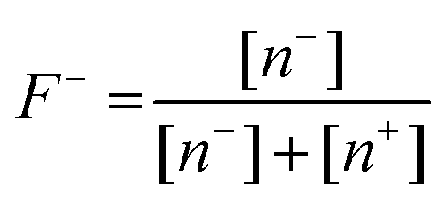
Complex coacervates are formed due to the interactions between the oppositely charged polymer chains, with the driving force being both entropy gain due to the release of counterions and electrostatic interaction. The fraction of the negative and positive charges is important for the total extent of CC formation. To narrow down the region of interest for evaluating the partitioning, we first evaluated the total CC formed as a function of the composition F− and looked at the water content for two CC compositions of interest.In Fig. 2A, it is shown that the largest amount of CC was formed around F− = 0.25 to 0.50, with the highest values found at 0.26 and 0.36 with 23.1 ± 3.3 mg and 22.4 ± 3.3 mg, respectively. Fig. 2B shows the photographs of the relative quantities of CC as a function of F−.
 | ||
| Fig. 2 Analysis of CC formation properties. (A) Total CC formed as a function of F− at a polyion concentration of 20 g L−1 and a NaCl concentration of 10 mM. Values are represented as average with standard deviations from triplicate experiments. (B) Photographs of different F− ratios with consistent amounts of total polyions. (C) The water content of the formed CCs was determined using TGA for F− = 0.26 and 0.36 at a polyion concentration of 10 g L−1 and a NaCl concentration of 10 mM. The left Y-axis shows the remaining mass fraction of the CC as the temperature presented on the right Y-axis is increased and water is vaporized. Values in (A) represent the average with standard deviations from triplicate experiments. | ||
As shown in Fig. 2C, we evaluated the water content of the two F− values with the highest CC formation as seen from Fig. 2A and found that for PEI/PAA CCs the water content varies drastically based on CC composition, with the water content for F− = 0.36 being 73.5%, and for F− = 0.26 being 51.9%. Comparing the remaining mass of the polyions in the CC to the total polyions added, it appears that for F− = 0.26 all the polyions form the CC mass, while for F− = 0.36 only approximately 60% of the polyions form the CC, with the rest presumably remaining in solution.
Intuitively, it might be expected that the largest volume of CC formation occurs at the composition F− = 0.50, where an equal amount of positive and negative monomers is present. However, this is not necessarily the case as demonstrated by the PEI/PAA CC system. One explanation for this discrepancy is that the interactions between polyions, water, and salts can affect the degree of ionization of the monomers.
Water content of CCs and PECs is typically reported to be between 60 and 80%.49–51 We found using TGA that for PEI/PAA CCs at a composition of F− = 0.36 the CCs fall within the reported range, though the water content at F− = 0.26 is approximately 10% lower than expected. The water content of CCs can impact the partitioning behaviour of solutes based on their preferential association with water. For example, lipases in general are known to prefer oil–water interfaces over fully aqueous environments.52 Both PEI and PAA are not expected to decompose at the given conditions, temperature, and timescale.53,54
Lipase enzyme distribution
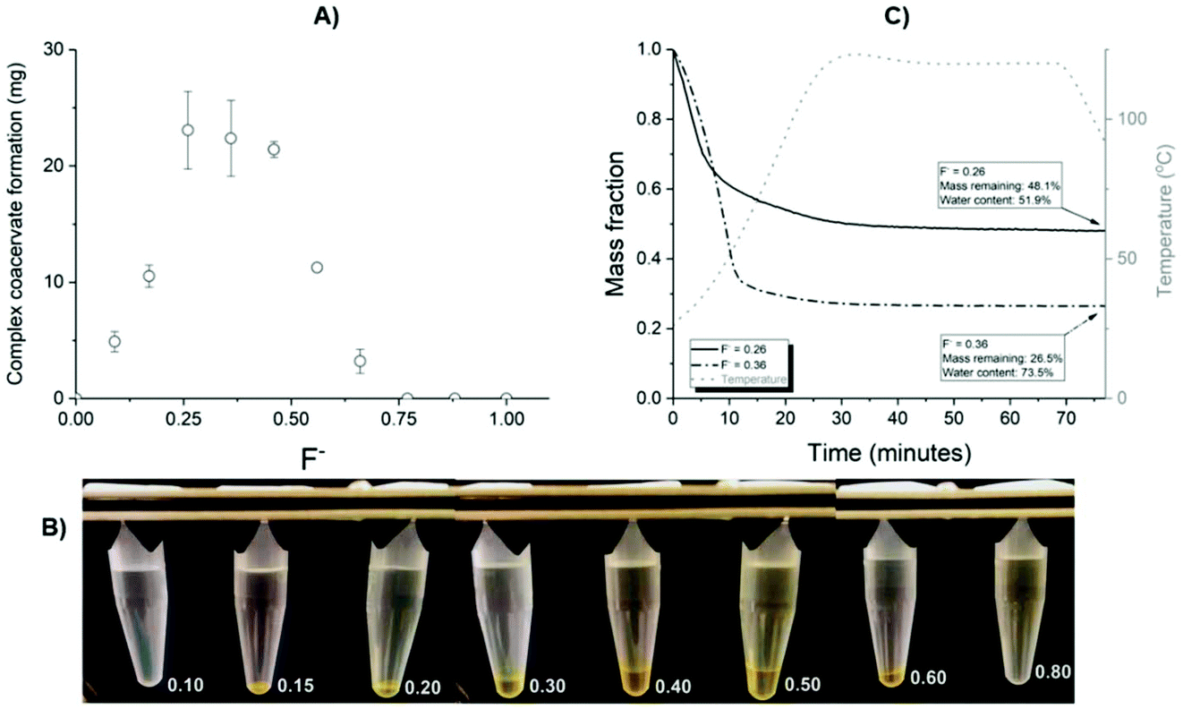
In this section, the partitioning of several types of lipases in the PEI/PAA complex coacervates is described. In Fig. 3, the distribution coefficients (KD) of three commonly used lipases PPL, CALB, and CALA as a function of the CC composition, the NaCl concentration, and the total polyion concentration are shown.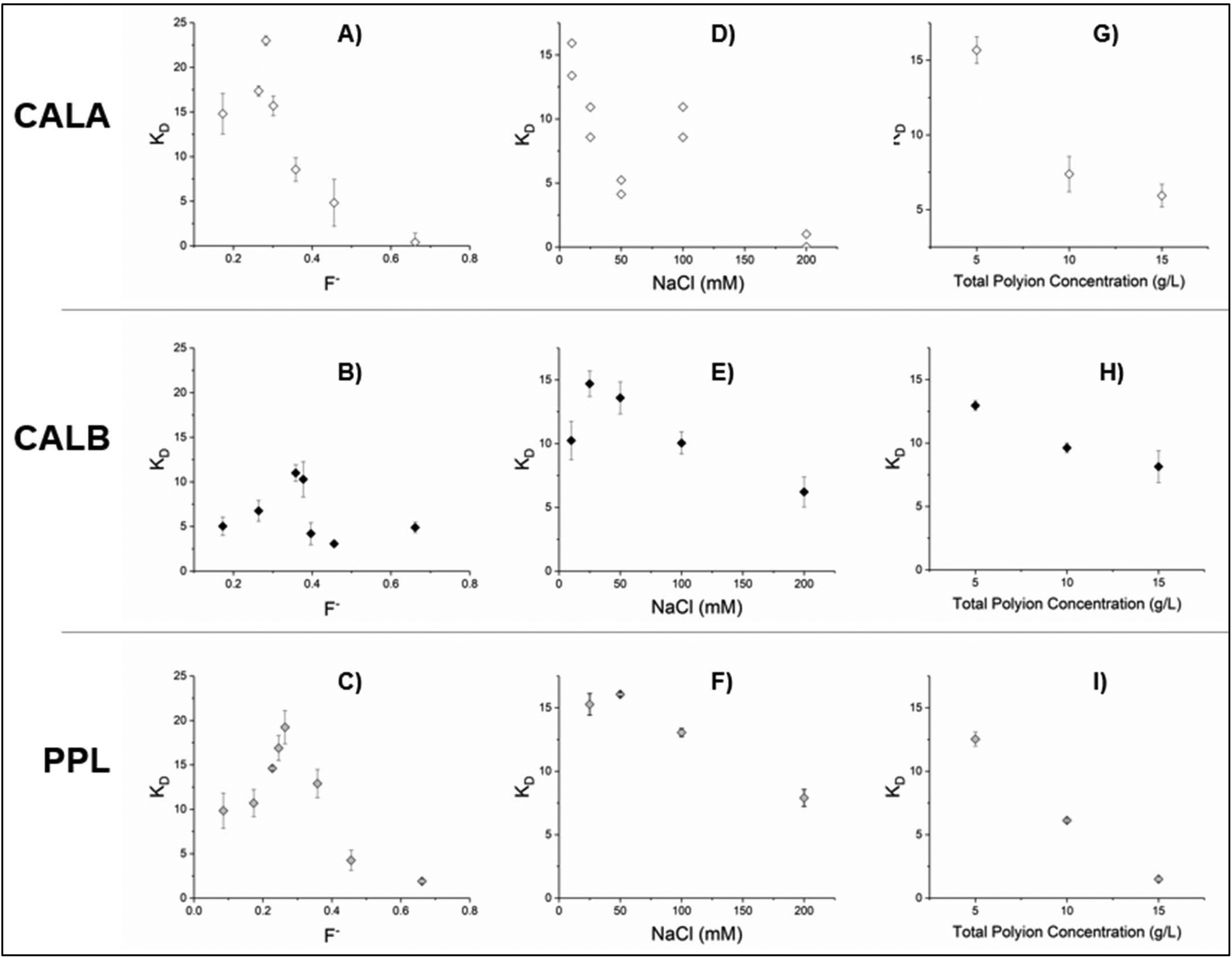 | ||
| Fig. 3 Distribution coefficients – KD for CALA (A, D and G), CALB (B, E and H), and PPL (C, F and I) as a function of CC composition (A–C), NaCl concentration (D–F), and polyion concentration (G–I). Unless otherwise specified, total polyion concentration is 5 g L−1, enzyme concentration is 67 μM, NaCl concentration is 10 mM, and F− is 0.36 for CALB and 0.26 for CALA and PPL. Measurements are shown as average with standard deviation for n = 3, except (D), which is shown as individual measurements. | ||
We found that the KD of all lipase types varies greatly as a result of the adjusted parameters. The charge ratio F− has the most significant consistent effect (Fig. 3A–C), showing distinct KD maxima at a composition of F− = 0.36 for CALB (KD maximum of 11.0 ± 0.9) and F− = 0.26 for CALA (KD maximum of 23.0 ± 0.5) and PPL (KD maximum of 19.2 ± 1.9). These maxima are partially consistent with the maximum values of CC formed (Fig. 2A); however, a small deviation in the composition results in a larger change in KD than that can be solely attributed to a difference in the CC quantity: for the region with the highest constant CC formation (F− ranging from 0.25 to 0.50), there are varieties in the KD for up to a factor 4 for CALA (Fig. 3A).
The distribution profiles were found to be dependent on the specific protein investigated. The results for these lipases corroborate the earlier studies that report similar nearly symmetrical distribution profiles (though centred around different F− values) for three proteins with poly(lysine)/poly(glutamate) CCs.26 Other proteins with different polyion pairs show completely different distribution profiles altogether that are not necessarily symmetrical.24,25 For now, there are no reliable methods to predict the distribution profile in advance as a result of the parameters. Many studies that look into the partitioning of proteins assume F− = 0.50 is the optimal composition for both PEC formation and partitioning and do not investigate the other charge ratios.27,28,30 Based on the results presented here, there might be opportunities for working at other compositions that result in more desired KD values.
Both CALB and PPL show a similar distribution profile as a function of the NaCl concentration (Fig. 3E and F) with a slight KD increase initially, followed by a decrease. CALA however shows an immediate decrease, followed by a local maximum (Fig. 3D) at a comparatively high salt concentration. By varying the NaCl concentration, the KD varies between approximately 5 and 15–20 for the investigated enzymes. We hypothesize that for CALB and PPL a partial screening of the polyion charges by the salt ions results in the CC being less densely packed, essentially increasing the distance between polyion chains and allowing the proteins (or other solutes) to enter the CC more easily. Polyion condensation in the presence of other ions (such as salt ions) results in ion association with charged monomer subunits of the polyion. This effectively screens the electrostatic interaction between the oppositely charged monomers of each polyion. Indeed, if the ionic strength of the solution becomes too high, the polyion structures dissolve completely as the degree of screening prevents the complex formation between polyions.46 Between complete complex dissolution and the absence of additional ions beyond the counterions brought in by the polyions, there is a concentration region where the salt ions prevent part of the oppositely charged polyions from associating. Subsequently, this can influence the behaviour of the condensates.
All three enzymes showed a similar trend of KD decrease as the total polyion concentration increased. A possible explanation is that as the total mass of CC increases, this does not result in a proportional increase of the CC–water interface, limiting the penetration of the solutes into the CC.
It is worth mentioning that there are other advantages of concentrating enzymes in CCs or PECs beyond extraction purposes. It has been reported that the activity of proteins may be enhanced in CCs compared to the same proteins in regular aqueous solutions.27,47 In addition, the polyions may protect the proteins from degradation, increasing the shelf life of (extracted) proteins.55 The mechanism for this is unknown, though the ability to both highly concentrate the enzymes and increase their activity is particularly interesting for industrial applications.
Lactic acid distribution
The partitioning of lactic acid into CCs was studied, as lactic acid is an industrially relevant small molecule. The effects of the CC composition, the NaCl concentration, the total polyion concentration, the initial LA concentration, and the temperature on the lactic acid KD were studied, and the results can be observed in Fig. 4. | ||
| Fig. 4 Lactic acid partitioning in PEI/PAA CC as a function of (A) CC composition, (B) NaCl concentration, (C) polyion concentration, (D) initial LA concentration, and (E) temperature. Unless otherwise specified, the experiments took place at approximately 20 °C, a polyion concentration of 5 g L−1, a LA concentration of 100 mM, a NaCl concentration of 10 mM, pH = 7, and F− = 0.26. Results are shown as average with standard deviations for n = 3. | ||
Unlike the distribution profiles for the lipase enzymes, we found only very little effect of the composition on the KD (Fig. 4A), which remained between 2 and 4. In contrast, the effect of NaCl on KD (Fig. 4B) of LA was more pronounced than those of CALB and PPL while following a similar distribution profile. Within our hypothesis of salt ions influencing the distance between polyion chains, the effect of the salt NaCl concentration may be more pronounced for LA, as it is substantially smaller than any of the lipases. By varying the NaCl concentration, we found the highest KD for LA at 7.4 ± 0.5 for 100 mM NaCl.
Similar to the trend with lipases, increasing polyion concentration had an adverse effect on the partitioning (Fig. 4C). However, altering the initial concentration of lactic acid only slightly affects the partitioning in the evaluated range (Fig. 4D), suggesting that the saturation point for the CC has not yet been reached as this would result in an expected decrease in KD at higher LA concentrations.24Fig. 4E shows that an increase in temperature has a small but consistent positive effect on the KD in the investigated range. This suggests that the extraction process is endothermic and that the driving force behind the partitioning is an increase in total entropy, perhaps similar to how an increase in entropy is the primary driving force for polyion–polyion association in the first place.23
The optimal KD for LA in our PEI/PAA CC system at approximately 100 mM NaCl is comparable to or greater than many other liquid–liquid extraction systems.44,56–58 A disadvantage of some of these reported systems is their reliance on low pH59 or the toxicity of the solvents.58 While some established extraction methods, such as tri-n-octylamine in 1-octanol,60 outperform CC systems with regard to LA for now, we show that the effects of system parameters for CC systems can substantially alter the KD. Interestingly, where the common method using tri-n-octylamine appears to decrease the distribution coefficient at higher temperatures, the opposite is true for PEI/PAA CCs.56 There are many additional parameters that can be further fine-tuned, suggesting the ability to achieve much higher KD values.
Butanol distribution, extraction, and back-extraction
We investigated the KD of butanol as a function of the CC composition (Fig. 5A) as well as the temperature (Fig. 5B). As butanol partitioning showed a remarkable temperature sensitivity, we evaluated the possibility of extraction and back-extraction of butanol using CCs by alternating between RT and 70 °C (Fig. 5D). | ||
| Fig. 5 Interaction between butanol and PEI/PAA CCs. (A) Distribution coefficients of butanol in PEI/PAA CCs as a function of CC composition shown as an average with standard deviation with n = 2. (B) Distribution of butanol as a function of temperature for F− = 0.26 and 0.36. For (A) and (B), the total polyion concentration was 5 g L−1, the NaCl concentration was 10 mM, and the butanol concentration was 400 mM. (C) Image of PEI/PAA CC at 50 g L−1 with (left) and without (right) the aqueous supernatant. (D) Butanol remaining in the CC at F− = 0.26 during RT to 70 °C extraction and 70 °C to RT back-extraction. Data are shown as average with n = 4. | ||
Contrary to the lipases, we observe the highest KD as a function of CC composition quite distant from the optimal CC formation, resulting in the highest value of KD of 22.7 ± 0.7 at F− = 0.56. This KD is very similar to that of a reported task-specific IL and substantially higher than the standard of oleyl alcohol, which are KD = 21 and 3.4, respectively.61,62
Whereas LA demonstrated only a minor temperature dependence of the KD (Fig. 4E), the butanol distribution shows a large difference between RT and 70 °C, roughly at a factor of 4–5. Out of the evaluated parameters, temperature is the most practical to change for the existing systems as it does not require the addition or removal of chemicals and is straightforward to implement. For this reason, we envisioned a PEI/PAA CC system that was able to partition butanol within the CC to a greater degree at high temperatures and could then be coaxed to release butanol into a separate aqueous environment at lower temperatures such as RT. To evaluate such a system for extraction and back-extraction of butanol, we prepared PEI/PAA CCs at higher concentrations of polyions (Fig. 5C). The resulting CCs had a mass of 62.2 ± 1.7 mg (average ± standard deviation, n = 4). A supernatant containing butanol was added to the CCs, and the temperature was increased from RT to 70 °C for butanol extraction. For back-extraction, the supernatant was replaced with fresh supernatant containing no butanol, and the temperature was decreased first to 60 °C, then to 40 °C, and finally to RT (Fig. 5D).
Consistent with the observations of Fig. 5B, increasing the temperature to 70 °C substantially increases the butanol content in the CC. Fig. 5B shows an approximate quadrupling of the KD, whereas Fig. 5D only shows a CC butanol increase from 8.80 ± 0.03 to 20.39 ± 0.80 mg, corresponding with a decrease of the supernatant butanol concentration from 4.39 ± 0.00% at RT to 2.28 ± 0.14% at 70 °C. A possible explanation for this discrepancy is the difference in the total polyion concentration, as Fig. 3G/H/I and 4C show that increased polyion concentrations do not necessarily lead to an increase in partitioning.
By replacing the supernatant and lowering the temperature in the steps from 70 °C back to RT, 21.1 ± 0.6% of the butanol extracted into the CC could be back-extracted into a new aqueous solution. Interestingly, reverting the temperature back to RT did not completely revert the butanol equilibrium and a fraction of butanol remains within the CC.
Considering the large number of tunable parameters, it is likely that with alterations a back-extraction higher than 21.1% is achievable. For example, increasing the salt concentration has been used to back-extract proteins from polyion micelles and polyion precipitates by disrupting the polyion complex,46 while varying the pH has been used to back-extract proteins, keeping the polyion precipitates intact.24 Other experimental parameters such as increasing the number of temperature steps or increasing the equilibration time may also prove to be beneficial. Further research should find improved recovery methods as well as better understanding of the physicochemical mechanisms allowing for a larger fraction of the CC-extracted butanol to be recovered.
Lactic acid and butanol distribution in the enzyme-filled complex coacervates
We hypothesized that the presence of additional components in the CCs can influence the partitioning behaviour of LA and butanol in those CCs. For this reason, we investigated the distribution of LA and butanol in PEI/PAA CCs that already contained PPL, CALB, or CALA enzymes. Similar to the presence of salt ions, the presence of relatively large enzymes in the CCs may change the structure of the polyion complex by altering the distance between polyions and the properties of the CC–water interface. We fixed the compositions of the systems to the F− at which the maximum KD was found; F− = 0.36 for PPL and CALB, and 0.26 for CALA. Then, LA (Fig. 6) and butanol (Fig. 7) partitioning was studied as a function of the ionic strength at 25 and 50 °C.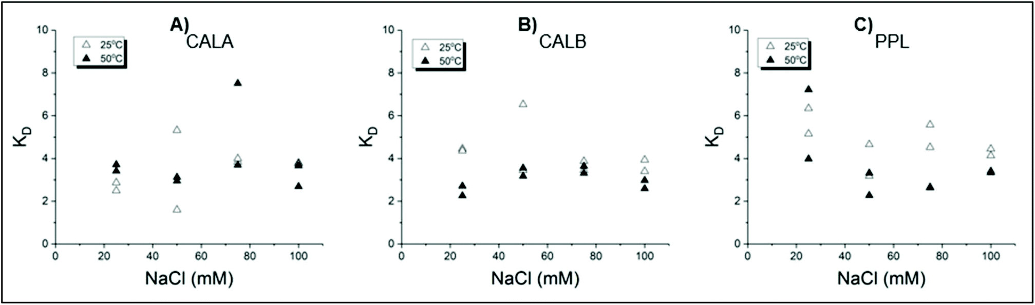 | ||
| Fig. 6 Distribution of LA as a function of NaCl concentration for PEI/PAA CC containing (A) CALA, (B) CALB, and (C) PPL lipase enzymes. Initial LA concentration was 100 mM, polyion concentration was 5 g L−1, and F− = 0.26 for CALA, and 0.36 for CALB and PPL. Results are shown as individual independent experiments. | ||
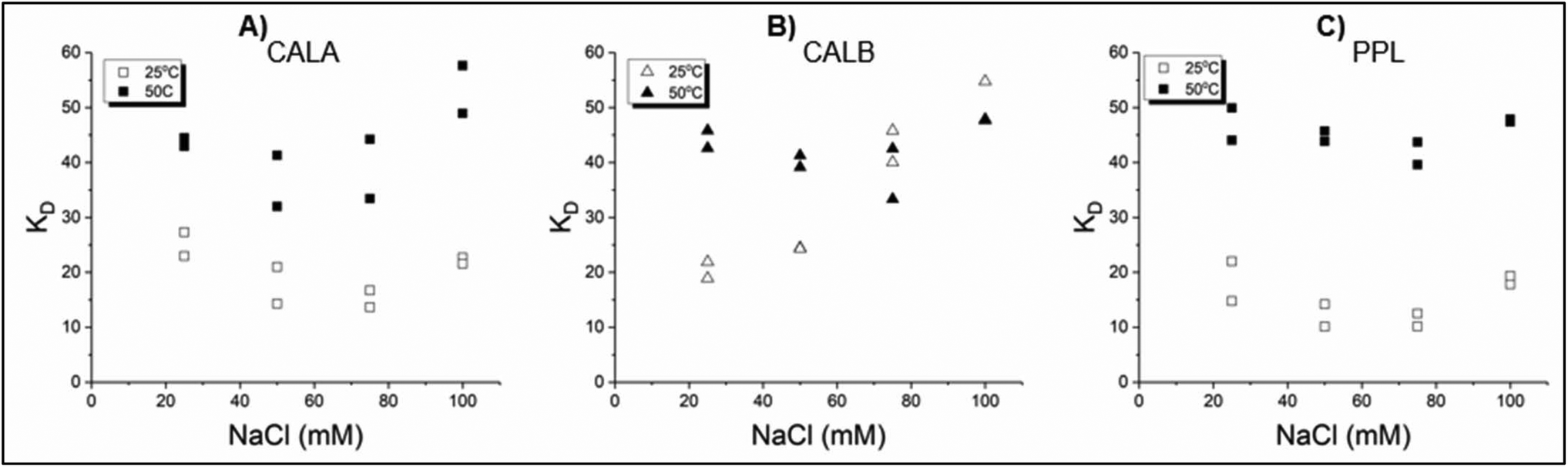 | ||
| Fig. 7 Distribution of butanol as a function of NaCl concentration for PEI/PAA CC containing (A) CALA, (B) CALB, and (C) PPL lipase enzymes. Initial butanol concentration was 400 mM, polyion concentration was 5 g L−1, and F− = 0.26 for CALA, and 0.36 for CALB and PPL. Results are shown as individual independent samples. | ||
In Fig. 6, we can see a stabilizing effect of the lipases on the LA distribution coefficients as they no longer strongly increase between 10 and 100 mM NaCl compared to the PEI/PAA CCs without the lipases shown in Fig. 4B. In addition, the presence of PPL slightly increases the ‘stable’ KD to approximately 5 compared to 3 without PPL. CALB increases the KD to approximately 4. For PPL and CALB, a higher temperature resulted in a slightly lower KD, comparable to values where the lipases were not present at all. Similar to Fig. 4E, there is no strongly noticeable difference between the investigated temperatures.
A much stronger effect is observed for the distribution of butanol shown in Fig. 7. For PPL and CALA, the KD values are comparable to CCs without the lipases at KD = 10–20, but with increased temperature the KD values increase to 40–50 for PPL and 30–50 for CALA. Interestingly, in the presence of CALB, the KD for butanol increases linearly with the NaCl concentration (Fig. 7B) at RT, but not at higher temperatures.
These ‘doped’ CCs show different distribution profiles than ‘empty’ CCs. Doped CCs may shield against the effect of increased NaCl concentration or simply increase the distribution coefficient by up to a factor of 3 compared to empty CCs. All in all, the concept of pre-filled CCs gives another parameter to tune and optimise the extraction potential of complex coacervates.
Conclusion and outlook
We present an exploratory study on new applications of complex coacervates. While the partitioning behaviour of CCs has been noted before, the step to develop them as an extraction medium has been absent. From previous studies as well as the results shown in this study, it has become clear that the partitioning behaviour of the compounds in CCs is a complex subject involving many tunable parameters that individually greatly influence the distribution coefficient between the aqueous environment and the CC.In our study, we showed that the distributions of lipase enzymes, lactic acid, and butanol in PEI/PAA complex coacervates are strongly affected by the CC composition, ionic strength as determined by the NaCl concentration, polyion concentration, temperature, and presence of other compounds in the CC. However, the effect of any of these parameters depends on the partitioned compound examined.
For example, we found that the CC composition has a great influence on the KD of lipases (Fig. 3A–C), while it has only a minimal effect on the KD of LA (Fig. 4A). Even within the category of lipases, the effect of the NaCl concentration on the KD of CALA is much stronger than on the KD of PPL (Fig. 3D and F, respectively). The only consistent influences of the parameter found were that higher concentrations of polyions above 5 g L−1 or high concentrations of NaCl led to lower KD values, though a small amount of NaCl was often (but not always) beneficial. The highest KD experimentally found and the corresponding parameters for the 5 compounds are presented in Table 1.
| Compound | Highest KD found | Figure | Composition (F−) | Compound concentration | NaCl concentration | Polyion concentration | Temperature |
|---|---|---|---|---|---|---|---|
| CALA | 23.0 ± 0.5 | Fig. 3A | 0.26 | 67 μM | 10 mM | 5 g L−1 | 20 °C |
| CALB | 14.7 ± 1.0 | Fig. 3E | 0.36 | 67 μM | 25 mM | 5 g L−1 | 20 °C |
| PPL | 16.1 ± 0.2 | Fig. 3F | 0.36 | 67 μM | 50 mM | 5 g L−1 | 20 °C |
| Lactic acid | 7.4 ± 0.5 | Fig. 4B | 0.26 | 100 mM | 100 mM | 5 g L−1 | 20 °C |
| Butanol | 53.3 ± 6.2 | Fig. 7A | 0.26 + CALA | 400 mM | 10 mM | 5 g L−1 | 50 °C |
We demonstrated that several relatively simple and tunable parameters can change the KD by a factor of 4 for lipases and butanol as a result of the changes in the CC composition (Fig. 3A–C) and temperature (Fig. 5B), respectively. It is unfortunate that many studies investigating the partitioning behaviour of solutes in CCs do not investigate different compositions, and instead fix it at F− = 0.50 where they might miss either compositions with greater partitioning or with greater PEC formation.27,28,30 As is demonstrated with the PEI/PAA system, we have shown that it is far from a safe assumption that the optimal polyion complex formation takes place at F− = 0.50, let alone the assumption that the desired partitioning properties are optimal at this composition.
Special emphasis has been laid on temperature as a parameter that is easily physically tunable without adding or removing chemicals to or from the system. Using temperature, we created a PEI/PAA CC temperature-swing extraction system that can extract approximately half the butanol from an aqueous supernatant at 70 °C, and then back-extract 21.1% of the extracted butanol back into a new aqueous phase at RT in a single-step system. In this way, CC extraction media can be considered analogous to, for example, cyclic CO2 absorption, where typical cyclic capacities are in the order of 5–15%.63
However, considering the number of tunable parameters, it is almost certain that the cyclic capacity can be made much more efficient, and that extraction/back-extraction of a variety of small molecules as well as proteins is possible. While the results for our butanol extraction were not directly comparable in efficiency to some of the results shown by ATPS systems,64 where up to 95% of a protein was purified in a single step, such high extraction numbers have been shown with different PECs for different proteins,24 suggesting that a similar potential for CCs exists.
There are several limitations of this study. Some of the experimental protocols in these experiments, such as centrifuging at 12500 g for 30 minutes, are impractical for industrial applications. These protocols were based on earlier fundamental research24,25 and it is likely (but not verified) that centrifuging at far lower speeds and durations is sufficient. Indeed, the butanol (back-)extraction was performed without additional centrifugation steps after the addition of butanol to the system.
The reasons for the variation in KD values and the mechanisms determining the distributions in CCs or other PECs are not well understood. The partitioning behaviour is currently not well understood and cannot yet be accurately predicted. Currently, this means that extensive testing for the individual compound, polyion pair, and tunable parameters is required in order to learn how the parameters influence partitioning. It would be extremely beneficial for the development of CCs as extraction media if the fundamental mechanisms of partitioning in CCs were better understood. The ability to predict the influence of (combinations of) parameters on partitioning prevents the necessity of high-throughput testing to optimise the parameters for the extraction of a particular desired compound. With a greater understanding of the underlying mechanisms, complex coacervates show promise as extraction media for a wide variety of compounds. The partitioning of solutes in CCs and PECs is the result of a complex interplay of at least 6 different compounds (polyanion, polycation, water, two salt ions, and the solute of interest), and the temperature will affect the interactions between all these compounds, making it difficult to predict the partitioning behaviour. For proteins, it is expected that the charge and charge distribution are important, and hydrophobic interactions will also play a role. The temperature-dependent partitioning of butanol is promising, but systematic studies are required to unravel the detailed molecular mechanism.
Conflicts of interest
The authors have no conflicts of interest to declare.Acknowledgements
The authors acknowledge and thank Corbion N.V. and Novozymes A/S for their generous donation of lactic acid, and CALA and CALB, respectively. The authors acknowledge funding from the Netherlands Organization for Scientific Research (NWO) VIDI grant (# 723.015.003).References
- T. Welton, Solvents and sustainable chemistry, Proc. R. Soc. A, 2015, 471(2183), 20150502, DOI:10.1098/rspa.2015.0502.
- M. Tobiszewski, J. Namieśnik and F. Pena-Pereira, Environmental risk-based ranking of solvents using the combination of a multimedia model and multi-criteria decision analysis, Green Chem., 2017, 19(4), 1034–1042, 10.1039/C6GC03424A.
- B. Schuur, Selection and design of ionic liquids as solvents in extractive distillation and extraction processes, Chem. Pap., 2015, 69(2), 245–253, DOI:10.1515/chempap-2015-0016.
- J. Marták and Š. Schlosser, New Mechanism and Model of Butyric Acid Extraction by Phosphonium Ionic Liquid, J. Chem. Eng. Data, 2016, 61(9), 2979–2996, DOI:10.1021/acs.jced.5b01082.
- D. J. G. P. van Osch, L. F. Zubeir, A. van den Bruinhorst, M. A. A. Rocha and M. C. Kroon, Hydrophobic deep eutectic solvents as water-immiscible extractants, Green Chem., 2015, 17(9), 4518–4521, 10.1039/C5GC01451D.
- A. I. Pratiwi, T. Yokouchi, M. Matsumoto and K. Kondo, Extraction of succinic acid by aqueous two-phase system using alcohols/salts and ionic liquids/salts, Sep. Purif. Technol., 2015, 155, 127–132, DOI:10.1016/j.seppur.2015.07.039.
- L. Castro, P. Pereira, M. Freire and A. Pedro, Progress in the Development of Aqueous Two-Phase Systems Comprising Ionic Liquids for the Downstream Processing of Protein-Based Biopharmaceuticals, Am. Pharm. Rev., 2019 Search PubMed.
- M. G. Freire, A. F. M. Cláudio, J. M. M. Araújo, J. A. P. Coutinho, I. M. Marrucho, J. N. Canongia Lopes and L. P. N. Rebelo, Aqueous biphasic systems: a boost brought about by using ionic liquids, Chem. Soc. Rev., 2012, 41(14), 4966–4995, 10.1039/c2cs35151j.
- J. S. Wilkes, A short history of ionic liquids—from molten salts to neoteric solvents, Green Chem., 2002, 4(2), 73–80, 10.1039/b110838g.
- M. L. Dietz, Ionic Liquids as Extraction Solvents: Where do We Stand?, Sep. Sci. Technol., 2006, 41(10), 2047–2063, DOI:10.1080/01496390600743144.
- B. Schuur, T. Brouwer, D. Smink and L. M. J. Sprakel, Green solvents for sustainable separation processes, Curr. Opin. Green Sustain. Chem., 2019, 18, 57–65, DOI:10.1016/j.cogsc.2018.12.009.
- S.-K. Mikkola, A. Robciuc, J. Lokajová, A. J. Holding, M. Lämmerhofer, I. Kilpeläinen, J. M. Holopainen, A. W. T. King and S. K. Wiedmer, Impact of Amphiphilic Biomass-Dissolving Ionic Liquids on Biological Cells and Liposomes, Environ. Sci. Technol., 2015, 49(3), 1870–1878, DOI:10.1021/es505725g.
- S. Bajkacz and J. Adamek, Evaluation of new natural deep eutectic solvents for the extraction of isoflavones from soy products, Talanta, 2017, 168, 329–335, DOI:10.1016/j.talanta.2017.02.065.
- M. Hayyan, C. Y. Looi, A. Hayyan, W. F. Wong and M. A. Hashim, In Vitro and In Vivo Toxicity Profiling of Ammonium-Based Deep Eutectic Solvents, PLoS One, 2015, 10(2), e0117934, DOI:10.1371/journal.pone.0117934.
- P. Xu, G.-W. Zheng, M.-H. Zong, N. Li and W.-Y. Lou, Recent progress on deep eutectic solvents in biocatalysis, Bioresour. Bioprocess., 2017, 4, 34, DOI:10.1186/s40643-017-0165-5.
- F. Bezold and M. Minceva, Liquid-liquid equilibria of n-heptane, methanol and deep eutectic solvents composed of carboxylic acid and monocyclic terpenes, Fluid Phase Equilib., 2018, 477, 98–106, DOI:10.1016/j.fluid.2018.08.020.
- M. Iqbal, Y. Tao, S. Xie, Y. Zhu, D. Chen, X. Wang, L. Huang, D. Peng, A. Sattar, M. Shabbir, H. Hussain, S. Ahmed and Z. Yuan, Aqueous two-phase system (ATPS): an overview and advances in its applications, Biol. Proced. Online, 2016, 18(18) DOI:10.1186/s12575-016-0048-8.
- Y. Chao and H. C. Shum, Emerging aqueous two-phase systems: from fundamentals of interfaces to biomedical applications, Chem. Soc. Rev., 2020, 49(1), 114–142, 10.1039/C9CS00466A.
- J. F. B. Pereira, M. G. Freire and J. A. P. Coutinho, Aqueous two-phase systems: Towards novel and more disruptive applications, Fluid Phase Equilib., 2020, 505, 112341, DOI:10.1016/j.fluid.2019.112341.
- A. G. Teixeira, R. Agarwal, K. R. Ko, J. Grant-Burt, B. M. Leung and J. P. Frampton, Emerging Biotechnology Applications of Aqueous Two-Phase Systems, Adv. Healthcare Mater., 2018, 7(6), 1701036, DOI:10.1002/adhm.201701036.
- N. Li, X. Yang, L. Nian, Z. Wang, L. Lei, K. Wang, H. Zhang, Ai. Yu and Z. Zhang, Determination of steroid hormones in milk using aqueous two-phase extraction coupled to liquid chromatography, Anal. Methods, 2015, 7(6), 2514–2522, 10.1039/C4AY03036B.
- S. Zimmermann, S. Gretzinger, M.-L. Schwab, C. Scheeder, P. K. Zimmermann, S. A. Oelmeier, E. Gottwald, A. Bogsnes, M. Hansson, A. Staby and J. Hubbuch, High-throughput downstream process development for cell-based products using aqueous two-phase systems, J. Chromatogr. A, 2016, 1464, 1–11, DOI:10.1016/j.chroma.2016.08.025.
- A. Melnyk, J. Namieśnik and L. Wolska, Theory and recent applications of coacervate-based extraction techniques, TrAC, Trends Anal. Chem., 2015, 71, 282–292, DOI:10.1016/j.trac.2015.03.013.
- J. J. van Lente, M. M. A. E. Claessens and S. Lindhoud, Charge-Based Separation of Proteins Using Polyelectrolyte Complexes as Models for Membraneless Organelles, Biomacromolecules, 2019, 20(10), 3696–3703, DOI:10.1021/acs.biomac.9b00701.
- S. Lindhoud and M. M. A. E. Claessens, Accumulation of small protein molecules in a macroscopic complex coacervate, Soft Matter, 2016, 12(2), 408–413, 10.1039/C5SM02386F.
- W. C. Blocher McTigue and S. L. Perry, Design rules for encapsulating proteins into complex coacervates, Soft Matter, 2019, 15(15), 3089–3103, 10.1039/C9SM00372J.
- P. M. McCall, S. Srivastava, S. L. Perry, D. R. Kovar, M. L. Gardel and M. V. Tirrell, Partitioning and Enhanced Self-Assembly of Actin in Polypeptide Coacervates, Biophys. J., 2018, 114(7), 1636–1645, DOI:10.1016/j.bpj.2018.02.020.
- K. A. Black, D. Priftis, S. L. Perry, J. Yip, W. Y. Byun and M. Tirrell, Protein Encapsulation via Polypeptide Complex Coacervation, ACS Macro Lett., 2014, 3(10), 1088–1091, DOI:10.1021/mz500529v.
- M. Zhao, S. A. Eghtesadi, M. B. Dawadi, C. Wang, S. Huang, A. E. Seymore, B. D. Vogt, D. A. Modarelli, T. Liu and N. S. Zacharia, Partitioning of Small Molecules in Hydrogen-Bonding Complex Coacervates of Poly(acrylic acid) and Poly(ethylene glycol) or Pluronic Block Copolymer, Macromolecules, 2017, 50(10), 3818–3830, DOI:10.1021/acs.macromol.6b02815.
- S. Huang, M. Zhao, M. B. Dawadi, Y. Cai, Y. Lapitsky, D. A. Modarelli and N. S. Zacharia, Effect of small molecules on the phase behavior and coacervation of aqueous solutions of poly(diallyldimethylammonium chloride) and poly(sodium 4-styrene sulfonate), J. Colloid Interface Sci., 2018, 518, 216–224, DOI:10.1016/j.jcis.2018.02.029.
- S. F. Banani, H. O. Lee, A. A. Hyman and M. K. Rosen, Biomolecular condensates: organizers of cellular biochemistry, Nat. Rev. Mol. Cell Biol., 2017, 18(5), 285–298, DOI:10.1038/nrm.2017.7.
- M. M. Fay and P. J. Anderson, The Role of RNA in Biological Phase Separations, J. Mol. Biol., 2018, 430(23), 4685–4701, DOI:10.1016/j.jmb.2018.05.003.
- A. Aguilera-Gomez and C. Rabouille, Membrane-bound organelles versus membrane-less compartments and their control of anabolic pathways in Drosophila, Dev. Biol., 2017, 428(2), 310–317, DOI:10.1016/j.ydbio.2017.03.029.
- S. Boeynaems, S. Alberti, N. L. Fawzi, T. Mittag, M. Polymenidou, F. Rousseau, J. Schymkowitz, J. Shorter, B. Wolozin, L. van den Bosch, P. Tompa and M. Fuxreiter, Protein Phase Separation: A New Phase in Cell Biology, Trends Cell Biol., 2018, 28(6), 420–435, DOI:10.1016/j.tcb.2018.02.004.
- E. Gomes and J. Shorter, The molecular language of membraneless organelles, J. Biol. Chem., 2018, 294(18), 7115–7127, DOI:10.1074/jbc.TM118.001192.
- F. Zhao, E. Repo, Y. Song, D. Yin, S. Ben Hammouda, L. Chen, S. Kalliola, J. Tang, K. C. Tam and M. Sillanpää, Polyethylenimine-cross-linked cellulose nanocrystals for highly efficient recovery of rare earth elements from water and a mechanism study, Green Chem., 2017, 19(20), 4816–4828, 10.1039/C7GC01770G.
- A. Zakeri, M. A. J. Kouhbanani, N. Beheshtkhoo, V. Beigi, S. M. Mousavi, S. A. R. Hashemi and A. Movahedpour, Polyethylenimine-based nanocarriers in co-delivery of drug and gene: a developing horizon, Nano Rev. Exp., 2018, 9(1), 1488497, DOI:10.1080/20022727.2018.1488497.
- G. Fang, F. Zeng, C. Yu and S. Wu, Low molecular weight PEIs modified by hydrazone-based crosslinker and betaine as improved gene carriers, Colloids Surf., B, 2014, 122, 472–481, DOI:10.1016/j.colsurfb.2014.07.007.
- A. Houde, A. Kademi and D. Leblanc, Lipases and Their Industrial Applications: An Overview, Appl. Biochem. Biotechnol., 2004, 118(1–3), 155–170, DOI:10.1385/ABAB:118:1-3:155.
- F. Hasan, A. A. Shah and A. Hameed, Industrial applications of microbial lipases, Enzyme Microb. Technol., 2006, 39(2), 235–251, DOI:10.1016/j.enzmictec.2005.10.016.
- D. Guerrand, Lipases industrial applications: focus on food and agroindustries, OCL, 2017, 24(4), D403, DOI:10.1051/ocl/2017031.
- M. Hajighasemi, B. P. Nocek, A. Tchigvintsev, G. Brown, R. Flick, X. Xu, H. Cui, T. Hai, A. Joachimiak, P. N. Golyshin, A. Savchenko, E. A. Edwards and A. F. Yakunin, Biochemical and Structural Insights into Enzymatic Depolymerization of Polylactic Acid and Other Polyesters by Microbial Carboxylesterases, Biomacromolecules, 2016, 17(6), 2027–2039, DOI:10.1021/acs.biomac.6b00223.
- V. Hábová, K. Melzoch and M. Rychtera, Modern method of lactic acid recovery from fermentation broth, Czech J. Food Sci., 2011, 22(3), 87–94, DOI:10.17221/3411-CJFS.
- A. Komesu, M. R. Wolf Maciel and R. Maciel Filho, Separation and Purification Technologies for Lactic Acid – A Brief Review, BioResources, 2017, 12(3), 6885–6901, DOI:10.15376/biores.12.3.6885-6901.
- H.-J. Huang, S. Ramaswamy, U. W. Tschirner and B. V. Ramarao, A review of separation technologies in current and future biorefineries, Sep. Purif. Technol., 2008, 62(1), 1–21, DOI:10.1016/j.seppur.2007.12.011.
- S. Lindhoud, R. de Vries, R. Schweins, M. A. Cohen Stuart and W. Norde, Salt-induced release of lipase from polyelectrolyte complex micelles, Soft Matter, 2009, 5(1), 242–250, 10.1039/B811640G.
- S. Lindhoud, W. Norde and M. A. Cohen Stuart, Effects of Polyelectrolyte Complex Micelles and Their Components on the Enzymatic Activity of Lipase, Langmuir, 2010, 26(12), 9802–9808, DOI:10.1021/la1000705.
- G. Rabbani, E. Ahmad, M. V. Khan, M. T. Ashraf, R. Bhat and R. H. Khan, Impact of structural stability of cold adapted Candida antarctica lipase B (CaLB): in relation to pH, chemical and thermal denaturation, RSC Adv., 2015, 5(26), 20115–20131, 10.1039/C4RA17093H.
- E. Spruijt, A. H. Westphal, J. W. Borst, M. A. Cohen Stuart and J. van der Gucht, Binodal Compositions of Polyelectrolyte Complexes, Macromolecules, 2010, 43(15), 6476–6484, DOI:10.1021/ma101031t.
- J. van der Gucht, E. Spruijt, M. Lemmers and M. A. Cohen Stuart, Polyelectrolyte complexes: Bulk phases and colloidal systems, J. Colloid Interface Sci., 2011, 361(2), 407–422, DOI:10.1016/j.jcis.2011.05.080.
- H. Bohidar, P. L. Dubin, P. R. Majhi, C. Tribet and W. Jaeger, Effects of Protein−Polyelectrolyte Affinity and Polyelectrolyte Molecular Weight on Dynamic Properties of Bovine Serum Albumin−Poly(diallyldimethylammonium chloride) Coacervates, Biomacromolecules, 2005, 6(3), 1573–1585, DOI:10.1021/bm049174p.
- M. Anvari, Extraction of lipase from Rhizopus microsporus fermentation culture by aqueous two-phase partitioning, Biotechnol. Biotechnol. Equip., 2015, 29(4), 723–731, DOI:10.1080/13102818.2015.1042406.
- V. V. Nedel’ko, B. L. Korsunskii, F. I. Dubovitskii and G. L. Gromova, The thermal degradation of branched polyethylenimine, Polym. Sci. U.S.S.R., 1975, 17(7), 1697–1703, DOI:10.1016/0032-3950(75)90172-0.
- I. C. McNeill and S. M. T. Sadeghi, Thermal stability and degradation mechanisms of poly(acrylic acid) and its salts: Part 1—Poly(acrylic acid), Polym. Degrad. Stab., 1990, 29(2), 233–246, DOI:10.1016/0141-3910(90)90034-5.
- A. Marin, D. P. DeCollibus and A. K. Andrianov, Protein Stabilization in Aqueous Solutions of Polyphosphazene Polyelectrolyte and Non-Ionic Surfactants, Biomacromolecules, 2010, 11(9), 2268–2273, DOI:10.1021/bm100603p.
- S. Lindhoud and M. A. Cohen Stuart, Relaxation Phenomena During Polyelectrolyte Complex Formation, in Polyelectrolyte Complexes in the Dispersed and SolidState I, ed. M. Müller, Springer Berlin Heidelberg, Red. Berlin, Heidelberg, 2012, vol. 255, pp. 139–172, DOI:10.1007/12_2012_178.
- H. G. Joglekar, I. Rahman, S. Babu, B. D. Kulkarni and A. Joshi, Comparative assessment of downstream processing options for lactic acid, Sep. Purif. Technol., 2006, 52(1), 1–17, DOI:10.1016/j.seppur.2006.03.015.
- K. L. Wasewar, A. B. M. Heesink, G. F. Versteeg and V. G. Pangarkar, Reactive extraction of lactic acid using alamine 336 in MIBK: equilibria and kinetics, J. Biotechnol., 2002, 97(1), 59–68, DOI:10.1016/S0168-1656(02)00057-3.
- N. Mungma, M. Kienberger and M. Siebenhofer, Reactive Extraction of Lactic Acid, Formic Acid and Acetic Acid from Aqueous Solutions with Tri-n-octylamine/1-Octanol/n-Undecane, ChemEngineering, 2019, 3(2), 43, DOI:10.3390/chemengineering3020043.
- A. Krzyżaniak, M. Leeman, F. Vossebeld, T. J. Visser, B. Schuur and A. B. de Haan, Novel extractants for the recovery of fermentation derived lactic acid, Sep. Purif. Technol., 2013, 111, 82–89, DOI:10.1016/j.seppur.2013.03.031.
- L. Y. Garcia-Chavez, C. M. Garsia, B. Schuur and A. B. de Haan, Biobutanol Recovery Using Nonfluorinated Task-Specific Ionic Liquids, Ind. Eng. Chem. Res., 2012, 51(24), 8293–8301, DOI:10.1021/ie201855h.
- L. Adhami, B. Griggs, P. Himebrook and K. Taconi, Liquid–Liquid Extraction of Butanol from Dilute Aqueous Solutions Using Soybean-Derived Biodiesel, J. Am. Oil Chem. Soc., 2009, 86(11), 1123–1128, DOI:10.1007/s11746-009-1447-7.
- I. M. Bernhardsen and H. K. Knuutila, A review of potential amine solvents for CO 2 absorption process: Absorption capacity, cyclic capacity and pKa, Int. J. Greenhouse Gas Control, 2017, 61, 27–48, DOI:10.1016/j.ijggc.2017.03.021.
- O. Cascone, B. A. Andrews and J. A. Asenjo, Partitioning and purification of thaumatin in aqueous two-phase systems, Enzyme Microb. Technol., 1991, 13(8), 629–635, DOI:10.1016/0141-0229(91)90076M.
| This journal is © The Royal Society of Chemistry 2021 |