
 Open Access Article
 This Open Access Article is licensed under a
 Creative Commons Attribution 3.0 Unported Licence
Correction: Transgenic PDGF-BB/sericin hydrogel supports for cell proliferation and osteogenic differentiation
Feng
Wang
ab,
Kai
Hou
a,
Wenjing
Chen
a,
Yuancheng
Wang
a,
Riyuan
Wang
a,
Chi
Tian
a,
Sheng
Xu
a,
Yanting
Ji
a,
Qianqian
Yang
a,
Ping
Zhao
a,
Ling
Yu
c,
Zhisong
Lu
c,
Huijie
Zhang
d,
Fushu
Li
e,
Han
Wang
e,
Baicheng
He
e,
David L.
Kaplan
*b and
Qingyou
Xia
*a
aBiological Science Research Center, Chongqing Key Laboratory of Sericultural Science, Chongqing Engineering and Technology Research Center for Novel Silk Materials, Southwest University, Chongqing, 400715, People's Republic of China. E-mail: Xiaqy@swu.edu.cn
bDepartment of Biomedical Engineering, Tufts University, Medford, MA 02155, USA. E-mail: David.kaplan@tufts.edu
cInstitute for Clean Energy & Advanced Materials, Faculty of Materials & Energy, Southwest University, Chongqing, 400715, People's Republic of China
dKey Laboratory of Biochemistry and Molecular Pharmacology of Chongqing, Chongqing Medical University, Chongqing, 400016, People's Republic of China
eDepartment of Pharmacology, School of Pharmacy, Chongqing Medical University, Chongqing, 400016, People's Republic of China
First published on 19th May 2021
Abstract
Correction for ‘Transgenic PDGF-BB/sericin hydrogel supports for cell proliferation and osteogenic differentiation’ by Feng Wang et al., Biomater. Sci., 2020, 8, 657–672, DOI: 10.1039/C9BM01478K.
The authors regret that the live/dead staining of cells seeded on TCP in Fig. 5C of the published manuscript was incorrect. The correct version is shown below. The authors note that this correction has no effect on the results reported, nor does this change any of the contents and conclusions of the paper.
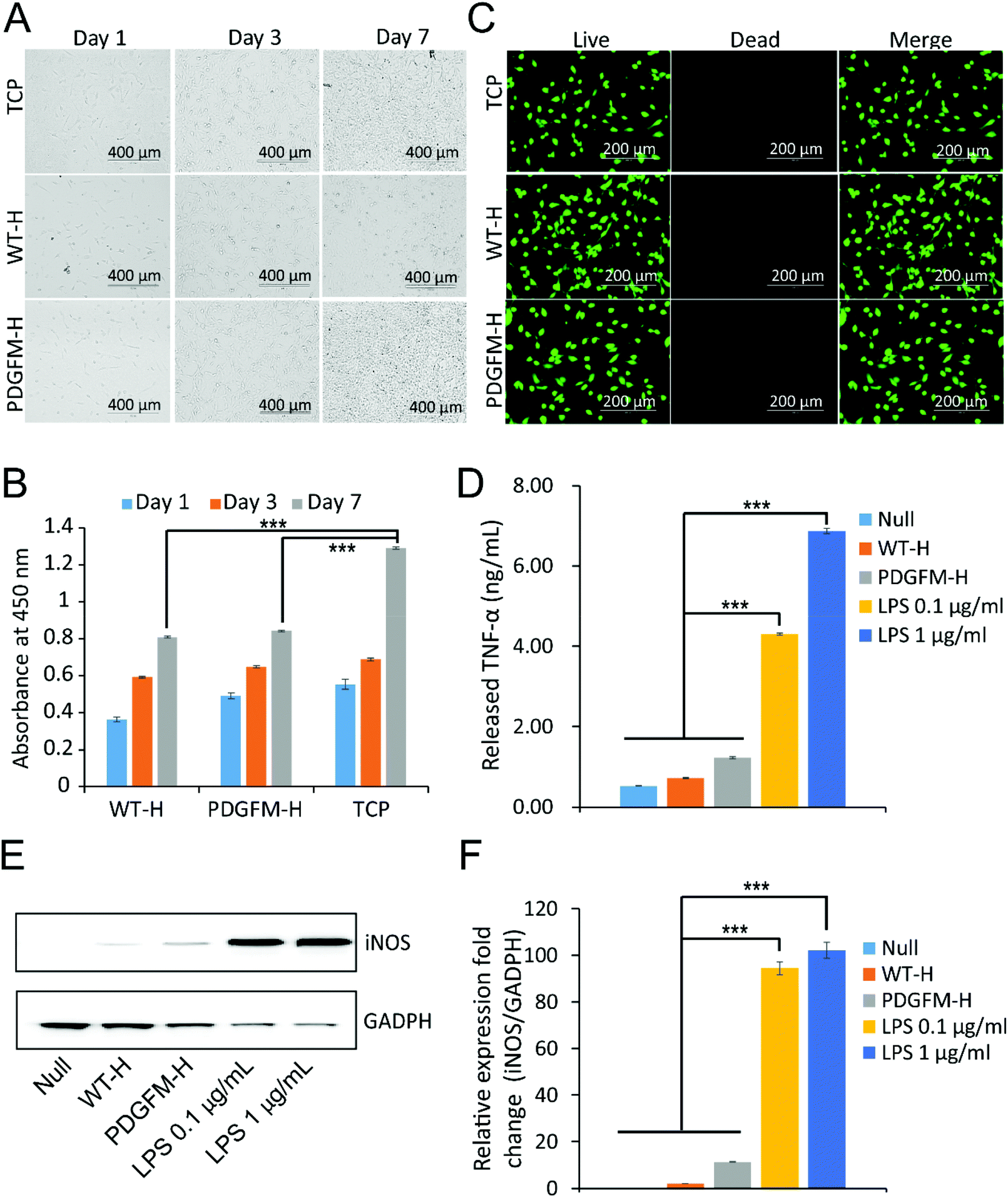 | ||
| Fig. 5 Biocompatibility and cellular inflammatory response of the PDGFM sericin hydrogels. (A and B) Optical microscopy and CCK-8 (cell counting kit-8) assay of growth of NIH/3T3 cells on TCP, WT and PDGFM sericin hydrogels at days 1, 3 and 7 after seeding in complete DMEM medium containing 10% FBS. Scale bars are 400 μm. The absorbance was measured at 450 nm. Measurements in triplicate and repeated independently three times. *P < 0.05, **P < 0.01, ***P < 0.001 compared to TCP group, student's t-test; (C) viability/cytotoxicity of NIH/3T3 cells seeded on the TCP, WT and PDGFM sericin hydrogels in complete DMEM medium containing 10% FBS after cultivation for 2 days. Live cells stained by calcein-AM dye and produced intense uniform green fluorescence in live cells (ex/em ∼495 nm/∼515 nm). Dead cells stained by EthD-1 dye and emitted bright red fluorescence (ex/em ∼495 nm/∼635 nm). The scale bars are 200 μm. (D) Released inflammatory factor TNF-α from the RAW264.7 cells in media induced by WT and PDGFM sericin hydrogels for 24 h; (E) expression analysis of iNOS in the RAW264.7 cells by western blot induced by WT and PDGFM sericin hydrogels for 24 h. GADPH was the internal control; (F) the gray intensity comparison of iNOS and GADPH expression in the RAW264.7 cells induced by WT and PDGFM sericin hydrogels for 24 h according to immunoblots. LPS induced inflammatory factors released with a dose effect (100 ng mL−1 and 1000 ng ml−1) were set as positive control. The measurements were in triplicate and repeated independently three times. *P < 0.05, **P < 0.01, ***P < 0.001 compared to the control group, student's t-test. WT-H and PDGFM-H represent the WT sericin hydrogel and PDGFM hydrogel, respectively. | ||
The Royal Society of Chemistry apologises for these errors and any consequent inconvenience to authors and readers.
| This journal is © The Royal Society of Chemistry 2021 |