DOI:
10.1039/D0BM90116D
(Correction)
Biomater. Sci., 2021,
9, 1044-1046
Correction: Prevention of pulmonary air leaks using a biodegradable tissue-adhesive fiber sheet based on Alaska pollock gelatin modified with decanyl groups
Received
18th December 2020
, Accepted 18th December 2020
First published on 6th January 2021
Abstract
Correction for ‘Prevention of pulmonary air leaks using a biodegradable tissue-adhesive fiber sheet based on Alaska pollock gelatin modified with decanyl groups’ by Hiroaki Ichimaru et al., Biomater. Sci., 2021, DOI: 10.1039/d0bm01302a.
The authors regret some errors in the materials and methods section of the article and the figure captions. The corrected versions are shown below.
2.15. Statistical analysis
Statistical analyses were carried out using student's t-tests. Statistically significant differences were considered as those for which p < 0.05. The data are reported as mean ± standard error of the mean (S.E.M.).
Fig. 2 Fabrication and physicochemical characterization of AdFSs. (a) Photograph of prepared 13C10-AdFS. (b) SEM images of Org-AdFS (left) and 13C10-AdFS (right). (c) Tensile strengths and (d) Young's moduli of C10-AdFSs with different modification ratios. (e) Swelling behavior of Org-AdFS and C10-AdFSs with different modification ratios after immersion in saline at 37 °C. (f) Water contact angle on Org-AdFS and C10-AdFSs with different modification ratios 5 s after applying the water droplet. The samples for all experiments were Org-AdFS (black), 13C10-AdFS (blue), 20C10-AdFS (red), and 37C10-AdFS (green) thermally cross-linked at 150 °C for 5 h. All AdFSs were 200 μm thick. The data are the mean ± S.E.M. of three or five samples (
n = 3 or 5). *
p < 0.05, **
p > 0.05.
Fig. 3 Burst strengths of AdFSs on porcine pleurae. (a) Burst strength of 13C10-AdFS with different hydration times. The AdFS had a thickness of 200 μm thickness and was cross-linked at 150 °C for 5 h. A PGA sheet was used as a control material. (b) Burst strengths of C10-AdFSs with different modification ratios and cross-linking times. The AdFS thickness was 200 μm. (c) Burst strengths of C10-AdFSs with different modification ratios and thickness. All AdFSs were cross-linked at 150 °C for 5 h. (d) Histological observation of Org-AdFS, C10-AdFS, and the PGA sheet after burst, showing a cross-section of the whole sample (top) and the upper surface (bottom). The scale bars are 500 μm (top) and 50 μm (bottom). (e) Photographs showing Org-AdFS, 13C10-AdFS, and PGA sheet at the time of rupture. The scale bars are 5 mm. (f) Fracture modes observed in the burst strength measurements of Org-AdFS, 13C10-AdFS, and the PGA sheet (
n = 5). The AdFSs were cross-linked at 150 °C for 5 h; the ADFS thickness was 200 μm. The AdFS (S) and tissue (T) are indicated in the image. The samples used in the burst strength measurements were Org-AdFS (black), 13C10-AdFS (blue), 20C10-AdFS (red), 37C10-AdFS (green), and PGA sheet (white). The data are the mean ± S.E.M. of five samples (
n = 5). *
p < 0.05, **
p > 0.05.
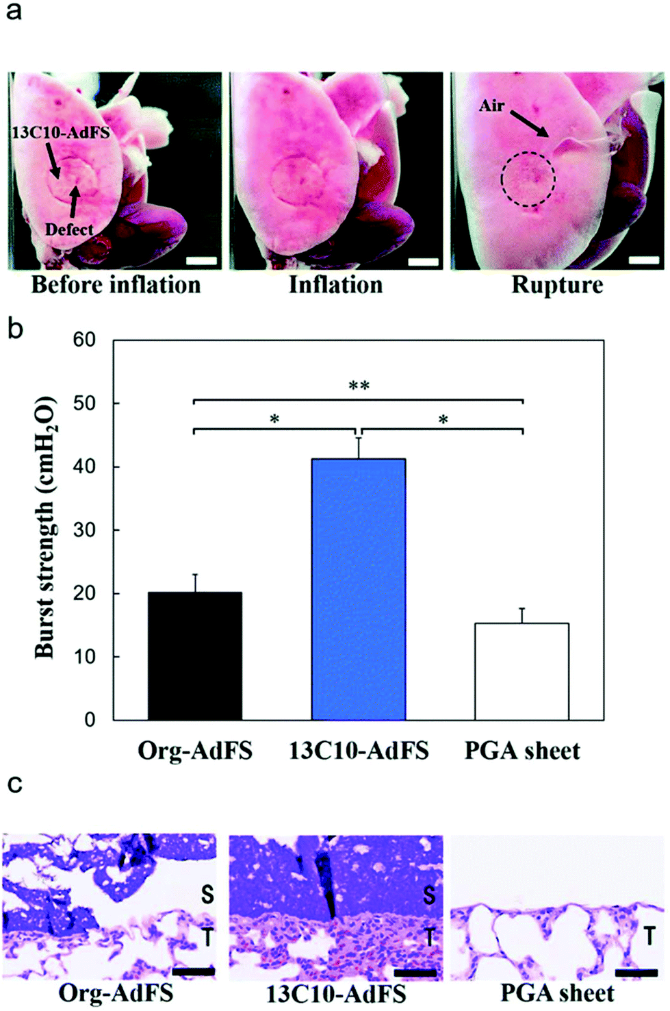
Fig. 4 Burst strengths of AdFSs on rat lung. (a) Photographs showing the burst strength measurement of 13C10-AdFS on rat lung. The scale bars are 5 mm. (b) Burst strengths of C10-AdFS, Org-AdFS, and a PGA sheet cross-linked at 150 °C for 5 h with thicknesses of 200 μm. (c) Histological observation of the AdFSs and the PGA sheet after burst strength measurement. The AdFS (S) and tissue (T) are indicated in the image. The scale bars are 50 μm. The data are the mean ± S.E.M. of five samples (
n = 5). *
p < 0.05, **
p > 0.05.
Fig. 6 Cytocompatibility of AdFS with L929 cells. (a) Cell viability of L929 cells incubated with lysates from AdFSs for 24 h. (b) Cell viability of L929 cells cultured on AdFSs for 24 h. (c) Actin/DAPI staining of L929 cells cultured on AdFS for 24 h. The data are the mean ± S.E.M. of three samples (
n = 3). *
p < 0.05, N.S.
p > 0.05.
The Royal Society of Chemistry apologises for these errors and any consequent inconvenience to authors and readers.
|
| This journal is © The Royal Society of Chemistry 2021 |
Click here to see how this site uses Cookies. View our privacy policy here. 
 Open Access Article
 This Open Access Article is licensed under a