 Open Access Article
Creative Commons Attribution 3.0 Unported Licence
Correction: In vivo migration of Fe3O4@polydopamine nanoparticle-labeled mesenchymal stem cells to burn injury sites and their therapeutic effects in a rat model
Xiuying
Li
a,
Zhenhong
Wei
a,
Binxi
Li
b,
Jing
Li
a,
Huiying
Lv
a,
Liya
Wu
a,
Hao
Zhang
b,
Bai
Yang
b,
Mingji
Zhu
*a and
Jinlan
Jiang
*a
aScientific Research Center, China-Japan Union Hospital of Jilin University, Changchun, Jilin, China. E-mail: jiangjinlan@jlu.edu.cn; zhumingji0822@q63.com; Tel: +86 18186811856
bState Key Laboratory of Supramolecular Structure and Materials, College of Chemistry, Jilin University, Changchun, Jilin, China
First published on 5th January 2021
Abstract
Correction for ‘In vivo migration of Fe3O4@polydopamine nanoparticle-labeled mesenchymal stem cells to burn injury sites and their therapeutic effects in a rat model’ by Xiuying Li et al., Biomater. Sci., 2019, 7, 2861–2872, DOI: 10.1039/C9BM00242A.
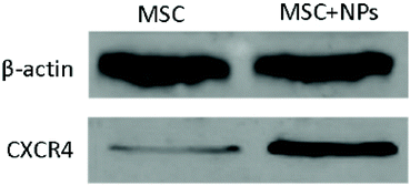
The authors regret errors in Fig. 1c, 3a and 5e in the original article. The corrected figures are shown below. In Fig. 1c, the authors wish to use a different TEM image to the one in Fig. 2a in their previous Biomaterials Science paper.1 In Fig. 3a, the 100, 150 and 200 μg ml−1 panels have been replaced as the original panels were incorrect leading to partial overlap with the 50 μg ml−1 panel. In Fig. 5e, the MSC and MSC + NPs bands for β-actin have been reversed as the original bands were incorrectly labelled due to being in the wrong orientation.
 | ||
| Fig. 1 Fe3O4@PDA nanoparticle preparation and internalization by MSCs. (C) The TEM image of a representative Fe3O4@PDA nanoparticle. Scale bar = 20 nm. | ||
The raw data for Fig. 3a and 5e were provided by the authors and reviewed by an independent expert.
The Royal Society of Chemistry apologises for these errors and any consequent inconvenience to authors and readers.
References
- L. Wu, F. Zhang, Z. Wei, X. Li, H. Zhao, H. Lv, R. Ge, H. Ma, H. Zhang, B. Yang, J. Li and J. Jiang, Biomater. Sci., 2018, 6, 2714 RSC.
| This journal is © The Royal Society of Chemistry 2021 |


