
Recent advances in microscale separation techniques for lipidome analysis

Abstract
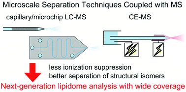
This review paper highlights the recent research on liquid-phase microscale separation techniques for lipidome analysis over the last 10 years, mainly focusing on capillary liquid chromatography (LC) and capillary electrophoresis (CE) coupled with mass spectrometry (MS). Lipids are one of the most important classes of biomolecules which are involved in the cell membrane, energy storage, signal transduction, and so on. Since lipids include a variety of hydrophobic compounds including numerous structural isomers, lipidomes are a challenging target in bioanalytical chemistry. MS is the key technology that comprehensively identifies lipids; however, separation techniques like LC and CE are necessary prior to MS detection in order to avoid ionization suppression and resolve structural isomers. Separation techniques using μm-scale columns, such as a fused silica capillary and microfluidic device, are effective at realizing high-resolution separation. Microscale separation usually employs a nL-scale flow, which is also compatible with nanoelectrospray ionization-MS that achieves high sensitivity. Owing to such analytical advantages, microscale separation techniques like capillary/microchip LC and CE have been employed for more than 100 lipidome studies. Such techniques are still being evolved and achieving further higher resolution and wider coverage of lipidomes. Therefore, microscale separation techniques are promising as the fundamental technology in next-generation lipidome analysis.
- This article is part of the themed collection: Analyst Review Articles 2021
 Please wait while we load your content...

