Surface oxidized two-dimensional antimonene nanosheets for electrochemical ammonia synthesis under ambient conditions

Munkhjargal Bat-Erdene a, Guangrui Xu bc, Munkhbayar Batmunkh ad, Abdulaziz S. R. Bati a, Jessica J. White d, Md J. Nine e, Dusan Losic e, Yu Chen c, Yun Wang d, Tianyi Ma b and Joseph G. Shapter *a
aAustralian Institute for Bioengineering and Nanotechnology, The University of Queensland, Brisbane, Queensland 4072, Australia. E-mail: j.shapter@uq.edu.au
bDiscipline of Chemistry, School of Environmental and Life Sciences, The University of Newcastle (UON), Callaghan, New South Wales 2308, Australia
cKey Laboratory of Macromolecular Science of Shaanxi Province, Key Laboratory of Applied Surface and Colloid Chemistry (Ministry of Education), Shaanxi Key Laboratory for Advanced Energy Devices, School of Materials Science and Engineering, Shaanxi Normal University, Xi'an 710062, PR China
dCentre for Clean Environment and Energy, School of Environment and Science, Griffith University, Gold Coast, Queensland 4222, Australia
eSchool of Chemical Engineering and Advanced Materials, The University of Adelaide, Adelaide, South Australia 5005, Australia

Received 10th December 2019 , Accepted 16th February 2020

First published on 17th February 2020


Abstract

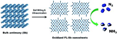
Two-dimensional (2D) antimonene nanosheets are prepared using a combination of ball milling and sonication-based liquid exfoliation and are used as an efficient electrocatalyst for the nitrogen reduction reaction (NRR). In 0.1 M KOH, a high NH3 yield of 180.4 μg h−1 mgCAT−1 and faradaic efficiency (FE) of 11.6% are achieved using our antimonene nanosheets. Theoretical calculations suggest that the oxidized species of antimonene act as the active catalytic sites for the NRR process. This work opens up a new avenue towards the development of 2D electrocatalysts for clean energy.


Ammonia (NH3) is one of the most produced chemical materials in the world owing to its importance in various sectors including agriculture, medicine, mining and in the household.1 NH3 has also drawn increasing attention as a promising energy carrier with high hydrogen density and low liquefying pressure.2 Although today's NH3 production requirements (>150 million tons per year) are principally met by the massive chemical plants based on Haber–Bosch process, this energy-intensive process consumes up to 5% of the global natural gas supply and is also responsible for a large amount of CO2 emission.3–5 These issues have led to significant efforts focused on finding alternative methods that are energy-efficient and environmentally friendly to produce NH3.6

Electrocatalytic nitrogen (N2) reduction reaction (NRR) driven by renewable energy sources offers a promising green process for sustainable production of NH3 under ambient conditions.7–9 Of particular importance in the NRR process is to sufficiently activate the N[triple bond, length as m-dash]N triple bond using efficient catalysts.7,10,11 Ideal catalytic materials should possess high electrocatalytic activity and selectivity, while still being abundantly available at low-cost. Noble metal catalysts (e.g., Au,12 Ag,13 Ru,14 and Rh15) have been the materials of choice for the NRR process. However, the scarcity and high cost of these catalysts hinder their widespread applications. Over the past few years, much attention has been paid to the development of non-noble-metal and metal-free catalysts such as Fe2O3–CNT,16 MoO3,17 Li+ incorporated PEBCD,18 Bi4V2O11/CeO2,19 N-doped porous carbon.20

Two-dimensional (2D) layered nanostructures have attracted increasing attention for use as electrocatalysts for the NRR.4,5,10,21–23 For instance, Zhang et al.5 explored MoS2 nanosheets as a noble-metal-free electrocatalyst for the NRR under ambient conditions. This MoS2 catalyst achieved a faradaic efficiency (FE) of 1.17% at −0.5 V versus reversible hydrogen electrode (RHE) in 0.1 M Na2SO4. Qiu et al.4 found that boron carbide (B4C) nanosheets can act as an excellent metal-free electrocatalyst toward NRR for NH3 synthesis, with a NH3 production yield of 26.57 μg h−1 mgCAT−1 and a FE of 15.95% at −0.75 V versus RHE when tested in 0.1 M hydrochloric acid (HCl). Recently, Zhang and coworkers used phosphorene nanosheets as a metal-free single element electrocatalyst for NRR.10 They used both indophenol blue and 15N2 isotope labelling methods to determine the NH3 yield. Intrinsic active sites of phosphorene facilitates the chemisorption of N2 molecules and provide sufficient electrons for activation of the inert N[triple bond, length as m-dash]N triple bond. Despite these great advances, the search for novel electrocatalysts that are readily available and exhibit high catalytic properties is still a very active area of research.

Herein, for the first time, we demonstrate that few-layer antimonene (FL-Sb) nanosheets prepared using a liquid-phase exfoliation (LPE) method can be an efficient NRR electrocatalyst for NH3 synthesis under ambient conditions. Previous studies found that 2D antimonene can be used as efficient catalyst for electrocatalytic water splitting.24–26 In our work, when tested in 0.1 M KOH electrolyte, our FL-Sb nanosheets without any supporting catalyst delivered a high NH3 yield of 180.4 μg h−1 mgCAT−1 at a potential of −0.1 V and a FE of 11.6% at a potential of +0.05 V versus the reversible hydrogen electrode (RHE). We used density functional theory (DFT) calculations to study the electrocatalytic mechanism for NRR on antimonene derivatives.

FL-Sb nanosheets were prepared in a 4[thin space (1/6-em)]:[thin space (1/6-em)]1 isopropanol/water mixture by exfoliating bulk Sb crystals using a combination of ball milling and ultrasonication (see ESI for details). It is well established that ball milling is a powerful strategy to functionalize 2D materials, creating abundant reactive sites, while it also helps to weaken the van der Waals interactions between the flakes.27 Moreover, Banks et al.28 found that the use of ball milling step helps to produce high yield homogeneous FL-Sb dispersions. The ball milled Sb flakes were further liquid exfoliated using ultrasonication, followed by centrifugation to obtain a stable dispersion of FL-Sb nanosheets with a concentration of around 0.068 g L−1 (as determined by UV-vis spectroscopy), which was much higher than previously reported values (1.74 × 10⁻³ g L−1 and 2.53 × 10⁻³ g L−1)29,30 including the FL-Sb prepared using a modified LPE involving ball milling (0.014 g L−1).28 This enhancement in the concentration of FL-Sb dispersion may be due to the fact that our ball milling was conducted using smaller ball size (1 mm) as compared to the previous report (5 mm),28 which results in more efficient exfoliation of bulk Sb. It should be noted that Gibaja et al.24 recently prepared an antimonene dispersion with a concentration of 0.368 g L−1 using a highly optimized production method. Atomic force microscopy (AFM) analysis (Fig. 1a) showed that FL-Sb sheets with flake dimensions in the range of a few hundreds of nanometers were produced due to the mechanochemical process induced by the ball milling.27,28 We further measured the thickness of our flakes deposited on a silicon substrate (Fig. 1a and S1). The measured heights of our FL-Sb sheets varied from 4 nm to 17 nm, with an average thickness of 10 nm (Fig. 1b). According to previous studies (considering a monolayer Sb sheet is 0.9 nm thick),31,32 our sample consists of few layers to multilayers sheets.


image file: c9ta13485a-f1.tif
Fig. 1 (a) Representative AFM image of the FL-Sb nanosheet. (b) Histogram of the height (thickness) of the FL-Sb nanosheets measured from 16 individual flakes. (c) UV-vis and (d) Raman spectrum of FL-Sb nanosheets.

As shown in Fig. 1c, the as-prepared FL-Sb dispersion showed a wide UV-vis absorption spectrum, which is consistent with previously reported theoretical results33 and experimentally measured spectra30 of 2D Sb. As shown in Fig. 1d, two characteristic Raman peaks centered at 114.1 cm−1 (Eg) and 149.2 cm−1 (A1g), which can be assigned to the in-plane vibrational mode and out-of-plane vibrational mode, respectively were observed for our FL-Sb and are consistent with the theoretically predicted characteristics of β-phase antimonene.34 However, the Raman modes centered at 190 cm−1 and 256 cm−1 were also observed, corresponding to the oxidized states of Sb (Sb2O3).34–36 Interestingly, all these Raman modes including the Sb2O3 peaks were also detected for the bulk Sb crystals (Fig. S2), suggesting that the bulk Sb already had some surface oxidation.

In order to gain further insight into the surface oxidation of both bulk Sb and FL-Sb, X-ray photoelectron spectroscopy (XPS) was used (Fig. 2a and S3a). As illustrated in Fig. 2b, the high-resolution XPS (Sb 3d) peak of our FL-Sb can be deconvoluted into two main spin-orbit split doublets with splitting of 9.40 eV,37 in addition to O 1s peak at around 532 eV.35,36 The small peaks centered at 528.0 eV and 537.4 eV can be assigned to the Sb 3d5/2 and Sb 3d3/2 of metallic Sb (Sb0), while two intense peaks at 530.4 eV and 539.8 eV are attributed to the oxidized Sb (Sb2O3).30,38 This suggests that our FL-Sb samples are strongly oxidized nanosheets and is consistent with previous reports.38–40 Interestingly, our bulk Sb before exfoliation also showed significant oxidation (Fig. S3b). Despite this, the Sb2O3 peak intensity of the bulk Sb was much lower than that of the FL-Sb, suggesting that further oxidation occurred during the FL-Sb preparation. This further increase in the oxidation of FL-Sb is believed to be due to the use of ball milling, creating abundant reactive sites during the mechanochemical process.27


image file: c9ta13485a-f2.tif
Fig. 2 (a) XPS survey scan, (b) high-resolution Sb 3d spectrum, (c) TEM and (d) HRTEM of FL-Sb nanosheets. (e) High-angle annular dark-field-scanning transmission electron microscope (HAADF-STEM) image of the FL-Sb flakes, and (f and g) the corresponding EDX elemental mapping and (h) EDX spectrum.

The transmission electron microscopy (TEM) image displayed in Fig. 2c reveals that the lateral size of our FL-Sb nanosheets is around 200 nm which is in agreement with the measured flake size from the AFM images. The lattice spacing of our FL-Sb nanosheet was 0.39 nm (Fig. 2d), which is consistent with previous literature.39,41 The energy-dispersive X-ray spectroscopy (EDX) elemental mapping illustrated in Fig. 2e–g indicates that our FL-Sb sample contains mainly Sb and O. Furthermore, the EDX spectrum of our FL-Sb exhibited a strong peak for “O” (Fig. 2h), which is an indication of Sb oxidation and consistent with the Raman and XPS results.

NRR experiments were performed in N2-saturated 0.1 M KOH solution in a gas-tight two-compartment cell under ambient conditions, which was separated by a Nafion membrane. Our FL-Sb nanosheets were used as the cathodic catalyst for the NRR. All potentials were reported on a reversible hydrogen electrode (RHE) scale. The NRR electrocatalytic performance of the FL-Sb nanosheets was first studied a linear sweep voltammetry (LSV) in both N2-saturated and Ar-saturated 0.1 M KOH solution (Fig. 3a). Although the LSV curves for both Ar- and N2-saturated solutions showed high current density due to the hydrogen evolution reaction (HER), noticeably higher current density (see Fig. 3a inset) obtained in a N2-saturated solution indicates that our FL-Sb nanosheets exhibit electrocatalytic activity for the NRR.42 Chronoamperometry measurements were carried out at different electrode potentials using our FL-Sb nanosheets in N2-saturated 0.1 M KOH solution. All the chronoamperometric curves showed negligible decay in the catalytic current densities, indicating the excellent stability of our FL-Sb nanosheets (Fig. 3b). It can be clearly seen that the current densities increased at the high electrode potentials due to the dominating HER process in the system.


image file: c9ta13485a-f3.tif
Fig. 3 (a) LSV curves of FL-Sb nanosheets in N2-saturated and Ar-saturated 0.1 M KOH electrolyte. Inset shows the zoomed in version (Y-axis: from −2 to −10 mA cm−2; and X-axis: from −0.3 to −0.5 V) of the LSV curve. (b) Chronoamperometric curves of FL-Sb nanosheets in N2-saturated 0.1 M KOH electrolyte at different potentials. (c) UV-vis absorption spectra of the KOH electrolyte stained with the indophenol indicator after charging at each given potential for 1 h in the presence of FL-Sb catalyst based working electrode, and (d) the corresponding NH3 yield rate and FE of FL-Sb nanosheets for the NRR (indophenol blue method). (e) NH3 yield rate and corresponding FE of the FL-Sb nanosheets determined by an ammonia selective electrode. (f) NH3 yield rate and FE of the FL-Sb nanosheets for the NRR under five consecutive cycles charging at −0.1 V (by indophenol blue method).

The indophenol blue method43 was used to determine the amount of NH3 produced (Fig. S4), and a possible by-product hydrazine (N2H4) was detected by the method of Watt and Chrisp (Fig. S5).44 Notably, no by-product N2H4 was detected (Fig. S6), suggesting the high selectivity of FL-Sb nanosheets for N2 toward NH3 production. After 1 h of the electrocatalytic reaction at constant potentials, the electrolyte containing the NH3 produced was colored with indophenol indicator and the UV-vis absorption spectra collected (Fig. 3c). The average NH3 yield and the corresponding FE values were calculated and plotted in Fig. 3d. Clearly, high NH3 yields and FEs were recorded at the lower potentials, while both the yield and FEs decreased when the potential is below −0.1 V due to the HER process. In particular, our FL-Sb exhibited a maximum NH3 yield of 180.39 μg h−1 mgCAT−1 when tested at −0.1 V, and its corresponding FE was 5.8%. The best recorded FE for our FL-Sb was 11.6% when tested at a potential of +0.05 V, while its NH3 yield was 133.1 μg h−1 mgCAT−1. The NH3 yields and FEs obtained for our 2D FL-Sb are remarkable and comparable to the state-of-the-art electrocatalysts including some noble metal containing catalysts.10,45,46 Table S1 compares the NRR performance obtained by our FL-Sb and other electrocatalysts reported in the literature.

While the indophenol blue is the most widely used method for NH3 quantification, we also used an ammonia selective electrode as it can provide a much lower detection limit and larger detection range.47 After plotting a calibration curve (Fig. S7), the measurements were carried out on the well stabilized electrolyte solution. It can be observed from Fig. 3e that the NH3 yields and FEs measured using this method were very consistent with the values determined using the indophenol blue method. In order to confirm the fact there is that no contribution to the NH3 yield from the electrode itself, we carried out NRR experiments using our bare supporting electrode (copper foam (CF)) and tested the yield using both indophenol blue and ammonia selective electrode methods. The yield rates were close to zero at all potentials, confirming that the CF electrode does not contribute to the production of NH3 during the catalysis (Fig. S8). We further evaluated the durability of our 2D catalyst for NRR over five consecutive cycles at −0.1 V (vs. RHE) (Fig. S9). As depicted in Fig. 3f, only slight decreases in both the yield rate and FE for NH3 production was observed after five cycles of chronoamperometric runs (about 90% performance retention at −0.1 V), indicating the robustness of our FL-Sb nanosheets for NRR electrocatalysis.

To identify the active catalytic species of our FL-Sb nanosheets, DFT calculations were carried out. Since the adsorption of N2 on the catalyst is one of the most important steps for the NRR, the adsorption properties of N2 both on β-antimonene and Sb2O3 (010) surface were analyzed. The β-antimonene was examined to provide a baseline of expected behaviour, while Sb2O3 was considered based on the XPS and Raman results obtained. It is well established that Sb2O3 consists of molecular units of Sb4O6 as shown in Fig. S10 and S11.48 By breaking the bonds in the plane (indicated by the dashed green line in Fig. S10), the (010) surface is formed and used for the calculation. Our theoretical results suggest a weak interaction between N2 and antimonene. As a comparison, the adsorption energy of N2 on Sb2O3 (010) surface was more than 2 times higher than that on antimonene monolayer (−0.06 and −0.13 eV for antimonene and Sb2O3, respectively), which suggests that the Sb2O3 is the active catalytic species in our FL-Sb nanosheets. The characterization results show that the topmost layers of antimonene have been oxidized to form Sb2O3 so it is clear that these active sites exist in the sample. Moreover, recent theoretical study by Wolff et al.49 reported that oxidized few-layer antimonene forms by itself a heterostructure consisting of semiconducting antimony oxide and semimetallic few-layer antimonene. The stronger adsorption can be ascribed to the increased polarity of Sb2O3 with respect to the non-polar antimonene monolayer. The strong adsorption on Sb2O3 (010) surface is further evidenced by the charge density difference plots, as shown in Fig. 4. There is almost no charge transfer between N2 and antimonene due to the considerably weak interaction. On the Sb2O3 (010) surface, partial charge is transferred from the N2 molecules to the region between adsorbate and surface after the adsorption. The transferred charges suggest the activation of the N[triple bond, length as m-dash]N triple bond, which is beneficial to the NRR.


image file: c9ta13485a-f4.tif
Fig. 4 Charge density difference of systems with adsorbed N2 molecule on (a) antimonene monolayer and (b) Sb2O3 (010) surface. The yellow and cyan contours indicate the enriched and depleted charge densities with the charge density value of ±0.001|e| Å−3, respectively. Color code: brown for Sb, red for O and dark blue for N.

Conclusions

In summary, surface-oxidized FL-Sb nanosheets were theoretically predicted and experimentally confirmed to be an active NRR electrocatalyst for electrochemical NH3 synthesis. When tested in 0.1 M KOH under ambient condition, our 2D antimonene nanosheets exhibited a high NH3 yield rate of 180.4 μg h−1 mgCAT−1 and FE of 11.6%. Due to the increased polarity, the adsorption energy of N2 on the surface-oxidized antimonene was more favorable as compared to the non-polar antimonene monolayer. As such, we found that the oxidized species of antimonene facilitates the activation of the N[triple bond, length as m-dash]N triple bond for the NRR process. We anticipate that the results reported in this work could open a new avenue in the development of monoelemental 2D catalysts for the electrocatalytic synthesis of NH3.

Conflicts of interest

There are no conflicts to declare.

Acknowledgements

The support of the Australian Research Council Discovery Program (DP160101301) is gratefully acknowledged. We acknowledge the use of the Queensland node of the Australian National Fabrication Facility (ANFF-Q) at the University of Queensland. This research was undertaken on the supercomputers in National Computational Infrastructure (NCI) in Canberra, Australia, which is supported by the Australian Commonwealth Government, and Pawsey Supercomputing Centre in Perth with the funding from the Australian Government and the Government of Western Australia. The authors thank Dr Ashley D. Slattery of Adelaide Microscopy at the University of Adelaide for his help for Titan TEM analysis.

Notes and references

  1. B. M. Hoffman, D. Lukoyanov, Z.-Y. Yang, D. R. Dean and L. C. Seefeldt, Chem. Rev., 2014, 114, 4041–4062 CrossRef CAS PubMed.
  2. D. J. Little, I. I. I. M. R. Smith and T. W. Hamann, Energy Environ. Sci., 2015, 8, 2775–2781 RSC.
  3. R. F. Service, Science, 2014, 345, 610 CrossRef CAS PubMed.
  4. W. Qiu, X.-Y. Xie, J. Qiu, W.-H. Fang, R. Liang, X. Ren, X. Ji, G. Cui, A. M. Asiri, G. Cui, B. Tang and X. Sun, Nat. Commun., 2018, 9, 3485 CrossRef PubMed.
  5. L. Zhang, X. Ji, X. Ren, Y. Ma, X. Shi, Z. Tian, A. M. Asiri, L. Chen, B. Tang and X. Sun, Adv. Mater., 2018, 30, 1800191 CrossRef PubMed.
  6. K. A. Brown, D. F. Harris, M. B. Wilker, A. Rasmussen, N. Khadka, H. Hamby, S. Keable, G. Dukovic, J. W. Peters, L. C. Seefeldt and P. W. King, Science, 2016, 352, 448–450 CrossRef CAS PubMed.
  7. X. Cui, C. Tang and Q. Zhang, Adv. Energy Mater., 2018, 8, 1800369 CrossRef.
  8. X. Guo, H. Du, F. Qu and J. Li, J. Mater. Chem. A, 2019, 7, 3531–3543 RSC.
  9. G.-F. Chen, S. Ren, L. Zhang, H. Cheng, Y. Luo, K. Zhu, L.-X. Ding and H. Wang, Small Methods, 2019, 3, 1800337 CrossRef.
  10. L. Zhang, L.-X. Ding, G.-F. Chen, X. Yang and H. Wang, Angew. Chem., Int. Ed., 2019, 58, 2612–2616 CrossRef CAS PubMed.
  11. K. Liu, J. Fu, L. Zhu, X. Zhang, H. Li, H. Liu, J. Hu and M. Liu, Nanoscale, 2020 10.1039/c9nr09117c.
  12. M.-M. Shi, D. Bao, B.-R. Wulan, Y.-H. Li, Y.-F. Zhang, J.-M. Yan and Q. Jiang, Adv. Mater., 2017, 29, 1606550 CrossRef PubMed.
  13. H. K. Lee, C. S. L. Koh, Y. H. Lee, C. Liu, I. Y. Phang, X. Han, C.-K. Tsung and X. Y. Ling, Sci. Adv., 2018, 4, eaar3208 CrossRef PubMed.
  14. Y. Yao, H. Wang, X.-z. Yuan, H. Li and M. Shao, ACS Energy Lett., 2019, 4, 1336–1341 CrossRef CAS.
  15. H.-M. Liu, S.-H. Han, Y. Zhao, Y.-Y. Zhu, X.-L. Tian, J.-H. Zeng, J.-X. Jiang, B. Y. Xia and Y. Chen, J. Mater. Chem. A, 2018, 6, 3211–3217 RSC.
  16. S. Chen, S. Perathoner, C. Ampelli, C. Mebrahtu, D. Su and G. Centi, Angew. Chem., Int. Ed., 2017, 56, 2699–2703 CrossRef CAS PubMed.
  17. J. Han, X. Ji, X. Ren, G. Cui, L. Li, F. Xie, H. Wang, B. Li and X. Sun, J. Mater. Chem. A, 2018, 6, 12974–12977 RSC.
  18. G.-F. Chen, X. Cao, S. Wu, X. Zeng, L.-X. Ding, M. Zhu and H. Wang, J. Am. Chem. Soc., 2017, 139, 9771–9774 CrossRef CAS PubMed.
  19. C. Lv, C. Yan, G. Chen, Y. Ding, J. Sun, Y. Zhou and G. Yu, Angew. Chem., Int. Ed., 2018, 57, 6073–6076 CrossRef CAS PubMed.
  20. Y. Liu, Y. Su, X. Quan, X. Fan, S. Chen, H. Yu, H. Zhao, Y. Zhang and J. Zhao, ACS Catal., 2018, 8, 1186–1191 CrossRef CAS.
  21. X. Yu, P. Han, Z. Wei, L. Huang, Z. Gu, S. Peng, J. Ma and G. Zheng, Joule, 2018, 2, 1610–1622 CrossRef CAS.
  22. P. Chen, N. Zhang, S. Wang, T. Zhou, Y. Tong, C. Ao, W. Yan, L. Zhang, W. Chu, C. Wu and Y. Xie, Proc. Natl. Acad. Sci. U. S. A., 2019, 116, 6635–6640 CrossRef CAS PubMed.
  23. Y. Luo, G.-F. Chen, L. Ding, X. Chen, L.-X. Ding and H. Wang, Joule, 2019, 3, 279–289 CrossRef CAS.
  24. C. Gibaja, M. Assebban, I. Torres, M. Fickert, R. Sanchis-Gual, I. Brotons, W. S. Paz, J. J. Palacios, E. G. Michel, G. Abellán and F. Zamora, J. Mater. Chem. A, 2019, 7, 22475–22486 RSC.
  25. X. Ren, Z. Li, H. Qiao, W. Liang, H. Liu, F. Zhang, X. Qi, Y. Liu, Z. Huang, D. Zhang, J. Li, J. Zhong and H. Zhang, ACS Appl. Energy Mater., 2019, 2, 4774–4781 CrossRef CAS.
  26. P. Ares, J. J. Palacios, G. Abellán, J. Gómez-Herrero and F. Zamora, Adv. Mater., 2018, 30, 1703771 CrossRef PubMed.
  27. J. Xu, I.-Y. Jeon, J.-M. Seo, S. Dou, L. Dai and J.-B. Baek, Adv. Mater., 2014, 26, 7317–7323 CrossRef CAS PubMed.
  28. E. Martínez-Periñán, M. P. Down, C. Gibaja, E. Lorenzo, F. Zamora and C. E. Banks, Adv. Energy Mater., 2018, 8, 1702606 CrossRef.
  29. C. Gibaja, D. Rodriguez-San-Miguel, P. Ares, J. Gómez-Herrero, M. Varela, R. Gillen, J. Maultzsch, F. Hauke, A. Hirsch, G. Abellán and F. Zamora, Angew. Chem., Int. Ed., 2016, 55, 14345–14349 CrossRef CAS PubMed.
  30. F. Zhang, J. He, Y. Xiang, K. Zheng, B. Xue, S. Ye, X. Peng, Y. Hao, J. Lian, P. Zeng, J. Qu and J. Song, Adv. Mater., 2018, 30, 1803244 CrossRef PubMed.
  31. P. Ares, F. Aguilar-Galindo, D. Rodríguez-San-Miguel, D. A. Aldave, S. Díaz-Tendero, M. Alcamí, F. Martín, J. Gómez-Herrero and F. Zamora, Adv. Mater., 2016, 28, 6332–6336 CrossRef CAS PubMed.
  32. F. Li, M. Xue, J. Li, X. Ma, L. Chen, X. Zhang, D. R. MacFarlane and J. Zhang, Angew. Chem., Int. Ed., 2017, 56, 14718–14722 CrossRef CAS PubMed.
  33. N. Wang, D. Cao, J. Wang, P. Liang, X. Chen and H. Shu, J. Mater. Chem. C, 2017, 5, 9687–9693 RSC.
  34. J. Ji, X. Song, J. Liu, Z. Yan, C. Huo, S. Zhang, M. Su, L. Liao, W. Wang, Z. Ni, Y. Hao and H. Zeng, Nat. Commun., 2016, 7, 13352 CrossRef CAS PubMed.
  35. G. Abellán, P. Ares, S. Wild, E. Nuin, C. Neiss, D. R.-S. Miguel, P. Segovia, C. Gibaja, E. G. Michel, A. Görling, F. Hauke, J. Gómez-Herrero, A. Hirsch and F. Zamora, Angew. Chem., Int. Ed., 2017, 56, 14389–14394 CrossRef PubMed.
  36. V. Lloret, M. Á. Rivero-Crespo, J. A. Vidal-Moya, S. Wild, A. Doménech-Carbó, B. S. J. Heller, S. Shin, H.-P. Steinrück, F. Maier, F. Hauke, M. Varela, A. Hirsch, A. Leyva-Pérez and G. Abellán, Nat. Commun., 2019, 10, 509 CrossRef PubMed.
  37. E. N. K. Glover, S. G. Ellington, G. Sankar and R. G. Palgrave, J. Mater. Chem. A, 2016, 4, 6946–6954 RSC.
  38. R. Gusmão, Z. Sofer, D. Bouša and M. Pumera, Angew. Chem., Int. Ed., 2017, 56, 14417–14422 CrossRef PubMed.
  39. C. C. Mayorga-Martinez, R. Gusmão, Z. Sofer and M. Pumera, Angew. Chem., Int. Ed., 2019, 58, 134–138 CrossRef CAS PubMed.
  40. Y. Gao, W. Tian, C. Huo, K. Zhang, S. Guo, S. Zhang, X. Song, L. Jiang, K. Huo and H. Zeng, J. Mater. Chem. A, 2019, 7, 3238–3243 RSC.
  41. X. Wang, J. He, B. Zhou, Y. Zhang, J. Wu, R. Hu, L. Liu, J. Song and J. Qu, Angew. Chem., Int. Ed., 2018, 57, 8668–8673 CrossRef CAS PubMed.
  42. H. Jin, L. Li, X. Liu, C. Tang, W. Xu, S. Chen, L. Song, Y. Zheng and S.-Z. Qiao, Adv. Mater., 2019, 31, 1902709 CrossRef PubMed.
  43. D. Zhu, L. Zhang, R. E. Ruther and R. J. Hamers, Nat. Mater., 2013, 12, 836 CrossRef CAS PubMed.
  44. G. W. Watt and J. D. Chrisp, Anal. Chem., 1952, 24, 2006–2008 CrossRef CAS.
  45. X. Zhang, T. Wu, H. Wang, R. Zhao, H. Chen, T. Wang, P. Wei, Y. Luo, Y. Zhang and X. Sun, ACS Catal., 2019, 9, 4609–4615 CrossRef CAS.
  46. L. Li, C. Tang, B. Xia, H. Jin, Y. Zheng and S.-Z. Qiao, ACS Catal., 2019, 9, 2902–2908 CrossRef CAS.
  47. C. Tang and S.-Z. Qiao, Chem. Soc. Rev., 2019, 48, 3166–3180 RSC.
  48. C. Svensson, Acta Crystallogr., Sect. B: Struct. Crystallogr. Cryst. Chem., 1975, 31, 2016–2018 CrossRef.
  49. S. Wolff, R. Gillen, M. Assebban, G. Abellán and J. Maultzsch, [Cond-mat.mtrl-sci], 2019, arXiv:1909.01204v01203.

Footnotes

Electronic supplementary information (ESI) available. See DOI: 10.1039/c9ta13485a
These two authors made an equal contribution to this work.

This journal is © The Royal Society of Chemistry 2020