
 Open Access Article
 This Open Access Article is licensed under a
 Creative Commons Attribution 3.0 Unported Licence
Correction: Live cell single molecule tracking and localization microscopy of bioorthogonally labeled plasma membrane proteins
Andres I.
König
ab,
Raya
Sorkin
c,
Ariel
Alon
ab,
Dikla
Nachmias
ab,
Kalyan
Dhara
d,
Guy
Brand
c,
Ofer
Yifrach
a,
Eyal
Arbely
 abd,
Yael
Roichman
ce and
Natalie
Elia
*ab
abd,
Yael
Roichman
ce and
Natalie
Elia
*ab
aDepartment of Life Sciences, Ben-Gurion University of the Negev, Beer Sheva 84105, Israel. E-mail: elianat@post.bgu.ac.il; Fax: +972-8-6477546; Tel: +972-8-6428735
bNational Institute for Biotechnology in the Negev (NIBN), Ben-Gurion University of the Negev, Beer Sheva 84105, Israel
cRaymond & Beverly Sackler School of Chemistry, Tel Aviv University, Tel Aviv 6997801, Israel
dDepartment of Chemistry, Ben-Gurion University of the Negev, Beer Sheva 84105, Israel
eRaymond & Beverly Sackler School of Physics, and the Center for light-matter interaction, Tel Aviv University, Tel Aviv 6997801, Israel
First published on 3rd September 2020
Abstract
Correction for ‘Live cell single molecule tracking and localization microscopy of bioorthogonally labeled plasma membrane proteins’ by Andres I. König et al., Nanoscale, 2020, 12, 3236–3248, DOI: 10.1039/C9NR08594G.
The authors regret that in the original manuscript, position 128 in the EGFR was referred to incorrectly as L128, rather than N128. This error occurred on page 3237 in the following sentence: “To this end, EGFR was mutated to carry a TAG codon at the previously published labeling site, Leu 128, and cloned into a single expression vector that encodes the cognate pair of tRNAcua:tRNA-synthetase17,18 (Fig. S1a†).”
The error also occurred in Fig. 1a, the corrected version of which is displayed below, along with the original, unaltered caption. These errors do not affect any of the experimental results and discussion or conclusions reported in the paper, only the display of the figure and text.
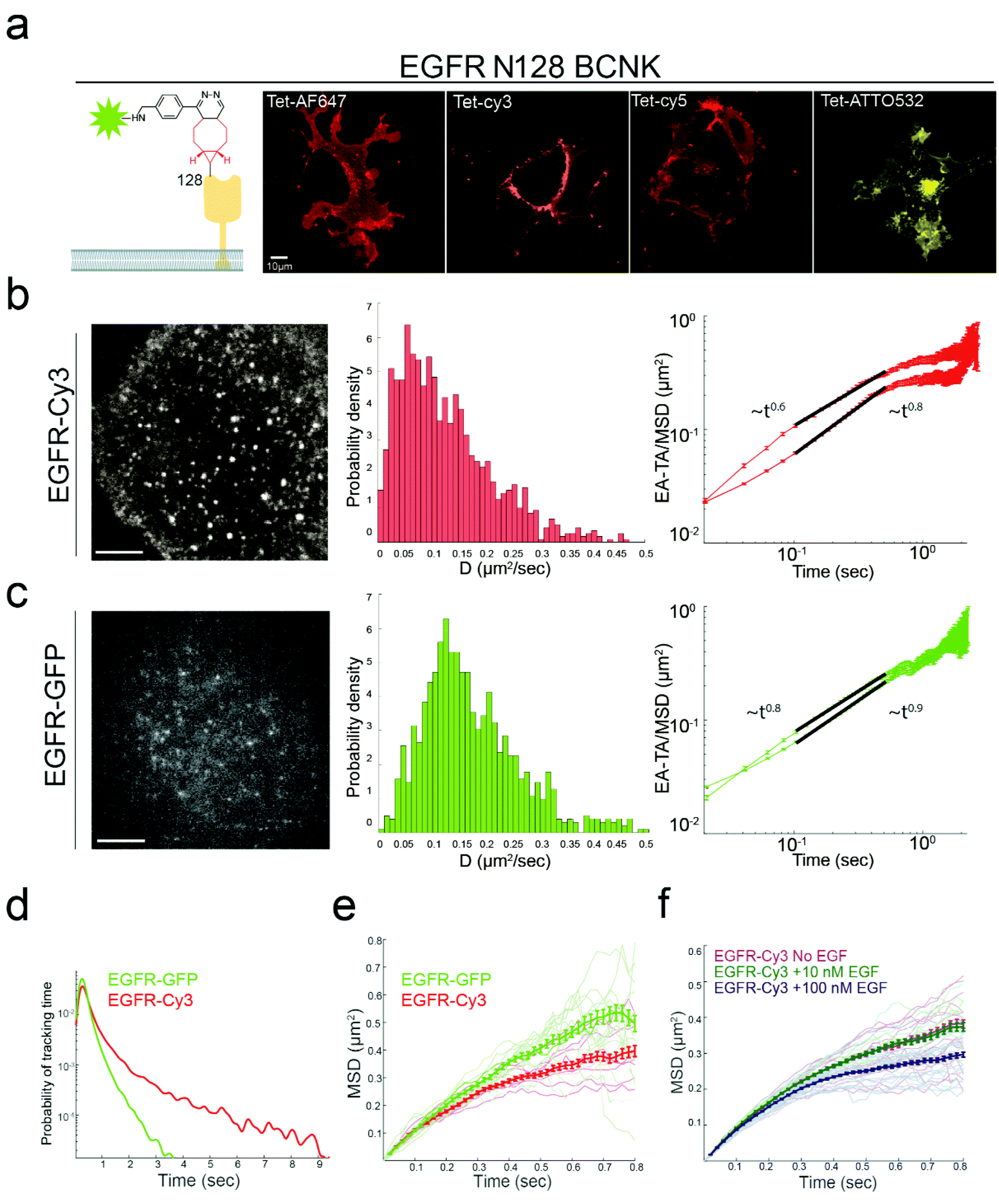 | ||
| Fig. 1 Bioorthogonal labeling with Fl-dyes enables single particle tracking of EGFR. (a) Left panel: Schematic representation of the labeling strategy. The extracellular domain of EGFR (yellow) was mutated to carry the ncAA BCNK (red) at position 128. Labeling was obtained via a bioorthogonal reaction between BCNK and a tetrazine conjugated Fl-dye (green star). Panels 2–5: Live COS7 cells expressing EGFR128BCNK together with GCE components (Fig. S1a†) were labeled with 1.5 μM of: Tet-AF647, Tet-Cy3, Tet-Cy5 or Tet-ATTO532 (Fig. S1b†) and imaged using a spinning disk confocal microscope. Shown are single slices taken from the center of the cell. Scale bar = 10 μm. (b and c) COS7 cells transfected with either GCE system plasmid with carrying EGFR128BCNK and labeled with Tet-Cy3 (b) or EGFR-GFP (c), were imaged in TIRF mode at 50 fps. Tracks were then obtained as described in methods section. Left panel: Representative images of the first frame (the complete video sequences are provided in Videos 1 and 2). Middle panels: The diffusion coefficient [D] of all particles tracked. EGFR-Cy3 median, 0.11 μm2 s−1 (with 95% confidence interval of 0.02–0.4 μm2 s−1, n = 1177). EGFR-GFP median, 0.15 μm2 s−1 (with 95% confidence interval of 0.04–0.47 μm2 s−1, n = 1018). Right panels: Ensemble-averaged MSD (top plots) and ensemble-averaged time-averaged MSD (bottom plots) in log scale, MSD ∼tα, where α is the anomalous exponent. (d) The tracking time probability density function of EGFR-GFP (green) or EGFR-Cy3 (red). (e) MSD curves of EGFR-GFP (green) or EGFR-Cy3 (red) as a function of time. Thick lines are the ensemble means for each labeling type with error bars representing SEM. Thin lines represent measurements obtained from individual cells (EGFR-Cy3 n = 9 cells, EGFR-GFP n = 17 cells, obtained from three independent experiments). (f) MSD curves obtained for EGFR-Cy3 under three different concentrations of EGF: no EGF (magenta), 10 nM EGF (dark green) or 100 nM EGF (blue) as a function of time. Thick lines are the ensemble means for each labeling type with error bars representing SEM. Thin lines represent measurements obtained from individual cells (no EGF n = 18 cells, 10 nM EGF n = 15 cells, and 100 nM EGF n = 18 cells). | ||
The Royal Society of Chemistry apologises for these errors and any consequent inconvenience to authors and readers.
| This journal is © The Royal Society of Chemistry 2020 |