 Open Access Article
Creative Commons Attribution 3.0 Unported Licence
Correction: Nanoparticle binding attenuates the pathobiology of gastric cancer-associated Helicobacter pylori
Dana
Westmeier
*a,
Gernot
Posselt
b,
Angelina
Hahlbrock
a,
Sina
Bartfeld
c,
Cecilia
Vallet
d,
Carmen
Abfalter
b,
Dominic
Docter
a,
Shirley K.
Knauer
d,
Silja
Wessler
b and
Roland H.
Stauber
*a
aDepartment of Nanobiomedicine/ENT, University Medical Center of Mainz, Langenbeckstrasse 1, 55101 Mainz, Germany. E-mail: roland.stauber@unimedizin-mainz.de; danawestmeier@uni-mainz.de
bDepartment of Molecular Biology, Paris-Lodron University of Salzburg, A-5020 Salzburg, Austria
cResearch Centre for Infectious Diseases, Institute of Molecular Infection Biology, University of Würzburg, Würzburg, Germany
dDepartment of Molecular Biology II, Centre for Medical Biotechnology (ZMB), University Duisburg-Essen, Universitätsstraße 5, 45117 Essen, Germany
First published on 8th January 2020
Abstract
Correction for ‘Nanoparticle binding attenuates the pathobiology of gastric cancer-associated Helicobacter pylori’ by Dana Westmeier et al., Nanoscale, 2018, 10, 1453–1463.
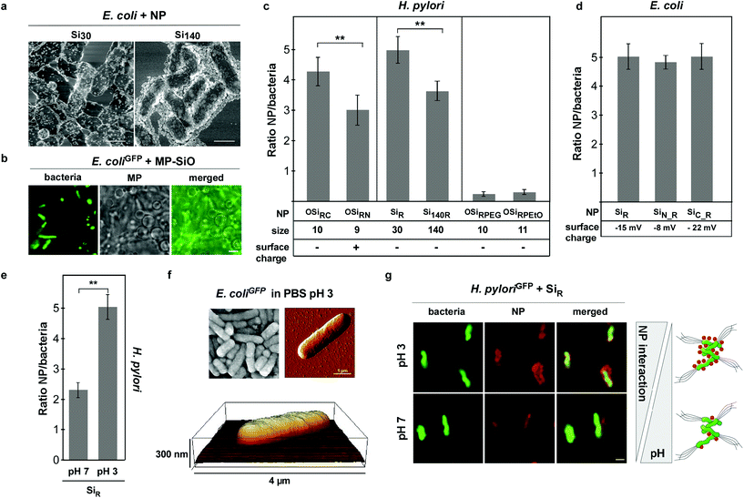
The authors have discovered that in the previously published Fig. 2, the electron microscopy image in panel 2a was generated in collaboration with Prof. Gunzer's group, for which we did not receive written confirmation to be used in the paper. In the revised version of Fig. 2, the electron microscopy image in panel 2a has been replaced with an alternative image, also showing the assembly of Si NPs with different sizes onto E. coli by scanning electron microscopy.
Although this error does not affect the conclusions and findings of this research paper, the authors sincerely apologize for the error and any confusion caused.
Please find below the corrected version of Fig. 2 below.
Furthermore, we wish to acknowledge the intellectual support received from Prof. Gunzer's group during the study. Hence, the acknowledgements section should therefore have read as follows:
Acknowledgements
We wish to acknowledge the intellectual support received from Prof. Gunzer's group and the Mainz imaging facility during the study.
The Royal Society of Chemistry apologises for these errors and any consequent inconvenience to authors and readers.
| This journal is © The Royal Society of Chemistry 2020 |

