
The Art in Science of MicroTAS 2019†
Gregory A.
Cooksey

National Institute of Standards and Technology (NIST), 100 Bureau Drive, Gaithersburg, MD 20899, USA. E-mail: gregory.cooksey@nist.gov
The committee thanks the contributors for invigorating the MicroTAS conference with their inspiring pieces—it was such a pleasure to consider these works of art and science.
Microfluidics and microscale research offer unique opportunities to view and express scientific discovery and creation beyond the scale of unaided human vision. This year's entries, like those from previous competitions, continue to serve that creative endeavor. We highlight many examples that enable us to encounter the hidden aesthetic of nature, to show novel creations from raw materials using new technologies, and to explore functional patterns that emerge when responsive biological components interact with a controlled environment.
We are very pleased to recognize the image “A Cell's World” by Joseph de Rutte from UCLA as the winner of the 2019 Art in Science Award (Fig. 1). The photomicrograph is a wonderful combination of color and shape that the author created from “uniform droplets formed using structured microparticles” that were “used to encapsulate single-cells and measure their secretions”. The basic characteristics and composition of the fabricated droplets are brilliantly depicted in the image, which inspires their use as both surrogates and vessels for cells. The award was presented at the RSC booth during a special ceremony (Fig. 2).
 | ||
| Fig. 1 “A Cell's World” by Joseph de Rutte from the University of California, Los Angeles (UCLA), USA, was the winning image from the 2019 Art in Science Competition. | ||
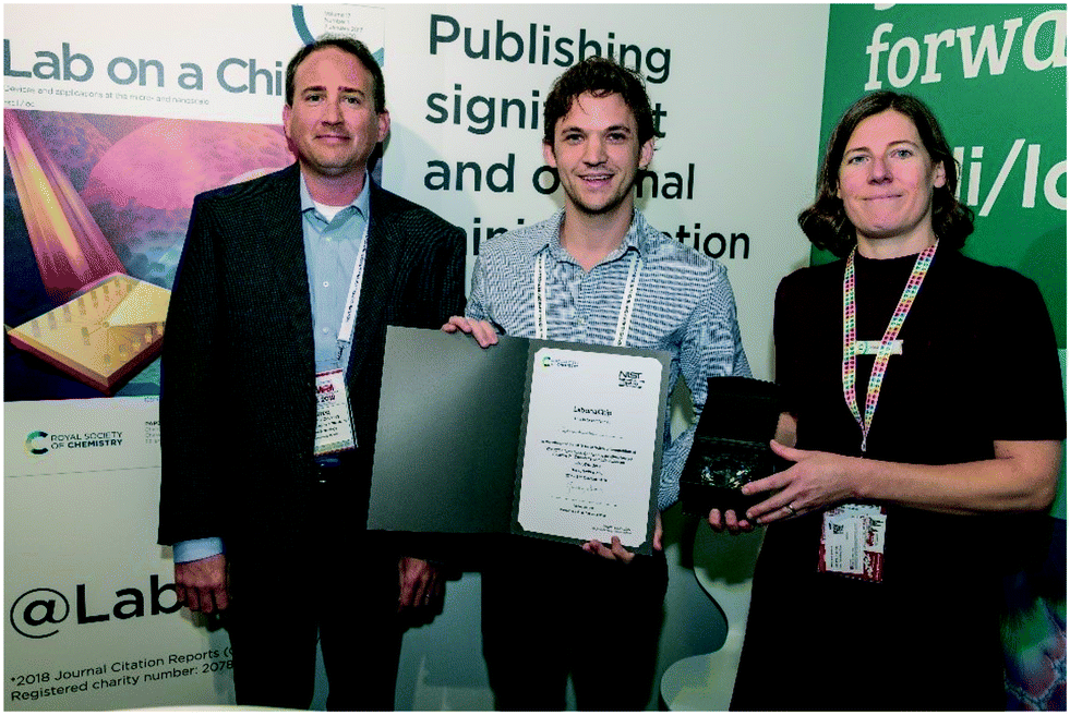 | ||
| Fig. 2 Greg Cooksey (left) and Maria Southall (Lab on a Chip Deputy Editor; right) present the Art in Science award to winner Joseph de Rutte at the RSC booth. | ||
First runner-up was awarded to Laura Barillas, Leibniz Institute for Plasma Science and Technology (INP), Germany, for the image “MicroQuasar” (Fig. 3). We thought the author's caption perfectly highlighted the similarity, despite the incredible difference in length scales, between the spiral plasma image and the radiation that one might visualize exiting a quasar.
 | ||
| Fig. 3 Laura Barillas from Leibniz Institute for Plasma Science and Technology (INP), Germany, was honored with first runner-up for her submission “MicroQuasar”. The author captured the spiral micropattern that developed during a maskless polymerization process. | ||
Michael Restaino from the University of Maryland, USA, earned second runner-up for “Sensing in Three-Dimensions”, for his extraordinary demonstration of the complex structures that can now be achieved with 3D printing technology (Fig. 4). The well-composed and colorized image shows interwoven metallic and fluidic structures.
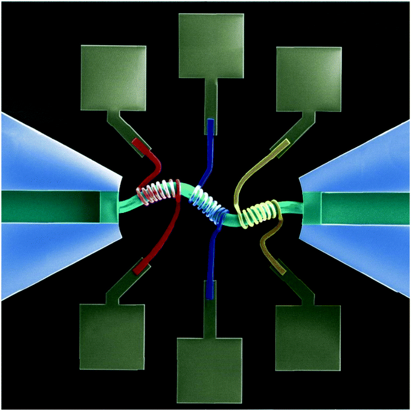 | ||
| Fig. 4 “Sensing in Three-Dimensions” earned second runner-up for Michael Restaino from the University of Maryland, USA. The scanning electron microscope (SEM) image provides intricate details of one of the most complicated 3D-printed devices ever realized on the microscale. | ||
Finally, the third runner-up honor was presented to Charlotte Yvanoff from Vrije Universiteit Brussel, Belgium, for the composition “Stars and Diamonds made out of bone cells” (Fig. 5). The image highlights the functional forms that are adapted by cells following interaction with a well-defined physical environment that limits sites for cell–substrate attachment. Such platforms have proven incredibly powerful in understanding various aspects of cell survival and function.1
 | ||
| Fig. 5 “Stars and Diamonds made out of bone cells” by Charlotte Yvanoff, Vrije Universiteit Brussel, Belgium. The fluorescence image shows how protein patterns (green) can direct the formation of actin stress fibres (red) and position of nuclei (blue) within cells attached to micropatterned substrates. | ||
The selection committee for the award consisted of Jeanne Andres, Lab on a Chip Executive Editor, Greg Cooksey, Project leader in the Microsystems and Nanotechnology Division at NIST, and Hang Lu, Lab on a Chip Associate Editor and Professor of Chemical and Biomolecular Engineering at the Georgia Institute of Technology.
Acknowledgements
The Art in Science competition is sponsored and supported by MicroTAS, the Chemical and Biological Microsystems Society (CBMS), the Lab on a Chip journal, and NIST. The 2019 award consisted of a monetary prize ($500), an award certificate, and a coveted front cover of the Lab on a Chip journal. Please check the MicroTAS 2020 conference website for further details regarding changes to the competition format and image submission deadline for the next MicroTAS Conference.References
- C. S. Chen, M. Mrksich, S. Huang, G. M. Whitesides and D. E. Ingber, Science, 1997, 276, 1425 CrossRef CAS PubMed
.
Footnote |
| † Any opinions or views expressed in this article are entirely those of the author and do not represent the views of the National Institute of Standards and Technology, the journal (Lab on a Chip), or the Royal Society of Chemistry. |
| This journal is © The Royal Society of Chemistry 2020 |